Patient data. Present illness. Past history. Initial Impression
|
|
|
- Lucy Morrison
- 5 years ago
- Views:
Transcription
1 GS-ER combine meeting 報告者 :R1 王帝皓指導者 :VS 連楚明 Patient data 25 y/o man Visited at 23:22 Chief complain: abd pain since 5:00 pm Triage: III Vital signs: BT: 35.7, PR: 75, RR: 18, BP: 121/70 SpO2: 99% E4M5V6 Present illness 吃了一顆水煎包後開始痛 Periumbilical region Colicky, persist 痛到站不直 Radiated to RLQ Not radiated to back No diarrhea/ vomit, no fever Past history Healthy No alcohol use Norecreation drug use No op history NKA PE Conscious: clear, alert Head & neck: supple Chest: Clear BS, RHB ABD: soft, no guarding, no rebounding. Normoactive BS Ext: warm Initial Impression Abd pain R/O diverticulitis R/O pancreatitis R/O appendicitis
2 Orders NPO CBC/DC/PLT PT, APTT Panel 1, CRP, lipase N/S run 60ml/hr Keto 1 amp IV st Bed side echo Bed side echo Bed side echo No hydronephrosis No GB distended, no wall thicken No free air Cecum wall edema 2 hrs later Still abd pain, VAS: 7 Rebound: mild 檢驗項檢驗值目名稱 檢驗項目名稱 檢驗值 檢驗值單位 PT 10.8 second Norma l 10.6 second control INR 1.02 Ratio APTT 33.0 second Norma l control 32.8 second Glucos e GOT( AST) 檢驗值單位 最小參考值 103 mg/dl 最大參考值 U/L BUN 16 mg/dl Creatin ine 0.8 mg/dl Na 146 meq/l K 4.1 meq/l egfr Lipase 60 U/L CRP mg/dl 檢驗項目名稱 檢驗值 檢驗值單位 Hb 15.9 gm/dl WBC 14.7 x1000/u l Differen tial count Segmen ted Neutro. Lympho cyte Monocy te Eosinop hil Basophi l ****** **** Platelet % 23.2 % 5.0 % 5.9 % 0.4 % x1000/u l Do abd CT 1.right sided small intestine almostly with segmental bowel dilatation, perienteric edema and short segmental diminished bowel wall enhancement associate small amount of the ascite. There is no intramural bowel gas and no extraluminal free air. Focal area of mesentery traction to one direction with mild torsion associate bird-beak like narrowed bowel segment disclsoed at RLQ. T-colon found almostly at left and lower abdomen. Cecum found at pelvic floor. 2.no apparent focal parenchymal nodule in the liver, spleen and pancreas. 3.symmetric perfusion of both kidneys. Mild pelviectasia on right side found. 4.no apparent biliary tree nor MPD dilatation. There is no detectable hyperdense or calcified biliary stone. GB appears to be normal. Pericholecystic space is clear. CT impression Mid-gut non-rotation or malrotation with RLQ internal hernia and probability of focal strangulation.
3 Orders IV fluid Cefa NPO Consult GS On NG decompression S-S enema BID Next morning Admit to GS ward Prepare for explore laparotomy OP findings: Operator: Dr. Right paraduodenal hernia Transition zone due to herniation and adhesion of proximal ileum at paracecal area and entrance of the sac. Operation time: 80 mins. Post-op course Smooth recovery, Try diet at D4 Discharged at post op day 7 OPD F/U: doing well Discussion Internal hernia
![Internal Hernia Hernias are of two main types, external and internal [1].](/docs-images/93/111399802/images/4-0.jpg "External hernias refer to prolapse of intestinal loops through a defect in the wall of the abdomen or pelvis internal hernias are defined by the protrusion of a viscus through a normal or abnormal")
4 Internal Hernia Hernias are of two main types, external and internal [1]. External hernias refer to prolapse of intestinal loops through a defect in the wall of the abdomen or pelvis internal hernias are defined by the protrusion of a viscus through a normal or abnormal peritoneal or mesenteric aperture within the confines of the peritoneal cavity. The orifice can be either ac- quired, such as a postsurgical, traumatic, or postinflammatory defect, or congenital, in- cluding both normal apertures. Incidence Although internal hernias have an overall incidence of less than 1%, they constitute up to 5.8% of all small-bowel obstructions, which, if left untreated, have been reported to have an overall mortality exceeding 50% if strangulation is present. Internal Hernia paraduodenal (53%), pericecal (13%), foramen of Winslow (8%), trans- mesenteric and transmesocolic (8%), inter- sigmoid (6%), and retroanastomotic (5%) (Fig. 1), with the overall incidence of internal hernias being %. Internal Hernia Fig. 1 Diagrammatic illustration shows various types of internal hernias: A = paraduodenal, B = foramen of Winslow, C = intersigmoid, D = pericecal, E = transmesenteric, and F = retroanastomotic. General Clinical Findings Clinically, internal hernias can be asymptomatic or cause significant discomfort ranging from constant vague epigastric pain to intermittent colicky periumbilical pain. Additional symptoms include nausea, vomiting (especially after a large meal), and recurrent intestinal obstruction. Symptom severity relates to the duration and reducibility of the hernia and the presence or absence of incarceration and strangulation. These symptoms may be altered or relieved by changes in patient position. General Imaging Findings on Radiography and CT General radiographic features with barium studies in- clude apparent encapsulation of distended bowel loops with an abnormal location, ar- rangement or crowding of small-bowel loops within the hernial sac, evidence of obstruction with segmental dilatation and stasis, with additional features of apparent fixation and re- versed peristalsis during fluoroscopic evaluation [1, 5] (Table1). On CT, additional findings include mesenteric vessel abnormalities, with engorgement, crowding, twisting, and stretching of these vessels commonly found and providing an important clue to the underlying diagnosis
![Paraduodenal Hernias accounting for approximately 53% of all cases [1].](/docs-images/93/111399802/images/5-0.jpg "Unlike most types of internal hernias, this subtype does have a sex predilection, being found more commonly in men by a ratio of 3:1.")
![There are two main types, left and right, with the former consisting of most (75%) cases [1, 6 8].](/docs-images/93/111399802/images/5-1.jpg "RadioGraphics EDUCATION EXHIBIT 997 CME FEATURE See accompanying test at http:// www.rsna.org /education /rg_cme.")
, 2 inferior duodenal fossa (fossa of Treitz) (75%), 3 paraduode-")


5 Paraduodenal Hernias accounting for approximately 53% of all cases [1]. Unlike most types of internal hernias, this subtype does have a sex predilection, being found more commonly in men by a ratio of 3:1. There are two main types, left and right, with the former consisting of most (75%) cases [1, 6 8]. RadioGraphics EDUCATION EXHIBIT 997 CME FEATURE See accompanying test at /education /rg_cme.html CT of Internal Hernias 1 Nobuyuki Takeyama, M D Takehiko Gokan, M D Yoshimitsu Ohgiya, MD Shuichi Satoh, MD Takashi Hashizume, MD Kiyoshi Hataya, MD Hiroshi Kushiro, M D M akoto Nakanishi, MD M itsuo Kusano, MD Hirotsugu Munechika, MD DUODENAL FOSSA 1 superior duodenal fossa (50%), 2 inferior duodenal fossa (fossa of Treitz) (75%), 3 paraduode- nal fossa (fossa of Landzert) (2%), 4 intermesocolic fossa (fossa of Broesike), 5 mesentericoparietal fossa (fossa of Waldeyer) (1%). (Adapted and reprinted, with permission, from reference 6.) Left paraduodenal hernias Left paraduodenal hernias have an overall incidence of approximately 40% of all internal hernias. They occur when bowel pro- lapses through Landzert s fossa, an aperture present in approximately 2% of the population Landzert s fossa is located be- hind the ascending or fourth part of the duodenum and is formed by the lifting up of a peritoneal fold by the inferior mesenteric vein and ascending left colic artery as they run along the lateral side of the fossa. Fig. 2 Graphic illustration of a left paraduodenal hernia depicts loop of small bowel prolapsing (curved arrow) through Landzert s fossa, located behind inferior mesenteric vein and ascending left colic artery (straight arrow). Herniated bowel loops are therefore located lateral to fourth portion of duodenum.
 behind pancreas (P) itself. Black arrow indicates stomach.")
 and left adrenal gland (arrow).")
 CT scan obtained 30 mm below a shows a horseshoelike configuration of collapsed jejunal loops (arrowheads) and dilated mesenteric vessels (arrow) between the pancreas (P) and stomach (S) without")


![This type of her- nia occurs more frequently in the setting of nonrotated small bowel [6, 8].](/docs-images/93/111399802/images/6-5.jpg "When com- pared with the left paraduodenal hernias, those on the right are usually larger and are more often fixed [5].")
6 Fig. 4 CT scans from six patients with left paraduodenal hernia. A, Axial contrast enhanced CT scan in 11 year old boy shows small bowel loops (arrows) between stomach (S) and pancreas (P). B, Axial contrast enhanced CT scan in 28 year old man shows small bowel loops (white arrow) behind pancreas (P) itself. Black arrow indicates stomach. C, Axial contrast enhanced CT scan in 36 year old man shows small bowel loops (arrows) displaying inferior mesenteric vein (arrowhead) to left. D, Coronal reconstruction of contrast enhanced CT data set in 28 year old man shows small bowel loops between transverse colon (T) and left adrenal gland (arrow). (a) Contrast enhanced CT scan of the upper abdomen shows a saclike mass of proximal jejunal loops (J). In this case, CT did not show the inferior mesenteric vein, which is a landmark for left PDH. (b) CT scan obtained 30 mm below a shows a horseshoelike configuration of collapsed jejunal loops (arrowheads) and dilated mesenteric vessels (arrow) between the pancreas (P) and stomach (S) without mass effect. Right Paraduodenal Hernia Right paraduodenal hernias Right paraduodenal hernias have an over- all incidence of approximately 13% and occur when bowel herniates through Waldeyer s fossa (representing a defect in the first part of the jejunal mesentery), be- hind the superior mesenteric artery and inferior to the transverse or third portion of the duodenum. In these situations, the herniated contents are located in the right half of the transverse mesocolon and behind the ascending mesocolon. This type of her- nia occurs more frequently in the setting of nonrotated small bowel [6, 8]. When com- pared with the left paraduodenal hernias, those on the right are usually larger and are more often fixed [5]. Because right- sided paraduodenal hernias are thought to be congenital, related to abnormalities of embryologic midgut rotation, there may be additional clues such as small-bowel nonro- tation, as evidenced by the superior mesen- teric vein occupying a more ventral and left- ward position and the absence of a normal horizontal duodenum [5, 8, 9]. The cecum, however, remains in its normal position. Vascular findings include jejunal branches of the superior mesenteric artery and superior mesenteric vein looping posteriorly and to the right of Fig. 5 Graphic illustration of the parent vessel to supply right the paraduodenal herniated loops shows loop of small bowel prolapsing (curved arrow) through Waldeyer s fossa, behind superior mesenteric artery (straight arrow) and inferior to third portion of duodenum (asterisk).


7 Pericecal hernia foramen of Winslow hernia Intersigmoid hernia, transmesocolon


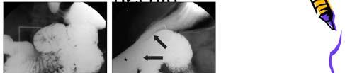
8 Embryonic anomaly A right paraduodenal hernia is formed when the prearterial limb fails to rotate around the superior mesenteric artery (SMA). The prearterial segment is the portion cephalic to the vitellomesenteric duct and comprises the distal duodenum and the entire small bowel to the distal ileum. Therefore, a portion of the small bowel remains to the right of the SMA. The result is a hernia orifice that is always to the right of the midline and usually faces medially and slightly downward. The mesentery of the ascending colon and a portion of the transverse colon make up the anterior wall of the sac, while the SMA and ileocolic artery lie in the free edge of the sac Case: A 41-year-old male patient, postprandial nausea, vomiting, abdominal pain and distension, which aggravated during the last 3 months, importantly affecting oral intake. CT scan an encapsulated cluster of small bowel loops occupying mainly the right upper quadrant, lateral to the duodenum, with the transverse colon located inferiorly. laparotomy; a large sac containing dilated small bowel loops Image OP finding A At abdominal inspection, a sac containing dilated small bowel loops lateral to the duodenum, displacing the colon inferiorly, was seen. B Dilated small bowel loops protruding through the fossa of Waldeyer. The superior mesenteric vessels are identified anteriorly. Thanks for your attension
A rare case of intestinal obstruction due to internal hernia. Dr. Jayanth 3 rd year PG Dept. Of General Surgery
 A rare case of intestinal obstruction due to internal hernia Dr. Jayanth 3 rd year PG Dept. Of General Surgery One of the common cause of acute abdomen May lead to high morbidity and mortality if not treated
Review of Internal Hernias: Radiographic and Clinical Findings
 Radiographic and Clinical Findings of Hernias Gastrointestinal Imaging Review C D E M N E U T R Y L I M C I G O F I N G Lucie C. Martin 1 Elmar M. Merkle William M. Thompson Martin LC, Merkle EM, Thompson
U Lecture Objectives. U Nordic Forum Trauma & Emergency Radiology. Bowel obstruction. U Bowel Obstruction: Etiologies
 Nordic Forum Trauma & Emergency Radiology Lecture Objectives Bowel Obstruction To illustrate the spectrum of acute obstruction of the small and the large bowel To explain how these bowel obstructions may
Nordic Forum - Trauma & Emergency Radiology. Bowel Obstruction: Imaging Update
 Nordic Forum - Trauma & Emergency Radiology Bowel Obstruction: Imaging Update Borut Marincek Institute of Diagnostic Radiology University Hospital Zurich, Switzerland Acute Abdomen Bowel Obstruction Bowel
The peritoneum. Prof. Oluwadiya KS, MBBS, FMCS(Orthop) Website:
 Website:") The peritoneum Prof. Oluwadiya KS, MBBS, FMCS(Orthop) Website: http://oluwadiya.com The peritoneum Serous membrane that lines the abdominopelvic cavity and invests the viscera The largest serous membrane
The peritoneum Prof. Oluwadiya KS, MBBS, FMCS(Orthop) Website: http://oluwadiya.com The peritoneum Serous membrane that lines the abdominopelvic cavity and invests the viscera The largest serous membrane
Case 72 y/o male Past hx : 1. Ampulla of Vater cancer s/p whipple operation 2. Liver abscess with K.P. 3. GI bleeding 4. DM No drug allergy
 Presenter : R2 周光緯 Supervisor :VS 連楚明 Case 72 y/o male Past hx : 1. Ampulla of Vater cancer s/p whipple operation 2. Liver abscess with K.P. 3. GI bleeding 4. DM No drug allergy 2012.08.15 2/45 ER visit
Perforation of a Duodenal Diverticulum. Elective Student S. C.
 Perforation of a Duodenal Diverticulum 2008 4 Elective Student S. C. Case History An elderly male presented to the Emergency Department with abdominal pain. Chief Complaint: Worsening, diffuse abdominal
Exploring Anatomy: the Human Abdomen
 Exploring Anatomy: the Human Abdomen PERITONEUM AND PERITONEAL CAVITY PERITONEUM The peritoneum is a thin serous membrane that lines the abdominal cavity and covers, in variable amounts, the viscera within
Christopher Lau Kings County Hospital SUNY Downstate Medical Center February 24, 2011
 Christopher Lau Kings County Hospital SUNY Downstate Medical Center February 24, 2011 37 year old male presented with 1 day history of abdominal pain Pain was diffuse but worst in the epigastric area No
Emergency presentation of hernias of the torso: What your surgeon wants to know.
 Emergency presentation of hernias of the torso: What your surgeon wants to know. Ken F Linnau, MD, MS Emergency Radiology UW Medicine Harborview Medical Center klinnau@uw.edu Nordic Forum 2017 Helsinki,
Development of pancreas and Small Intestine. ANATOMY DEPARTMENT DR.SANAA AL-AlSHAARAWY DR.ESSAM Eldin Salama
 Development of pancreas and Small Intestine ANATOMY DEPARTMENT DR.SANAA AL-AlSHAARAWY DR.ESSAM Eldin Salama OBJECTIVES At the end of the lecture, the students should be able to : Describe the development
General Data. 王 X 村 78 y/o 男性
 General Data 王 X 村 78 y/o 男性 Chief Complaint Vomiting twice this early morning Fever up to 38.9ºC was noted Present Illness (1) Old CVA with left side weakness for more than 10 years and with bed ridden
GI Grand Rounds. A A Lifetime of Abdominal Pain 12/9/2004 Tim Edwards
 GI Grand Rounds A A Lifetime of Abdominal Pain 12/9/2004 Tim Edwards PMH Aug 25, 1992 4 year old male presents to a pediatric gastroenterologist for primary complaint of anorexia, intermittent abdominal
Nasogastric tube. Stomach. Pylorus. Duodenum 1. Duodenum 2. Duodenum 3. Duodenum 4
 Esophagus Barium Swallow Stomach and Duodenum 4 year old Upper GI Nasogastric tube Stomach and Duodenum 4 year old Upper GI Nasogastric tube Stomach Pylorus Duodenum 1 Duodenum 2 Duodenum 3 Duodenum 4
Case Report Intestinal Malrotation: A Rare Cause of Small Intestinal Obstruction
 Case Reports in Surgery, Article ID 453128, 4 pages http://dx.doi.org/10.1155/2014/453128 Case Report Intestinal Malrotation: A Rare Cause of Small Intestinal Obstruction Mesut Sipahi, 1 Kasim Caglayan,
The abdominal Esophagus, Stomach and the Duodenum. Prof. Oluwadiya KS
 The abdominal Esophagus, Stomach and the Duodenum Prof. Oluwadiya KS www.oluwadiya.com Viscera of the abdomen Abdominal esophagus: Terminal part of the esophagus The stomach Intestines: Small and Large
Pathology of Intestinal Obstruction. Dr. M. Madhavan, MBBS., MD., MIAC, Professor of Pathology Saveetha Medical College
 Pathology of Intestinal Obstruction Dr. M. Madhavan, MBBS., MD., MIAC, Professor of Pathology Saveetha Medical College Pathology of Intestinal Obstruction Objectives list the causes of intestinal obstruction
Anatomy of the Large Intestine
 Large intestine Anatomy of the Large Intestine 2 Large Intestine Extends from ileocecal valve to anus Length = 1.5-2.5m = 5 feet Regions Cecum = 2.5-3 inch Appendix= 3-5 inch Colon Ascending= 5 inch Transverse=
Anatomy of the SMALL INTESTINE. Dr. Noman Ullah Wazir PMC
 Anatomy of the SMALL INTESTINE Dr. Noman Ullah Wazir PMC SMALL INTESTINE The small intestine, consists of the duodenum, jejunum, and illium. It extends from the pylorus to the ileocecal junction were the
Peritoneum: Def. : It is a thin serous membrane that lines the walls of the abdominal and pelvic cavities and clothes the viscera.
 Peritoneum: Def. : It is a thin serous membrane that lines the walls of the abdominal and pelvic cavities and clothes the viscera. Layers of the peritoneum: 1. Outer Layer ( Parietal Peritoneum) : lines
TRANSOMENTAL HERNIATION CAUSING ACUTE INTESTINAL OBSTRUCTION N. Suresh Kumar 1, Rahul Rai 2, P. Kulandai Velu 3
 TRANSOMENTAL HERNIATION CAUSING ACUTE INTESTINAL OBSTRUCTION N. Suresh Kumar 1, Rahul Rai 2, P. Kulandai Velu 3 HOW TO CITE THIS ARTICLE: N. Suresh Kumar, Rahul Rai, P. Kulandai Velu. Transomental Herniation
Lab Monitor Images Dissection of the Abdominal Vasculature + Lower Digestive System
 Lab Monitor Images Dissection of the Abdominal Vasculature + Lower Digestive System Stomach & Duodenum Frontal (AP) View Nasogastric tube 2 1 3 4 Stomach Pylorus Duodenum 1 Duodenum 2 Duodenum 3 Duodenum
Case Report Multi-slice computed tomography diagnosis of left paraduodenal hernia in an adult complicated by volvulus: a case report
 Int J Clin Exp Med 2016;9(11):22428-22433 www.ijcem.com /ISSN:1940-5901/IJCEM0036446 Case Report Multi-slice computed tomography diagnosis of left paraduodenal hernia in an adult complicated by volvulus:
Cecal Volvulus: Case Presentation and Review of CT Findings
 August 2011 Cecal Volvulus: Case Presentation and Review of CT Findings Omar Pardesi, Harvard Medical School Year III Our Patient LD: History & Physical HPI: 28 y.o. female presents with diffuse abdominal
Hirschprung s. Meconium plug R/S >1 R/S <1
 NEONATAL ABDOMINAL EMERGENCIES LOW OBSTRUCTION HIGH OBSTRUCTION INTESTINAL OBSTRUCTION High obstruction - proximal to mid-ileumileum Few dilated, air filled bowel loops Complete obstruction diagnosed by
Dr. Zahiri. In the name of God
 Dr. Zahiri In the name of God small intestine = small bowel is the part of the gastrointestinal tract Boundaries: Pylorus Ileosecal junction Function: digestion and absorption of food It receives bile
Embryology of the Midgut and Hind gut
 Embryology of the Midgut and Hind gut Prof. Abdulameer Al-Nuaimi E-mail: a.al-nuaimi@sheffield.ac.uk E-mail: abdulameerh@yahoo.com Abdominal organs www.google.co.uk/search? Development of Duodenum The
Fareed Khdair, MD Assistant Professor Chief, Section of Pediatric Gastroenterology, Hepatology, and Nutrition University of Jordan School of Medicine
 Fareed Khdair, MD Assistant Professor Chief, Section of Pediatric Gastroenterology, Hepatology, and Nutrition University of Jordan School of Medicine Outline Lecture one : Gut formation Foregut: esophagus,
Midgut. Over its entire length the midgut is supplied by the superior mesenteric artery
 Gi Embryology 3 Midgut the midgut is suspended from the dorsal abdominal wall by a short mesentery and communicates with the yolk sac by way of the vitelline duct or yolk stalk Over its entire length the
International Surgery Strangulated lesser omentum hiatus hernia: a rare case report and a literature review
 International Surgery Strangulated lesser omentum hiatus hernia: a rare case report and a literature review --Manuscript Draft-- Manuscript Number: Full Title: Article Type: Keywords: Corresponding Author:
An Interesting case of Retrocaecal internal herniation causing Small bowel Obstruction
 Accepted Manuscript An Interesting case of Retrocaecal internal herniation causing Small bowel Obstruction A.O. Rae, Core trainee,year 2 General Surgery, A. Kalyanaraman, SpR in General Surgery, A.E. Ward,
Home FAQ Archives ABP Topics NeoReviews.org My Bookmarks CME Information Help. Print this Page Add to my Bookmarks Page 3 of 10
 Welcome Kristin Ingstrup [ Logout ] SEARCH Home FAQ Archives ABP Topics NeoReviews.org My Bookmarks CME Information Help Overview Editorial Board My Learning Plan January February March May June July August
Request Card Task ANSWERS
 Request Card Task ANSWERS Medical Student Workbook Author: Dr Sam Leach, SpR Case 1 What differential diagnoses are most likely? Which investigation is most appropriate? Case 1 The most likely diagnosis
Key words: anomaly of intestine, reversed rotation, adult
 Key words: anomaly of intestine, reversed rotation, adult n92+ 4 n Fig. 1 Barium enema obstruction of the arrows) and mobile seen. (prone position, : Extrinsic transvers colon (between cecum (round arrow)
Visit ER at 10:43. Case conference. Past history. Present illlness. Physical examination. Impression
 Visit ER at 10:43 Case conference Supervisor: VS 吳柏衡 Presentor: R1 劉邦民 102.12.16 60 y/o male Chief complaint: syncope Triage: I T/P/R:33.2/81/18, BP=78/41mmHg, SpO2=92% Conscious: E4V5M6 Present illlness
Intestinal Obstruction Clinical Presentation & Causes
 Intestinal Obstruction Clinical Presentation & Causes V Chidambaram-Nathan Consultant Transplant and General Surgeon Sheffield Kidney Institute Northern General Hospital Intestinal Obstruction One of the
Development of the Digestive System. W.S. O The University of Hong Kong
 Development of the Digestive System W.S. O The University of Hong Kong Plan for the GI system Then GI system in the abdomen first develops as a tube suspended by dorsal and ventral mesenteries. Blood
Mousa Salah. Dr. Mohammad Al. Mohtasib. 1 P a g e
 8 Mousa Salah Dr. Mohammad Al. Mohtasib 1 P a g e In the previous lecture we talked about the peritoneum, and we said that the peritonium is a serous sac, and it consists of two layers, visceral and parietal.
Para-duodenal hernia: a report of five cases and review of literature
 Shadhu et al. BMC Surgery (2018) 18:32 https://doi.org/10.1186/s12893-018-0365-8 CASE REPORT Para-duodenal hernia: a report of five cases and review of literature Kamleshsingh Shadhu 1,2, Dadhija Ramlagun
Abdominal radiology 腹部放射線學
 Abdominal radiology 腹部放射線學 台北醫學大學 - 市立萬芳醫院 留偉順 laowilson@hotmail.com The Normal Abdominal Series Chest Supine abdomen Erect abdomen Left lateral decubitus abdomen Learning objectives Understanding normal
Introduction and Definitions
 Bowel obstruction Introduction and Definitions Accounts for 5% of all acute surgical admissions Patients are often extremely ill requiring prompt assessment, resuscitation and intensive monitoring Obstruction
د. عصام طارق. Objectives:
 GI anatomy Lecture: 5 د. عصام طارق Objectives: To describe anatomy of stomach, duodenum & pancreas. To list their main relations. To define their blood & nerve supply. To list their lymph drainage. To
The jejunum and the Ileum. Prof. Oluwadiya KS
 The jejunum and the Ileum Prof. Oluwadiya KS www.oluwadiya.siteled.com Introduction Introduction The small intestine (SI) comprises of the duodenum, jejunum and the ileum The jejunum is the second part
Volvulus of the Gastrointestinal Tract: x-ray and CT imaging
 Volvulus of the Gastrointestinal Tract: x-ray and CT imaging Poster No.: C-0076 Congress: ECR 2013 Type: Educational Exhibit Authors: E. Papadaki, S. Paschalidou, S. GIANNOU ; Rethymno, CR/ 1 2 2 3 1 3
The embryonic endoderm initially is widely connected with the yolk sac. As a consequence of cephalocaudal and lateral folding, a portion of the
 DIGESTIVE SYSTEM The embryonic endoderm initially is widely connected with the yolk sac. As a consequence of cephalocaudal and lateral folding, a portion of the endoderm-lined yolk sac cavity is incorporated
Internal Hernia After Gastric Bypass: Sensitivity and Specificity of Seven CT Signs with Surgical Correlation and Controls
 CT of Hernia After Gastric Bypass Abdominal Imaging Original Research Mark E. Lockhart 1 Franklin N. Tessler 1 Cheri L. Canon 1 J. Kevin Smith 1 Matthew C. Larrison 1 Naomi S. Fineberg 2 Brandon P. Roy
Lesser sac: Anatomy and non-neoplastic processes
 Lesser sac: Anatomy and non-neoplastic processes Poster No.: C-0027 Congress: ECR 2013 Type: Educational Exhibit Authors: E. Papadaki, R. Moschona, S. Paschalidou ; Rethymno, CR/ 1 2 2 3 1 3 GR, Rethymno/GR,
Patient Information. Age: 8 y/o Sex: Female. Date of Admission: Date of Discharge:
 Patient Information Age: 8 y/o Sex: Female Date of Admission: 92-10-08 Date of Discharge: 92-10-18 Chief Complaint Severe admominal pain and vomiting with dysuria since last afternoon Present Illness Lower
Objectives. Pediatric Mortality. Another belly pain. Gastroenteritis. Spewing & Pooing Child 4/18/16
 Gastro-tastrophies A Review of Pediatric GI Emergencies Objectives Discuss common presentations of Pediatric Abdominal Pain complaints Discuss work up and physical exam findings Discuss care, management
Gastrointestinal Pathology. August 2007
 Gastrointestinal Pathology August 2007 Case 1 Dysphagia and halitosis Case 1 Dilatation of the oesophagus with a smooth narrowing of its lower end. The large volume of contained fluid indicates delayed
ER-GS COMBINE CONFERENCE
 ER-GS COMBINE CONFERENCE 報告者 :R3 許力云指導者 :VS 連楚明 101.07.18 Patient Data 44 y/o, male E4V5M6 TPR: 37.1 /087/18 BP:141/075 mmhg SpO2: 96% 檢傷主訴 : 病患來診為腹痛 Triage: 2 History RUQ pain for 2 days Persistent and
A novel plain abdominal radiograph sign to diagnose malrotation with volvulus
 A novel plain abdominal radiograph sign to diagnose malrotation with volvulus Nataraja RM 1, Mahomed AA 1* 1. Department of Paediatric Surgery, Royal Alexandra Hospital for Sick Children, Brighton,UK *
-Ensherah Mokheemer. -Shatha Al-Jaberi محمد المحتسب- 1 P a g e
 9-9 -Ensherah Mokheemer -Shatha Al-Jaberi محمد المحتسب- 1 P a g e Small intestine has three regions: ( االثني عشر( The duodenum The jejunum The ileum Small intestine Duodenum: -c-shaped -The concavity
- Tamara Wahbeh. - Fareed Khdair. 0 P a g e
 -1 - Tamara Wahbeh - - Fareed Khdair 0 P a g e GI Embryology Note: I included everything in the records and slides; anything in the slide not included in this sheet was not mentioned by the doctor during
Jhia Anjela D. Rivera 1 1. BS Biology, Department of Biology, College of Science, Polytechnic University of the Philippines
 DIGESTIVE SYSTEM Jhia Anjela D. Rivera 1 1 BS Biology, Department of Biology, College of Science, Polytechnic University of the Philippines DIGESTIVE SYSTEM Consists of the digestive tract (gastrointestinal
Case Report Transmesenteric Internal Herniation Leading to Small Bowel Obstruction Postlaparoscopic Radical Nephrectomy
 Hindawi Case Reports in Surgery Volume 2017, Article ID 5128246, 4 pages https://doi.org/10.1155/2017/5128246 Case Report Transmesenteric Internal Herniation Leading to Small Bowel Obstruction Postlaparoscopic
-12. -Renad Habahbeh. -Dr Mohammad mohtasib
 -12 -Renad Habahbeh - -Dr Mohammad mohtasib The Gallbladder -The gallbladder has a body, a fundus (a rounded end), a neck, Hartmann s pouch before the neck and a cystic duct that meets the common hepatic
UNDERSTANDING X-RAYS: ABDOMINAL IMAGING THE ABDOMEN
 UNDERSTANDING X-RAYS: ABDOMINAL IMAGING THE ABDOMEN Radiology Enterprises radiologyenterprises@gmail.com www.radiologyenterprises.com STOMACH AND SMALL BOWEL STOMACH AND SMALL BOWEL Swallowed air is a
ABDOMEN - GI. Duodenum
 TALA SALEH ABDOMEN - GI Duodenum - Notice the shape of the duodenum, it looks like capital G shape tube which extends from the pyloroduodenal junction to the duodenojejunal junction. - It is 10 inches
Small Plicae Circularis. Short Closely packed together. Sparse, completely absent at distal part Lymphoid Nodule
 Intestines Differences Between Jejunum and Ileum Types Jejunum Ileum Color Deeper red Paler pink Calibre Bigger Smaller Thickness of wall Thick and Heavy Thin and Lighter Vascularity Highly vascularised
Documentation Dissection
 History of Present Illness: Documentation Dissection The patient is a 50-year-old male c/o symptoms for past 4 months 1, severe 2 bloating and stomach cramps, some nausea, vomiting, diarrhea. In last 3
Accessory Glands of Digestive System
 Accessory Glands of Digestive System The liver The liver is soft and pliable and occupies the upper part of the abdominal cavity just beneath the diaphragm. The greater part of the liver is situated under
Block 3: DISSECTION 2 CELIAC TRUNK, JEJUNUM/ILEUM, LARGE INTESTINE, DUODENUM, PANCREAS, PORTAL VEIN; MOBILIZATION OF THE LIVER
 1 Block 3: DISSECTION 2 CELIAC TRUNK, JEJUNUM/ILEUM, LARGE INTESTINE, DUODENUM, PANCREAS, PORTAL VEIN; MOBILIZATION OF THE LIVER Attempt to complete as much as you can of the dissection explained in the
In the name ofgod. Abdomen 3. Dr. Zahiri
 In the name ofgod Abdomen 3 Dr. Zahiri Peritoneum Peritoneum It is the serous membrane(a type of loose connective tissue and is covered by mesothelium) that lines the abdominal cavity. Extensions of the
The Whipple Operation Illustrations
 The Whipple Operation Illustrations Fig. 1. Illustration of the sixstep pancreaticoduodenectomy (Whipple operation) as described in a number of recent text books by Dr. Evans. The operation is divided
Bushra Arafa Zayed & Hanan Jamal. - Dana AF
 - 10 - Bushra Arafa Zayed & Hanan Jamal - Dana AF - Mohammad Al Muhtaseb Notes: This sheet was written in the same order as the slides, and everything in the slides is mentioned in this sheet. Pictures
Development of the Digestive System. W.S. O School of Biomedical Sciences, University of Hong Kong.
 Development of the Digestive System W.S. O School of Biomedical Sciences, University of Hong Kong. Organization of the GI tract: Foregut (abdominal part) supplied by coeliac trunk; derivatives include
Duodenum retroperitoneal
 Duodenum retroperitoneal C shaped Initial region out of stomach into small intestine RETROperitoneal viscus Superior 1 st part duodenal cap ; moves upwards and backwards to lie on the R crura medial to
Volvulus characterization in radiology: A review
 Volvulus characterization in radiology: A review Poster No.: C-1677 Congress: ECR 2010 Type: Topic: Educational Exhibit GI Tract Authors: C. Antunes, M. Seco, A. Canelas, C. Ruivo, C. Paulino, F. Cruz,
ASSESSING THE PLAIN ABDOMINAL RADIOGRAPH M A A M E F O S U A A M P O F O
 ASSESSING THE PLAIN ABDOMINAL RADIOGRAPH M A A M E F O S U A A M P O F O Introduction The abdomen (less formally called the belly, stomach, is that part of the body between the thorax (chest) and pelvis,
Anatomy: Know Your Abdomen
 Anatomy: Know Your Abdomen Glossary Abdomen - part of the body below the thorax (chest cavity); separated by the diaphragm. Anterior - towards the front of the body. For example, the umbilicus is anterior
Guidelines, Policies and Statements D5 Statement on Abdominal Scanning
 Guidelines, Policies and Statements D5 Statement on Abdominal Scanning Disclaimer and Copyright The ASUM Standards of Practice Board have made every effort to ensure that this Guideline/Policy/Statement
Adult bowel obstruction with acute abdomen: spectrum of CT findings
 Adult bowel obstruction with acute abdomen: spectrum of CT findings Poster No.: C-1571 Congress: ECR 2013 Type: Educational Exhibit Authors: L. Turturici, G. Gherarducci, F. Bianchi, R. Pascale, M. Tonerini,
Good morning! July 24, 2014
 Good morning! July 24, 2014 Prep #1 A 2-year-old boy presents to your office with a 2-day history of swelling of the right eye. He has been otherwise well. There are scattered insect bites on his body,
Acute Abdomen. Nirav Patel MD, FACS Banner University Medical Center - Phoenix
 Acute Abdomen Nirav Patel MD, FACS Banner University Medical Center - Phoenix ? Diffuse periumbilical with localization to RLQ + Nausea, anorexia, fevers - Diarrhea, emesis Exacerbated by movement, bumps
Gastrointestinal Tract. Anatomy of GI Tract. Anatomy of GI Tract. (Effective February 2007) (1%-5%)
 (1%-5%)") Gastrointestinal Tract (Effective February 2007) (1%-5%) Anatomy of GI Tract Esophagus bulls-eye or target EG junction seen on sagittal scan posterior to left lobe of liver and anterior to aorta Anatomy
Gastrointestinal Tract (Effective February 2007) (1%-5%) Anatomy of GI Tract Esophagus bulls-eye or target EG junction seen on sagittal scan posterior to left lobe of liver and anterior to aorta Anatomy
Basic Abdominal Sonography
 24S Basic Abdominal Sonography Procedural Overview JOHN FATCHETT II, RDMS is provided. Patient preparation (i.e., fasting) scanning techniques, spleen, transducer. evaluation of abdominal anatomy in the
Al-Mohtaseb. Saba Alfayoumi. Mo Alfarra
 8 Al-Mohtaseb Saba Alfayoumi Mo Alfarra For the comparison purposes refer to the last page where you can find a table that summarizes them. Enjoy Jejunum and Ileum -They're intraperitoneal and freely mobile
Case Presentation SIGMOID VOLVULUS
 Case Presentation SIGMOID VOLVULUS By, Dr. ANSARI SANA AFREEN 1 yr PG Dept. of General Surgery KIMS Narketpally Sathish a 18yr old male presented to the EMD on 10-06- 2015 COMPLAINTS AND DURATION: Pain
Management of Small Bowel Obstruction: An Update. Case Presentation
 Management of Small Bowel Obstruction: An Update The Postgraduate Course in General Surgery March 20-23, 2011 Jonathan Carter, MD Assistant Professor of Surgery Case Presentation 67 year old otherwise
3/21/2011. Case Presentation. Management of Small Bowel Obstruction: An Update. CT abdomen and pelvis. Abdominal plain films
 Case Presentation 67 year old otherwise healthy woman presents to the ED with a chief complaint of abdominal pain, nausea and vomiting for five days. Management of Small Bowel Obstruction: An Update The
Abdominal Pain. Luke Donnelly, MD Emergency Medicine
 Abdominal Pain Luke Donnelly, MD Emergency Medicine Objectives Approach to abdominal pain Evaluation Critical diagnoses and treatments Abdominal Pain Most Common ER Complaint Broad Differential Can often
Plain abdomen The standard films are supine & erect AP views (alternative to erect, lateral decubitus film is used in ill patients).
.") Plain abdomen The standard films are supine & erect AP views (alternative to erect, lateral decubitus film is used in ill patients). The stomach can be readily identified by its location, gastric rugae
Plain abdomen The standard films are supine & erect AP views (alternative to erect, lateral decubitus film is used in ill patients). The stomach can be readily identified by its location, gastric rugae
1 Right & left Hepatic ducts Gastric Impression of spleen
 Pancreatic Model 1 Right & left Hepatic ducts 14 Gastric Impression of spleen 2 Common hepatic duct 15 Renal Impression of spleen 3 Cystic Duct 16 Colic Impression of spleen 4 Common Bile Duct 17 Splenic
The Foregut. At first the esophagus is short. but with descent of the heart and lungs it lengthens rapidly
 GI embryology 2 The Foregut At first the esophagus is short but with descent of the heart and lungs it lengthens rapidly The muscular coat, which is formed by surrounding splanchnic mesenchyme, is striated
Congenital Internal Hernia Presented with Life Threatening Extensive Small Bowel Strangulation
 pissn: 2234-8646 eissn: 2234-8840 http://dx.doi.org/10.5223/pghn.2013.16.3.190 Pediatric Gastroenterology, Hepatology & Nutrition 2013 September 16(3):190-194 Case Report PGHN Congenital Internal Hernia
Adults intestinal malrotations: a simple approach to recognize the most frequent types with MDCT
 Adults intestinal malrotations: a simple approach to recognize the most frequent types with MDCT Poster No.: C-2484 Congress: ECR 2012 Type: Educational Exhibit Authors: F. Macrì, R. Argirò, B. SACCONI,
CLINICAL PRESENTATION AND RADIOLOGY QUIZ QUESTION
 Donald L. Renfrew, MD Radiology Associates of the Fox Valley, 333 N. Commercial Street, Suite 100, Neenah, WI 54956 6/23/2012 Radiology Quiz of the Week # 78 Page 1 CLINICAL PRESENTATION AND RADIOLOGY
STRUCTURAL BASIS OF MEDICAL PRACTICE EXAMINATION 3. October 17, 2014
 STRUCTURAL BASIS OF MEDICAL PRACTICE EXAMINATION 3 October 17, 2014 PART l. Answer in the space provided. (12 pts) 1. Identify the structures. (2 pts) A. B. A B C. D. C D 2. Identify the structures. (2
STRUCTURAL BASIS OF MEDICAL PRACTICE EXAMINATION 3. October 16, 2015
 STRUCTURAL BASIS OF MEDICAL PRACTICE EXAMINATION 3 October 16, 2015 PART l. Answer in the space provided. (12 pts) 1. Identify the structures. (2 pts) A. B. A B C. D. C D 2. Identify the structures. (2
A Frame of Reference for Anatomical Study. Anatomy and Physiology Mr. Knowles Chapter 1 Liberty Senior High School
 A Frame of Reference for Anatomical Study Anatomy and Physiology Mr. Knowles Chapter 1 Liberty Senior High School Anatomical Terms of Direction and Position Created for communicating the direction and
Sonographic Appearances of Common Gut Pathology in Paediatric Patients: Comparison with Plain Abdominal Radiography
 3668 Radiographer Text 1/4/04 2:57 PM Page 11 The Radiographer vol. 51: 11-17 Sonographic Appearances of Common Gut Pathology in Paediatric Patients: Comparison with Plain Abdominal Radiography Lino Piotto
Preview from Notesale.co.uk Page 1 of 34
 Abdominal viscera and digestive tract Digestive tract Abdominal viscera comprise majority of the alimentary system o Terminal oesophagus, stomach, pancreas, spleen, liver, gallbladder, kidneys, suprarenal
Title small intestine: a fatal case invol. Author(s) Munetaka; Tsuruyama, Tatsuaki; Tama. Citation Forensic science international (201
 Munetaka; Tsuruyama, Tatsuaki; Tama. Citation Forensic science international (201") Title Transmesenteric hernia due to doubl small intestine: a fatal case invol Author(s) Kakimoto, Yu; Abiru, Hitoshi; Kotan Munetaka; Tsuruyama, Tatsuaki; Tama Citation Forensic science international (201
Title Transmesenteric hernia due to doubl small intestine: a fatal case invol Author(s) Kakimoto, Yu; Abiru, Hitoshi; Kotan Munetaka; Tsuruyama, Tatsuaki; Tama Citation Forensic science international (201
Intestinal Malrotation
 Intestinal Malrotation Poster No.: C-1368 Congress: ECR 2015 Type: Educational Exhibit Authors: K. Carpentier, B. De Foer, F. Deckers, P. Leyman, M. 1 1 1 2 1 1 1 2 Pouillon, P. M. Parizel ; Wilrijk/BE,
Left Paraduodenal Hernia Successfully Treated with Laparoscopic Surgery: A Case Report
 71 Left Paraduodenal Hernia Successfully Treated with Laparoscopic Surgery: A Case Report Tuyoshi Shoji Raisuke Nishiyama Koji Oba Masaki Azuma Department of Surgery, Haibara General Hospital, Makinohara
Urinary System VASTACCESS, INC.
 Urinary System www.vastaccess.com 2 Urinary Tract Kidney Ureter Urinary Bladder Urethra Prostate (male) Membranous (male) Spongy (male) 3 Kidney Relations Suprarenal (Adrenal) Glands Liver Duodenum Transverse
ACUTE ABDOMEN IN OLDER CHILDREN. Carlos J. Sivit M.D.
 ACUTE ABDOMEN IN OLDER CHILDREN Carlos J. Sivit M.D. ACUTE ABDOMEN Clinical condition characterized by severe abdominal pain developing over several hours ACUTE ABDOMINAL PAIN Common childhood complaint
Consecutive, Bilateral Obturator Hernia in a Single Case HO Aydın¹, EHA Soy¹, T Avcı¹, T Tezcaner¹, S Yıldırım ABSTRACT
 Consecutive, Bilateral Obturator Hernia in a Single Case HO Aydın¹, EHA Soy¹, T Avcı¹, T Tezcaner¹, S Yıldırım ABSTRACT Obturator hernia (OH) is a rare pelvic hernia. It is diffucult to make an early diagnosis