Role of modern imaging methods in maxillofacial diagnostics
|
|
|
- Jocelyn Dorsey
- 5 years ago
- Views:
Transcription

1 SEMMELWEIS EGYETEM Department of Radiology Role of modern imaging methods in maxillofacial diagnostics Department of Diagnostic Imaging Semmelweis University, Budapest Diagnosztikus képalkotó eljárások anatómiája FOK V. 09/04/2018.


2 50 years old female 3 days ago accidentally was injected with disinfectant instead of local anesthetics, which caused tissue necrosis swollen, painful face subfebrile erythema on the neck
3 Which diagnostic method would help here??? MR vizsgálat
4 Questions that could be considered? Extent of inflammation? State of the organs in the region? Crepitation- fluctuation? Abscess formation? Most important: is there any Abscess? If so = surgery
5 Specific examinations Definite question calls for a straight answer Step 1: Ultrasound examination of he neck Step 2: crossectional imaging method non contrast and contrast enhanced CT Abscess?


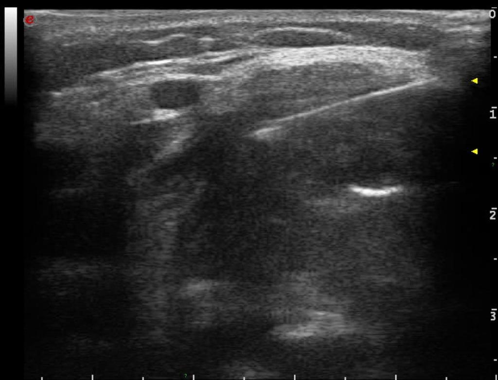
6 US


7 CT examination Phlegmone (cellulitis). No abscess.
8 Diagnostioc Imaging methods Ultrasound Conventional X-ray (+ fluoroscopy) Non-contrast Contrast enhanced CT (Multislice, cone-beam) / MR Angiography DSA CT-, MR-Angiography Molecular medicine scintigraphy SPECT (single photon emission computed tomography) PET (positron emission tomography), PET-CT
9 US Image is based on soundwave reflections Bones and gases cannot be examined solid cystic DD -excellent Lower depth higher resolution Flow related measurements (Doppler) Direction, speed stenosis, vascularization inflammation, tumor biopsy guidance (FNAB, core)
10 US Indications face Floor of the mouth Superficial layers of the neck Above bones Main lymph node regions Preparation Jewelry needs to be taken off Bandage removal tracheostomy line if possible removed Consent in case of contrast enhanced examinations or biopsies

11 Extraoral


12


13 Bucca


14 Submandibular region

15 Floor of the mouth


16 Thyroid left lobe


17 Lgl


18


19

20 20
21 Elastography Qunatitatiove measurement of tissue characteristics: Inner stimulus or external compression the tissues move Elastic vs. Tough tissue Tumors are tougher: less elastic Color code
22 Color Tissue Greyscale Soft, elastic tough

23

24


25
26 Conventional X-ray indications: panoramic X-ray and dental X-ray less valuable since CT: face bones, orbit, temporal bone, sinuses Preparation


27 Dental
28 Conventional X-ray w/ contrast Swallow Sialography (sialolithiasis) Still valuable
29 Conventional X-ray Contrast enhanced Indications FB -obstruction Perforation diverticulum Sclerotic lesions tumor stenosis Preparations jewelry empty stomach cooperation


30 sialography Swallow x-ray


31 nyelés rtg

32 CT concepts


33 CT concepts DVT digital volumenography CBCT cone beam CT MSCT multislice CT DSCT dual source CT MDCT Multidetector row CT HRCT high-resolution CT

34 Non-contrast and contrast enhanced CT Uses X-ray NC and CE + multiple rows of detectors CTA option Better soft tissue resolution than X-ray, but lower spatial resolution /512x512 px/ High dose of ionizing radiation
")

35 Non-contrast and contrast enhanced CT Indications tumor TNM Deep spaces of the HN region Bones (fractures, paranasal sinuses) Suprahyoid regions - MR is more useful Inrahyoid neck very good; Preparation Remove metal pieces Consent Kidney function iv. contrast DM (metformin)


36


37
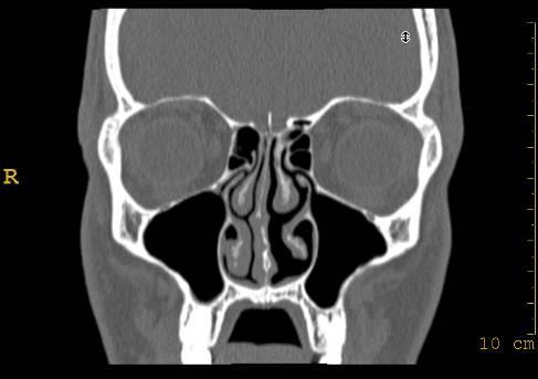
38 Face CT Helical MDCT Resolution High Collimation 16 x 0.75 Pitch Rot FOV Filter 200 / 150/ 100 mm D (bone) kv 120 mas 250 Sliceth/ rec WC; WW 2mm/ 1mm 200/ 2000 HU matrix 512


39 MDCT primary planes coronal axial

40 coronal


41 Axial Soft tissue window coronal MPR


42


43 natív CT postprocessing: MPR = multiplanáris rekonstrukció sagittalis coronalis


44 Submandibular gl. tumor


45


46 sinonasal polyposis


47 MD- vs. CBCT

48

49 CBCT NewTom 3G / i-cat / ILUMA/ ProMax 3D CB MercuRay / Dental CBCT / 3D Accuitomo / Sirona Gelileos


50


51 kv mas CTDIvol (mgy) Face MDCT Face CBCT MDCT CBCT


52 MDCT CBCT Significantly lower radiation max 100 μsv ( MDCT kb μsv ) Panoramic X-ray μsv (daily background radiation 8 μsv)
53
54 Advantages- Disadvantages MDCT CBCT Tissue contrast Excellent Moderate Density measurement (HU) precise Not reliable Windowing Based on HU Brightness and contrast FOV changeable Standard Ionising radiation High (min. 10x) low Voxel Isovolumetric 0.625mm Isovolumetric 0.125mm Duration of the examination Irrelevant (10-30sec) Sensitive to motion (<40sec) Klausztrofóbia Not likely (vs MR) No Space needed Larger Smaller Metal artefacts stronger weaker Expences High low Covered by health insurance yes no
55 CBCT clinical indications implantiology oro- and maxillofacialis diseases TMJ Surgical planning fogszabályozás és orthognathia impactált fogak DMF

56


57

58

59


60


61

62
63 Non contast and contrast enhanced MR Maps the Hydrogen atoms of tissues best soft tissue resolution No ionizing radiation More expensive, less available Longer examination time (30-50 min) Problematic next to metallic objects, bones harder to assess
64 Non contrast and contrast enhanced MR Indications Suprahyoid neck Skull base (foramens) Tumor extent (scar tissue recurring tumor) Preparation Metallic objects removed Consent Contraindicated metallic prosthetics, pacemaker claustrophobia


65 MR T2W MR T1W MR T2W


66


67 US MR T2W MR T2W
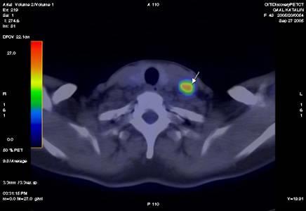
68 DSA Digital X-ray + subtraction High radiation, semi-invasive Regional vascular structure Problems are usually related to contrast agents
69 Digitális szubtrakciós angiográfia Indications Vascular structure of tumors possible interventions (RFA-, chemoembolisation, chemoablation, embolsation, paraganglioma glomus tumors Preparation Consent empty stomach Sterile environment


70


71 Molecular imaging Poor morphologic resolution Good temporal / functional res. Tc isotope Gamma camera (thyroid) SPECT single photon emission CT FDG-PET F18 glucose positron. Tumor metastasis, primary malignancy or inflammation PET-CT fused images - better morphology


72 Ectopic parthyroid MIBI 5 perc MIBI 120 perc Kontroll célzott UH Botos anita pertechnetát SPECT sagittal 21 éves nő, PTH: 58 pg/ml (!), seca: 2,85 mmol/l adenoma


73


74


75 tu. rad. linguae + met lgg. colli


76 FDG-PET: metastasis
Head-and-Neck Radiology
 Head-and-Neck Radiology Péter Magyar MD. Diagnosztikus képalkotó Lecture eljárások anatómiája 4th year 2007. Semmelweis University Department of Diagnostic Radiology and Oncology Guide-line Parts and borders
Computed tomography. Department of Radiology, University Medical School, Szeged
 Computed tomography Department of Radiology, University Medical School, Szeged voxel +1-4 +2 +5 +3 +1 0-2 pixel -2 0 +1-4 -6 +5 +2 +1 Department of Radiology, University Medical School, Szeged
X-Ray & CT Physics / Clinical CT
 Computed Tomography-Basic Principles and Good Practice X-Ray & CT Physics / Clinical CT INSTRUCTORS: Dane Franklin, MBA, RT (R) (CT) Office hours will be Tuesdays from 5pm to 6pm CLASSROOM: TIME: REQUIRED
Cardiac Imaging Tests
 Cardiac Imaging Tests http://www.medpagetoday.com/upload/2010/11/15/23347.jpg Standard imaging tests include echocardiography, chest x-ray, CT, MRI, and various radionuclide techniques. Standard CT and
Combined Anatomical and Functional Imaging with Revolution * CT
 GE Healthcare Case studies Combined Anatomical and Functional Imaging with Revolution * CT Jean-Louis Sablayrolles, M.D. Centre Cardiologique du Nord, Saint-Denis, France Case 1 Whole Brain Perfusion and
Medical imaging X-ray, CT, MRI, scintigraphy, SPECT, PET Györgyi Műzes
 Medical imaging X-ray, CT, MRI, scintigraphy, SPECT, PET Györgyi Műzes Semmelweis University, 2nd Dept. of Medicine Medical imaging: definition technical process of creating visual representations about
Imaging Work-Up of a Neck Mass - Adults & Children
 Disclosures Imaging Work-Up of a Neck Mass - Adults & Children I have nothing to disclose Christine M Glastonbury MBBS Professor of Radiology & Biomedical Imaging Otolaryngology-Head & Neck Surgery and
2
 1 2 3 4 5 6 7 8 9 10 11 12 13 Cine loop of tomosynthesis slice images through the chest. 14 Standard PA chest radiograph (left) and single slice from the tomosynthesis image dataset (right) of a patient
CNS Imaging. Dr Amir Monir, MD. Lecturer of radiodiagnosis.
 CNS Imaging Dr Amir Monir, MD Lecturer of radiodiagnosis www.dramir.net Types of radiological examinations you know Plain X ray X ray with contrast GIT : barium (swallow, meal, follow through, enema) ERCP
PARATHYROID IMAGING. James Lee, MD Chief, Endocrine Surgery Co-Director NY Thyroid-Parathyroid Center Columbia University Medical Center
 PARATHYROID IMAGING James Lee, MD Chief, Endocrine Surgery Co-Director NY Thyroid-Parathyroid Center Columbia University Medical Center NO DISCLOSURES Overview The hallmarks of the ideal test Benefits
Practical CT and MRI Anthony J. Fischetti, DVM, MS, DACVR Department Head of Diagnostic Imaging The Animal Medical Center, New York OBJECTIVE:
 Practical CT and MRI Anthony J. Fischetti, DVM, MS, DACVR Department Head of Diagnostic Imaging The Animal Medical Center, New York OBJECTIVE: This lecture describes the most common indications for referred
PULMONARY EMBOLISM ANGIOCT (CTA) ASSESSMENT OF VASCULAR OCCLUSION EXTENT AND LOCALIZATION OF EMBOLI 1. BACKGROUND
 ASSESSMENT OF VASCULAR OCCLUSION EXTENT AND LOCALIZATION OF EMBOLI 1. BACKGROUND") JOURNAL OF MEDICAL INFORMATICS & TECHNOLOGIES Vol. 11/2007, ISSN 1642-6037 Damian PTAK * pulmonary embolism, AngioCT, postprocessing techniques, Mastora score PULMONARY EMBOLISM ANGIOCT (CTA) ASSESSMENT
JOURNAL OF MEDICAL INFORMATICS & TECHNOLOGIES Vol. 11/2007, ISSN 1642-6037 Damian PTAK * pulmonary embolism, AngioCT, postprocessing techniques, Mastora score PULMONARY EMBOLISM ANGIOCT (CTA) ASSESSMENT
Introduction to Radiology
 Introduction - Lecture 1 436 Teams Introduction to Radiology Objectives Introduce the various Medical Imaging Modalities. Understand the basics of image generation. Relate imaging to gross anatomy. Appreciate
Nuclear Medicine in the Diabetic Foot
 26.11.2015, Uniklinik Balgrist Nuclear Medicine in the Diabetic Foot Martin Hüllner Nuklearmedizin und Neuroradiologie, USZ / UZH Outline A. Imaging modalities brief technical overview B. Nuclear medicine
declipse SPECT Imaging Probe Worldwide first registration-free ultrasound fusion with high-resolution 3D SPECT images
 declipse SPECT Imaging Probe Worldwide first registration-free ultrasound fusion with high-resolution 3D SPECT images high-resolution 3D SPECT live ultrasound realtime fusion of SPECT / ultrasound Hybrid
Bone PET/MRI : Diagnostic yield in bone metastases and malignant primitive bone tumors
 Bone PET/MRI : Diagnostic yield in bone metastases and malignant primitive bone tumors Lars Stegger, Benjamin Noto Department of Nuclear Medicine University Hospital Münster, Germany Content From PET to
The Role of. 54 MARCH 2016 // dentaltown.com. radiology feature. by Deepak Gupta, MDS
 The Role of Imaging by Deepak Gupta, MDS Dr. Deepak Gupta completed his post graduation in oral medicine and maxillofacial radiology in 2011. He is currently an Associate Professor/ Reader in the Department
HEALTHFIRST 2011 RADIOLOGY PROGRAM CODE LIST
 HEALTHFIRST 2011 RADIOLOGY PROGRAM CODE LIST Outpatient Radiology utilization call Carecore at 1-877-773-6964 Modality CPT CODE Description CT SCANS 70450 CT HEAD/BRAIN W/O CONTRAST CT SCANS 70460 CT HEAD/BRAIN
Radiology Codes Requiring Authorization*
 70336 Magnetic resonance (eg, proton) imaging, temporomandibular joint(s) 70450 Computed tomography, head or brain; without contrast material 70460 Computed tomography, head or brain; with contrast material(s)
Radiologic Imaging Magnetic Resonance Imaging (MRI)
") Radiologic Imaging X-ray has always been the golden rule in diagnosing and treating podiatric patients. Unfortunately, for some patients the diagnosis is not as evident. That is when we need to utilize
Radiologic Imaging X-ray has always been the golden rule in diagnosing and treating podiatric patients. Unfortunately, for some patients the diagnosis is not as evident. That is when we need to utilize
COMENIUS-Project: SM&CLIL Radiation & Medicine
 Medical imaging refers to the techniques and processes used to create images of the human body (or parts thereof) for clinical purposes. Thanks to modern mathematics and computer technology, medical imaging
Cigna - Prior Authorization Procedure List: Radiology & Cardiology
 Cigna - Prior Authorization Procedure List: Radiology & Cardiology Category CPT Code CPT Code Description 93451 Right heart catheterization 93452 Left heart catheterization 93453 Combined right and left
PARATHYROID NUCLEAR MEDICINE IMAGING REVIEW DISCLOSURES
 PARATHYROID NUCLEAR MEDICINE IMAGING REVIEW Miguel Hernandez Pampaloni, M.D., Ph.D. Chief, Nuclear Medicine Assistant Professor of Radiology UCSF Department of Radiology and Biomedical Imaging DISCLOSURES
High Tech Imaging Quick Reference Guide
 High Tech Imaging Quick Reference Guide 1 High Tech Imaging Authorizations may now be requested through our secure provider portal, BlueAccess. Getting Started Step 1: Log into BlueAccess from www.bcbst.com
HIP RADIOLOGY PROGRAM CODE LISTS
 EFFECTIVE OCTOBER 1, 2012 70336 MAGNETIC RESONANCE IMAGING TMJ 70450 COMPUTED TOMOGRAPHY HEAD/BRAIN WITHOUT 70460 COMPUTED TOMOGRAPHY HEAD/BRAIN WITH 70470 COMPUTED TOMOGRAPHY HEAD/BRAIN WITHOUT AND WITH
Outline. Biological Psychology: Research Methods. Dr. Katherine Mickley Steinmetz
 Biological Psychology: Research Methods Dr. Katherine Mickley Steinmetz Outline Neuroscience Methods Histology Electrophysiological Recordings Lesion Neuroimaging Neuroanatomy Histology: Brain structure
POSITRON EMISSION TOMOGRAPHY (PET)
") Status Active Medical and Behavioral Health Policy Section: Radiology Policy Number: V-27 Effective Date: 08/27/2014 Blue Cross and Blue Shield of Minnesota medical policies do not imply that members should
Status Active Medical and Behavioral Health Policy Section: Radiology Policy Number: V-27 Effective Date: 08/27/2014 Blue Cross and Blue Shield of Minnesota medical policies do not imply that members should
Imaging in gastric cancer
 Imaging in gastric cancer Gastric cancer remains a deadly disease because of late diagnosis. Adenocarcinoma represents 90% of malignant tumors. Diagnosis is based on endoscopic examination with biopsies.
PET IMAGING (POSITRON EMISSION TOMOGRAPY) FACT SHEET
 FACT SHEET") Positron Emission Tomography (PET) When calling Anthem (1-800-533-1120) or using the Point of Care authorization system for a Health Service Review, the following clinical information may be needed to
Positron Emission Tomography (PET) When calling Anthem (1-800-533-1120) or using the Point of Care authorization system for a Health Service Review, the following clinical information may be needed to
Lung Perfusion Analysis New Pathways in Lung Imaging. Case Study Brochure PLA 309 Hospital
 Lung Perfusion Analysis New Pathways in Lung Imaging Case Study Brochure PLA 309 Hospital http://www.toshibamedicalsystems.com Toshiba Medical Systems Corporation 2012 all rights reserved. Design and specifications
Applicable Neuroradiology
 For the Clinical Neurology Clerkship LSU Medical School New Orleans Amy W Voigt, MD Clerkship Director Introduction The field of Radiology first developed following the discovery of X-Rays by Wilhelm Roentgen
General Imaging. Imaging modalities. Incremental CT. Multislice CT Multislice CT [ MDCT ]
![General Imaging. Imaging modalities. Incremental CT. Multislice CT Multislice CT [ MDCT ]](/thumbs/76/74079340.jpg "General Imaging. Imaging modalities. Incremental CT. Multislice CT Multislice CT [ MDCT ]") General Imaging Imaging modalities Conventional X-rays Ultrasonography [ US ] Computed tomography [ CT ] Radionuclide imaging Magnetic resonance imaging [ MRI ] Angiography conventional, CT,MRI Interventional
General Imaging Imaging modalities Conventional X-rays Ultrasonography [ US ] Computed tomography [ CT ] Radionuclide imaging Magnetic resonance imaging [ MRI ] Angiography conventional, CT,MRI Interventional
Imaging of the Paranasal Sinuses
 14. Sommerschule Imaging of the Paranasal Sinuses Bettlach 24.08.2018 Christoph Schlegel Conventional Radiology NNH-Status: okzipito-frontal: frontal sinus, anterior ethmoid okzipito-nasal : maxillary
CT of the Head, Spine, and Cerebral Vessels
 CT of the Head, Spine, and Cerebral Vessels Objectives Determine specific imaging plane used to acquire or reformat CT scan, i.e. sagittal, coronal, transverse, and offaxis or oblique. Assess and evaluate
HONG KONG COLLEGE OF RADIOLOGISTS. Higher Training (Radiology) Subspecialty Training in Computed Tomography
 Subspecialty Training in Computed Tomography") HONG KONG COLLEGE OF RADIOLOGISTS Higher Training (Radiology) Subspecialty Training in Computed Tomography [The following guidelines should be read in conjunction with the General Guidelines on Higher
HONG KONG COLLEGE OF RADIOLOGISTS Higher Training (Radiology) Subspecialty Training in Computed Tomography [The following guidelines should be read in conjunction with the General Guidelines on Higher
LYMPHATIC DRAINAGE IN THE HEAD & NECK
 LYMPHATIC DRAINAGE IN THE HEAD & NECK Like other parts of the body, the head and neck contains lymph nodes (commonly called glands). Which form part of the overall Lymphatic Drainage system of the body.
Advances in Head and Neck Imaging
 Advances in Head and Neck Imaging Tao Ouyang, MD a, Barton F. Branstetter IV, MD b, * KEYWORDS Computed tomography Magnetic resonance imaging Positron emission tomography Radiology in dental and craniomaxillofacial
I AM DEMANDING Type CMOS Flat Panel CMOS CMOS ø 40 x 40 mm, ø 60 x 60 mm, ø 80 x 80 mm, ø 110 x 80 mm
 TECHNICAL SPECIFICATIONS 1168 1501 1978 1237 1551-2351 Ø 1090 PANORAMIC CBCT CEPHALOMETRIC X-RAY SOURCE Tube type High frequency DC generator 2.8 mmal / 85 kv 7.0 mmal / 90 kv 2.8 mmal / 85 kv Operation
FOR CMS (MEDICARE) MEMBERS ONLY NATIONAL COVERAGE DETERMINATION (NCD) FOR MAGNETIC RESONANCE IMAGING:
 MEMBERS ONLY NATIONAL COVERAGE DETERMINATION (NCD) FOR MAGNETIC RESONANCE IMAGING:") National Imaging Associates, Inc. Clinical guidelines BONE MARROW MRI Original Date: July 2008 Page 1 of 5 CPT Codes: 77084 Last Review Date: September 2014 NCD 220.2 MRI Last Effective Date: July 2011
National Imaging Associates, Inc. Clinical guidelines BONE MARROW MRI Original Date: July 2008 Page 1 of 5 CPT Codes: 77084 Last Review Date: September 2014 NCD 220.2 MRI Last Effective Date: July 2011
Course specification
 Al-Azhar University Faculty of Medicine for Men Course specification For Master of Radiodiagnosis ( 2014 2015 ) University : Al-Azhar Faculty : Medicine for men Course specification - Programmers on which
3D Cone beam CT & Digital Radiography Dedicated to Otorhinolaryngology
 3D Cone beam CT & Digital Radiography Dedicated to Otorhinolaryngology Multi-functional imaging solution3 RAYSCAN m is an unique 2-in-1 imaging solution, combining Cone Beam CT and Digital Radiography,
PET-MRI in malignant bone tumours. Lars Stegger Department of Nuclear Medicine University Hospital Münster, Germany
 PET-MRI in malignant bone tumours Lars Stegger Department of Nuclear Medicine University Hospital Münster, Germany Content From PET to PET/MRI General considerations Bone metastases Primary bone tumours
MODAL QUESTION PAPER X-RAYTECHNIC1AN X-RAY EQUIPMENT & RADIATION PHYSIC S 1 ST YEAR PAPER I Time: 3 Hours Max. Marks: 50.
 X-RAY EQUIPMENT & RADIATION PHYSIC S 1 ST YEAR PAPER I 1. Define Electric Current and give its Sl Units? 2. Define the Unit ROENTGEN? 3. Mention the advantage of Grid s? 4. Name the various types of Transformers?
Nuclear Medicine and PET. D. J. McMahon rev cewood
 Nuclear Medicine and PET D. J. McMahon 150504 rev cewood 2018-02-15 Key Points Nuclear Medicine and PET: Imaging: Understand how Nuc Med & PET differ from Radiography & CT by the source of radiation. Be
Low Dose Molecular Breast Imaging
 Low Dose Molecular Breast Imaging Dr. M.K. O Connor Conflict of Interest Royalties - Gamma Medica Research funding GE Healthcare Research support MTTI Michael O Connor, Ph.D Dept. of Radiology Mayo Clinic
General Cardiovascular Magnetic Resonance Imaging
 2 General Cardiovascular Magnetic Resonance Imaging 19 Peter G. Danias, Cardiovascular MRI: 150 Multiple-Choice Questions and Answers Humana Press 2008 20 Cardiovascular MRI: 150 Multiple-Choice Questions
Cone-Beam CT for MSK Extremities
 8/6/0 Diagnostic Image Quality Evaluation of an Extremity Cone-Beam CT Scanner: Pre-Clinical and First Clinical Results Abdullah Muhit Wojciech Zbijewski, J Webster Stayman John Yorkston, Nathan Packard,
NEURO PROTOCOLS MRI NEURO PROTOCOLS (SIEMENS SCANNERS)
") Page 1 NEURO PROTOCOLS Brain Stroke Brain Brain with contrast Brain for seizures Brain for MS Brain for Pineal gland Sella FAST Scan for hydrocephalus MRA/MRV Brain MRA carotids 8 th nerve Cranial nerves
Page 1 NEURO PROTOCOLS Brain Stroke Brain Brain with contrast Brain for seizures Brain for MS Brain for Pineal gland Sella FAST Scan for hydrocephalus MRA/MRV Brain MRA carotids 8 th nerve Cranial nerves
THE TUFFEST STUFF CT REGISTRY REVIEW Live Lecture Seminar SATURDAY CURRICULUM
 1. The CT Imaging Chain-10 major components & their functions a. The x-ray tube b. Generator c. Filter d. Pre-patient collimator e. Pre-detector collimator f. Detector system g. Analog to digital converter
STRUCTURED EDUCATION REQUIREMENTS EFFECTIVE: JANUARY 1, 2016
 Computed Tomography The purpose of structured education is to provide the opportunity for individuals to develop mastery of discipline-specific knowledge that, when coupled with selected clinical experiences,
FOR CMS (MEDICARE) MEMBERS ONLY NATIONAL COVERAGE DETERMINATION (NCD) FOR MAGNETIC RESONANCE IMAGING:
 MEMBERS ONLY NATIONAL COVERAGE DETERMINATION (NCD) FOR MAGNETIC RESONANCE IMAGING:") National Imaging Associates, Inc. Clinical guidelines SINUS MRI Original Date: November 2007 Page 1 of 5 CPT Codes: 70540, 70542, 70543 Last Review Date: July 2014 NCD 220.2 MRI Last Effective Date: July
National Imaging Associates, Inc. Clinical guidelines SINUS MRI Original Date: November 2007 Page 1 of 5 CPT Codes: 70540, 70542, 70543 Last Review Date: July 2014 NCD 220.2 MRI Last Effective Date: July
AMERICAN IMAGING MANAGEMENT
 2012 CPT Codes Computerized Tomography (CT) CPT Description Abdomen 74150 CT abdomen; w/o 74160 CT abdomen; with 74170 CT abdomen; w/o followed by Chest 71250 CT thorax; w/o 71260 CT thorax; with 71270
AMERICAN IMAGING MANAGEMENT
 2010 BCBS of Georgia CPT Codes With Grouper Numbers Computerized Tomography (CT) CPT Description Abdomen 74150 CT abdomen; w/o contrast 6 74160 CT abdomen; with contrast 74170 CT abdomen; w/o contrast
SPECIFIC PRINCIPLES FOR DOSE REDUCTION IN HEAD CT IMAGING. Rajiv Gupta, MD, PhD Neuroradiology, Massachusetts General Hospital Harvard Medical School
 SPECIFIC PRINCIPLES FOR DOSE REDUCTION IN HEAD CT IMAGING Rajiv Gupta, MD, PhD Neuroradiology, Massachusetts General Hospital Harvard Medical School OUTLINE 1 st Presentation: Dose optimization strategies
I. Cancer staging problem
 Instrumentation Lab (Craig Levin) Angela Craig Peter Jin Frezghi Guillem Billie Garry Research interests (by imaging modality) I. High resolution radionuclide imaging : positron emission tomography (PET)
In-Office Cone Beam Computerized Tomography: Technology Review and Clinical Examples Michael Tischler, DDS
 Page 1 of 8 Issue Date: June 2008, Posted On: 6/26/2008 In-Office Cone Beam Computerized Tomography: Technology Review and Clinical Examples Michael Tischler, DDS Electronic Medical Record Research EMR
ADI Procedure Codes. August 2016 Revised April 2017 Page 1 of 7 ADI Procedure Codes
 Code Description 70450 CT Head without contrast 70460 CT Head with contrast 70470 CT Head with & without contrast 70480 CT Orbit, et al without contrast 70481 CT Orbit, et al with contrast 70482 CT Orbit,
Brain Tumors. What is a brain tumor?
 Scan for mobile link. Brain Tumors A brain tumor is a collection of abnormal cells that grows in or around the brain. It poses a risk to the healthy brain by either invading or destroying normal brain
TOPICS FOR PRACTICAL LESSONS, DISCIPLINE RADIOLOGY For the IIIrd year students Faculty of Medicine, university year
 TOPICS FOR PRACTICAL LESSONS, DISCIPLINE RADIOLOGY For the IIIrd year students Faculty of Medicine, university year 2018-2019 I. Evolution of radiology. Notion of Radiophysics. 1. Medical imaging definition.
Austin Radiological Association Nuclear Medicine Procedure PET SODIUM FLUORIDE BONE SCAN (F-18 NaF)
") Austin Radiological Association Nuclear Medicine Procedure PET SODIUM FLUORIDE BONE SCAN (F-18 NaF) Overview Indication Sodium Fluoride F18 injection is a radioactive diagnostic agent for positron emission
Austin Radiological Association Nuclear Medicine Procedure PET SODIUM FLUORIDE BONE SCAN (F-18 NaF) Overview Indication Sodium Fluoride F18 injection is a radioactive diagnostic agent for positron emission
User Guide for Dental and Maxillofacial Cone Beam Computed Tomography (CBCT)
") User Guide for Dental and Maxillofacial Cone Beam Computed Tomography (CBCT) Poster No.: C-0756 Congress: ECR 2014 Type: Educational Exhibit Authors: J. Ukkonen, J. Asp; Helsinki/FI Keywords: Education
User Guide for Dental and Maxillofacial Cone Beam Computed Tomography (CBCT) Poster No.: C-0756 Congress: ECR 2014 Type: Educational Exhibit Authors: J. Ukkonen, J. Asp; Helsinki/FI Keywords: Education
Original Research THE USE OF REFORMATTED CONE BEAM CT IMAGES IN ASSESSING MID-FACE TRAUMA, WITH A FOCUS ON THE ORBITAL FLOOR FRACTURES
 DOI: 10.15386/cjmed-601 Original Research THE USE OF REFORMATTED CONE BEAM CT IMAGES IN ASSESSING MID-FACE TRAUMA, WITH A FOCUS ON THE ORBITAL FLOOR FRACTURES RALUCA ROMAN 1, MIHAELA HEDEȘIU 1, FLOAREA
Multislice CT versus cone beam CT: dosimetric and image quality comparision.
 Multislice CT versus cone beam CT: dosimetric and image quality comparision. Poster No.: C-2589 Congress: ECR 2015 Type: Authors: Scientific Exhibit C. Dionisi 1, A. Crispo 2, E. Stefani 3, A. Lovaglio
Anthem Blue Cross and Blue Shield Virginia Advanced Imaging Procedures Requiring Precertification Revised 02/13/2013
 Anthem Blue Cross and Blue Shield Virginia Advanced Imaging Procedures Requiring Precertification Revised 02/13/2013 Modality and CT Head CTA Head: Cerebrovascular MRI Head MRA Head: Cerebrovascular Functional
Present status and future of Proton beam therapy
 Present status and future of Proton beam therapy Description At present, the types of proven treatment for cancer are surgery, radiotherapy, and chemotherapy. Depending on the characteristics of cancer
Radionuclides in Medical Imaging. Danielle Wilson
 Radionuclides in Medical Imaging Danielle Wilson Outline Definitions History and development Radionuclide applications & techniques in imaging Conclusion Definition #1 : Radionuclide An unstable nucleus
Flexible Easy Competitive. SCANORA 3Dx - The in-office large field-of-view Cone Beam CT system for Head and Neck imaging
 Flexible Easy Competitive SCANORA 3Dx - The in-office large field-of-view Cone Beam CT system for Head and Neck imaging SCANORA 3Dx. The solution. SCANORA 3Dx makes advanced 3D imaging easy in the head
Chapter 5 Section 1.1. Diagnostic Radiology (Diagnostic Imaging)
") Radiology Chapter 5 Section 1.1 Issue Date: March 7, 1986 Authority: 32 CFR 199.4(a), (b)(2)(x), (c)(2)(viii), (e)(14) and 32 CFR 199.6(d)(2) 1.0 CPT 1 PROCEDURE CODES 70010-72292, 73000-76499, 77071-77084,
Radiology Chapter 5 Section 1.1 Issue Date: March 7, 1986 Authority: 32 CFR 199.4(a), (b)(2)(x), (c)(2)(viii), (e)(14) and 32 CFR 199.6(d)(2) 1.0 CPT 1 PROCEDURE CODES 70010-72292, 73000-76499, 77071-77084,
Chief Radiographer TEI Clinical Associate 2016
 MDCT Principles i and Applications Ε ΑGADAKOS MSc Ε. ΑGADAKOS MSc Chief Radiographer TEI Clinical Associate 2016 Aim To understand d recent technological advances in MSCT and how they can be effectively
ESOR COURSES FOR EDiR 2018
 ESOR COURSES FOR EDiR 2018 Education in partnership myesr.org/esor myesr.org/esor Course information In 2018, ESOR is continuing to organise foundation courses to prepare/assist those entering examinations
Arteriogram An X-ray of an artery after the injection of dye.
 A Abscess A localized collection of pus in any part of the body, usually surrounded by inflamed tissue. Anesthetic An agent that causes loss of sensation with or without the loss of consciousness. Angiography,
05/02/ CPT Preauthorization Groupings Effective May 2, Computerized Tomography (CT) Abdomen 6. CPT Description SEGR CT01
 Abdomen 6. CPT Description SEGR CT01") Computerized Tomography (CT) 6 & 101 5 Upper Extremity 11 Lower Extremity 12 Head 3 Orbit 1 Sinus 2 Neck 4 7 Cervical Spine 8 Thoracic Spine 9 Lumbar Spine 10 Colon 13 CPT Preauthorization Groupings CPT
Computerized Tomography (CT) 6 & 101 5 Upper Extremity 11 Lower Extremity 12 Head 3 Orbit 1 Sinus 2 Neck 4 7 Cervical Spine 8 Thoracic Spine 9 Lumbar Spine 10 Colon 13 CPT Preauthorization Groupings CPT
BlueAdvantage SM. & BlueChoice SM Radiology Prior Authorization Program Code List CPT /HCPS
 BlueAdvantage SM & BlueChoice SM Radiology Prior Authorization Program Code List CPT /HCPS 70336 MRI TMJ 70450 CT Head Without Contrast 70460 CT Head With Contrast 70470 CT Head Without & With Contrast
Laura Tormoehlen, M.D. Neurology and EM-Toxicology Indiana University
 Laura Tormoehlen, M.D. Neurology and EM-Toxicology Indiana University Disclosures! No conflicts of interest to disclose Neuroimaging 101! Plain films! Computed tomography " Angiography " Perfusion! Magnetic
Austin Radiological Association Nuclear Medicine Procedure BONE MINERAL STUDY (Tc-99m-MDP, Tc-99m-HMDP)
") Austin Radiological Association Nuclear Medicine Procedure BONE MINERAL STUDY (Tc-99m-MDP, Tc-99m-HMDP) Overview The Bone Mineral Study, with either Tc-99m-MDP or Tc-99m-HMDP, depicts the distribution
Austin Radiological Association Nuclear Medicine Procedure BONE MINERAL STUDY (Tc-99m-MDP, Tc-99m-HMDP) Overview The Bone Mineral Study, with either Tc-99m-MDP or Tc-99m-HMDP, depicts the distribution
Dosimetry of recently introduced CBCT Units for Oral and Maxillofacial Radiology
 Dosimetry of recently introduced CBCT Units for Oral and Maxillofacial Radiology John B Ludlow, Laura E Davies-Ludlow, André Mol University of North Carolina, Chapel Hill, NC Background CBCT is seeing
Image Diagnostic Technology Ltd IDT Ireland, 15 Market Street, Kinsale, Co. Cork, Ireland P17 XN65 Tel: Mob: IRL:
 Image Diagnostic Technology Ltd IDT Ireland, 15 Market Street, Kinsale, Co. Cork, Ireland P17 XN65 Tel: +44 20 8819 9158 Mob: +44 7767 366596 IRL: +353 21 470 9501 Web: www.simplantscans.com Email: info@ctscan.co.uk
COGNITIVE SCIENCE 17. Peeking Inside The Head. Part 1. Jaime A. Pineda, Ph.D.
 COGNITIVE SCIENCE 17 Peeking Inside The Head Part 1 Jaime A. Pineda, Ph.D. Imaging The Living Brain! Computed Tomography (CT)! Magnetic Resonance Imaging (MRI)! Positron Emission Tomography (PET)! Functional
Head & Neck Clinical Sub Group. Network Agreed Imaging Guidelines for UAT and Thyroid Cancer. Measure Nos: 11-1C-105i & 11-1C-106i
 Greater Manchester, Lancashire & South Cumbria Strategic Clinical Network & Senate Head & Neck Clinical Sub Group Network Agreed Imaging Guidelines for UAT and Thyroid Cancer Measure Nos: 11-1C-105i &
Cigna - Prior Authorization Procedure List: Radiology & Cardiology
 Cigna - Prior Authorization Procedure List: Radiology & Cardiology Product Category CPT Code CPT Code Description Radiology MR 70336 MRI Temporomandibular Joint(s), (TMJ) Radiology CT 70450 CT Head or
Dental Cone Beam CT. What is Dental Cone Beam CT?
 Scan for mobile link. Dental Cone Beam CT Dental cone beam computed tomography (CT) is a special type of x-ray equipment used when regular dental or facial x-rays are not sufficient. Your doctor may use
Basics of nuclear medicine
 Basics of nuclear medicine Prof. dr. Davor Eterović Prof. dr. Vinko Marković Radioisotopes are used both in diagnostics and in therapy Diagnostics gamma emitters are used since gamma rays can penetrate
Assessment of Bone Density Measurements Using Cone Beam Computed Tomography and Multislice Computed Tomography (Experimental Study)
") Assessment of Bone Density Measurements Using Cone Beam Computed Tomography and Multislice Computed Tomography (Experimental Study) Thesis Submitted to the Faculty of Oral and Dental Medicine, Cairo University,
Assessment of Bone Density Measurements Using Cone Beam Computed Tomography and Multislice Computed Tomography (Experimental Study) Thesis Submitted to the Faculty of Oral and Dental Medicine, Cairo University,
An Introduction to Dual Energy Computed Tomography
 An Introduction to Dual Energy Computed Tomography Michael Riedel University of Texas Health Science Center at San Antonio Introduction The idea of computed tomography (CT) was first introduced in the
PERSPECTIVES OF THE TOOTH RESTORATION TECHNOLOGY BASED ON THE COMPUTED TOMOGRAPHY DATA
 PERSPECTIVES OF THE TOOTH RESTORATION TECHNOLOGY BASED ON THE COMPUTED TOMOGRAPHY DATA Maxim Putrik *, Vladimir Ivanov, Igor Antsygin Federal State Autonomous Educational Institution of Higher Education
Ultrasound-Guided Transcutaneous Needle Biopsy of the Base of the Tongue and Floor of the Mouth From a Submental Approach
 TECHNICAL INNOVATION Ultrasound-Guided Transcutaneous Needle Biopsy of the Base of the Tongue and Floor of the Mouth From a Submental Approach Jason M. Wagner, MD, Rachel D. Conrad, MD, Trinitia Y. Cannon,
Pediatric CT Protocols (18 years old or less)
") Pediatric CT Protocols (18 years old or less) Ped1: Head CT Ped2: Cervical spine CT Ped3: Sinus CT Ped4: Neck CT Ped5: Chest CT Ped6: Abdomen and pelvis CT Ped7: Thoracic or lumbar spine CT Ped8: Extremity
Pediatric CT Protocols (18 years old or less) Ped1: Head CT Ped2: Cervical spine CT Ped3: Sinus CT Ped4: Neck CT Ped5: Chest CT Ped6: Abdomen and pelvis CT Ped7: Thoracic or lumbar spine CT Ped8: Extremity
Head to new heights with your imaging SCANORA 3D
 SCANORA 3D Head to new heights with your imaging Benefits at a glance The solution for dentomaxillofacial and ENT imaging Easy Patient seated for added stability during exposure. Clear, self-explinatory
ADVANCES IN RADIATION TECHNOLOGIES IN THE TREATMENT OF CANCER
 ADVANCES IN RADIATION TECHNOLOGIES IN THE TREATMENT OF CANCER Bro. Dr. Collie Miller IARC/WHO Based on trends in the incidence of cancer, the International Agency for Research on Cancer (IARC) and WHO
5G XL - R EN ENGLISH
 5G XL - R15.0 - EN ENGLISH Sede legale ed amministrativa - Headquarters QR srl - Via Selice Provinciale, 23/a - 40026 Imola - Bo (Italy) Stabilimento - Plant Via Fermi, 40-37136 Verona (Italy) Tel. +39
Option D: Medicinal Chemistry
 Option D: Medicinal Chemistry Basics - unstable radioactive nuclei emit radiation in the form of smaller particles alpha, beta, positron, proton, neutron, & gamma are all used in nuclear medicine unstable
SPECT-CT: Τι πρέπει να γνωρίζει ο Καρδιολόγος
 SPECT-CT: Τι πρέπει να γνωρίζει ο Καρδιολόγος Δρ Αναστασία Κίτσιου Διευθύντρια, Καρδιολογική Κλινική, Σισμανόγλειο ΓΝΑ Chair, Education Committee, Section on Nuclear Cardiology & Cardiac CT, EACVI, ESC
Prospective Study of Computed Tomography and Ultrasound Imaging for Palpable Neck Lesions at a Tertiary Care Teaching Hospital
 Original article Prospective Study of Computed Tomography and Ultrasound Imaging for Palpable Neck Lesions at a Tertiary Care Teaching Hospital Dr. Sunil Kumar Associate Professor, Department of Radiodiagnosis,
Head and Neck Cancer. What is head and neck cancer?
 Scan for mobile link. Head and Neck Cancer Head and neck cancer is a group of cancers that usually originate in the squamous cells that line the mouth, nose and throat. Typical symptoms include a persistent
Clinical Image Gallery Next Generation Volume 1
 Clinical Image Gallery Next Generation Volume 1 Dr. Russell Bull Royal Bournemouth Hospital, Bournemouth, United Kingdom After long experience with the first generation, a next generation Aquilion ONE
CT Radiation Risks and Dose Reduction
 CT Radiation Risks and Dose Reduction Walter L. Robinson, M.S. D.A.B.S.N.M., D.A.B.M.P., D.A.B.R. Consultant Certified Medical Radiation Health & Diagnostic Imaging Physicist Medical Radiation and Children
Alice Fung, MD Oregon Health and Science University
 Alice Fung, MD Oregon Health and Science University Disclosure Comments The speaker Alice Fung, MD Has relevant financial relationships to disclose. Received honorarium from (Guerbet). This individual
Austin Radiological Association Nuclear Medicine Procedure WHITE BLOOD CELL MIGRATION STUDY (In-111-WBCs, Tc-99m-HMPAO-WBCs)
") Austin Radiological Association Nuclear Medicine Procedure WHITE BLOOD CELL MIGRATION STUDY (In-111-WBCs, Tc-99m-HMPAO-WBCs) Overview Indications The White Blood Cell Migration Study demonstrates the distribution
Austin Radiological Association Nuclear Medicine Procedure WHITE BLOOD CELL MIGRATION STUDY (In-111-WBCs, Tc-99m-HMPAO-WBCs) Overview Indications The White Blood Cell Migration Study demonstrates the distribution
Chapter 16 Worksheet Code It
 Name: Class: Date: ID: A Chapter 16 Worksheet 3 2 1 Code It True/False Indicate whether the statement is true or false. 1. CT scans generate three-dimensional images. 2. An ultrasound produces images of
DE UK. ultra low dose. your dental equipment sales, service and training partner. Planmeca
 DE UK your dental equipment sales, service and training partner Planmeca ultra low dose Pioneering low dose 3D imaging Planmeca ProMax 3D units offer a unique Planmeca Ultra Low Dose imaging protocol that