EN PRODUCT LAUNCH DOCUMENT. I-MAX 3D 2016 Révision : V Product launch document EN Page 1 sur 29. Version 02, OCTOBER 2016
|
|
|
- Ashlynn Booker
- 5 years ago
- Views:
Transcription

1 Product launch document EN Page 1 sur 29 EN PRODUCT LAUNCH DOCUMENT Version 02, OCTOBER 2016
2 Product launch document EN Page 2 sur 29 INDEX 1. PRODUCT IDENTITY AND POSITIONNING TECHNICAL CHARACTERISTICS SENSORS AND XRAY GENERATOR CHARACTERISTICS COMPUTER CHARACTERISTICS UNIT DIMENSIONS D EXAMINATION MODES D EXAMINATION MODES USER SOFTWARE INTERFACE QUICKVISION 3D STAGES OF GUIDED SURGERY... 22


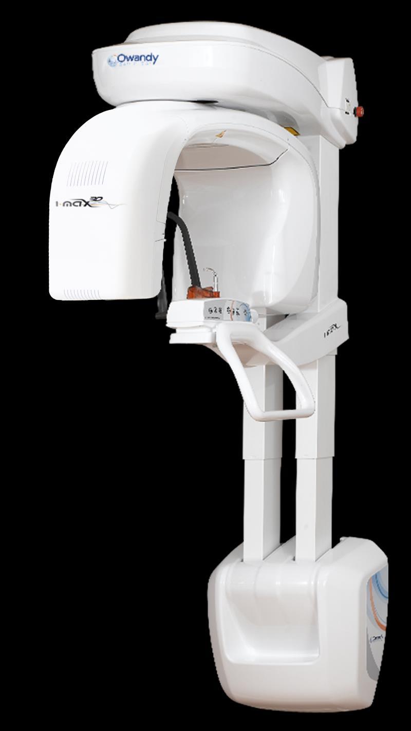
3 Product launch document EN Page 3 sur PRODUCT IDENTITY AND POSITIONNING Key points Unit design µ
4 Product launch document EN Page 4 sur 29 Main competitors advantages 1. Multi FOV 3D Cone Beam Can be adapted for use with all kinds of dental practices Implantology: 93x86mm (full mouth), 50x86mm (full arch) Endodontic: 50x50mm 2. HD images 3D sensor resolution : voxel 87 µm (smallest cross-section) D programmes Complete dental volume Left / right TMJ Sinus Maxillary volume / Mandibular volume Frontal maxillary Left / right premolar maxillary Left / right molar maxillary Frontal mandibular Left / right premolar mandibular Left / right molar mandibular 4. Creation of surgical guides Scan of impression trays, plaster models and radiological guides Overlay of STL and DICOM files With the I-Max 3D / Quickvision 3D combination, fully autonomous creation of surgical guides 5. Lightweight and sophisticated design The lightest 3D panoramic unit on the market: 66,5kg Attach it to a wall and you have an intraoral generator Takes up zero floor space Stylish: makes surgery look good to patients 6. Easy to use Face-to-face patient positioning Easy to handle equipment Demo video: Intuitive user interface Imaging tools and enhancing filters automatically integrated into the c1lr-0 control software. 7. Quick and easy to install in your surgery Demo video: Pz_Uwy4 Lightweight and compact unit delivered as one single package Exclusive Easy-To-Install system: the unit is delivered fully assembled with an intelligent system, requiring just one technician to fix it easily to the wall High degree of electronic optimisation; one main control device: easy maintenance, breakdown and troubleshooting operations Unit can be fully operated remotely 8. Controlled budget Unit optimised for manufacturing Lower installation costs, economical shipping costs Low breakdown rate The best Investment / Performance ratio 9. CAD / CAM ready Imports STL files from a dental impressions camera or lab scan Exports STL files for printing to a 3D printer or milling machine


 Full")
 Maxillary")

")


5 Product launch document EN Page 5 sur 29 FOCUS : 16 multi FOV 3D programmes: 86 x 93 mm (Ø x H) Full dental exam 86 x 50 mm (Ø x H) Maxillary 86 x 50 mm (Ø x H) Mandibular 86 x 93 mm (Ø x H) Left TMJ 86 x 93 mm (Ø x H) Right TMJ 86 x 93 mm (Ø x H) Sinus Maxillary left molars Maxillary left premolars Maxillary frontal 50 x 50 mm (Ø x H) Maxillary right premolars Maxillary right molars Mandibulary left molars Mandibulary left premolars Mandibulary frontal 50 x 50 mm (Ø x H) Mandibulary right premolars Mandibulary right molars
6 Product launch document EN Page 6 sur TECHNICAL CHARACTERISTICS General features Manufacturer Class Protection degree Line frequency OWANDY RADIOLOGY Croissy-Beaubourg, France Class II-b for European Directive for Medical Devices 93/42 Class II for Canadian MDR Class I with type B applied parts according to IEC Class II according to 21CFR-subchapter J (for V version) IPX0 standard device 50/60Hz Maximum line current V 50/60 Hz Power consumption V 50/60 Hz Line apparent resistance 0.5 Ω max -- Line voltage regulation -- < 3% à 99 V ~ Rated output voltage (kvp) Anodic current Mechanical characteristics kv, with 2 kv steps Focus-receptor distance 52 cm (20.5") Telescopic motorized column run 66 cm (26") Maximum total height 218 cm (86") Weight (complete unit, wall mounted version) Weight of optional unit support Working conditions Minimum room size Recommended room size ma, according to r20 scale 66,5 kg 6 kg 120x120cm (47.2"x47.2") 120x140cm (47.2"x55.1") Unit footprint dimensions (mm) 1107(wall side) x 953mm = 1m² Maximum working temperature range Relative working humidity (RH) range 30% 75% Temperature range for transport and storing Humidity range for transport and storing Minimum atmospheric pressure for transport and storing < 95% without condense 630 hpa
7 Product launch document EN Page 7 sur SENSORS AND XRAY GENERATOR CHARACTERISTICS Tube-head features Model MPV 05 Manufacturer Maximum tube voltage with accuracy 86 kv ± 8 % Maximum anodic current with accuracy 12.5 ma ± 10 % Duty cycle 1:16 Nominal power Total filtration HVL (Half value layer) Transformer insulation Cooling V.S.M. S.p.A Buccinasco (Milan) Italie kw (86 kvp ma) 2.5 mm Al 86 kvp > 3.2 mm Al 86 kv p Oil bath By convection Leakage radiation at 1 m < kvp ma - 3s duty cycle 1/16 Tube-head features Manufacturer CEI (Italie) Type OPX 105 Nominal focus size Inherent filtration Anode tilt 5 Anode material Nominal maximum voltage Filament max current Filament max voltage Anode thermal capacity Digital Sensor features Sensible area (H x L) 0.5 mm 0.5 mm Tungsten 105 kvp 4 A 8.0 V 30 KJ Voxel 87 µm Pixel (H) 120 x 120 µm Laser centering devices CMOS sensor 144 x 118 mm 2 laser beams are used for patient positioning. Beams align mid Sagittal and Frankfurt planes. Class 2 laser product according to IEC Standard :2007. Wave length Divergence Optical power on the working surface 650 nm ± 10 nm < 2.0 mrad < 1 mw
8 Product launch document EN Page 8 sur COMPUTER CHARACTERISTICS Recommended configuration Operating system Processor Memory Graphic board Main drive Speed network connection practice Other Windows bits Core i7 (4 coeurs 8 threads) 3 GHz or higher 8 Go nvidia 4 Go (ex : GTX 9 Go) SSD or SATA 1 Gbit Slot for Ethernet 1 Gbit board (PCI-Express 4X minimum)
9 Product launch document EN Page 9 sur UNIT DIMENSIONS Wall mounted version
10 Product launch document EN Page 10 sur D EXAMINATION MODES Exposure time Panoramic (PAN) Emi panoramic Improved orthogonality Panoramic Reduced dose Panoramic Frontal dentition Bitewing TMJ mouth closed/open Sinus P/A projection Image magnification Adult / Child standard Panoramic 14.4 s PAN Adult / 13.3 s Child 7.8 s Adult / 7.3 s Child 11.9 s Adult / Child 11.9 s Adult / 10.8 s Child 4.4 s Adult / Child 3.2 s (half bitewing) 6.3 s (standard bitewing) 4.8 s per image for left and right joint in open and closed condition 9.4 s Geometric magnification 1 : 1.28 (constant over dentition part) Magnification after software correction 1 : 1 (*) TMJ open/closed mouth 1 : 1.25 (nominal) 1 : 1 (*) Sinus 1 : 1.27 (nominal) 1 : 1 (*) Programs Examination selection type Automatic selection for Adult and Child, 3 Sizes Manual selection also possible for each programs Collimator with automatic positioning (*) The declared image magnification value is valid after proper software calibration.

11 Product launch document EN Page 11 sur 29 PANORAMIc Programs range Standard Panoramic Reduced dose Panoramic Half Panoramic Left Half Panoramic Right frontal dentition Improved orthogonality Panoramic BITEWING Programs range Standard Bitewing Half Bitewing Left Half Bitewing Right SINUS program Sinus P/A projection
12 Product launch document EN Page 12 sur 29 TMJ (Temporal Mandibular Joint) Programs range Standard TMJ, open/closed mouth Half TMJ sequence exam
13 Product launch document EN Page 13 sur D EXAMINATION MODES Exposure time 3D exams (except TMJ 3D) Sinus 3D TMJ 3D Half 3D volume Exposure time accuracy 11.2 s 11.2 s 10.8 s 11.2 s ± 5 % or ± 20ms whichever is greater


14 Product launch document EN Page 14 sur 29 3D Programmes range Full volume: 9x9 cm Half volume : 9x5 cm Small volume: 5x5 cm Scan of radiological guides Scan of impression trays Scan of plasters models


15 Product launch document EN Page 15 sur USER SOFTWARE INTERFACE Main settings window: default exam selected automatically.. Main window with complete program selection menu, in extended view.

16 Product launch document EN Page 16 sur 29 Main window with the image live preview
17 Product launch document EN Page 17 sur QUICKVISION 3D DICOM VIEWER Independent rotation of the various axes in each of the 4 screens, to display the area required. Identifies the nerve and selects the right size and shape of implant. Demo video: Coronal View Sagittal View Selection of nerve colour and diameter according to preferences Axial View 3D Volume
18 Product launch document EN Page 18 sur 29 Sectioning plans automatically default to coplanar views with the global system (axial, sagittal and coronal) Sectioning plan angles can be adjusted by entering the line relating to the section. Because these plans can be rotated, it s easy to analyse cross-sections from any position. The original view can be restored by using the re-set command. Demo video: Lines symbolizing the section which can be grabbed and rotated
19 Product launch document EN Page 19 sur 29 The mandibular nerve can be drawn in, to avoid touching it during the operation. 3 stages to obtain the mandibular nerve: Sectioning plan positioning so as to highlight foramina Selecting the point to highlight the section of the canal for various cross-sections Confirming operation Demo video: Foramen Option to create implants or import them from a library (Nobel, etc.). Option to rotate radiological images around the implant. Demo video:


20 Product launch document EN Page 20 sur 29 Overlaying In this view, various objects can be overlaid when reconstructing the 3D patient volume. This can be very useful, in order to correctly position the plaster mould used to model the surgical guide on, or to adjust the implant fixations or other positioning elements. Demo video: Plaster mould This can hide the reconstruction of the patient's 3D volume in order to be able to see one or more elements.


21 Product launch document EN Page 21 sur 29 Surgical Guide Design Demo video: Draw the mandibular nerve. Select the fixations, position them and adjust their size. Overlay the plaster model scan. Add the ring, the insert and the surgical guide. Finalize the surgical guide by adapting it to the plaster model. 3 4 Guide Drill trajectory Fixture Inserte Sleeve Select a view that will display the final model of the surgical guide. Download the project in order to generate an STL file and to print your drill template in 3D. Alternatively, you can send the patient s plaster mould and 3D volume to the specialist centre, which will be able to both plan the implant and create the drill template for you.

22 Product launch document EN Page 22 sur STAGES OF GUIDED SURGERY STAGE 1 3D imaging to produce a DICOM image Full-mouth imaging in just one exposure (3D I-Max). Integrated and optimised system for implant planning.

 Convert the DICOM file into an STL file Traditional dental")
 Import the STL file into QuickVision Result of the 4 methods:")
23 Product launch document EN Page 23 sur 29 STAGE 2 Create an STL file of the dental impression 4 méthods to obtain an STL file Method 1 Method 2 Method 3 Method 4 Traditional dental impression Create the plaster model Scan plaster model in a lab to obtain an STL file Import the STL file into QuickVision Traditional dental impression Create the plaster model Scan plaster model with I-Max 3D (DICOM) Convert the DICOM file into an STL file Traditional dental impression Scan dental impression with 3D I-Max (DICOM) Convert the DICOM file into an STL file Dental impression taken directly using an intraoral camera (STL file) Import the STL file into QuickVision Result of the 4 methods: STL file


24 Document Lancement produit FR Page 24 sur 29 STAGE 3 QuickVision 3D : superposition, planification et création du guide Overlay DICOM and STL files to obtain a complete image with soft and hard tissue. Demo video available on our YouTube channel Owandy Radiology (Superimpositioning_OWANDY RADIOLOGY_QuickVision 3D) Quick, easy and intuitive implant treatment planning Demo video available on our YouTube channel Owandy Radiology (Implant create and place_owandy RADIOLOGY_QuickVision 3D)
")
25 Document Lancement produit FR Page 25 sur 29 Quick, easy and intuitive implant treatment planning Demo video available on our YouTube channel Owandy Radiology (Implant create and place_owandy RADIOLOGY_QuickVision 3D)

 type printer, or by the")

26 Document Lancement produit FR Page 26 sur 29 STAGE 4 3D printing of the surgical guide on a Form 2 (Formlabs) type printer, or by the laboratory Top quality guided surgery. Time-savings (no subcontracting).


27 Document Lancement produit FR Page 27 sur 29 STAGE 5 Implant placement: a safe and accurate surgical operation The guide is placed in the mouth and its position controlled using the windows. A circular scalpel is used to mark the gums, to carry out flapless surgery or, in this case, surgery with a small flap. The implant is positioned using the guide, which ensures the perfect axial, vertical and rotational positioning with regard to the indexation.


28 Document Lancement produit FR Page 28 sur 29 The laboratory screw is removed. The prosthetic is placed and screwed onto the implant. It finds the right position in alignment with the other teeth. Connective tissue roll is used to repair gum tissue. Stitches are done using PTFE 4/0 Cytoplast thread. The occlusal command is used to check the underbite.
29 Document Lancement produit FR Page 29 sur 29 End of the document
3D/2D WALL MOUNTED UNIT
 Product launch document EN Page 1 sur 30 EN PRODUCT LAUNCH DOCUMENT REV03, September 2018 3D/2D WALL MOUNTED UNIT Product launch document EN Page 2 sur 30 INDEX 1. PRODUCT IDENTITY AND POSITIONNING...
2D AND 3D/2D WALL-MOUNTED PANORAMIC UNITS
 2D AND 3D/2D WALL-MOUNTED PANORAMIC UNITS KEEP YOUR CLINIC ONE STEP AHEAD! Wall-mounted concept: zero foot print 62kg - the lightest unit on the market Face to face positioning High Definition The fruit
PAN CEPH 3D CONE BEAM
 PAN CEPH 3D CONE BEAM 2D - 3D panoramic units PANORAMIC CEPHALOMETRIC 3D CONE BEAM IMAGING I-MAX TOUCH Tactile & naturally intuitive panoramic imaging Discover the simplicity and efficiency this unit can
Dental Line. 3D digital panoramic system. radiology ahead
 Dental Line 3D digital panoramic system radiology ahead new generation 3D digital panoramic unit 3D imaging s value available for anyone Following the incredible success of the innovative digital panoramic
I AM DEMANDING Type CMOS Flat Panel CMOS CMOS ø 40 x 40 mm, ø 60 x 60 mm, ø 80 x 80 mm, ø 110 x 80 mm
 TECHNICAL SPECIFICATIONS 1168 1501 1978 1237 1551-2351 Ø 1090 PANORAMIC CBCT CEPHALOMETRIC X-RAY SOURCE Tube type High frequency DC generator 2.8 mmal / 85 kv 7.0 mmal / 90 kv 2.8 mmal / 85 kv Operation
Head to new heights with your imaging SCANORA 3D
 SCANORA 3D Head to new heights with your imaging Benefits at a glance The solution for dentomaxillofacial and ENT imaging Easy Patient seated for added stability during exposure. Clear, self-explinatory
CS 9300 Family. The power of flexibility
 CS 9300 Family The power of flexibility The new CS 9300 digital imaging system from Carestream Dental take the guesswork out of examinations The all-in-one CS 9300 is the most versatile multimodality imaging
THE WAIT IS OVER CS D. 3D imaging is now available for everyone
 THE WAIT IS OVER CS 8100 3D 3D imaging is now available for everyone COMPLEXITY IS NO LONGER THE STANDARD NOW THERE ARE MANY REASONS TO MOVE TO 2D/3D IMAGING Now it s possible to experience nothing but
XPan 3D Plus. FONA Every dental solution you need. Advanced dental technology. Headquarters THE ULTIMATE DIAGNOSTIC SOLUTION DIGITAL DENTISTRY
 FONA Every dental solution you need Through decades of experience and deep understanding of the dental profession, we deliver complete, reliable and accessible solutions.regardless of country or specialisation,
- RCS Paris B
 Technical specifications PANORAMIC CBCT CEPHALOMETRIC X-ray source Tube type High frequency DC generator Total filtration >2.5 mm Al @ 90 kv Mode of operation Continuous Pulsed Continuous Tube voltage
Profound understanding of anatomy
 ENGLISH Profound understanding of anatomy Planmeca ProMax 3D, the intelligent and multipurpose X-ray unit, is designed to obtain complete information on patient anatomy in the minutest detail. The unit
Versatility And Expandability In One Panoramic.
 Orthoralix 9200 / 9200 DDE Versatility And Expandability In One Panoramic. Panoramic X-ray Systems Intraoral X-ray Systems Digital Intraoral Sensors Digital X-ray Phosphor Plates Intraoral Cameras Imaging
VistaVox S 3D from Dürr Dental
 VistaVox S 3D from Dürr Dental 3D and 2D X-ray images with exceptional image quality COMPRESSED AIR SUCTION IMAGING DENTAL CARE HYGIENE Taking diagnostics to the next level VistaVox S combines diagnostic
Digital Dentistry Solution
 Digital Dentistry Solution Digital Dentistry Solution 02 Digital Dentistry Solution CAD/CAM System Mill - Clinic Optimized for glass-ceramic High speed and steady quality in milling Simple and elegant
3D Cone beam CT & Digital Radiography Dedicated to Otorhinolaryngology
 3D Cone beam CT & Digital Radiography Dedicated to Otorhinolaryngology Multi-functional imaging solution3 RAYSCAN m is an unique 2-in-1 imaging solution, combining Cone Beam CT and Digital Radiography,
True Low Dose. Exact time to display image on screen may vary upon computer and network configuration.
 RAYSCAN ALPHA PLUS True Low Dose Cone Beam CT Industry Leading Resolution High resolution images provide all the clinical information needed while keeping radiation exposure low. Endodontics - Smallest
Profound understanding of anatomy
 ENGLISH Profound understanding of anatomy The unique Planmeca ProMax 3D product family offers equipment for all maxillofacial imaging. All volumes sizes from the smallest special cases to whole head images
DIAGNOSTIC IMAGING. OPTIMIZED.
 ABOUT LED DENTAL SEE THE DIFFERENCE Using our years of business insight and clinical experience as a foundation, LED Dental takes the uncertainty out of your imaging purchase decision. We offer our clients
X X X. GXS-700 Direct USB Digital Intraoral Sensors. Buy a Sensor Combo and a Digital Pan Unit, Receive $600 Off! GO.BENCO benco.
 8 0 0. G O. B E N C O b e n c o. c o m GS-700 Direct USB Digital Intraoral Sensors Designed to make migrating from film, or upgrading an existing digital system, easier than ever High quality image capture
ORTHOPHOS XG 3 DS. X-ray systems. ORTHOPHOS XG 3 Digital panoramic X-ray for practical diagnostics.
 ORTHOPHOS XG 3 DS X-ray systems ORTHOPHOS XG 3 Digital panoramic X-ray for practical diagnostics. ORTHOPHOS XG 3 Standard panoramic X-ray with proven technology. competent successful Many years of competence
STELLARIS 3D 4 IN 1 CBCT SOLUTION FOR ADVANCED DIAGNOSTICS
 STELLARIS 3D 4 IN CBCT SOLUTION FOR ADVANCED DIAGNOSTICS 3 STELLARIS 3D 4 IN CBCT SOLUTION FOR ADVANCED DIAGNOSTICS Stellaris 3D is a complete and compact, fully upgradeable 3D CBCT for a patient, Panoramic
GUIDED SURGERY TECHNIQUE
 GUIDED SURGERY TECHNIQUE INDEX WORKFLOW...4 OUR SOLUTIONS...5 PLANNING PROTOCOL - PARTIAL EDENTULISM...6 - TOTAL EDENTULISM...9 SOFTWARE FEATURES...12 SURGICAL COMPONENTS FOR GUIDED IMPLANTOLOGY...14
3D/2D. Hyperion X5 Suspended imaging system. Data subject to change without notice. 03/2017 MX53DGB171S00
 www.my-ray.com Data subject to change without notice. 03/2017 MX53DGB171S00 According to the regulations in force, some products and/or features may have different availability and characteristics in areas
T h e D e n t a l C o m p a n y FROM DIAGNOSTIC SCAN TO SURGERY, WE SHAPE THE FUTURE OF DENTISTRY.
 T h e D e n t a l C o m p a n y FROM DIAGNOSTIC SCAN TO SURGERY, WE SHAPE THE FUTURE OF DENTISTRY. SIDEXIS SOFTWARE ORTHOPHOS SL D/D SEAMLESS THE NEW STANDARD IN CLINICAL DIAGNOSIS AND PATIENT COMMUNICATION
OP 3D Vision The upgradable 3D X-ray system for the strictest demands.
 OP 3D Vision The upgradable 3D X-ray system for the strictest demands. The solution for every task: KaVo OP 3D Vision. Regardless of which dental query you may have, the KaVo ORTHOPANTOMOGRAPH OP 3D Vision
Easy operation. Numerous diagnostic options. X-rays you can rely on: the ORTHOPHOS XG device family. All ORTHOPHOS XG 3Dready programs at a glance.
 CAD /CAM Systems Instruments Hygiene Systems Treatment Centers Imaging Systems Subject to technical changes and errors in the text, Order No. A91100-M47-B346-01-7600, Printed in Germany, Dispo-Nr. 04602,
Profound understanding of anatomy
 ENGLISH Profound understanding of anatomy The unique Planmeca ProMax 3D product family offers equipment for all maxillofacial imaging. All volume sizes from the smallest special cases to whole head images
ENGLISH. Cefla s.c. - Via Selice Provinciale 23/a, Imola - Italy Tel
 09-2017 NVGEGB161S00 According to the regulations in force, some products and/or features may have different availability and characteristics in areas outside of the European Union. Please contact your
English. Perfect Vision
 English Perfect Vision Everything becomes clearer and simpler with a big F.O.V. The complete dentomaxillofacial volume ready for your diagnosis One scan provides you with an incredible amount of information
ORTHOPANTOMOGRAPH OP200 D ORTHOCEPH OC200 D. True dynamo leading through the decades.
 ORTHOPANTOMOGRAPH OP200 D ORTHOCEPH OC200 D True dynamo leading through the decades. 1 You can t dublicate the legacy. 1946 Professor Y.V. Paatero publishes his first paper on Panoramic Tomography. 1951
Digital Dentistry Solution
 Digital Dentistry Solution Digital Dentistry Solution 02 Digital Dentistry Solution CAD/CAM System Optimized for glass ceramic High speed and steady quality in milling Simple and elegant design Bar, Disk
For technical assistance: Tel Working time: 8.30 am pm
 I For technical assistance: Tel. +39 059 392827 Working time: 8.30 am - 5.30 pm Index JD Digital solutions for Intraoral Scanning JD Implant Libraries JD Guided Surgery Kit JD Surgical Guide and 3D Model
Low Dose Excellent Image Quality Rapid Reconstruction
 Low Dose Excellent Image Quality Rapid Reconstruction Efficient 3 in 1 Dental X-ray System CBCT > Precise 3-D Anatomical structures - Accurate diagnosis for doctors - Safe implant for patients > Significant
For true visualisation
 ENGLISH For true visualisation Planmeca ProModel is a patient-specific physical model for high-end maxillofacial operations and dental surgery. By reproducing the anatomy of the patient in real-size, Planmeca
The Power Of Choice Pan. Ceph. 3D. Your Imaging Future Starts Today.
 NEW from Gendex! The Power Of Choice Pan. Ceph. 3D. Your Imaging Future Starts Today. Cone Beam 3D Imaging Systems Panoramic X-ray Systems Intraoral X-ray Systems Digital Intraoral Sensors Digital X-ray
The Optimum Choice for Implantologist
 The Optimum Choice for Implantologist What is essential for your practice? What s the best way to choose a 3D X-ray machine for implant treatment planning? 02 Doctor says.. There are diagnostic limitations
CT Scanning Protocol For V2R Guided Surgery Solutions
 CT Scanning Protocol For V2R Guided Surgery Solutions 2 V2R CT Scanning Protocol \\ Contents Contents General requirements... 3 V2R Dual Scan Protocol... 5 V2R Single Scan Protocol... 8 Overview... 10
» Guided Surgery. by MEDENTiKA « MEDENTIGUIDE SURGERY MANUAL QUATTROCONE QUATTROCONE30. IPS Implant systems
 » Guided Surgery by MEDENTiKA «IPS Implant systems MEDENTIGUIDE QUATTROCONE QUATTROCONE30 SURGERY MANUAL » MedentiGuide «MedentiGuide drill sleeves support the surgeon in preparing the implant bed for
Guided immediate loading implant surgery planned with Implant Studio D.D.S. Jae-min, Lee
 Guided immediate loading implant surgery planned with Implant Studio D.D.S. Jae-min, Lee Jung-plant Dental office 1 PROLOGUE How can we deal with the immediate loading implant cases easier and more accurate
I AM EXCLUSIVE TECHNICAL SPECIFICATIONS. The first personal imaging plate scanner IMAGING PLATES. System. Windows recommended configuration
 TECHNICAL SPECIFICATIONS System Resolution... 20 lp/mm Scan Time (fast mode)...1,6s - 2,7s Scan Time (high definition mode)...2,1s - 3,6s Connection... Ethernet RJ-45 Dimensions... L. 154 x D. 204 x H.
Planmeca ProMax 3D s Planmeca ProMax 3D ENGLISH
 Planmeca ProMax 3D s Planmeca ProMax 3D ENGLISH Genuine all-in-one unit Planmeca ProMax 3D s and Planmeca ProMax 3D units are designed to obtain complete information on patient anatomy in the minutest
» Guided Surgery. by MEDENTiKA « MEDENTIGUIDE SURGERY MANUAL MICROCONE. IPS Implant systems
 » Guided Surgery by MEDENTiKA «IPS Implant systems MEDENTIGUIDE MICROCONE SURGERY MANUAL » MedentiGuide «MedentiGuide drill sleeves support the surgeon in preparing the implant bed for MEDENTiKA implants.
GUIDED SURGERY SOFTWARE
 GUIDED SURGERY SOFTWARE PREDICTABLE, RELIABLE, AND REPEATABLE RESULTS 15+ years of clinical success 150,000+ guided cases TRANSFORMING CREATIVITY DIGITIZE YOUR WORKFLOW AND TAKE ADVANTAGE OF EXCITING OPPORTUNITIES
Veraviewepocs 3D R100 & F40
 Veraviewepocs 3D R100 & F40 Innovative 3D Reuleaux Full Arch FOV Thinking ahead. Focused on life. Veraviewepocs 3D R100 A New Frontier in X-ray Diagnostics Veraviewepocs 3D R100 has changed the shape of
3D Panoramic Cephalometric. Innovation, in reach. KODAK 9000 Extraoral Imaging System
 3D Panoramic Cephalometric Innovation, in reach 9000 KODAK 9000 Extraoral Imaging System Cephalometric Innovation made simple Innovation made simple We believe in innovation. We always have. In fact, our
SpiderX. Portable Residual Stress X-Ray Diffractometer.
 Portable Residual Stress X-Ray Diffractometer www.gnr.it About SpiderX GNR Analytical Instrument offers equipment based on X-Ray Diffraction to measure residual stress state and retained austenite content.
BIOBank Innovation: The Customized Bone Graft
 Preserved and regenerated human bone Innovation: The Customized Bone Graft Concept A Customized Graft for bone apposition BENEFITS FOR SURGEONS: Easier planning of the surgery: properties and positioning
3Shape X1 Scanning redefined
 3Shape X1 Scanning redefined Why choose the X1 Give your patients a great experience No head fixation and sleek design create a comfortable scanning experience for your patient High image quality low dose
RaySafe X2 Solo DENT. X-ray meter for dental applications
 RaySafe X2 Solo DENT X-ray meter for dental applications Quality Assurance in Dental X-ray Hundreds of millions of X-ray exposures are done every year in dental clinics around the globe. The majority are
BIOTECH DENTAL GROUP, 2.0 DENTAL OFFICE PARTNER.
 BROCHURE BIOTECH DENTAL GROUP, 2.0 DENTAL OFFICE PARTNER. Since its creation in 1987, Biotech Dental has been committed to develop a strong relationship of trust with dental surgeons and dental technicians.
The Quality Leader in 3D Cone Beam CT
 The Quality Leader in 3D Cone Beam CT The Complete 2-in-1 or 3-in-1 Multi-modality Solution PreXion, with over 15 years of innovation in the medical and dental fields, introduces the PreXion3D Eclipse.
NewTom GiANO HR PERFECT.VISION
 CEFLA s.c. Via Selice Provinciale 23/a 40026 Imola Italy t. +39 045 8202727 045 583500 info@newtom.it newtom.it 06/2018 NHRGB181S00 According to the standards in force, in extra-eu areas the availability
Dimensional stability in composite cone beam computed tomography
 (2010) 39, 512 516 2010 The British Institute of Radiology http://dmfr.birjournals.org TECHNICAL REPORT Dimensional stability in composite cone beam computed tomography S Kopp* and P Ottl Department of
Guided Surgery Service Manual POWERED BY
 Guided Surgery Service Manual POWERED BY IN LABORATORY Neway laboratory scanner IN STUDIO Intraoral scanner CS 3600 SCANNING AND DATA ACQUISITION DIGITAL UNIVERSE* IN SWEDEN & MARTINA Industrial milling
Introducing the Pala 3D-printed Denture
 ebook Create your best workflow and optimize production with cara 3D printing technology. Introducing the Pala 3D-printed Denture Powered by cara Print 4.0 and dima Print materials. < 2 hrs. Giving a hand
3Shape X1 Scanning redefined
 3Shape X1 Scanning redefined Why choose the X1 Give your patients a great experience No head fixation and sleek design create a comfortable scanning experience for your patient High image quality low dose
Hyperion X9 3-in-1 Imaging System
 Hyperion X9 3-in-1 Imaging System 2 Hyperion X9, full imaging. 3 Hyperion X9, just right for me. The present and the future of my work. In three dimensions. Hyperion X9 offers me multiple possibilities
ORTHOPANTOMOGRAPH OP 3D Pro A platform for changing needs
 ORTHOPANTOMOGRAPH OP 3D Pro A platform for changing needs ORTHOPANTOMOGRAPH OP 3D Pro OP 3D Pro is the most comprehensive 3-in-1 platform designed for today and tomorrow, covering the entire maxillofacial
EasyLineImplant. Guided Surgery
 EasyLineImplant Guided Surgery Guided surgery Guided Surgery represents an improvement in the traditional surgery, increasing the advantages for clinicians and patients. The guided procedure allows reaching
ORTHOPANTOMOGRAPH OP300. A platform for changing needs.
 ORTHOPANTOMOGRAPH OP300 A platform for changing needs. 1 Panoramic with cephalometric digital image Xray machine For me, peace of mind means a patient trusting in my care, time after time Leading the way
3D Accuitomo XYZ Slice View Tomograph. Super High-Resolution Images of Region of Interest
 3D Accuitomo XYZ Slice View Tomograph. Super High-Resolution Images of Region of Interest Thinking ahead. Focused on life. 2 Cone Beam X-Ray CT Imaging X-Ray Tube Imaging Intensifier Imaging Volume Voxel
RaySafe i3 INSTALLATION & SERVICE MANUAL
 RaySafe i3 INSTALLATION & SERVICE MANUAL 2017.06 Unfors RaySafe 5001104-1.1 All rights are reserved. Reproduction or transmission in whole or in part, in any form or by any means, electronic, mechanical
Xelis Dental - What New in
 Xelis Dental - What New in 1.0.6.1 Clinical Needs Why Xelis-Dental? Panorama Cephalography Intra-oral Digital Camera Traditional imaging systems - 2-Dimension view Incorrect anatomical information - Distortion
HOW TO USE PALTOP DIGITAL SERVICE?
 HOW TO USE PALTOP DIGITAL SERVICE? **the service is currently available in Israel only** PALTOP Digital offers planning and production services according to your personal needs. Orders may be initiated
IMPLANT 3D REL. 8.0 INSTRUCTIONS FOR USE
 IMPLANT 3D REL. 8.0 INSTRUCTIONS FOR USE Implant 3D Rel. 8.0 IFU (MU-I3DE8.0 - Rev. 1-30/04/2015) CONTENTS IMPLANT 3D REL. 8.0... 5 INTRODUCING IMPLANT 3D... 6 SYSTEM REQUIREMENTS... 8 INSTALLATION...
Nuclear Associates
 Nuclear Associates 37-013 GARD Users Manual March 2005 Manual No. 37-013-1 Rev. 2 2004, 2005 Fluke Corporation, All rights reserved. Printed in U.S.A. All product names are trademarks of their respective
Reinvent Your Practice and Your TEAM through 3D Guided Implantology By Patrick Hayden, M.Ed
 Reinvent Your Practice and Your TEAM through 3D Guided Implantology By Patrick Hayden, M.Ed The Dental Market In 2011 there were over half a billion missing teeth in adults 18-65*. Two million of those
D3D CBCT. See more Do More
 D3D CBCT See more Do More BIOLASE DaVinci Imaging D3D For capturing superior 3D image acquisitions with the lowest minimal dose for patient safety The BIOLASE DaVinci Imaging D3D has one of the lowest
TABLE OF CONTENTS P. 4-5 P. 6-7 P. 8-9 P P P P
 Full Smile Solution TABLE OF CONTENTS P. 4-5 P. 6-7 P. 8-9 P. 10-11 P. 12-13 P. 14-15 P. 16-17 FULL SMILE SOLUTION MGUIDE PROCESS ADVANTAGES CUSTOM SOLUTIONS MGUIDE SURGICAL SETS MCENTER REQUIRED SOURCES
Flexible Easy Competitive. SCANORA 3Dx - The in-office large field-of-view Cone Beam CT system for Head and Neck imaging
 Flexible Easy Competitive SCANORA 3Dx - The in-office large field-of-view Cone Beam CT system for Head and Neck imaging SCANORA 3Dx. The solution. SCANORA 3Dx makes advanced 3D imaging easy in the head
INTRAORAL SCANNER TRANSFORMING CREATIVITY
 INTRAORAL SCANNER TRANSFORMING CREATIVITY COMING SOON! POWDER-FREE SCANNING* Increased efficiency, accuracy, and comfort for your patient. Gesture control module for infection control INTRAORAL SCANNER
THE USE OF KEYSTONE EASYGUIDE CT SCANNING SOFTWARE FOR DIAGNOSIS, DIRECTION AND DEPTH DETERMINATION
 CT DIAGNOSTICS IN 3D IMPLANT TREATMENT PLANNING THE USE OF KEYSTONE EASYGUIDE CT SCANNING SOFTWARE FOR DIAGNOSIS, DIRECTION AND DEPTH DETERMINATION Timothy Kosinski, DDS, MAGD Assistant Clinical Professor
CS 8100 FAMILY / CS D FAMILY / CS 9300 FAMILY EXTRAORAL SOLUTIONS EXTRAORDINARY POSSIBILITIES
 CS 8100 FAMILY / CS 8100 3D FAMILY / CS 9300 FAMILY EXTRAORAL SOLUTIONS EXTRAORDINARY POSSIBILITIES A SOLUTION FOR EVERY CLINIC AND ANY CONSULTATION The CS 8100 does more than my old machine and is half
The Key to Confidence
 WITH The Key to Confidence The Key to Confidence More detail, better accuracy, greater confidence The G-scan Brio is a revolutionary MRI approach for all musculoskeletal applications, which allows you
The Definitive Dental 3D Printing Solution
 The Definitive Dental 3D Printing Solution Dental Pro Introducing the worlds fastest Dental 3D Printer Speed 3D printing now exists in an elegant desktop solution Dental models produced faster than pouring
Connect your Scanner to SomnoMed Canada. SOMGauge Protrusive Bite Recording - Manual. Scanning Impressions - Lower and Upper
 IOS Instructions How to create and submit the best scans to SomnoMed Canada for the creation of a custom SomnoDent Sleep Apnea Appliance Its a simple process: STEP 1 Connect your Scanner to SomnoMed Canada
Planmeca ProMax 3D s Planmeca ProMax 3D ENGLISH
 Planmeca ProMax 3D s Planmeca ProMax 3D ENGLISH Learn more: Planmeca Imaging for ipad Genuine all-in-one unit Planmeca ProMax 3D s and Planmeca ProMax 3D units are designed to obtain complete information
inlab Software 18.0 New digital options dentsplysirona.com/inlab
 inlab Software 18.0 New digital options dentsplysirona.com/inlab 02 I 03 inlab CAD Software 18.0 Dental design requires good software The new inlab CAD Software 18.0 focuses even more closely on the requirements
I AM SAFER. Reliable X-ray technology that reduces radiation exposure
 I AM SAFER Reliable X-ray technology that reduces radiation exposure EN 40 cm / 80 cm / 110 cm (16 / 31 / 43 ) 70 cm (27 ) 9 cm (3,5 ) 36 cm (14 ) 79 cm / 119 cm / 149 cm (31 / 46 / 59 ) 87 cm (34 ) 81
ADVANCED 3D IMAGING. CEFLA s.c. Via Selice Provinciale 23/a Imola Italy t newtom.
 CEFLA s.c. Via Selice Provinciale 23/a 40026 Imola Italy t. +39 045 8202727 045 583500 info@newtom.it newtom.it 05/2018 NVGEGB181S00 According to the standards in force, in extra-eu areas the availability
ENGLISH. Advanced tools for orthodontics
 ENGLISH Advanced tools for orthodontics A complete solution for orthodontics One unit one software Panoramic Cephalometric CBCT image 3D photo Planmeca ProMax unit 3D model scan Planmeca offers a complete
NewTom 5G XL EXTRA.VISION
 CEFLA s.c. Via Selice Provinciale 23/a 40026 Imola Italy t. +39 045 8202727 045 583500 info@newtom.it newtom.it 06/2018 N5GXGB181S00 According to the standards in force, in extra-eu areas the availability
Immediate loading and implant surgery with digital workflow
 Mirero Dental Clinic Dr. Jaemin Lee Immediate loading and implant surgery with digital workflow Solutions featured: 3Shape TRIOS 3Shape Dental System 3Shape Implant Studio Case information Patient, 32-year
About Dental Wings. DWOS Dental Wings Digital Dentistry Platform
 About Dental Wings Dental Wings is a leading provider of dental CAD / CAM solutions. The company is specialized in the development of 3D scanners and CAD software dedicated to different dental market segments.
Quick guide for 3shape order form
 Quick guide for 3shape order form Elos Accurate Library Elos Accurate Library December 2017 Content: Introduction 2 Elos Accurate Hybrid Base Kit order form 3 Elos Accurate Hybrid Base Bridge order form
5 reasons to choose TRIOS
 5 reasons to choose TRIOS Superior scanning Flexible hardware Widest range of Open and flexible Shade measurement and technologies configurations indications for any lab setup integrated intraoral camera
STL. inside. ineos Blue: Precision, Speed and Control. THE SCANNER WITH Bluecam TECHNOLOGY
 CAD/CAM SYSTEMS INSTRUMENTS HYGIENE SYSTEMS TREATMENT CENTERS IMAGING SYSTEMS THE SCANNER WITH Bluecam TECHNOLOGY ineos Blue: Precision, Speed and Control. STL inside T h e D e n t a l C o m p a n y ineos
Veraviewepocs 3D. F40 and R100 with innovative 3D Reuleaux Full Arch FOV. Thinking ahead. Focused on life.
 Veraviewepocs 3D F40 and R100 with innovative 3D Reuleaux Full Arch FOV Thinking ahead. Focused on life. Veraviewepocs 3D R100 A New Frontier in X-ray Diagnostics Veraviewepocs 3D R100 has changed the
Straight to the point with your diagnosis.
 CAD/CAM SYSTEMS INSTRUMENTS HYGIENE SYSTEMS TREATMENT CENTERS IMAGING SYSTEMS HELIODENT DS INTRAORAL X-RAY WITH EXCELLENT IMAGE QUALITY Straight to the point with your diagnosis. T h e D e n t a l C o
3Shape TRIOS Engage and excite your patients
 3Shape TRIOS Engage and excite your patients Create patient excitement with 3Shape TRIOS 3Shape TRIOS allows you to enhance patient experience with exciting engagement tools, reduce chair-time and embrace
Visualise a better practice
 Visualise a better practice Powerful & portable I m itero Element 2 and I bring innovation to visualisation itero Element 2 delivers faster scan processing than the current itero Element scanner, enhanced
3D-MODEL CUSTOM-MADE MODELS SEGMENTATION AND PRODUCTION SERVICE OF BONE MODELS WITH HIGHEST 3D PRINTING RESOLUTION
 CUSTOM-MADE MODELS 3D-MODEL SEGMENTATION AND PRODUCTION SERVICE OF BONE MODELS WITH HIGHEST 3D PRINTING RESOLUTION FOLLOW US ON CUSTOM-MADE MODELS 3D-MODEL From a CT or CBCT scan, 3D-model service provides
PRODUCT DOCUMENTATION FOR SMOP TEMPLATE DESIGNER
 PRODUCT DOCUMENTATION FOR SMOP TEMPLATE DESIGNER smop is a product and brand of Swissmeda AG, www.mysmop.com document version 181119 for smop version 2.14.2 1. DENTISTS WORKFLOW WITH SMOP 1. Create Case
High-end solution STEP-BY-STEP USER GUIDE
 High-end solution STEP-BY-STEP USER GUIDE System Contents REAL TIME Restoration Guided Conical drill Multi-Unit abutment, Conical connection Guided kit for conical connection implant procedure C1 & V3
NEODENT GUIDED SURGERY
 NEODENT GUIDED SURGERY MANUAL Grand Morse Connection Helix Implant GRAND POSSIBILITIES WITH A LIMITLESS SOLUTION. GRAND MORSE NEODENT GUIDED SURGERY. 4 5 6 CONTENTS 1. CLINICAL STEP BY STEP OF GRAND
Dolphin 3D Imaging 11.7 beta
 Dolphin 3D Imaging 11.7 beta Dolphin 3D Surgery Dolphin s 3D Surgery has expanded to include even more features! This revolutionary treatment planning and presentation software module now includes a fully
Digital Imaging from a new perspective
 TREATMENT CENTRES HANDPIECES HYGIENE SYSTEMS X-RAY SYSTEMS CEREC TREATMENT CENTRES HANDPIECES HYGIENE SYSTEMS X-RAY SYSTEMS CEREC SIRONA CREATING AND MAINTAINING VALUE. You are right to expect a great
PREVIEW 2018 WELCOME TO DIGITAL DENTISTRY. 3D Printer for prosthodontists and dentists.
 PREVIEW 2018 WELCOME TO DIGITAL DENTISTRY. 3D Printer for prosthodontists and dentists. ENTER THE DIGITAL AGE Digital dentistry and 3D printing are transforming the dental industry, changing the workflow
Everything. Supplying everything dental for your practice needs
 Everything Connect Dental Supplying everything dental for your practice needs henryschein.co.uk Highly accurate, durable and 3D Conebeam digital image ORTHOPHOS XG3D/SL Practice Management Software Secure
HANDS-FREE ULTRASOUND
 The recently introduced Sonopuls 190 from Enraf-Nonius is a highly appreciated device for ultrasound therapy with multi-frequency treatment heads. A modern device with an extremely quick start-up and a