Conflicts of interest
|
|
|
- Susan McCarthy
- 5 years ago
- Views:
Transcription


 «Diseases of the nail and their management» R Baran, D")
 are 100% kept under the responsibility of the Direction de la Recherche des")
1 Dermoscopy of acral melanoma Luc THOMAS Service de Dermatologie Université Claude Bernard, Lyon 1 Centre Hospitalier Lyon Sud Lyon, France Conflicts of interest Jean Marie Naeyaert Full-time Employee of the French Ministry for Education and Research, no private practice and no personal financial relationship with pharmaceutical or bio-medical industry PI skin oncology clinical trials (VICAL, ROCHE, GSK, BMS, INTUISKIN, PIXIENCE, NOVARTIS, GENENTECH, GALDERMA, MERCK-SERONO) no personal honorarium. (Public declaration of interest(s) available on HAS website : Author of «Atlas de Dermoscopie» RP Braun et L Thomas Elsevier Masson Paris 2007 (French, Portuguese (Brazil) & Polish) «Précis de dermatologie et IST» JH Saurat, JM Lachapelle, D Lipsker et L Thomas 5eme édition, Elsevier, Masson, Paris 2009 (French & Italian) «Diseases of the nail and their management» R Baran, D de Berker, M Holzberg & L Thomas Willey-Blackwell, London 2012 «Dermatologie chirurgicale» JM Amici, D Egasse, M Beylot Barry et L Thomas Elsevier Masson Paris 2012 Funding(s) Université Claude Bernard Lyon 1 Ligue Contre le cancer du Rhône et de l Ain Fondation APICIL Funds (mainly honoraria of clinical trials) are 100% kept under the responsibility of the Direction de la Recherche des Hospices Civils de Lyon and of Lyon 1 - Claude Bernard University as well as expenses The best quality for a chief is his ability to be rewarded for things that happen just by themselves Scott Adams Ezus 1


2 Alice Phan Stéphane Dalle Nicolas Poulalhon Sébastien Debarbieux Clélia Moulin Mona Amini-Adlé 2


3 Fanny Julia Marie Perrier-Muzet Olivier Béatrix Brigitte Balme Robert Baran All pictures presented during this lecture, except when specifically mentioned, belong to the collection of the department of dermatology of Lyon 1 University. When applicable, credit for borrowed pictures is mentioned on the slides. All patients gave their consent for photographs shown. Reproduction, publication, public use of any image without permission is strictly prohibited. 3


4 Dermoscopy A Dermoscopy reflects the anatomy M Exceptions Face Palms and soles Scars Nail Courtesy of S. Puig and J. Malvehy Mucous membranes Two-steps diagnostic strategy «Special location» pigmented lesions Pigmented skin lesion 1 st step Algorithm Melanocytic Non melanocytic 2 nd step Benign Suspicious Malignant ABCD rule Menzies s 7 points checklist Pattern analysis 4


 seems to confer a poorer prognosis mainly because")


5 Palms & soles Nails Harald Kittler Palms and soles CLINICAL AND LABORATORY INVESTIGATIONS DOI /j x Acral lentiginous melanoma: a clinicoprognostic study of 126 cases A. Phan, S. Touzet,* S. Dalle, S. Ronger-Savlé, B. Balme and L. Thomas Department of Dermatology, Hôtel Dieu, Claude Bernard University, Lyon cedex 02, France *Technology Assessment Unit, Department of Medical Information, Hôpitaux de Lyon, Claude Bernard University, Lyon cedex 03, France Summary Correspondence Luc Thomas. luc.thomas@chu-lyon.fr Accepted for publication 20 February 2006 Key words achromic melanoma, acral lentiginous melanoma, melanoma, prognosis, skin cancer Conflicts of interest None declared. Background Although the histopathological subtype of melanoma has not been clearly proven to carry independent prognostic significance, acral lentiginous melanoma (ALM) seems to confer a poorer prognosis mainly because disease is often more advanced at the time of diagnosis. Objectives To investigate the distinctive epidemiological and clinical characteristics of ALM, a peculiar histological entity, and to identify prognostic factors. Methods We performed a register-based review of cases from a single large referral centre, the University Hospital Department of Dermatology, Lyons, France. We reviewed patient demographics, the initial presentation of the lesion, and clinical outcome. ALM-specific and disease-free survival were estimated using the Kaplan Meier method and compared using the log-rank test. A Cox model was used to identify prognostic factors. Wallace s line Palms and soles Anatomy of volar skin Eccrine duct opening Crista superficialis (ridge) Sulcus superficialis (furrow) ldelanoma Nevus Crista superficiatis limitans Crista profunda intermedia Courtesy of T. Saida 5


6 Furrows Ridges Furrow Furrow Furrow Ridge Ridge Nevus crista profunda limitans Melanoma canal sudoripare canal sudoripare Palleschi et al, Clin Exp Derm 2006;31: Saïda et al, Clin Derm 2002;20: Two malignant patterns Two malignant patterns Two important rules Parallel ridge pattern Parallel ridge pattern Saida et al, Arch Dermatol 2004;140: Irregular diffuse pigmentation 6
 162")

:1618-20.")


7 A. Phan et al. Br J Dermatol (2010) 162 : Nevus Melanoma Braun RP et al, Arch Dermatol 2008 Dec; 144(12): Irregular diffuse pigmentation Saida et al, Arch Dermatol 2004;140: A. Phan et al. Br J Dermatol (2010) 162 :


8 Melanoma ALM subtype Clark s III, 0.3 mm Two important rules Rule N # 1 : Never be reinsured by the presence of a benign pattern within others patterns Rule N # 2 : Always consider the presence of a malignant pattern, within others, as a warning sign 8


 usually shows a bluish, quite homogeneous pigmentation involving the ridges.")
. The multiplicity, bilateral location, and chronicity of the lesions are suggestive of ethnic-type")


9 SECTION EDITOR: JAMES M. GRICHNIK, MD, PhD; ASSISTANT SECTION EDITORS: ASHFAQ A. MARGHOOB, MD; ALON SCOPE, MD A A B Figure 1. B A B Figure 3. R-P. Braun et al. Dermatology (2013) skinsight Benign Dermoscopic Parallel Ridge Pattern Variants T Alice Phan, MD; Stéphane Dalle, MD; Marie-Cécile Marcilly, MD; Jean-Pierre Bergues, MD; Luc Thomas, MD, PhD; Centre Hospitalier Lyon-Sud, Claude Bernard University, Pierre Bénite, France HE PARALLEL RIDGE PATTERN (PRP) IS A VOlar dermoscopic pattern that is characterized by an accentuated pigmentation on the ridges of the skin markings, while the furrows are hypopigmented or unpigmented. It has been demonstrated to be highly specific for acral melanoma, and its presence must be considered a warning sign of melanoma. However, rare cases of benign acral melanocytic nevus can exhibit the PRP, as in the edge of the compound melanocytic nevus shown in Figure 1A. The acral blue nevus (Figure 1B) usually shows a bluish, quite homogeneous pigmentation involving the ridges. Ethnic pigmentation clinically presents as pigmented macules with a typical PRP on the palms and soles (Figure 2A). The multiplicity, bilateral location, and chronicity of the lesions are suggestive of ethnic-type pigmentation. The presentation is similar in Laugier-Hunziker or Peutz-Jeghers syndrome. Ectasic papillary capillaries of acral angioma present as regular red-blue globules along the ridges (Figure 2B). Repetitive microtrauma can cause extravasation of erythrocytes in the cornified layer, with subsequent misleading globular reddish brown pigmentation on the ridges, as seen in Figure 3A, which shows a posttraumatic purpura due to the friction of the fifth finger caused by regular use of the computer mouse. The PRP was also found in subcorneal hematoma. The lesion appears as well-demarcated roundish macule, with reddish brown pigmentation along the ridges (Figure 3B). Deposit of exogenous pigmentation on the ridges, especially in areas where the stratum corneum is thicker, can also simulate melanoma, as shown in Figure 4,withtheuse of a self-tanning preparation by a woman with localized palmar hyperkeratosis. A. Phan et al. Arch Dermatol (2011) 147 :


10 Robertson et al, Austral J Dermatol 2010;51: Nails


11 Melanonychia striata Ethnic-type pigmentation Melanocytic nevus Melanoma Sub-ungual haemorrhage Laugier Hutziker disease Drug-induced pigmentation Onychomycosis Trauma-induced pigmentation 11


 Change")


12 Bowen s disease Onychomatricoma Clinical algorithm Clinical algorithm Acquired during childhood Polydactylic Stable Another (good) explanation for pigmentation (drug, ethnic, etc ) No periungual involvement Onset during adulthood Monodactylic Changes over time Peri-ungual involvement (Hutchinson s sign) Polychromia Triangular shaped (adult) Change of the plate Childhood onset Polydactylic / other explanation Rather benign Clinical algorithm Clinical algorithm Hutchinson s sign Change over time Polychromia Plate change Rather Malignant M 0 M 18 Triangular shape Rather Malignant 12


13 Dermoscopy Arch Dermatol 2002, 138, A. Phan et al. Br J Dermatol (2010) 162 : Known from dermoscopy Blood spots Brown color of the background 13


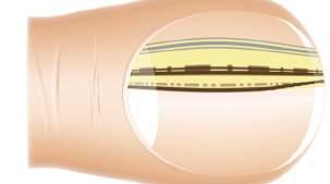

14 Regular pattern of the longitudinal lines Irregular pattern of the longitudinal lines Gray-yellowish color of the background 14


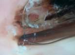
15 Micro-Hutchinson s sign Longitudinal leuco/ xanthonychia Distal subungual hyperkeratosis Linear micro-hemorrhages 15
")
 Micro-Hutchinson s sign")


16 Diagnoses Pigmented melanoma Brown background (95%) Irregular pattern of lines (95%) Micro-Hutchinson s sign (15%) Blood spots (5%) Nail-matrix melanocytic nevus Brown Background (100%) Regular pattern of lines (94%) Irregular lines (6%) 16

")

17 Subungual hemorrhage Blood spot (100 %) M Absence of any other symptom M Follow-up : clearance of the lesion within 3 to 6 months Nail unit lentigo and lentiginosis Identical in isolated lentigo and in lentiginoses of different types Gray line (93 %) No other change of the nail plate (92 %) Drug-induced nail pigmentation Dermoscopical pattern of a lentigo : Homogeneous gray line (93 %) 17
 Repetitive trauma-induced nail pigmentation Dermoscopical pattern of a")
 Blood spots, micro-hemorrhages Change")
 Longitudinal leuco-xanthonychia (60%) Polychromia (45%) Triangular distal")
 Irregular linear telangiectasias (30%) Triangular")
18 Ethnic-type nail pigmentation Dermoscopical pattern of a lentigo : Homogeneous gray line (87 %) Repetitive trauma-induced nail pigmentation Dermoscopical pattern of a lentigo : Homogeneous gray line (no statistical evaluation available) Trauma-induced changes (no statistical evaluation available) Blood spots, micro-hemorrhages Change of the nail plate surface Pigmented Bowen s / SCC Subungual distal hyperkeratosis (70%) Longitudinal leuco-xanthonychia (60%) Polychromia (45%) Triangular distal onycholysis, convex deformity and yellow spot (40%) Disappearance of the lunula and linear microhemorrahges (35%) Irregular linear telangiectasias (30%) Triangular lunula (20%) 18

 Linear")
 Convex")


 Triangular")
")


19 Onychomatricoma Subungual distal hyperkeratosis (73%) Linear microhemorrahges (67%) Convex deformity of the nail plate, longitudinal superimposition above the lunula and longitudinal leucoxanthonychia (53%) Triangular distal onycholysis (47%) Longitudinal erythronychia, irregular linear telangiectasias (40%) Digital follow-up M 0 M 0 M 0 ALM, Clark s II, 0.18mm M 18 M 0 M 3 In situ ALM 19


20 Onychomycosis First Evaluation M 0 Atypical Subungual hemorrhage Non Atypical persistance Clearance 4 months control Surgical Biopsy modification No modification Annual follow-up M 0 M 6 (after treatment) Intra-operative dermoscopy J Am Acad Dermatol 2005, 53, In vivo Ex vivo Non melanocytic Melanocytic 20


21 Non melanocytic Melanocytic Benign Patterns Lines regular and globules Non melanocytic Lines regular Lines regular and globules Non melanocytic Lines regular Lines regular and globules Benign Patterns Malignant pattern Lines irregular & irregular globules Lines irregular & irregular globules 21


22 Lines irregular & irregular globules Lines irregular & irregular globules Nevus Lines irregular & irregular globules Melanoma Melanoma 22


23 M 7 Squamous cell carcinoma of the nail bed Limitation(s) Ex-vivo reflectance confocal microscopy 23


24 In-vivo reflectance confocal microscopy Ex vivo in vivo ex vivo in vivo ex vivo 24


25 Melanoma Male patient 48 year old Skin type IIIb Melanonychia striata Monodactylic Present for about 3 years Changing over time Melan A Male patient 48 year old Skin type IIIb Melanonychia striata Monodactylic Present for about 3 years Changing over time Male patient 48 year old Skin type IIIb Melanonychia striata Monodactylic Present for about 3 years Changing over time Dermoscopy Nail matrix dermoscopy Male patient 48 year old Skin type IIIb Melanonychia striata Monodactylic Present for about 3 years Changing over time Male patient 48 year old Skin type IIIb Melanonychia striata Monodactylic Present for about 3 years Changing over time In-vivo RCM In-vivo RCM 25


26 Male patient 48 year old Skin type IIIb Melanonychia striata Monodactylic Present for about 3 years Changing over time Male patient 48 year old Skin type IIIb Melanonychia striata Monodactylic Present for about 3 years Changing over time In-vivo RCM Male patient 48 year old Skin type IIIb Melanonychia striata Monodactylic Present for about 3 years Changing over time M 2 In press Br J Dermatol


27 You already know Paris Visit Lyons! 27
Acral and Mucosal Dermoscopy
 Acral and Mucosal Dermoscopy Caroline C. Kim, MD Assistant Professor, Department of Dermatology Harvard Medical School Director, Pigmented Lesion Clinic Associate Director, Cutaneous Oncology Program Beth
Phoebe Rich MD Adjunct Professor OHSU Portland, Oregon
 Nail Tips for Diagnosis and Management of Nail Disorders Winter Clinical Dermatology Conference 2017 Hawaii Phoebe Rich MD Adjunct Professor OHSU Portland, Oregon Objectives diagnostic clues for benign
Appendix : Dermoscopy
 Go Back to the Top To Order, Visit the Purchasing Page for Details APP Appendix : Dermoscopy Dermoscopy, also known as dermatoscopy, epiluminoscopy and epiluminescent microscopy, is an effective non-invasive
Dermoscopy: Recognizing Top Five Common In- Office Diagnoses
 Dermoscopy: Recognizing Top Five Common In- Office Diagnoses Vu A. Ngo, DO Department of Family Medicine and Dermatology Choctaw Nation Health Services Authority Learning Objectives Introduction to dermoscopy
DERMATOLOGY PRACTICAL & CONCEPTUAL. Introduction. Dermoscopy. Hiroshi Sakai 1, Kyoko Tonomura 1, Hirotsugu Shirabe 1, Masaru Tanaka 2
 DERMATOLOGY PRACTICAL & CONCEPTUAL www.derm101.com Assessment of the colors of melanin pigment in acral compound nevus by using a novel dermoscopy technique with surgical light illumination and saturation
Acral lentiginous melanoma in the Turkish population and a new dermoscopic clue for the diagnosis
 DERMATOLOGY PRACTICAL & CONCEPTUAL www.derm101.com Acral lentiginous melanoma in the Turkish population and a new dermoscopic clue for the diagnosis Fezal Ozdemir 1, Micol A. Errico 2, Banu Yaman 3, Isil
Age-related prevalence of dermatoscopic patterns of acral melanocytic nevi
 DERMATOLOGY PRACTICAL & CONCEPTUAL www.derm101.com Age-related prevalence of dermatoscopic patterns of acral melanocytic nevi Reiko Suzaki 1, Sumiko Ishizaki 1, Hitoshi Iyatomi 2, Masaru Tanaka 1 1 Department
MODULE 1. LOCAL AND GENERAL CRITERIA IN PIGMENTED MELANOCYTIC LESIONS.
 DERMOSCOPY TEACHING PROGRAMME Dermoscopy Teaching Programme Module 1 MODULE 1. LOCAL AND GENERAL CRITERIA IN PIGMENTED MELANOCYTIC LESIONS. Dermoscopy is a non-invasive in vivo technique that provides
Basics in Dermoscopy
 Basics in Dermoscopy Manal Bosseila Professor of Dermatology, Cairo University Member of European Academy Dermatology & Venereology EADV Member of International Dermoscopy Society IDS Member of Aesthetic
Nail apparatus melanoma: dermoscopic and histopathologic correlations on a series of 23 patients from a single centre
 DOI: 10.1111/jdv.14568 JEADV ORIGINAL ARTICLE Nail apparatus melanoma: dermoscopic and histopathologic correlations on a series of 23 patients from a single centre M. Starace, E. Dika, P.A. Fanti, A. Patrizi,
Disclosure. Objectives. PAFP CME Conference Lou Mancano MD, FAAFP Reading Health System November 18, 2016
 PAFP CME Conference Lou Mancano MD, FAAFP Reading Health System November 18, 2016 1 Disclosure The speaker has no conflict of interest, financial agreement, or working affiliation with any group or organization.
Early Detection of Subungual Melanoma In Situ: Proposal of ABCD Strategy in Clinical Practice Based on Case Series
 pissn 1013-9087ㆍeISSN 2005-3894 Ann Dermatol Vol. 30, No. 1, 2018 https://doi.org/10.5021/ad.2018.30.1.36 ORIGINAL ARTICLE Early Detection of Subungual Melanoma In Situ: Proposal of ABCD Strategy in Clinical
STUDY. Dermoscopic Characteristics of Congenital Melanocytic Nevi Affecting Acral Volar Skin
 STUDY Dermoscopic Characteristics of Congenital Melanocytic Nevi Affecting Acral Volar Skin Akane Minagawa, MD; Hiroshi Koga, MD; Toshiaki Saida, MD, PhD Objective: To characterize the dermoscopic features
22/04/2015. Dermoscopy of Melanoma. Ilsphi Browne. Overview
 Dermoscopy of Melanoma Ilsphi Browne Overview The device Dermoscopic criteria (terminology) Colour Patterns Global features Local features Approach to diagnosing pigmented lesions Other uses in general
STUDY. Dermoscopic Examination of Nail Pigmentation
 STUDY Dermoscopic Examination of Nail igmentation Sandra Ronger, MD, hd; Sandrine Touzet, MD; Claire Ligeron, MD; Brigitte Balme, MD; Anne Marie Viallard, MD; Danièle Barrut, MD; Cyrille Colin, MD, hd;
STUDY. Dermatologists Accuracy in Early Diagnosis of Melanoma of the Nail Matrix
 STUDY Dermatologists Accuracy in Early Diagnosis of Melanoma of the Nail Matrix Nilton Di Chiacchio, MD; Sergio Henrique Hirata, MD; Mauro Yoshiaki Enokihara, MD; Nilceo S. Michalany, MD; Gabriella Fabbrocini,
What is Dermoscopy? Early Dermoscopes. Deciphering Dermoscopy: Terminology, Features & Algorithms 6/17/2018
 Deciphering Dermoscopy: Terminology, Features & Algorithms Where did it come from and why do we use it? Jennie T. Clarke, MD Associate Professor of Dermatology University of Utah School of Medicine What
STUDY. Characteristic Epiluminescent Microscopic Features of Early Malignant Melanoma on Glabrous Skin
 Characteristic Epiluminescent Microscopic Features of Early Malignant Melanoma on Glabrous Skin A Videomicroscopic Analysis STUDY Shinji Oguchi, MD; Toshiaki Saida, MD, PhD; Yoko Koganehira, MD; Sachiko
6/17/2018. Breaking Bad (Part 1) Dermoscopy of Brown(ish) Things. Bad?
 Dermoscopy of Brown(ish) Things. Bad?") Breaking Bad (Part 1) Dermoscopy of Brown(ish) Things Jennie T. Clarke, MD ssociate Professor of Dermatology University of Utah School of Medicine Bad? 1 Brown(ish) Things Bad Melanoma Pigmented basal
Breaking Bad (Part 1) Dermoscopy of Brown(ish) Things Jennie T. Clarke, MD ssociate Professor of Dermatology University of Utah School of Medicine Bad? 1 Brown(ish) Things Bad Melanoma Pigmented basal
Introduction to Dermoscopy. Nicholas Compton, MD June 16, 2010
 Introduction to Dermoscopy Nicholas Compton, MD June 16, 2010 Overview What is dermoscopy Brief history Types of dermoscopy General approach to lesion of interest 2 step algorithm 3-point checklist Practice
Palmoplantar melanocytic nevi: dermoscopic and histopathological correlation
 Research Article Palmoplantar melanocytic nevi: dermoscopic and histopathological correlation Mónica Andrea Barengo 1, María Paula Gutiérrez 1, Enrique Valente 2, Alejandro Ruiz Lascano 3 Abstract Introduction.
Description of Some Dermatoscopic Features of Acral Pigmented Lesions in Iranian Patients: A Preliminary Study
 ORIGINAL REPORT Description of Some Dermatoscopic Features of Acral Pigmented Lesions in Iranian Patients: A Preliminary Study Reza Nemati Ahmadabad 1, Hayede Ghaninezhad 1, Homayoon Moslehi 2, Sahar Azizahari
Melanoma and Dermoscopy. Disclosure Statement: ABCDE's of melanoma. Co-President, Usatine Media
 Melanoma and Dermoscopy Richard P. Usatine, MD, FAAFP Professor, Family and Community Medicine Professor, Dermatology and Cutaneous Surgery Medical Director, University Skin Clinic University of Texas
Onychoscopy. André Lencastre, MD a,, Ana Lamas, MD b, Daniel Sá, MD b, Antonella Tosti, MD c. Introduction. Methods of nail dermatoscopy.
 Clinics in Dermatology (2013) 31, 587 593 André Lencastre, MD a,, Ana Lamas, MD b, Daniel Sá, MD b, Antonella Tosti, MD c a Serviço de Dermatologia, Hospital de Santo António dos Capuchos, Centro Hospitalar
Malignant non-melanocytic lesions
 Malignant non-melanocytic lesions Course C023: Fundamentals of Dermoscopy March 4, 2019, 11:20 AM - 11:50 PM Room: 146B Jason B. Lee, MD Professor & Vice Chair Director of Dermatopathology & Pigmented
The impact of GP sub-specialisation and dermatoscopy use on diagnostic accuracy for melanomas in Australia
 The impact of GP sub-specialisation and dermatoscopy use on diagnostic accuracy for melanomas in Australia Cliff Rosendahl, Gail Williams, Diann Eley, Tobias Wilson, Greg Canning, Jeffrey Keir, Ian McColl,
Key factors in successfully integrating dermoscopy into your clinical practice
 Key factors in successfully integrating dermoscopy into your clinical practice S051 Dilemmas and challenges in skin cancer therapies and management Monday, March 4 th 2019 (9AM-12PM) Room 209A 10:56-11:09AM
Clinical and Dermoscopic Features of Thin Nodular Melanoma
 Clinical and Dermoscopic Features of Thin Nodular Melanoma A study of the International Dermoscopy Society Coordinator: Dr. Alexander J. Stratigos and colleagues, alstrat2@gmail.com ** Extended to May
Dermoscopy of Acral Melanoma: A Multicenter Study on Behalf of the International Dermoscopy Society
 Original Paper Received: June 12, 2013 Accepted after revision: October 6, 2013 Published online: November 23, 2013 Dermoscopy of Acral Melanoma: A Multicenter Study on Behalf of the International Dermoscopy
Acral Lentiginous Melanoma Developing during Long-standing Atypical Melanosis: Usefulness of Dermoscopy for Detection of Early Acral Melanoma
 Ann Dermatol Vol. 23, No. 3, 2011 DOI: 10.5021/ad.2011.23.3.400 CASE REPORT Acral Lentiginous Melanoma Developing during Long-standing Atypical Melanosis: Usefulness of Dermoscopy for Detection of Early
Mole mapping and monitoring. Dr Stephen Hayes. Associate Specialist in Dermatology, University Hospital Southampton
 Mole mapping and monitoring Dr Stephen Hayes Associate Specialist in Dermatology, University Hospital Southampton Outline of presentation The melanoma epidemic Benefits of early detection Risks of the
Mohs surgery for the nail unit
 Mohs surgery for the nail unit olivier.cogrel@chu-bordeaux.fr Dermatologic surgery, Mohs surgery and lasers unit CHU Bordeaux, France Squamous cell carcinoma +++ Acral lentiginous melanoma Lichte et al.
Skin Cancer A Personal Approach. Dr Matthew Strack Dunedin New Zealand
 Skin Cancer A Personal Approach Dr Matthew Strack Dunedin New Zealand Outline Dermoscopy Instruments and setup Photochemosurgery Clinical Aim: Leave with 2-3 ideas JLE Benign Junctional Nevus Management
Multiple Primary Melanoma in a Thai Male: A Case Report
 Case Report Multiple Primary Melanoma in a Thai Male: A Case Report J Med Assoc Thai 2014; 97 (Suppl. 2): S234-S238 Full text. e-journal: http://www.jmatonline.com Kittisak Payapvipapong MD*, Pinyapat
Abrupt Intralesional Color Change on Dermoscopy as a New Indicator of Early Superficial Spreading Melanoma in a Japanese Woman
 Published online: June 24, 2015 1662 6567/15/0072 0123$39.50/0 This is an Open Access article licensed under the terms of the Creative Commons Attribution-NonCommercial 3.0 Unported license (CC BY-NC)
Regression 2/3/18. Histologically regression is characterized: melanosis fibrosis combination of both. Distribution: partial or focal!
 Regression Margaret Oliviero MSN, ARNP Harold S. Rabinovitz MD Histologically regression is characterized: melanosis fibrosis combination of both Distribution: partial or focal! Dermatoscopic terminology
F006 Imaging in Dermatology Melanocytic Neoplasia Clinical-Confocal-Pathological-Correlations
 F006 Imaging in Dermatology Melanocytic Neoplasia Clinical-Confocal-Pathological-Correlations Melissa Gill, MD SkinMedical Research and Diagnostics Dobbs Ferry, NY, USA Department of Pathology SUNY Downstate
Subungual blue naevus presenting with elkonyxis
 Hong Kong J. Dermatol. Venereol. (2016) 24, 87-91 Case Report Subungual blue naevus presenting with elkonyxis S Do an, N Atakan, H Khurami, O Gökoz, O Bitik Common blue naevi usually occur on the skin
LENTIGO SIMPLEX. Epidemiology
 LENTIGO SIMPLEX Epidemiology The frequency of lentigo simplex in children and adults has not been determined. There does not appear to be a racial or gender predilection. Lentigo simplex is the most common
It can be helpful in some cases of actinic keratosis, Bowen s disease and squamous cell carcinoma
 Dermoscopy Introduction, Terminology and Structures (to be read in conjunction with the Diagnostic Dermoscopic Algorithm) Copyright to Cunliffe TP (Jan. 2017) All rights reserved Introduction Dermoscopy
Dermoscopy STFM Richard Usatine, MD 5/2/16. Disclosure Statement: Some Dermatoscopes. Dermoscopy Video. Thanks to Dr.
 Disclosure Statement: Dermoscopy STFM 2016 Richard P. Usatine, MD, FAAFP Professor, Family and Community Medicine Professor, Dermatology and Cutaneous Surgery Medical Director, Clinic University of Texas
Dermoscopy. Enhanced Diagnostic Ability: Pigmented Lesions. Ted Rosen, MD Baylor College of Medicine Houston, Texas
 Dermoscopy Enhanced Diagnostic Ability: Pigmented Lesions Ted Rosen, MD Baylor College of Medicine Houston, Texas Faculty Disclosure Statement No conflicts relevant to this workshop! Sir William Osler
INTRODUCTION HOUSEKEEPING June 11 th Dr John Adams Dermatologist/Dermoscopist MOLEMAP NZ/Australia MOLESAFE USA
 INTRODUCTION HOUSEKEEPING June 11 th 2015 Dr John Adams Dermatologist/Dermoscopist MOLEMAP NZ/Australia MOLESAFE USA Program Skin cancer statistics. Dermoscopy description and usefulness. Patient /lesion
Clinical characteristics
 Skin Cancer Fernando Vega, MD Seattle Healing Arts Clinical characteristics Precancerous lesions Common skin cancers ACTINIC KERATOSIS Precancerous skin lesions Actinic keratoses Dysplastic melanocytic
Dermoscopy. Sir William Osler. Dermoscopy. Dermoscopy. Melanoma USA Primary Care Update Faculty Disclosure Statement
 Diagnostic Ability: Pigmented Lesions Ted Rosen, MD Baylor College of Medicine Houston, Texas Enhanced 2010 Primary Care Update Faculty Disclosure Statement Ted Rosen, MD Speakers Bureau: Abbott, Amgen,
Dermoscopy Quiz 3-Point Checklist Algorithm
 Dermoscopy Quiz 3-Point Checklist Algorithm GLOBAL PATTERN Globular LOCAL CRITERIA Aggregated globules Milia-like cysts 3 POINT CHECK LIST Symmetrical No abnormal net Slight Blue-white veil BENIGN MELANOCYTIC
This is a repository copy of Easily missed? Amelanotic melanoma. White Rose Research Online URL for this paper:
 This is a repository copy of Easily missed? Amelanotic melanoma. White Rose Research Online URL for this paper: http://eprints.whiterose.ac.uk/127789/ Version: Accepted Version Article: Muinonen-Martin,
Benign versus Cancerous Lesions How to tell the difference FMF 2014 Christie Freeman MD, CCFP, DipPDerm, MSc
 1 Benign versus Cancerous Lesions How to tell the difference FMF 2014 Christie Freeman MD, CCFP, DipPDerm, MSc Benign lesions Seborrheic Keratoses: Warty, stuck-on Genetics and birthdays Can start in late
50 interactive dermoscopic case discussions Dr Stephen Hayes
 50 interactive dermoscopic case discussions Dr Stephen Hayes Annotations will be found on your memory drive, as will 100 case discussions and other learning material Melanoma 2mm thick Ugly duckling-one
Finding Melanoma. Is not easy!
 Finding Melanoma Is not easy! Finding Melanoma Victoria mean depth at diagnosis is 1.5 mm. Melanoma 1.5mm Has Stage 1B Mortality 10% Melanoma Spotting a killer! Spotting a killer Visual Clues What are
Management of patients with melanocytic and non-melanocytic neoplasms
 Management of patients with melanocytic and non-melanocytic neoplasms Ashfaq Marghoob MD Harold Rabinovitz MD Margaret Oliviero ARNP Harald Kittler MD Jupiter Cancer Centrer Characteristic Dermoscopic
Identifying Skin Cancer. Mary S. Stone MD Professor of Dermatology and Pathology University of Iowa Carver College of Medicine March, 2018
 Identifying Skin Cancer Mary S. Stone MD Professor of Dermatology and Pathology University of Iowa Carver College of Medicine March, 2018 American Cancer Society web site Skin Cancer Melanoma Non-Melanoma
Diagnosis of Lentigo Maligna Melanoma. Steven Q. Wang, M.D. Memorial Sloan-Kettering Cancer Center Basking Ridge, NJ
 Diagnosis of Lentigo Maligna Melanoma Steven Q. Wang, M.D. Memorial Sloan-Kettering Cancer Center Basking Ridge, NJ Conflict of Interest: None Topics Epidemiology and Natural History Clinical and Histologic
Dermoscopy in everyday practice. What and Why? When in doubt cut it out? Trilokraj Tejasvi MD
 Dermoscopy in everyday practice Trilokraj Tejasvi MD Assistant Professor, Department of Dermatology, Director Teledermatology services, University of Michigan, Faculty Associate, GLOBAL REACH, Michigan
The Integumentary System. Mosby items and derived items 2010, 2006, 2002, 1997, 1992 by Mosby, Inc., an affiliate of Elsevier Inc.
 The Integumentary System The Skin Structure two primary layers called epidermis and dermis Epidermis Outermost and thinnest primary layer of skin Composed of several layers of stratified squamous epithelium
Describe the functions of the vertebrate integumentary system. Discuss the structure of the skin and how it relates to function.
 Chapter 5 Describe the functions of the vertebrate integumentary system. Discuss the structure of the skin and how it relates to function. Explain the basis for different skin colors. Describe the structure
Nails Examination and Disorders. Overview. Case 1 15/09/2016. Samantha Eisman. 25 year old woman Noticed at pedicure Single toe
 Nails Examination and Disorders Samantha Eisman Dermatologist MBChB/ MRCP/ FCDerm(SA)/ FACD Demystify nails Overview QUIZ Talk Examination nails and and site specific disease QUIZ answers and cover common
Multispectral Digital Skin Lesion Analysis. Summary
 Subject: Multispectral Digital Skin Lesion Analysis Page: 1 of 8 Last Review Status/Date: March 2016 Multispectral Digital Skin Lesion Analysis Summary There is interest in noninvasive devices that will
Supplementary Online Content
 Supplementary Online Content Chernoff KA, Marghoob AA, Lacouture ME, Deng L, Busam KJ, Myskowski PL. Dermoscopic findings in cutaneous metastases. JAMA Dermatol. Published online January 15, 2014. doi:10.1001/jamadermatol.2013.8502
Chronology of lichen planus-like keratosis features by dermoscopy: a summary of 17 cases
 DERMATOLOGY PRACTICAL & CONCEPTUAL www.derm101.com Chronology of lichen planus-like keratosis features by dermoscopy: a summary of 17 cases Soko Watanabe 1, Mizuki Sawada 1, Itaru Dekio 1, Sumiko Ishizaki
Principles of Anatomy and Physiology
 Principles of Anatomy and Physiology 14 th Edition CHAPTER 5 The Integumentary System Introduction The organs of the integumentary system include the skin and its accessory structures including hair, nails,
Learning Objectives. Tanning. The Skin. Classic Features. Sun Reactive Skin Type Classification. Skin Cancers: Preventing, Screening and Treating
 Learning Objectives Skin Cancers: Preventing, Screening and Treating Robert A. Baldor, MD, FAAFP Professor, Family Medicine & Community Health University of Massachusetts Medical School Distinguish the
SURGERY OF THE HAND. Atypical Presentation of Subungal Melanoma INTRODUCTION CASE REPORT. Seung Hwan Hwang, Sujin Bahk, SuRak Eo
 CASE REPORT pissn 1598-3889 eissn 2234-0998 J Korean Soc Surg Hand 2017;22(1):68-72. http://doi.org/10.12790/jkssh.2017.22.1.68 JOURNAL OF THE KOREAN SOCIETY FOR SURGERY OF THE HAND Atypical Presentation
Chapter 5 The Integumentary System. Copyright 2009, John Wiley & Sons, Inc. 1
 Chapter 5 The Integumentary System Copyright 2009, John Wiley & Sons, Inc. 1 Introduction The organs of the integumentary system include the skin and its accessory structures including hair, nails, and
Yes. Breaking Bad II: Dermoscopy of Pink-ish Things. Does it Fit? Yes 6/17/2018. Yes. Joslyn Kirby, MD, MS, MEd
 Breaking Bad II: Dermoscopy of Pink-ish Things Joslyn Kirby, MD, MS, MEd Yes Observe Yes Step 2. Fit a Benign Nevus Pattern? Does it Fit? Step 1: Melanocytic? pigment network, globules, homogeneous? No
Nail matrix melanoma: consecutive cases in a general practice
 DERMATOLOGY PRACTICAL & CONCEPTUAL www.derm101.com Nail matrix melanoma: consecutive cases in a general practice Cliff Rosendahl MBBS 1, Alan Cameron MBBS 1, David Wilkinson MBBS 1, Paul Belt, FRACS 2,
SUBUNGUAL MALIGNANT MELANOMA ON THE RIGHT INDEX IN A DENTIST AFTER PROLONGED OCCUPATIONAL EXPOSURE TO X-RAYS
 SUBUNGUAL MALIGNANT MELANOMA ON THE RIGHT INDEX IN A DENTIST AFTER PROLONGED OCCUPATIONAL EXPOSURE TO X-RAYS J. HATZIS*, V. MAKROPOULOS**, N. AGNANTIS*** * Department of Skin and Venereal Diseases, University
Chromonychia is an abnormality in the colour of the. ActaDV ActaDV. Distinct Patterns and Aetiology of Chromonychia
 108 CLINICAL REPORT Distinct Patterns and Aetiology of Chromonychia Soo Hyeon BAE, Lee Min YOUNG and Jee-Bum LEE Department of Dermatology, Chonnam National University Medical School, Gwangju, South Korea
F109 Imaging in Dermatology Melanocytic Neoplasia Clinical-Confocal-Pathological-Correlations
 F109 Imaging in Dermatology Melanocytic Neoplasia Clinical-Confocal-Pathological-Correlations Melissa Gill, MD SkinMedical Research and Diagnostics Dobbs Ferry, NY, USA Department of Pathology SUNY Downstate
Malignant Melanoma Early Stage. A guide for patients
 This melanoma patient brochure is designed to help educate melanoma patients and their caregivers. It was developed under the guidance of Dr. Michael Smylie, Professor, Department of Oncology, University
DIFFERENCES IN DERMOSCOPIC IMAGES FROM NON-POLARIZED DERMOSCOPE AND POLARIZED DERMOSCOPE INFLUENCE THE DIAGNOSTIC ACCURACY AND CONFIDENCE LEVEL.
 DIFFERENCES IN DERMOSCOPIC IMAGES FROM NON-POLARIZED DERMOSCOPE AND POLARIZED DERMOSCOPE INFLUENCE THE DIAGNOSTIC ACCURACY AND CONFIDENCE LEVEL. 1. Steven Q. Wang MD 1 (wangs@mskcc.org) 2. Stephen W. Dusza
Nail diseases This page outlines the terms used by dermatologists to describe diseases of the fingernails and toenails.
 Nail diseases This page outlines the terms used by dermatologists to describe diseases of the fingernails and toenails. Abnormalities of the nail plate surface Nail discolouration Abnormalities of the
IT S FUNDAMENTAL MY DEAR WATSON! A SHERLOCKIAN APPROACH TO DERMATOLOGY
 IT S FUNDAMENTAL MY DEAR WATSON! A SHERLOCKIAN APPROACH TO DERMATOLOGY Skin, Bones, and other Private Parts Symposium Dermatology Lectures by Debra Shelby, PhD, DNP, FNP-BC, FADNP, FAANP Debra Shelby,
Talking to Your Clients About Skin Cancer. Objectives 9/9/2017. Amanda Friedrichs, MD, FAAD AMTA National Conference September 14, 2017
 Talking to Your Clients About Skin Cancer Amanda Friedrichs, MD, FAAD AMTA National Conference September 14, 2017 Objectives Provide general information about skin cancer and how skin cancers commonly
Cover Page. The handle holds various files of this Leiden University dissertation.
 Cover Page The handle http://hdl.handle.net/1887/22172 holds various files of this Leiden University dissertation. Author: Rhee, Jasper Immanuel van der Title: Clinical characteristics and management of
Acral Melanoma in Japan
 Acral Melanoma in Japan MAKOTO SEUI, M.D., HIDEAKI TAKEMATSU, M.D., MICHIKO HOSOKAWA, M.D., MASAAKI OBATA, M.D., YASUSHI TOMITA, M.D., TAIZO KATO, M.D., MASAAKI TAKAHASHI, M.D., AND MARTIN C. MIHM, JR.,
Pigmented lesions of the Oral cavity
 Oral medicine أ.م.د احسان عبد هللا كميل Pigmented lesions of the Oral cavity Pigmented oral lesions are a large group of disorders in which the dark or brown color is the essential clinical characteristic.
Conjunctival Melanoma: A New Clinical and Therapeutical Approach
 149 Conjunctival Melanoma: A New Clinical and Therapeutical Approach M. Rodríguez-Martín a J. Rodríguez-Martín c N. Merino de Paz a P. Contreras Ferrer a P. Rocha Cabrera c B. Rodríguez Martín d G. Gordillo
Skin and Body Membranes
 4 Skin and Body Membranes PowerPoint Lecture Slide Presentation by Jerry L. Cook, Sam Houston University ESSENTIALS OF HUMAN ANATOMY & PHYSIOLOGY EIGHTH EDITION ELAINE N. MARIEB Skin and Body Membranes
Page: 1 of 16. Optical Diagnostic Devices for Evaluating Skin Lesions Suspected of Malignancy
 Last Review Status/Date: December 2014 Page: 1 of 16 Skin Lesions Suspected of Malignancy Description There is interest in non-invasive devices that will improve the diagnosis of malignant skin lesions.
Supplementary Online Content
 Supplementary Online Content Foley P, Gordon K, Griffiths, CEM, et al. Efficacy of guselkumab compared with adalimumab and placebo for psoriasis in specific body regions: a secondary analysis of 2 randomized
Dermatoscopic features of cutaneous non-facial non-acral lentiginous growth pattern melanomas
 DERMATOLOGY PRACTICAL & CONCEPTUAL www.derm101.com Dermatoscopic features of cutaneous non-facial non-acral lentiginous growth pattern melanomas Jeff Keir 1 1 Department of Dermatology, School of Medicine,
Revised Pattern Analysis: a method for the accurate diagnosis of pigmented skin lesions
 Dermatoscopy for Students A concise outline of: Revised Pattern Analysis: a method for the accurate diagnosis of pigmented skin lesions And Chaos and Clues: a decision algorithm for routine practice to
Simulators of melanoma
 Simulators of melanoma Philip E. LeBoit, M.D. Depts. of Pathology and Dermatology University of California, San Francisco Simulators of melanoma Simulators of melanoma in situ Melanocytic Non-melanocytic
Optical Diagnostic Devices for Evaluating Skin Lesions Suspected of Malignancy. Original Policy Date
 MP 2.01.29 Optical Diagnostic Devices for Evaluating Skin Lesions Suspected of Malignancy Medical Policy Section Medicine Issue 12:2013 Original Policy Date 12:2013 Last Review Status/Date Reviewed with
DERMATOLOGY PRACTICAL & CONCEPTUAL. Gabriel Salerni 1,2, Teresita Terán 3, Carlos Alonso 1,2, Ramón Fernández-Bussy 1 ABSTRACT
 DERMATOLOGY PRACTICAL & CONCEPTUAL www.derm101.com The role of dermoscopy and digital dermoscopy follow-up in the clinical diagnosis of melanoma: clinical and dermoscopic features of 99 consecutive primary
Dermatology for the PCP Deanna G. Brown, MD, FAAD Susong Dermatology Consulting Staff at CHI Memorial
 Dermatology for the PCP Deanna G. Brown, MD, FAAD Susong Dermatology Consulting Staff at CHI Memorial Cutaneous Oncology for the PCP Deanna G. Brown, MD, FAAD Susong Dermatology Consulting Staff at CHI
Triage amalgamated dermoscopic algorithm (TADA) for skin cancer screening
 for skin cancer screening") DERMATOLOGY PRACTICAL & CONCEPTUAL www.derm101.com Triage amalgamated dermoscopic algorithm (TADA) for skin cancer screening Tova Rogers 1, Maria Marino 1, Stephen W. Dusza 1, Shirin Bajaj 1, Michael A.
DERMATOLOGY PRACTICAL & CONCEPTUAL www.derm101.com Triage amalgamated dermoscopic algorithm (TADA) for skin cancer screening Tova Rogers 1, Maria Marino 1, Stephen W. Dusza 1, Shirin Bajaj 1, Michael A.
Chapter 6 Skin and the Integumentary System. Skin Cells. Layers of Skin. Epidermis Dermis Subcutaneous layer beneath dermis not part of skin
 Chapter 6 Skin and the Integumentary System Composed of several tissues Maintains homeostasis Protective covering Retards water loss Regulates body temperature Houses sensory receptors Contains immune
Surgical Margins in Cutaneous Melanoma (2 cm Versus 5 cm for Lesions Measuring Less Than 2.1-mm Thick)
") 1941 Surgical Margins in Cutaneous Melanoma (2 cm Versus 5 cm for Lesions Measuring Less Than 2.1-mm Thick) Long-Term Results of a Large European Multicentric Phase III Study David Khayat, M.D., Ph.D.
1941 Surgical Margins in Cutaneous Melanoma (2 cm Versus 5 cm for Lesions Measuring Less Than 2.1-mm Thick) Long-Term Results of a Large European Multicentric Phase III Study David Khayat, M.D., Ph.D.
Cancer Council Australia Wiki Guidelines 2017
 WHAT IS THE ROLE OF SEQUENTIAL DIGITAL DERMOSCOPY IMAGING IN MELANOMA DIAGNOSIS? Cancer Council Australia Wiki Guidelines 2017 SHORT-TERM MONITORING 3 months Any change leads to excision Any melanocytic
Springer Healthcare. Staging and Diagnosing Cutaneous Melanoma. Concise Reference. Dirk Schadendorf, Corinna Kochs, Elisabeth Livingstone
 Concise Reference Staging and Diagnosing Cutaneous Melanoma Dirk Schadendorf, Corinna Kochs, Elisabeth Livingstone Extracted from Handbook of Cutaneous Melanoma: A Guide to Diagnosis and Treatment Published
Katsuhiro Yamada, Natsuko Noguti, Masaaki Tsuda, Hazime Nagato, Naoko Hasunuma, Yoshihiro Umebayashi and Motomu Manabe
 Akita J Med 36 : 45-52, 2009 45 Katsuhiro Yamada, Natsuko Noguti, Masaaki Tsuda, Hazime Nagato, Naoko Hasunuma, Yoshihiro Umebayashi and Motomu Manabe (Received 22 December 2008, Accepted 15 January 2009)
Chapter 05. Lecture Outline. See separate PowerPoint slides for all figures and tables pre-inserted into PowerPoint without notes.
 Chapter 05 Lecture Outline See separate PowerPoint slides for all figures and tables pre-inserted into PowerPoint without notes. Copyright The McGraw-Hill Companies, Inc. Permission required for reproduction
VACAVILLE DERMATOLOGY
 Connecting the Dots on those Spots NANDAN V. KAMATH, M.D. VACAVILLE DERMATOLOGY Sources All of the photos were taken with permission from the Dermnet NZ website - Dermnet New Zealand after communicating
Dermoscopy, the use of a handheld
 ONLINE EXCLUSIVE Dermoscopy in family medicine: A primer Dermoscopy allows you to see deeper into the skin than with the naked eye. Here s how you can make use of it to spot malignant conditions sooner.
Longitudinal erythronychia is a relatively common
 Longitudinal erythronychia: Suggestions for evaluation and management Nathaniel J. Jellinek, MD East Greenwich, Rhode Island Longitudinal erythronychia is a frequent nail presentation with a limited differential
comedo-like openings (clods, brown or orange & circles) milia-like cysts (dots or clods, white) 1/29/18 Dotted vessels are also commonly seen in SCC
 milia-like cysts (dots or clods, white) 1/29/18 Dotted vessels are also commonly seen in SCC") Brown circles Dotted vessels are also commonly seen in SCC Step1 1. Nevus (unequivocal) 2. DF/IDN 3. BCC 4. SCC Network Patchy network Peripheral network & central hypopigmentation DF: network with central
Brown circles Dotted vessels are also commonly seen in SCC Step1 1. Nevus (unequivocal) 2. DF/IDN 3. BCC 4. SCC Network Patchy network Peripheral network & central hypopigmentation DF: network with central
Tips and tricks in the dermoscopy of pigmented lesions
 Kaminska-Winciorek and Spiewak BMC Dermatology 2012, 12:14 CORRESPONDENCE Open Access Tips and tricks in the dermoscopy of pigmented lesions Grazyna Kaminska-Winciorek * and Radoslaw Spiewak Abstract Dermoscopy