Dermoscopy. Sir William Osler. Dermoscopy. Dermoscopy. Melanoma USA Primary Care Update Faculty Disclosure Statement
|
|
|
- Austin Owen
- 5 years ago
- Views:
Transcription

1 Diagnostic Ability: Pigmented Lesions Ted Rosen, MD Baylor College of Medicine Houston, Texas Enhanced 2010 Primary Care Update Faculty Disclosure Statement Ted Rosen, MD Speakers Bureau: Abbott, Amgen, Centocor, Galderma, Graceway, GSK, Pharmaderm, Stiefel None of these is relevant to this workshop! Sir William Osler There is no more difficult art to acquire than the art of observation. Many look, but few see. Melanoma USA 2010 Lifetime Risk Invasive MM 1/74 1/60 1/50 1/1500 1/600 1/250 1/150 1/100 Projected Dermatoscopy Epiluminescence microscopy (ELM) Surface microscopy Skin surface microscopy Magnified diascopy Oil immersion diascopy Render the superficial epidermal component transparent View deeper epidermis and dermis Oil + Magnification Polarized light + Magnification Polarized light superior in examination of pigmented lesions Dermatol Surg 34:1389, 2008 Almost ART! Semi-SCIENCE! SCIENCE! Takes time and experience ADJUNCTIVE procedure, never replaces clinical judgment


2 Welch-Allyn Episcope ~$375-$500 $500 Heine Deltascope ~$500 Oil-Based Rechargeable (Lithium Ion) $995 Switchable LED to polarizer 10x magnification Lumio 2x magnification LED + Polarizer Replaceable batteries Good for general skin exam or large areas $495


3 Automated Analysis Digital magnified image to computer Lesion assessed by pre-set criteria Lesion scored Cancer probability Images saved MelaFind MoleMax SIAscope, SolarScan Of note: computer assisted dermoscopy may have HIGHER false positive rate than hand held procedure! Up to false positive 26% Br J Dermatol 143:1016, 2003 At best, it is equivalent to hand-held units with experienced clinicians Br J Dermatol 157:926, 2007 and 161:591, 2009 There is still a role for the human being in diagnosis! DO YOU NEED DERMOSCOPY FOR EVERY PIGMENTED LESION? No Needed! Of course not! Some lesions are OBVIOUS Helps to raise or lower index of suspicion with equivocal lesions Pick the Melanoma Pick the Melanoma A C A C B B


4 : Utility I Does it REALLY help? Increased diagnostic accuracy among family physicians by 40% Derm 143:1016, 2000 J Clin Oncol 24:1877, 2006 Are there FALSE POSITIVES? Yes, but these are uncommon 2.5-8% false positives JEADV 15:24, 2001 Melanoma Res 13:179, 2003 Are there FALSE NEGATIVES? but these are very rare Dermatol 46:957, 2002 Br J Yes, J Am Acad : Utility II When compared to naked eye examination of pigmented lesions, dermoscopy increases PROPER diagnosis of melanoma by up to 15x baseline, more so with more experience using technique Br J Dermatol 159:669, 2008 (Meta- analysis of all prior studies) Texts * Buy one. Any one. Read it. Re-read it. * GENERAL CHARACTERISTICS Homogeneous v. Heterogeneous Symmetrical v. Asymmetrical SPECIFIC FEATURES Consensus Conference 1990 identified 22 distinct features These have been condensed into various checklists Heterogeneous Homogeneous Asymmetrical Symmetircal
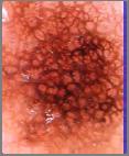
5 Regular pigment net Irregular pigment net Prominent pigment net Discrete pigment net Wide pigment net Narrow pigment net Broad pigment net Delicate pigment net Pseudopod Radial streaming White veil Black dots Brown globules White scar-like area Blue-grey area Hypopigmentation Reticular depigment Milia-like cyst Comedo-like opening Telangiectasia Red-blue areas Maple-leaf areas : Algorithms ABCD rules Stolz: Semin Cutan Med Surg 22:9, 2003 Menzies scoring method Menzies: Arch Dermatol 132:1178, 1996 CASH Algorithm Henning: J Am Acad Derm 56:45, point checklist system Argenziano: Arch Dermatol 134:1563, 1998 All systems perform equally! Arch Derm 141:1008, 2005 The first three are cumbersome. The 7-point checklist is predominant method. : Algorithms ABCD rules 77.5% Menzies scoring method 84.6% CASH Algorithm 68.4% 7-point checklist system 89.4% Relative sensitivity in the hands of a NON-expert Arch Dermatol 141:1008, Point Checklist MAJOR CRITERIA (2 points) Atypical pigment network Blue-white veil Atypical vascular pattern MINOR CRITERIA (1 point) Irregular streaks (streaming, pseudopods) Irregular pigmentation Irregular dots or globules Regression structures Total score > 3 highly suspect MM 7 Point Checklist MAJOR CRITERIA (2 points) Atypical pigment network Blue-white veil Atypical vascular pattern MINOR CRITERIA (1 point) Irregular streaks (streaming, pseudopods) Irregular pigmentation Irregular dots or globules Regression structures Total score > 3 highly suspect MM Fundamental structure Grid of connected pigmented lines Due to: pigmented rete ridges in the lower epidermis Pigment Network


6 Pigment Network Irregular Pigment Network Irregular Pigment Network Irregular Pigment Network Not symmetric and Not homogeneous Irregular (Atypical) Network Dots and Globules Fundamental structure Large to small spot-like pigment Due to: nests of pigment-laden cells at DEJ or in upper dermis Specificity 80-86% Sensitivity 30-35%


7 Dots and Globules Dots and Globules Irregular Dots and Globules Irregular Pigmentation Specificity 80% Sensitivity 50% Irregular shape Irregular distribution Multiple colors Due to: very random pigment throughout epidermis and dermis Specificity 46% Sensitivity approaches 100% Irregular Pigmentation Irregular Streaks At lesional edge Well defined structures Should NOT be there Radial streaming Finger-like extensions Pseudopods Bulbous extensions Due to peripheral expansion along DEJ

 A B C 96%")


8 Radial Streaming Radial Streaming Radial Streaming Pseudopods (types) A B C 96% specificity 18% sensitivity D E F-I Pseudopods Blue-White Veil Whitish-colored film overlying amorphous blue to blue-grey White: thickened epidermis Blue: heavily pigmented dermal melanocytes Should not be there in normal nevocellular or other growths Relatively specific for melanoma


9 Blue-White Veil Blue-White Veil Specificity 97% Sensitivity 51% Blue-White Veil Blue-White Veil Regression Structures Regression Structures Bone white areas Represent tumor regression? Response to immune attack Due to: loss of melanin fibrosis and/or


10 Regression Structures Atypical Vascular Pattern Specificity 92-93% Sensitivity 36-46% Least common Hardest to show Linear or dotted blood vessels Due to: dermal neovascularization That is: tumor induces its own blood supply Specificity 60-70% Sensitivity 8% in Action in Action Regression? 1 TOTAL SCORE = 2 BIOPSY = DYSPLASTIC NEVUS in Action Irregular Pigment

11 in Action TOTAL SCORE = 5 BOPSY: SSMM 1.1mm in Action TOTAL SCORE = 0 in Action in Action TOTAL SCORE = 0 BIOPSY = JUNCT L NEVUS in Action TOTAL SCORE = 5 BIOPSY = SSMM 0.33mm

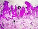
12 in Action in Action?Irregular pigment1 TOTAL SCORE = 0-1 BIOPSY = JUNCT L NEVUS Seborrheic keratosis Seborrheic Keratosis Comedone-like Plugs Milia-like Cysts


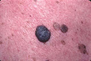
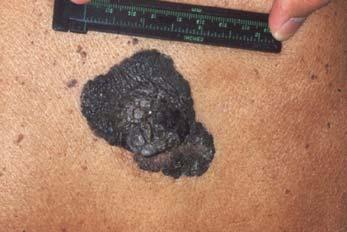
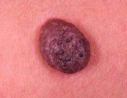
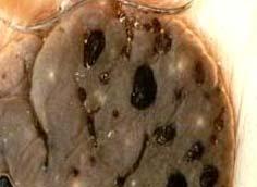
13 Seborrheic Keratoses & MM Search Carefully! SK MM Where is the dangerous lesion? What is this lesion? NOTE: Multiple red-blue globules This is pathognomonic for hemangioma What is this lesion? Senile angioma (Cherry angioma)
14 Easy to do and non-invasive Augments direct observation Careful scoring leads to reproducible results Helps distinguish lesions that must be removed or biopsied from those that remain or be observed can Other New Techniques Multispectral imaging Confocal scanning laser microscopy Ultrasound Optical coherence tomography Magnetic resonance imaging J Am Acad Derm 49:777-97, 2003 Johann Goethe What is the most difficult thing of all? That which seems the easiest: to see with your eyes that which lies right before your eyes.
Dermoscopy. Enhanced Diagnostic Ability: Pigmented Lesions. Ted Rosen, MD Baylor College of Medicine Houston, Texas
 Dermoscopy Enhanced Diagnostic Ability: Pigmented Lesions Ted Rosen, MD Baylor College of Medicine Houston, Texas Faculty Disclosure Statement No conflicts relevant to this workshop! Sir William Osler
22/04/2015. Dermoscopy of Melanoma. Ilsphi Browne. Overview
 Dermoscopy of Melanoma Ilsphi Browne Overview The device Dermoscopic criteria (terminology) Colour Patterns Global features Local features Approach to diagnosing pigmented lesions Other uses in general
Appendix : Dermoscopy
 Go Back to the Top To Order, Visit the Purchasing Page for Details APP Appendix : Dermoscopy Dermoscopy, also known as dermatoscopy, epiluminoscopy and epiluminescent microscopy, is an effective non-invasive
Disclosure. Objectives. PAFP CME Conference Lou Mancano MD, FAAFP Reading Health System November 18, 2016
 PAFP CME Conference Lou Mancano MD, FAAFP Reading Health System November 18, 2016 1 Disclosure The speaker has no conflict of interest, financial agreement, or working affiliation with any group or organization.
Dermoscopy: Recognizing Top Five Common In- Office Diagnoses
 Dermoscopy: Recognizing Top Five Common In- Office Diagnoses Vu A. Ngo, DO Department of Family Medicine and Dermatology Choctaw Nation Health Services Authority Learning Objectives Introduction to dermoscopy
Basics in Dermoscopy
 Basics in Dermoscopy Manal Bosseila Professor of Dermatology, Cairo University Member of European Academy Dermatology & Venereology EADV Member of International Dermoscopy Society IDS Member of Aesthetic
6/17/2018. Breaking Bad (Part 1) Dermoscopy of Brown(ish) Things. Bad?
 Dermoscopy of Brown(ish) Things. Bad?") Breaking Bad (Part 1) Dermoscopy of Brown(ish) Things Jennie T. Clarke, MD ssociate Professor of Dermatology University of Utah School of Medicine Bad? 1 Brown(ish) Things Bad Melanoma Pigmented basal
Breaking Bad (Part 1) Dermoscopy of Brown(ish) Things Jennie T. Clarke, MD ssociate Professor of Dermatology University of Utah School of Medicine Bad? 1 Brown(ish) Things Bad Melanoma Pigmented basal
Dermoscopy Quiz 3-Point Checklist Algorithm
 Dermoscopy Quiz 3-Point Checklist Algorithm GLOBAL PATTERN Globular LOCAL CRITERIA Aggregated globules Milia-like cysts 3 POINT CHECK LIST Symmetrical No abnormal net Slight Blue-white veil BENIGN MELANOCYTIC
Introduction to Dermoscopy. Nicholas Compton, MD June 16, 2010
 Introduction to Dermoscopy Nicholas Compton, MD June 16, 2010 Overview What is dermoscopy Brief history Types of dermoscopy General approach to lesion of interest 2 step algorithm 3-point checklist Practice
What is Dermoscopy? Early Dermoscopes. Deciphering Dermoscopy: Terminology, Features & Algorithms 6/17/2018
 Deciphering Dermoscopy: Terminology, Features & Algorithms Where did it come from and why do we use it? Jennie T. Clarke, MD Associate Professor of Dermatology University of Utah School of Medicine What
INTRODUCTION HOUSEKEEPING June 11 th Dr John Adams Dermatologist/Dermoscopist MOLEMAP NZ/Australia MOLESAFE USA
 INTRODUCTION HOUSEKEEPING June 11 th 2015 Dr John Adams Dermatologist/Dermoscopist MOLEMAP NZ/Australia MOLESAFE USA Program Skin cancer statistics. Dermoscopy description and usefulness. Patient /lesion
Malignant non-melanocytic lesions
 Malignant non-melanocytic lesions Course C023: Fundamentals of Dermoscopy March 4, 2019, 11:20 AM - 11:50 PM Room: 146B Jason B. Lee, MD Professor & Vice Chair Director of Dermatopathology & Pigmented
Clinical and Dermoscopic Features of Thin Nodular Melanoma
 Clinical and Dermoscopic Features of Thin Nodular Melanoma A study of the International Dermoscopy Society Coordinator: Dr. Alexander J. Stratigos and colleagues, alstrat2@gmail.com ** Extended to May
Fundamentals of dermoscopy
 Fundamentals of dermoscopy Learning objectives Upon completion of this session, participants should be able to: describe the basic principles of dermoscopy identify features associated with pigmented and
MODULE 1. LOCAL AND GENERAL CRITERIA IN PIGMENTED MELANOCYTIC LESIONS.
 DERMOSCOPY TEACHING PROGRAMME Dermoscopy Teaching Programme Module 1 MODULE 1. LOCAL AND GENERAL CRITERIA IN PIGMENTED MELANOCYTIC LESIONS. Dermoscopy is a non-invasive in vivo technique that provides
Total body photography in high risk patients
 Total body photography in high risk patients Doug Grossman, MD, PhD Department of Dermatology Huntsman Cancer Institute University of Utah Summer AAD F032 Practical Considerations for Patients with Melanoma
Skin Cancer A Personal Approach. Dr Matthew Strack Dunedin New Zealand
 Skin Cancer A Personal Approach Dr Matthew Strack Dunedin New Zealand Outline Dermoscopy Instruments and setup Photochemosurgery Clinical Aim: Leave with 2-3 ideas JLE Benign Junctional Nevus Management
DIFFERENCES IN DERMOSCOPIC IMAGES FROM NON-POLARIZED DERMOSCOPE AND POLARIZED DERMOSCOPE INFLUENCE THE DIAGNOSTIC ACCURACY AND CONFIDENCE LEVEL.
 DIFFERENCES IN DERMOSCOPIC IMAGES FROM NON-POLARIZED DERMOSCOPE AND POLARIZED DERMOSCOPE INFLUENCE THE DIAGNOSTIC ACCURACY AND CONFIDENCE LEVEL. 1. Steven Q. Wang MD 1 (wangs@mskcc.org) 2. Stephen W. Dusza
comedo-like openings (clods, brown or orange & circles) milia-like cysts (dots or clods, white) 1/29/18 Dotted vessels are also commonly seen in SCC
 milia-like cysts (dots or clods, white) 1/29/18 Dotted vessels are also commonly seen in SCC") Brown circles Dotted vessels are also commonly seen in SCC Step1 1. Nevus (unequivocal) 2. DF/IDN 3. BCC 4. SCC Network Patchy network Peripheral network & central hypopigmentation DF: network with central
Brown circles Dotted vessels are also commonly seen in SCC Step1 1. Nevus (unequivocal) 2. DF/IDN 3. BCC 4. SCC Network Patchy network Peripheral network & central hypopigmentation DF: network with central
STUDY. Scott W. Menzies, MB,BS, PhD; Karin Westerhoff, MD; Harold Rabinovitz, MD; Alfred W. Kopf, MD; William H. McCarthy, MBBS, MEd; Brian Katz
 STUDY Surface Microscopy of Pigmented Basal Cell Carcinoma Scott W. Menzies, MB,BS, PhD; Karin Westerhoff, MD; Harold Rabinovitz, MD; Alfred W. Kopf, MD; William H. McCarthy, MBBS, MEd; Brian Katz Objectives:
10/3/2018. Dermoscopy: Looking beneath the surface of the skin. Dermoscopy for Family Medicine 10/11/2018
 Dermoscopy for Family Medicine 10/11/2018 Jane M. Grant-Kels, MD, FAAD Founding Chair Emeritus, Dept of Dermatology Professor of Dermatology, Pathology & Pediatrics Director of the Cut Oncology Ctr & Melanoma
STUDY. Epiluminescence Microscopy for the Diagnosis of Doubtful Melanocytic Skin Lesions
 STUDY Epiluminescence Microscopy for the Diagnosis of Doubtful Melanocytic Skin Lesions Comparison of the ABCD Rule of Dermatoscopy and a New 7-Point Checklist Based on Pattern Analysis Giuseppe Argenziano,
Dermoscopy in everyday practice. What and Why? When in doubt cut it out? Trilokraj Tejasvi MD
 Dermoscopy in everyday practice Trilokraj Tejasvi MD Assistant Professor, Department of Dermatology, Director Teledermatology services, University of Michigan, Faculty Associate, GLOBAL REACH, Michigan
Yes. Breaking Bad II: Dermoscopy of Pink-ish Things. Does it Fit? Yes 6/17/2018. Yes. Joslyn Kirby, MD, MS, MEd
 Breaking Bad II: Dermoscopy of Pink-ish Things Joslyn Kirby, MD, MS, MEd Yes Observe Yes Step 2. Fit a Benign Nevus Pattern? Does it Fit? Step 1: Melanocytic? pigment network, globules, homogeneous? No
The impact of GP sub-specialisation and dermatoscopy use on diagnostic accuracy for melanomas in Australia
 The impact of GP sub-specialisation and dermatoscopy use on diagnostic accuracy for melanomas in Australia Cliff Rosendahl, Gail Williams, Diann Eley, Tobias Wilson, Greg Canning, Jeffrey Keir, Ian McColl,
Dermoscopy-a BRIEF introduction
 Dermoscopy-a BRIEF introduction Aim of presentation -to tell you what dermoscopy is -to show some of what it can do -point the interested learner to further resources Overview of dermoscopy Dermoscopy
Key factors in successfully integrating dermoscopy into your clinical practice
 Key factors in successfully integrating dermoscopy into your clinical practice S051 Dilemmas and challenges in skin cancer therapies and management Monday, March 4 th 2019 (9AM-12PM) Room 209A 10:56-11:09AM
Dermoscopy STFM Richard Usatine, MD 5/2/16. Disclosure Statement: Some Dermatoscopes. Dermoscopy Video. Thanks to Dr.
 Disclosure Statement: Dermoscopy STFM 2016 Richard P. Usatine, MD, FAAFP Professor, Family and Community Medicine Professor, Dermatology and Cutaneous Surgery Medical Director, Clinic University of Texas
Abrupt Intralesional Color Change on Dermoscopy as a New Indicator of Early Superficial Spreading Melanoma in a Japanese Woman
 Published online: June 24, 2015 1662 6567/15/0072 0123$39.50/0 This is an Open Access article licensed under the terms of the Creative Commons Attribution-NonCommercial 3.0 Unported license (CC BY-NC)
Non-Melanocytic Pattern Dermoscopy
 Non-Melanocytic Pattern Dermoscopy I have no conflicts of interest to disclose Except that I LOVE dermoscopy Michelle Tarbox, MD Assistant Professor of Dermatology and Dermatopathology Texas Tech University
Dermoscopy. Synonyms. Dermoscopy. Definition. Dermoscopy opens up a world of colour and structure that can t be seen with the naked eye
 Synonyms Dermoscopy Australasian College of Dermatologists G.P Training Module Dermoscopy Dermatoscopy Epiluminescence microscopy Skin surface microscopy Incident light microscopy Oil immersion microscopy
Diagnosis of Lentigo Maligna Melanoma. Steven Q. Wang, M.D. Memorial Sloan-Kettering Cancer Center Basking Ridge, NJ
 Diagnosis of Lentigo Maligna Melanoma Steven Q. Wang, M.D. Memorial Sloan-Kettering Cancer Center Basking Ridge, NJ Conflict of Interest: None Topics Epidemiology and Natural History Clinical and Histologic
Non-melanocytic Patterns
 Non-melanocytic Lesions Non-melanocytic Patterns Michelle Tarbox, MD Assistant Professor of Dermatology and Dermatopathology Texas Tech University Health Sciences Center 2018 Seborrheic keratoses Acanthotic
Regression 2/3/18. Histologically regression is characterized: melanosis fibrosis combination of both. Distribution: partial or focal!
 Regression Margaret Oliviero MSN, ARNP Harold S. Rabinovitz MD Histologically regression is characterized: melanosis fibrosis combination of both Distribution: partial or focal! Dermatoscopic terminology
Dermoscopy, the use of a handheld
 ONLINE EXCLUSIVE Dermoscopy in family medicine: A primer Dermoscopy allows you to see deeper into the skin than with the naked eye. Here s how you can make use of it to spot malignant conditions sooner.
STUDY. Identification of Clinically Featureless Incipient Melanoma Using Sequential Dermoscopy Imaging
 STUDY Identification of Clinically Featureless Incipient Melanoma Using Sequential Dermoscopy Imaging Harald Kittler, MD; Pascale Guitera, MD; Elisabeth Riedl, MD; Michelle Avramidis, MD; Ligia Teban,
STUDY. Characteristic Epiluminescent Microscopic Features of Early Malignant Melanoma on Glabrous Skin
 Characteristic Epiluminescent Microscopic Features of Early Malignant Melanoma on Glabrous Skin A Videomicroscopic Analysis STUDY Shinji Oguchi, MD; Toshiaki Saida, MD, PhD; Yoko Koganehira, MD; Sachiko
Acral and Mucosal Dermoscopy
 Acral and Mucosal Dermoscopy Caroline C. Kim, MD Assistant Professor, Department of Dermatology Harvard Medical School Director, Pigmented Lesion Clinic Associate Director, Cutaneous Oncology Program Beth
Diagnostic Dermatology & Dermatologic Surgery Overview
 Diagnostic Dermatology & Dermatologic Surgery Overview A comprehensive range of innovative products & accessories for dermatologic diagnosis & surgery Clinical imaging The Fully integrated patient management
Graph-based Pigment Network Detection in Skin Images
 Graph-based Pigment Network Detection in Skin Images M. Sadeghi a,b, M. Razmara a, M. Ester a, T. K. Lee a,b,c, M. S. Atkins a a Simon Fraser University, 8888 University Drive, Burnaby, BC, Canada, V5A1S6;
Case Report Micromelanomas: A Review of Melanomas 2mmand a Case Report
 Case Reports in Oncological Medicine, Article ID 206260, 4 pages http://dx.doi.org/10.1155/2014/206260 Case Report Micromelanomas: A Review of Melanomas 2mmand a Case Report Sharad P. Paul 1,2,3 1 Skin
It can be helpful in some cases of actinic keratosis, Bowen s disease and squamous cell carcinoma
 Dermoscopy Introduction, Terminology and Structures (to be read in conjunction with the Diagnostic Dermoscopic Algorithm) Copyright to Cunliffe TP (Jan. 2017) All rights reserved Introduction Dermoscopy
Multiple Primary Melanoma in a Thai Male: A Case Report
 Case Report Multiple Primary Melanoma in a Thai Male: A Case Report J Med Assoc Thai 2014; 97 (Suppl. 2): S234-S238 Full text. e-journal: http://www.jmatonline.com Kittisak Payapvipapong MD*, Pinyapat
Development and validation of a scoring system for SIAscopic diagnosis of pigmented skin lesions in primary care
 Development and validation of a scoring system for SIAscopic diagnosis of pigmented skin lesions in primary care J Hunter 1,2, FM Walter 3,5, M Moncrieff 1, S Cotton 4 PN Hall 1, J Emery 3,5 1 Dept of
Optical Diagnostic Devices for Evaluating Skin Lesions Suspected of Malignancy. Original Policy Date
 MP 2.01.29 Optical Diagnostic Devices for Evaluating Skin Lesions Suspected of Malignancy Medical Policy Section Medicine Issue 12:2013 Original Policy Date 12:2013 Last Review Status/Date Reviewed with
MELANOMA: HANDS-ON OR HANDS-OFF?
 MELANOMA: HANDS-ON OR HANDS-OFF? M SCHAMM MBChB (Pret), FCS (SA) Endocrine and Transplant Surgeon, Department of Surgery, University of the Witwatersrand; and Clinical Head Transplant Surgery, Charlotte
Aspects on in vivo imaging techniques for diagnostics of pigmented skin lesions
 Thesis for the Degree of Doctor of Philosophy Aspects on in vivo imaging techniques for diagnostics of pigmented skin lesions Karin Terstappen (Westerhoff) Department of Dermatology and Venereology Institure
Pathology of the skin. 2nd Department of Pathology, Semmelweis University
 Pathology of the skin 2nd Department of Pathology, Semmelweis University Histology of the skin Epidermis: Stratum corneum Stratum granulosum Stratum spinosum Stratum basale Dermis: papillary and reticular
Page: 1 of 16. Optical Diagnostic Devices for Evaluating Skin Lesions Suspected of Malignancy
 Last Review Status/Date: December 2014 Page: 1 of 16 Skin Lesions Suspected of Malignancy Description There is interest in non-invasive devices that will improve the diagnosis of malignant skin lesions.
Melanoma and Dermoscopy. Disclosure Statement: ABCDE's of melanoma. Co-President, Usatine Media
 Melanoma and Dermoscopy Richard P. Usatine, MD, FAAFP Professor, Family and Community Medicine Professor, Dermatology and Cutaneous Surgery Medical Director, University Skin Clinic University of Texas
Associate Clinical Professor of Dermatology MUSC
 Re-excision of Moderately Dysplastic Nevi: Should we or shouldn t we? John C. Maize, Jr, M.D. Dermatologist and Dermatopathologist Trident Dermatology, Charleston SC Associate Clinical Professor of Dermatology
Benign and malignant epithelial lesions: Seborrheic keratosis: A common benign pigmented epidermal tumor occur in middle-aged or older persons more
 Benign and malignant epithelial lesions: Seborrheic keratosis: A common benign pigmented epidermal tumor occur in middle-aged or older persons more common on the trunk; but extremities, head and neck are
F006 Imaging in Dermatology Melanocytic Neoplasia Clinical-Confocal-Pathological-Correlations
 F006 Imaging in Dermatology Melanocytic Neoplasia Clinical-Confocal-Pathological-Correlations Melissa Gill, MD SkinMedical Research and Diagnostics Dobbs Ferry, NY, USA Department of Pathology SUNY Downstate
Description of Some Dermatoscopic Features of Acral Pigmented Lesions in Iranian Patients: A Preliminary Study
 ORIGINAL REPORT Description of Some Dermatoscopic Features of Acral Pigmented Lesions in Iranian Patients: A Preliminary Study Reza Nemati Ahmadabad 1, Hayede Ghaninezhad 1, Homayoon Moslehi 2, Sahar Azizahari
Introduction to Dermoscopy. Disclosure. Introduction
 Introduction to Dermoscopy 1 Disclosure Dr. Deborah Bren has no conflict of interest, financial agreement, or working affiliation with any group or organization. 2 Introduction Deborah A. Bren, DO Family
Optical Diagnostic Devices for Evaluating Skin Lesions Suspected of Malignancy
 2.01.42 Optical Diagnostic Devices for Evaluating Skin Lesions Suspected of Malignancy Section 2.0 Medicine Subsection Effective Date November 26, 2014 Original Policy Date November 26, 2014 Next Review
Dermatopathology: The tumor is composed of keratinocytes which show atypia, increase mitoses and abnormal mitoses.
 Squamous cell carcinoma (SCC): A common malignant tumor of keratinocytes arising in the epidermis, usually from a precancerous condition: 1- UV induced actinic keratosis, usually of low grade malignancy.
Cover Page. The handle holds various files of this Leiden University dissertation.
 Cover Page The handle http://hdl.handle.net/1887/22172 holds various files of this Leiden University dissertation. Author: Rhee, Jasper Immanuel van der Title: Clinical characteristics and management of
IV.4. Early Evolution of Melanoma (Small-Diameter Melanoma)
") Chapter Early Evolution of Melanoma (Small-Diameter Melanoma) Robert J. Friedman, Melanie Warycha, Michele Farber, Dina Gutkowicz-Krusin, Harold Rabinovitz, David Polsky, Margaret Oliviero, Darrell S.
Chapter Early Evolution of Melanoma (Small-Diameter Melanoma) Robert J. Friedman, Melanie Warycha, Michele Farber, Dina Gutkowicz-Krusin, Harold Rabinovitz, David Polsky, Margaret Oliviero, Darrell S.
المركب النموذج--- سبيتز وحمة = Type Spitz's Nevus, Compound SPITZ NEVUS 1 / 7
 SPITZ NEVUS 1 / 7 Epidemiology An annual incidence rate of 1.4 cases of Spitz nevus per 100,000 individuals has been estimated in Australia, compared with 25.4 per 100,000 individuals for cutaneous melanoma
Prior Authorization Review Panel MCO Policy Submission
 Prior Authorization Review Panel MCO Policy Submission A separate copy of this form must accompany each policy submitted for review. Policies submitted without this form will not be considered for review.
Revised Pattern Analysis: a method for the accurate diagnosis of pigmented skin lesions
 Dermatoscopy for Students A concise outline of: Revised Pattern Analysis: a method for the accurate diagnosis of pigmented skin lesions And Chaos and Clues: a decision algorithm for routine practice to
Dermoscopic Features of Non-Pigmented Eccrine Poromas in. Department of Dermatology, Shinshu University School of Medicine,
 Original article Dermoscopic Features of Non-Pigmented Eccrine Poromas in Association with their Histopathological Features Akane Minagawa, Hiroshi Koga,* Masaomi Takahashi, + Kenji Sano, + Ryuhei Okuyama,
Age-related prevalence of dermatoscopic patterns of acral melanocytic nevi
 DERMATOLOGY PRACTICAL & CONCEPTUAL www.derm101.com Age-related prevalence of dermatoscopic patterns of acral melanocytic nevi Reiko Suzaki 1, Sumiko Ishizaki 1, Hitoshi Iyatomi 2, Masaru Tanaka 1 1 Department
Benign versus Cancerous Lesions How to tell the difference FMF 2014 Christie Freeman MD, CCFP, DipPDerm, MSc
 1 Benign versus Cancerous Lesions How to tell the difference FMF 2014 Christie Freeman MD, CCFP, DipPDerm, MSc Benign lesions Seborrheic Keratoses: Warty, stuck-on Genetics and birthdays Can start in late
EARLY ONLINE RELEASE
 EARLY ONLINE RELEASE Note: This article was posted on the Archives Web site as an Early Online Release. Early Online Release articles have been peer reviewed, copyedited, and reviewed by the authors. Additional
50 interactive dermoscopic case discussions Dr Stephen Hayes
 50 interactive dermoscopic case discussions Dr Stephen Hayes Annotations will be found on your memory drive, as will 100 case discussions and other learning material Melanoma 2mm thick Ugly duckling-one
ORIGINAL ARTICLE. 980 Journal of Investigative Dermatology (2006), Volume 126 & 2006 The Society for Investigative Dermatology
, Volume 126 & 2006 The Society for Investigative Dermatology") ORIGINAL ARTICLE Results from an Observational Trial: Digital Epiluminescence Microscopy Follow-Up of Atypical Nevi Increases the Sensitivity and the Chance of Success of Conventional Dermoscopy in Detecting
ORIGINAL ARTICLE Results from an Observational Trial: Digital Epiluminescence Microscopy Follow-Up of Atypical Nevi Increases the Sensitivity and the Chance of Success of Conventional Dermoscopy in Detecting
BJD British Journal of Dermatology. Summary. What s already known about this topic? CLINICAL AND LABORATORY INVESTIGATIONS
 CLINICAL AND LABORATORY INVESTIGATIONS BJD British Journal of Dermatology Pigmented nodular melanoma: the predictive value of dermoscopic features using multivariate analysis M.A. Pizzichetta, 1 H. Kittler,
The most common mistakes on dermatoscopy of melanocytic lesions
 Review paper The most common mistakes on dermatoscopy of melanocytic lesions Grażyna Kamińska-Winciorek 1, Waldemar Placek 2 1 The Center for Diagnostics and Treatment of Skin Diseases, Katowice, Poland
Phoebe Rich MD Adjunct Professor OHSU Portland, Oregon
 Nail Tips for Diagnosis and Management of Nail Disorders Winter Clinical Dermatology Conference 2017 Hawaii Phoebe Rich MD Adjunct Professor OHSU Portland, Oregon Objectives diagnostic clues for benign
Toby Maurer, MD University of California, San Francisco. Lifetime risk of an American developing melanoma
 Distinguishing Pigmented Skin Lesions and Melanoma Toby Maurer, MD University of California, San Francisco Epidemiology of Melanoma Lifetime risk of an American developing melanoma 1935: 1 in 1500 1980:
DERMATOLOGY PRACTICAL & CONCEPTUAL. Introduction. Dermoscopy. Hiroshi Sakai 1, Kyoko Tonomura 1, Hirotsugu Shirabe 1, Masaru Tanaka 2
 DERMATOLOGY PRACTICAL & CONCEPTUAL www.derm101.com Assessment of the colors of melanin pigment in acral compound nevus by using a novel dermoscopy technique with surgical light illumination and saturation
Toby Maurer, MD University of California, San Francisco. Lifetime risk of an American developing melanoma
 Distinguishing Pigmented Skin Lesions and Melanoma Toby Maurer, MD University of California, San Francisco Epidemiology of Melanoma Lifetime risk of an American developing melanoma 1935: 1 in 1500 1980:
Features Causing Confusion between Basal Cell Carcinoma and Squamous Cell Carcinoma in Clinical Diagnosis
 TH Ryu, et al pissn 1013-9087ㆍeISSN 2005-3894 Ann Dermatol Vol. 30, No. 1, 2018 https://doi.org/10.5021/ad.2018.30.1.64 ORIGINAL ARTICLE Features Causing Confusion between Basal Cell Carcinoma and Squamous
INCREASE IN incidence and mortality rates for
 Skin Research and Technology 2005; 11: 236 241 Copyright & Blackwell Munksgaard 2005 Printed in Denmark. All rights reserved Skin Research and Technology Pigment distribution in melanocytic lesion images:
The Evolution of Melanoma Diagnosis: 25 Years Beyond the ABCDs
 CA CANCER J CLIN 2010;60:301 316 The Evolution of Melanoma Diagnosis: 25 Years Beyond the ABCDs Darrell S. Rigel, MD 1 ; Julie Russak, MD 2 ; Robert Friedman, MD 3 Abstract Early detection of malignant
Case Report Dermoscopy Clues in Pigmented Bowen s Disease
 Dermatology Research and Practice Volume 2010, Article ID 464821, 9 pages doi:10.1155/2010/464821 Case Report Dermoscopy Clues in Pigmented Bowen s Disease Daniela Gutiérrez-Mendoza, 1 Roberto Narro-Llorente,
Morphologic Features of Melanocytes, Pigmented Keratinocytes, and Melanophages by In Vivo Confocal Scanning Laser Microscopy
 Morphologic Features of Melanocytes, Pigmented Keratinocytes, and Melanophages by In Vivo Confocal Scanning Laser Microscopy Klaus J. Busam, M.D., Carlos Charles, M.D., Grace Lee, M.D., Allan C Halpern,
NEW. DELTA 20 Plus Dermatoscope Head
 04 MOre details FOR an even better diagnosis: POLARISATION, HIGH PERFORMANCE LEDs and HIGH RESOLUTION optics. While the rate of new cases of malignant melanoma is increasing worldwide, mortality rates
DERMATOLOGY PRACTICAL & CONCEPTUAL. Gabriel Salerni 1,2, Teresita Terán 3, Carlos Alonso 1,2, Ramón Fernández-Bussy 1 ABSTRACT
 DERMATOLOGY PRACTICAL & CONCEPTUAL www.derm101.com The role of dermoscopy and digital dermoscopy follow-up in the clinical diagnosis of melanoma: clinical and dermoscopic features of 99 consecutive primary
Multispectral Digital Skin Lesion Analysis. Summary
 Subject: Multispectral Digital Skin Lesion Analysis Page: 1 of 8 Last Review Status/Date: March 2016 Multispectral Digital Skin Lesion Analysis Summary There is interest in noninvasive devices that will
MECHANISMS OF HUMAN DISEASE: LABORATORY SESSION PATHOLOGY OF THE SKIN LAB. Friday, February 13, :30 am 11:00 am
 MECHANISMS OF HUMAN DISEASE: LABORATORY SESSION PATHOLOGY OF THE SKIN LAB Friday, February 13, 2009 9:30 am 11:00 am FACULTY COPY GOALS: Describe the basic clinical and morphologic features of various
Multispectral Digital Skin Lesion Analysis
 Multispectral Digital Skin Lesion Analysis Policy Number: 2.01.101 Last Review: 2/2018 Origination: 2/2016 Next Review: 8/2018 Policy Blue Cross and Blue Shield of Kansas City (Blue KC) will not provide
MECHANISMS OF HUMAN DISEASE: LABORATORY SESSION PATHOLOGY OF THE SKIN LAB. Friday, February 12, :30 am 11:00 am
 MECHANISMS OF HUMAN DISEASE: LABORATORY SESSION PATHOLOGY OF THE SKIN LAB Friday, February 12, 2012 9:30 am 11:00 am FACULTY COPY GOALS: Describe the basic clinical and morphologic features of various
Mole mapping and monitoring. Dr Stephen Hayes. Associate Specialist in Dermatology, University Hospital Southampton
 Mole mapping and monitoring Dr Stephen Hayes Associate Specialist in Dermatology, University Hospital Southampton Outline of presentation The melanoma epidemic Benefits of early detection Risks of the
Algorithmic reproduction of asymmetry and border cut-off parameters according to the ABCD rule for dermoscopy
 JEADV ISSN 1468-3083 Blackwell Publishing Ltd ORIGINAL ARTICLE Algorithmic reproduction of asymmetry and border cut-off parameters according to the ABCD rule for dermoscopy G Pellacani,* C Grana, S Seidenari
Dermatoscopic features of cutaneous non-facial non-acral lentiginous growth pattern melanomas
 DERMATOLOGY PRACTICAL & CONCEPTUAL www.derm101.com Dermatoscopic features of cutaneous non-facial non-acral lentiginous growth pattern melanomas Jeff Keir 1 1 Department of Dermatology, School of Medicine,
Common Benign Lesions and Skin Cancers. 22nd May 2015 Dr Mark Foley
 Common Benign Lesions and Skin Cancers 22nd May 2015 Dr Mark Foley Thank you for downloading this file. This intended to supplement the presentation given at the NZ Wound Care Conference, it is not intended
*Please see amendment for Pennsylvania Medicaid at the end
 1 of 32 Number: 0188 Policy *Please see amendment for Pennsylvania Medicaid at the end of this CPB. I. Aetna considers total body photography (TBP) and dermoscopy (also known as total body imaging, digital
Prediction without Pigment: a decision algorithm for non-pigmented skin malignancy
 DERMATOLOGY PRACTICAL & CONCEPTUAL www.derm101.com Prediction without Pigment: a decision algorithm for non-pigmented skin malignancy Cliff Rosendahl 1, Alan Cameron 1, Philipp Tschandl 2, Agata Bulinska
HEINE Dermatoscopes. Magnification Magnification x 40x* (digital)
") [ 079 ] HEINE DELTA 20 T ic 1 NEW: NC 2 mini 3000 Magnification Magnification 10 16 x 40x* (digital) 6 10 x 30x* (digital) 10 x Illumination illumination XHL illumination Toggle function Examination mode
[ 079 ] HEINE DELTA 20 T ic 1 NEW: NC 2 mini 3000 Magnification Magnification 10 16 x 40x* (digital) 6 10 x 30x* (digital) 10 x Illumination illumination XHL illumination Toggle function Examination mode
:- HEINE IC1 NEW! 21st Century Digital Dermatoscopy
 :- HEINE IC1 21st Century Digital Dermatoscopy The HEINE ic1 is a new image capturing device to record clinical and dermatoscopic images for documenting, monitoring and supporting differential diagnosis
Evaluation of electrical impedance spectroscopy as an adjunct to dermoscopy in short-term monitoring of atypical melanocytic lesions
 DERMATOLOGY PRACTICAL & CONCEPTUAL www.derm101.com Evaluation of electrical impedance spectroscopy as an adjunct to dermoscopy in short-term monitoring of atypical melanocytic lesions Hannah Ceder 1, Alexandra
Supplementary Online Content
 Supplementary Online Content Chernoff KA, Marghoob AA, Lacouture ME, Deng L, Busam KJ, Myskowski PL. Dermoscopic findings in cutaneous metastases. JAMA Dermatol. Published online January 15, 2014. doi:10.1001/jamadermatol.2013.8502
Triage amalgamated dermoscopic algorithm (TADA) for skin cancer screening
 for skin cancer screening") DERMATOLOGY PRACTICAL & CONCEPTUAL www.derm101.com Triage amalgamated dermoscopic algorithm (TADA) for skin cancer screening Tova Rogers 1, Maria Marino 1, Stephen W. Dusza 1, Shirin Bajaj 1, Michael A.
DERMATOLOGY PRACTICAL & CONCEPTUAL www.derm101.com Triage amalgamated dermoscopic algorithm (TADA) for skin cancer screening Tova Rogers 1, Maria Marino 1, Stephen W. Dusza 1, Shirin Bajaj 1, Michael A.
R J M E Romanian Journal of Morphology & Embryology
 Rom J Morphol Embryol 2013, 54(2):315 320 ORIGINAL PAPER R J M E Romanian Journal of Morphology & Embryology http://www.rjme.ro/ Correlation of dermatoscopy with the histopathological changes in the diagnosis
Skin lesions The Good and the Bad. Dr Virginia Hubbard Ipswich Hospital NHS Trust Barts and the London School of Medicine and Dentistry
 Skin lesions The Good and the Bad Dr Virginia Hubbard Ipswich Hospital NHS Trust Barts and the London School of Medicine and Dentistry Case 1 32 year old woman Australian Lesion on back New hair growing
Conflicts. Objectives. University of Texas Health Science Center at San Antonio. Pediatrics Grand Rounds 24 August Pediatric Dermatology 101
 Pediatric Dermatology 101 John C. Browning, MD, FAAD, FAAP Conflicts Investigator: ViroXis Advisor: ViroXis Advisory Board: TopMD Speaker: Galderma Objectives Understand the meaning and importance of cutaneous