50 interactive dermoscopic case discussions Dr Stephen Hayes
|
|
|
- Donna Crawford
- 6 years ago
- Views:
Transcription
1 50 interactive dermoscopic case discussions Dr Stephen Hayes Annotations will be found on your memory drive, as will 100 case discussions and other learning material
2
3
4
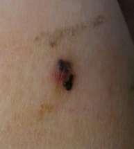
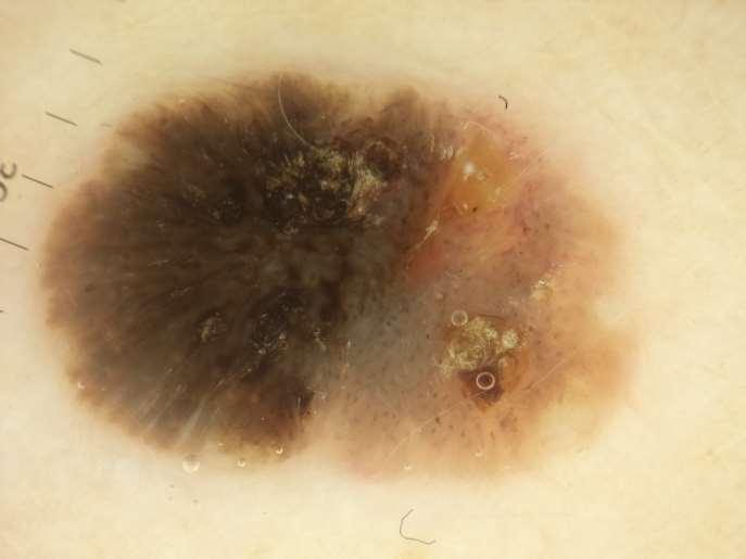
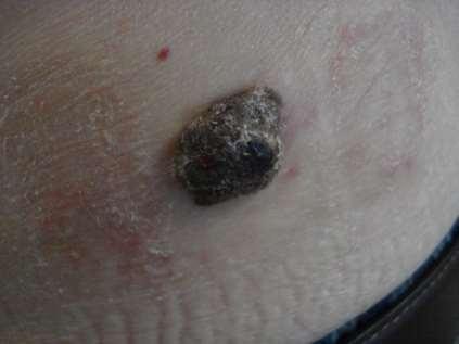
5 Melanoma 2mm thick Ugly duckling-one look is enough Gross asymmetry of shape, colour Dermoscopy is chaotic 2 distinct sections of lesion Inverse/reverse network Shiny white streaks PINK COLOUR BEWARE
6

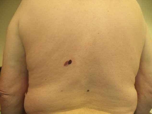
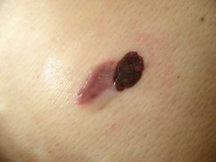
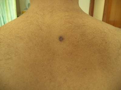
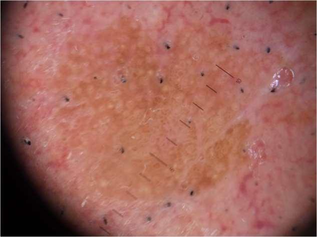
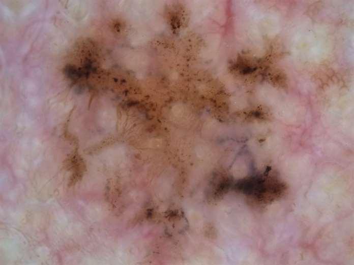
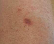
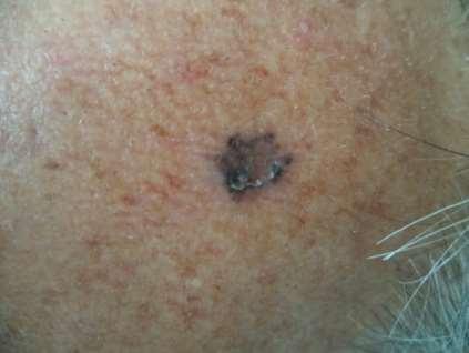
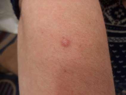
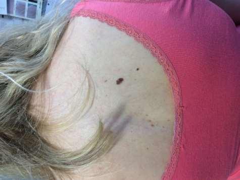
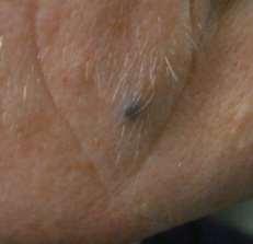
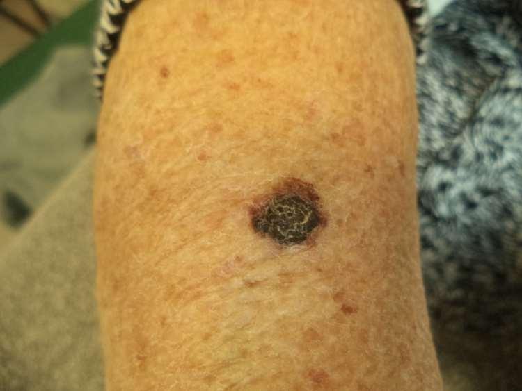
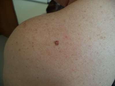
7 Lichen planus (? Lichenoid reaction to Solitary new, changing lesion in deeply pigmented skin-do not rule out a skin cancer although not common -however, a decision was made to biopsy rather than excise, to get right margns and also in case it was inflammatory rather than neoplastic (avoid keloid scar). This proved a wise decision. insect bite)
8
9
10
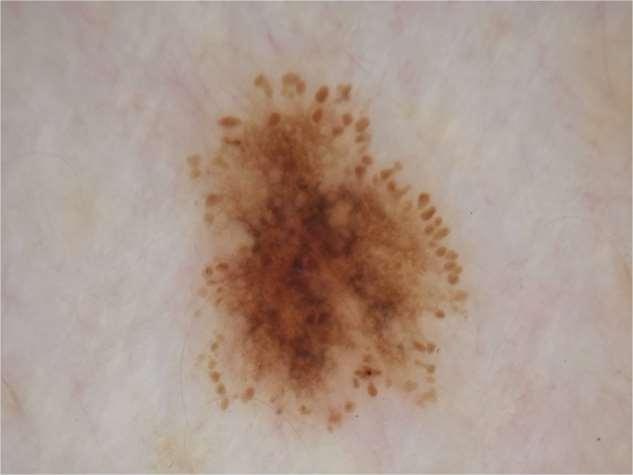
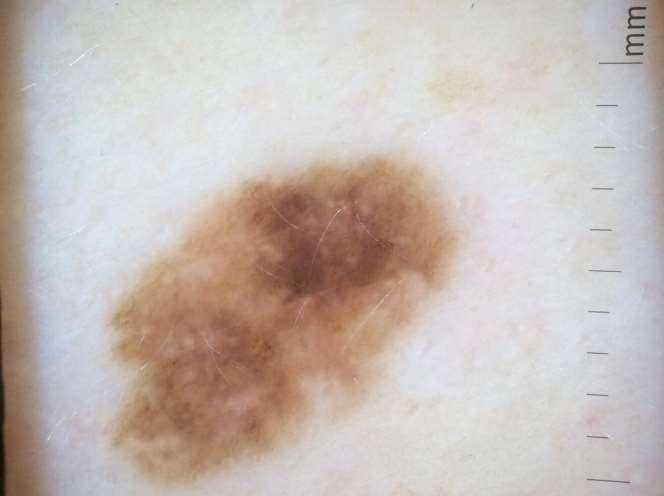
11 Dysplastic naevus
12
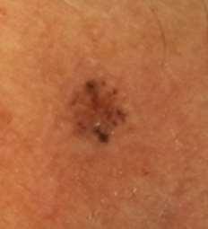
13 Traumatised seb keratosis Well organised keratin Looped vessels Well defined border
14
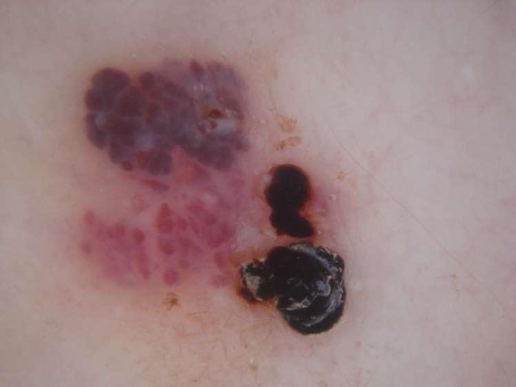
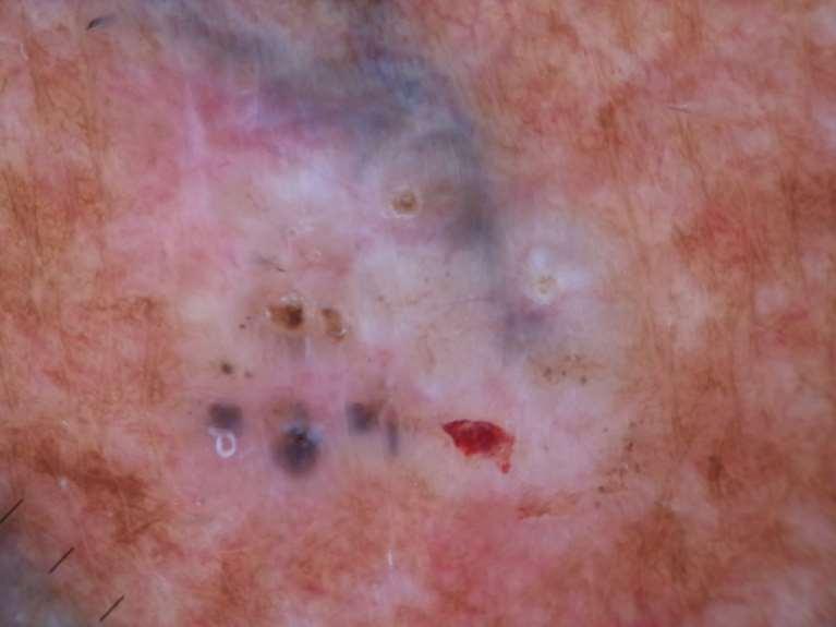
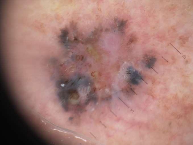
15 Chaotic-multiple structures and colours Black blotches/clods and 5 and 11 o clock Irregular dots and globules scattered through lesion NB 2 experienced doctors were unsure whether pigmented BCC or lentigo maligna, but both agreed urgent excision was needed Lentigo maligna
16

17 Sebaceous gland hyperplasia Often multiple Typically seen on elderly forehead Rarely bigger than 4mm Can be confused with small popular BCCs White or yellow clods crown vessels, do not cross centre of lesion
18
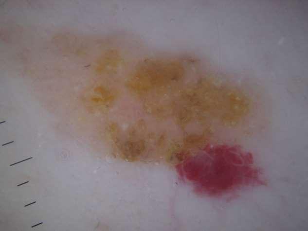
19
 or BCC-therefore melanoma by default.")
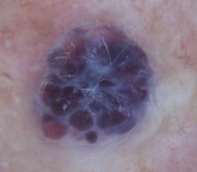
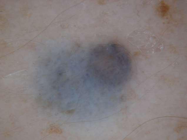
20 melanoma Ugly duckling +++ Muktiple colours including blue, white, brown Shiny white structures Impossible to classify as a benign entity (e.g. haemangioma, DF, wart) or BCC-therefore melanoma by default.
21 Chaotic? What clues?

22 Traumatised seb k Keratin plates lower third Rest of lesion has many grey dots and dashes, which are typical of thrombosed short looped vessels Photography and review
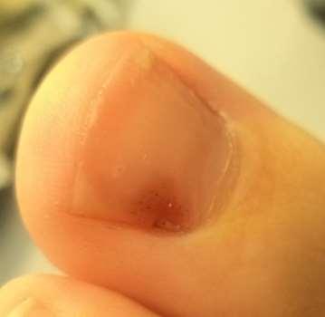
23 chaos? Is it a seb k? what clues?

24 melanoma Part of a very large lesion (see graticule) Multiple colours and patterns Irregular dots/globules AND asymmetrical network seen together on lower edge of lesion
25

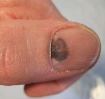
26 Collision lesion, seb k and haemangioma Excised as very odd The red section is homogenous within itself The darker section has distinct if irregular borders Comedo like openings and milia like cysts are seen
27
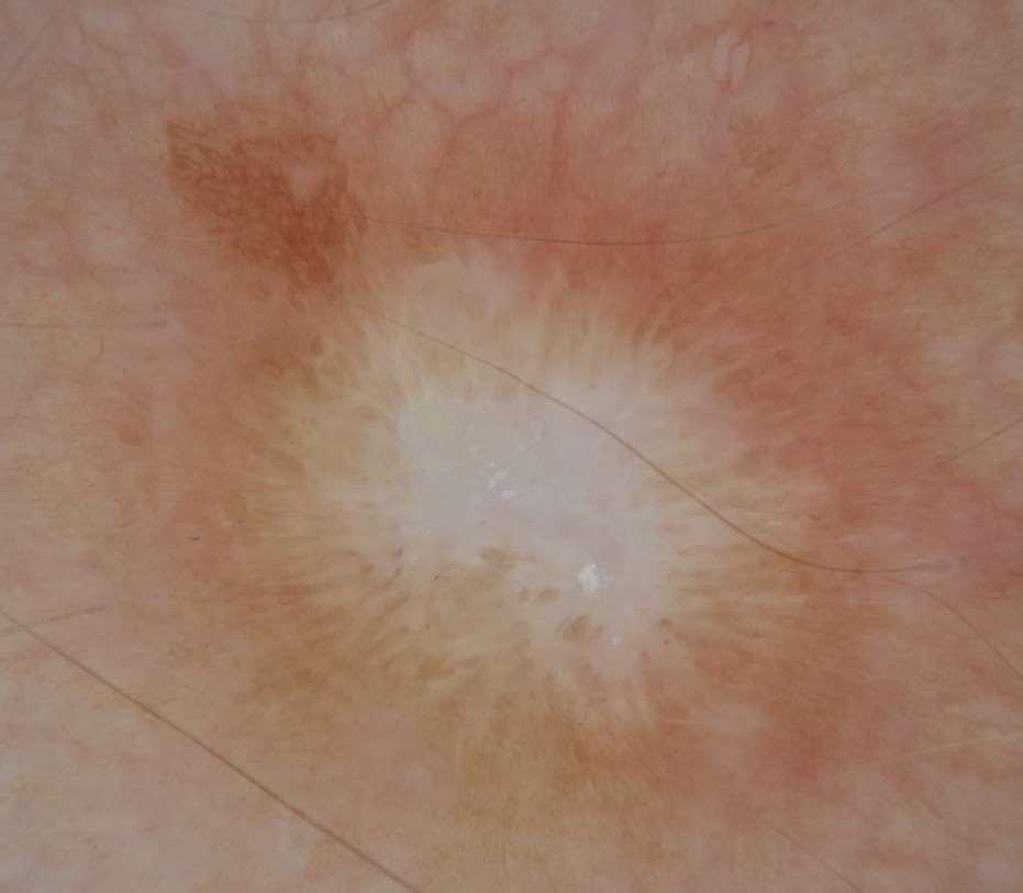
28

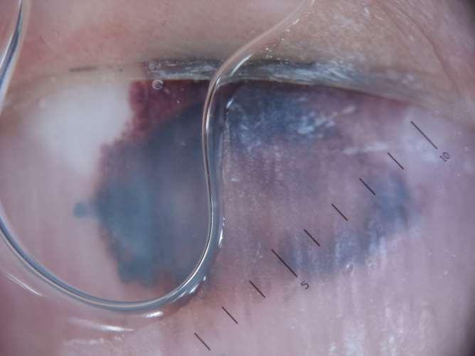
29 Same lesion, 2 views
30

31 Blood under the nail is MANY TIMES more common than acral melanoma, even in the absence of a history of trauma Think of march fractures of the metatarsal, you don t need a direct injury to cause trauma Blood clots grow out slowly, if there is a gap, however small, between the pigment and the proximal nail fold, it s 99% likely to be blood, not a melanoma Brown and red globules signify blood
32


33 Blood under nail (the onycholysis is irrelevant, probably psoriatic)
34 Red flag features of nail unit melanoma 1) destruction of nail 2) tumour 3) Hutchinson s sign (pigment in skin adjacent to nail). Absence of these features does not eliminate the possibility of melanoma (or SCC which can also cause nail unit disease) but is reassuring and should be recorded.
35 Check these images on line -of-nail-unit/ img=pmc _ &req=4 1.jpg Or just Google nail unit melanoma (I don t have any images I can share-sh)
36
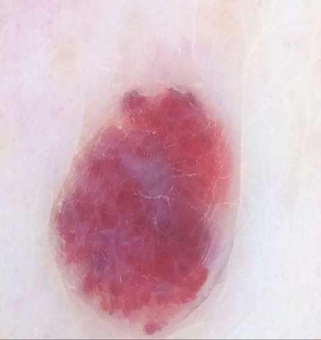
 on the RIDGES Trivial thickening of furrows right of centre is irrelevant in view of overall benign")
37 Benign acral naevus Parallel FURROW pattern One colour, one pattern, no chaos Furrows defined by the pale circles (sweat gland openings) on the RIDGES Trivial thickening of furrows right of centre is irrelevant in view of overall benign pattern

38 The tiny pale dots mark the RIDGES, therefore the pigment is mainly in the FURROWS which is a benign pattern. For more examples of benign and malignant acral lesions go to Dr Eric Ehrsam s excellent blog
39


40 Excised as suspected spindle cell naevus Histology was Benign junctional Acral naevus

41 How worried are you?
42
43
44
 Grey and brown are not allowed Collision lesions involving haemangiomas are not rare and may")
45 Haemangiomas= blood in fibrous stroma Blood is allowed to be blue, red, purple/mauve or black (if clotted) Grey and brown are not allowed Collision lesions involving haemangiomas are not rare and may confuse
46
47

48 Collision lesions These are statistically inevitable due to the random distribution of benign and malignant skin lesions. Probably the most common are seb k/haemangioma, but any combination is possible, including the seb k/bcc collision seen here (observe the vessels)
49
50 Biopsy proven traumatised seb k Note homogenous looped vessels in pale halos Small blood clot in right lower quarter Some keratin in top left quarter
51
52

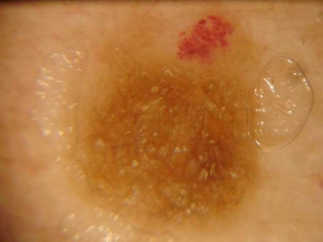
53 Lichenoid keratosis Benign, but can look worrying History of recent change in colour, may itch Lentigo undergoing regression, lichen planus like histology Typical dermoscopy, homogenous grey dots and dashes, some pink. Excision is excusable, the confident dermoscopist may prefer to photo and review
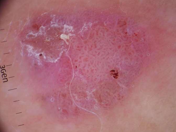
54
55 melanoma Chaos Gross asymmetry Multiple colours and patterns Branched streaks at left lower edge (particularly) Eccentric black blotches


56 Compare and contrast chaos
57
58 BCC Pink and white background Micro erosion (red clod lower right of centre) Blue clods, lower left Vessels NB the large blue clod at the top is a tattoo!
59 2 pigmented lesions, same patient
60 Both harmless flat seb ks, showing homogenous fat fingers or clod patterns. Note the skin lines. Over time these lesions will get thicker and develop more 3-dimensional seb k structures such as milia like cysts and comedo like openings
61
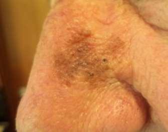
62 Lentigo maligna (LM) This growing flat pigmented lesion on an older white person s face has to be something. Benign options are seb k and benign lentigo (solar lentigo, lentigo simplex). We can t prove either of those, so lentigo maligna must be excluded. It can t be, due to the presence of grey and blue. The angular, deeply pigmented clods at 2 and 7 o clock are highly suggestive of LM, as confirmed.
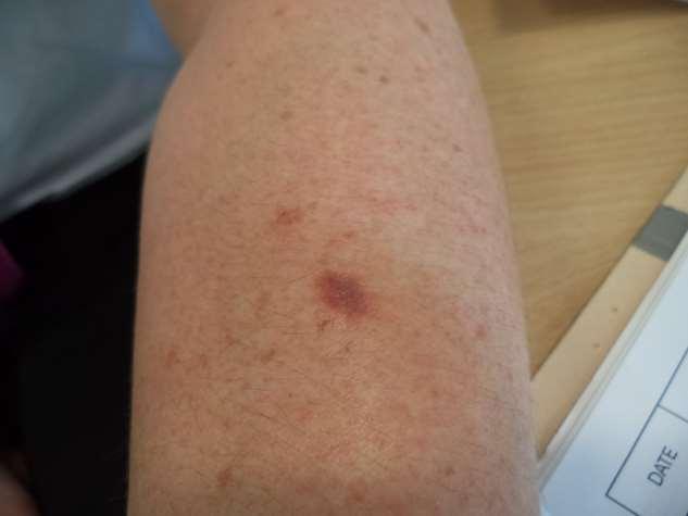
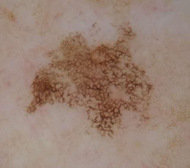
63
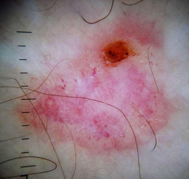
64 BCC Orange clod is a micro erosion (orange=fibrin) Note pink background and sharply focussed irregular vessels NB you cannot in my opinion confidently exclude amelanotic melanoma on this image, prompt excision would be wise.
65
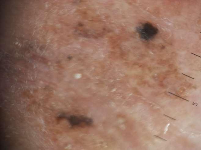
66 BCC with pigment Micro erosions (the irregular yellowish clods) Irregular clods of grey pigment Sharply focussed vessels (NB not classic arborising, they aren t always) It is wise to excise suspected pigmented BCCs urgently, sometimes they are melanomas
67

68 Gross asymmetry Multiple colours and patterns spoke wheel structures at 12, 1 and 11 o clock HOWEVER can you really tell these with confidence from the radial streaks see in melanoma? Excise urgently to rule out melanoma Pigmented BCC
69
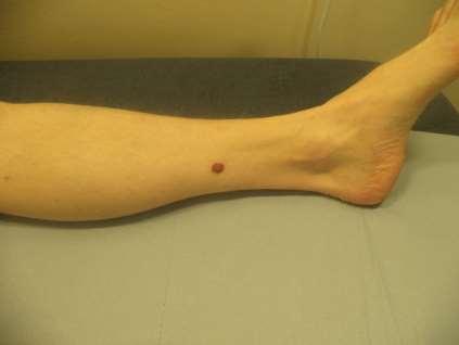
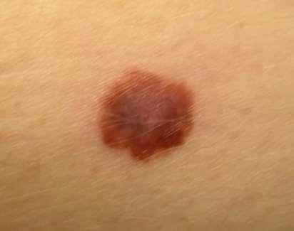
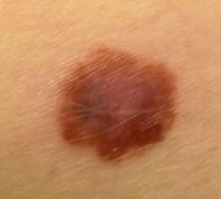
70 dermatofibroma Rare under age 20 Probably due to fibrotic response to insect bite Most common round shoulders and below knees Bidigital palpation=hard as a button Dimple sign Dermoscopy-central irregular white scar, peripheral pseudolattice brown circles See examples in 100 cases in USB drive
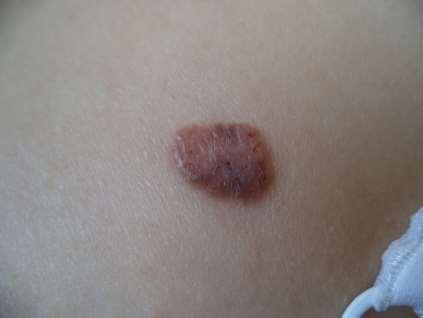
71
72

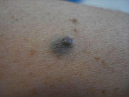
73 Biopsy proven dysplastic naevus Reticular network, so melanocytic Several shades of brown Somewhat uneven Appearance of a black blotch forming up in the upper centre of lesion It may be legitimate for experienced dermoscopists to photograph and follow up FLAT mildly suspicious melanocytic lesions, but if you do this you are responsible for fail safe record labelling, storage and safeguarding. NEVER FOLLOW UP A SUSPICIOUS NODULAR LESION. NB there is little no evidence that dysplastic naevi are premalignant, see Harald Kittler s YouTube video The myth of dysplastic naevi

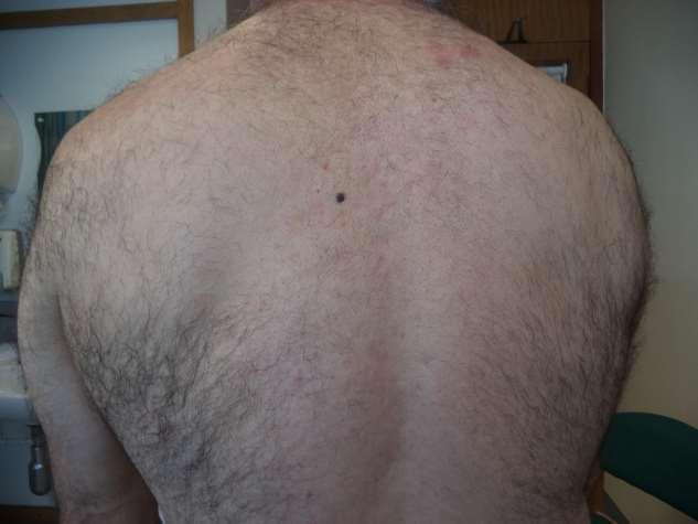
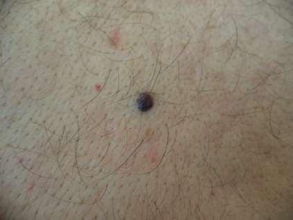
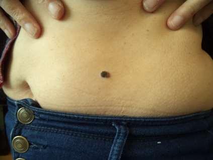
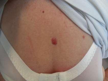
74 7mm lesion upper back, recent change
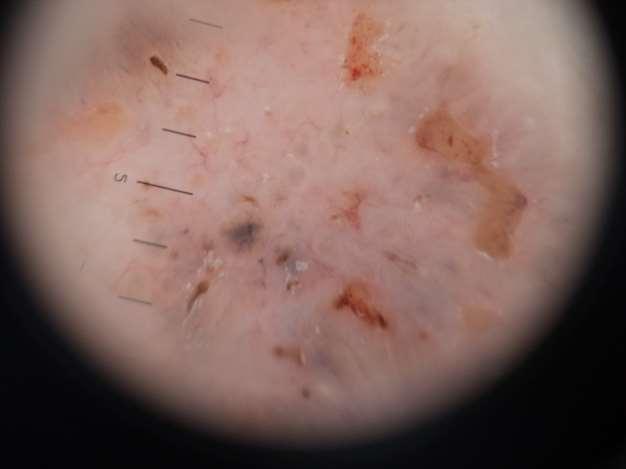
75

76 Benign, spindle cell naevus with epithelioid features You will NEVER get down to a ratio of 1 melanoma per suspicious pigmented lesion excised, and it would be dangerous to try too hard. An acceptable ratio is probably about 4:1, but this is debateable. Anything above 10 is, in my opinion, too high.
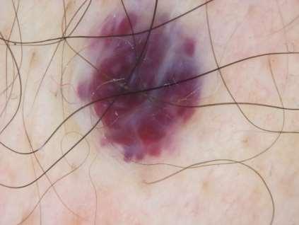
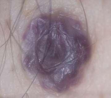
77
78 Mildly dysplastic naevus No colour other than brown ugly duckling No first-rank features of melanoma Photo and review may have been a reasonable course of action
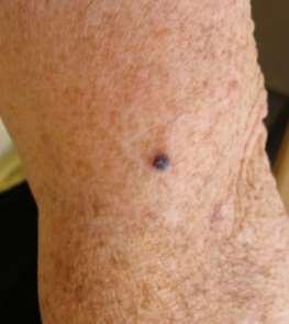
79

80 Benign compound naevus Not excised Felt soft (wobble sign) Brown clods are reasonably even is size, colour and distribution Well defined border No first rank melanoma features
81 Solitary lesion, grown over 2 years or so
82
83 The key is chaos (multiple colours, Multiple patterns) Achtung! Verrucous melanoma mimicking seborrhoeic wart
84
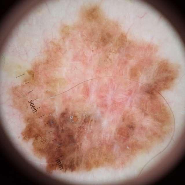
85 Recent change. Excise or leave?
86

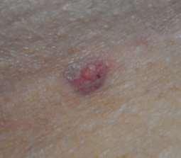
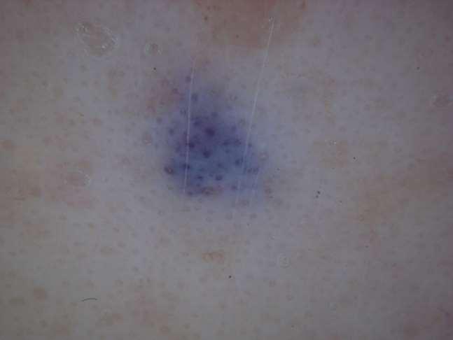
87 Excised due to recent change, histology was benign blue naevus Blue naevi typically appear before age 30 A bit of white or brown is OK, they don t have to be perfectly homogenous Most commonly appear as solitary papules <5mm Common locations hands, wrist, foot, ankle, head If small and stable, no concerns, but beware if NFG The sudden appearance of multiple blue naevi may signify metastatic melanoma
88

89 Ink spot lentigo Irregular shape, but the colour and pattern are very even/homogenous Ink spot lentigos are not premalignant, but tend to be seen on sun damaged skin Often multiple Must be flat
90
91

92 Thick melanoma Dermoscopy is irrelevant Chaos (ABCD fail +++) Multiple colours Irregular edge Shiny white structures on black background Just looks EVIL!
93
94

95 Reported as dysplastic naevus Reticular net, therefore melanocytic (2 step algorithm) Can we confidently say its benign? Not really Network quite irregular, suggestion of eccentric black blotch beginning to form at 10 o clock Too odd to leave, but if it had been a melanoma would have been a very thin one
96

97 Pink background Sharply focussed tapering vessels No features for any kind of non-bcc lesion NB the absence of micro erosions, pigmented structures or shiny white streaks does not matter. Beware of diagnostic greed (expecting a full house of all possible features for any given lesion type) Nodular BCC
98
 Can we say for sure its harmless?")
99 melanoma ABCD fail Multiple colours and patterns including shiny white streaks, network, eccentric blotch, atypical vessels. Network present (therefore melanocytic) Can we say for sure its harmless?( 2 step algorithm) No we cannot
100 On line resources International Dermoscopy Society join for free, on line discussions, YouTube vidoes, 5 th World Dermoscopy Congress in Thessaloniki, th June See you there? Great discount for early bookers and IDS members. free on line dermoscopy course best blog my blog
Dermoscopy-a BRIEF introduction
 Dermoscopy-a BRIEF introduction Aim of presentation -to tell you what dermoscopy is -to show some of what it can do -point the interested learner to further resources Overview of dermoscopy Dermoscopy
It can be helpful in some cases of actinic keratosis, Bowen s disease and squamous cell carcinoma
 Dermoscopy Introduction, Terminology and Structures (to be read in conjunction with the Diagnostic Dermoscopic Algorithm) Copyright to Cunliffe TP (Jan. 2017) All rights reserved Introduction Dermoscopy
Mole mapping and monitoring. Dr Stephen Hayes. Associate Specialist in Dermatology, University Hospital Southampton
 Mole mapping and monitoring Dr Stephen Hayes Associate Specialist in Dermatology, University Hospital Southampton Outline of presentation The melanoma epidemic Benefits of early detection Risks of the
22/04/2015. Dermoscopy of Melanoma. Ilsphi Browne. Overview
 Dermoscopy of Melanoma Ilsphi Browne Overview The device Dermoscopic criteria (terminology) Colour Patterns Global features Local features Approach to diagnosing pigmented lesions Other uses in general
comedo-like openings (clods, brown or orange & circles) milia-like cysts (dots or clods, white) 1/29/18 Dotted vessels are also commonly seen in SCC
 milia-like cysts (dots or clods, white) 1/29/18 Dotted vessels are also commonly seen in SCC") Brown circles Dotted vessels are also commonly seen in SCC Step1 1. Nevus (unequivocal) 2. DF/IDN 3. BCC 4. SCC Network Patchy network Peripheral network & central hypopigmentation DF: network with central
Brown circles Dotted vessels are also commonly seen in SCC Step1 1. Nevus (unequivocal) 2. DF/IDN 3. BCC 4. SCC Network Patchy network Peripheral network & central hypopigmentation DF: network with central
Skin lesions The Good and the Bad. Dr Virginia Hubbard Ipswich Hospital NHS Trust Barts and the London School of Medicine and Dentistry
 Skin lesions The Good and the Bad Dr Virginia Hubbard Ipswich Hospital NHS Trust Barts and the London School of Medicine and Dentistry Case 1 32 year old woman Australian Lesion on back New hair growing
Benign versus Cancerous Lesions How to tell the difference FMF 2014 Christie Freeman MD, CCFP, DipPDerm, MSc
 1 Benign versus Cancerous Lesions How to tell the difference FMF 2014 Christie Freeman MD, CCFP, DipPDerm, MSc Benign lesions Seborrheic Keratoses: Warty, stuck-on Genetics and birthdays Can start in late
Common Benign Lesions and Skin Cancers. 22nd May 2015 Dr Mark Foley
 Common Benign Lesions and Skin Cancers 22nd May 2015 Dr Mark Foley Thank you for downloading this file. This intended to supplement the presentation given at the NZ Wound Care Conference, it is not intended
Dermoscopy Quiz 3-Point Checklist Algorithm
 Dermoscopy Quiz 3-Point Checklist Algorithm GLOBAL PATTERN Globular LOCAL CRITERIA Aggregated globules Milia-like cysts 3 POINT CHECK LIST Symmetrical No abnormal net Slight Blue-white veil BENIGN MELANOCYTIC
Fundamentals of dermoscopy
 Fundamentals of dermoscopy Learning objectives Upon completion of this session, participants should be able to: describe the basic principles of dermoscopy identify features associated with pigmented and
Dermoscopy in everyday practice. What and Why? When in doubt cut it out? Trilokraj Tejasvi MD
 Dermoscopy in everyday practice Trilokraj Tejasvi MD Assistant Professor, Department of Dermatology, Director Teledermatology services, University of Michigan, Faculty Associate, GLOBAL REACH, Michigan
Dermoscopy: Recognizing Top Five Common In- Office Diagnoses
 Dermoscopy: Recognizing Top Five Common In- Office Diagnoses Vu A. Ngo, DO Department of Family Medicine and Dermatology Choctaw Nation Health Services Authority Learning Objectives Introduction to dermoscopy
Disclosure. Objectives. PAFP CME Conference Lou Mancano MD, FAAFP Reading Health System November 18, 2016
 PAFP CME Conference Lou Mancano MD, FAAFP Reading Health System November 18, 2016 1 Disclosure The speaker has no conflict of interest, financial agreement, or working affiliation with any group or organization.
INTRODUCTION HOUSEKEEPING June 11 th Dr John Adams Dermatologist/Dermoscopist MOLEMAP NZ/Australia MOLESAFE USA
 INTRODUCTION HOUSEKEEPING June 11 th 2015 Dr John Adams Dermatologist/Dermoscopist MOLEMAP NZ/Australia MOLESAFE USA Program Skin cancer statistics. Dermoscopy description and usefulness. Patient /lesion
The impact of GP sub-specialisation and dermatoscopy use on diagnostic accuracy for melanomas in Australia
 The impact of GP sub-specialisation and dermatoscopy use on diagnostic accuracy for melanomas in Australia Cliff Rosendahl, Gail Williams, Diann Eley, Tobias Wilson, Greg Canning, Jeffrey Keir, Ian McColl,
VACAVILLE DERMATOLOGY
 Connecting the Dots on those Spots NANDAN V. KAMATH, M.D. VACAVILLE DERMATOLOGY Sources All of the photos were taken with permission from the Dermnet NZ website - Dermnet New Zealand after communicating
6/17/2018. Breaking Bad (Part 1) Dermoscopy of Brown(ish) Things. Bad?
 Dermoscopy of Brown(ish) Things. Bad?") Breaking Bad (Part 1) Dermoscopy of Brown(ish) Things Jennie T. Clarke, MD ssociate Professor of Dermatology University of Utah School of Medicine Bad? 1 Brown(ish) Things Bad Melanoma Pigmented basal
Breaking Bad (Part 1) Dermoscopy of Brown(ish) Things Jennie T. Clarke, MD ssociate Professor of Dermatology University of Utah School of Medicine Bad? 1 Brown(ish) Things Bad Melanoma Pigmented basal
Key factors in successfully integrating dermoscopy into your clinical practice
 Key factors in successfully integrating dermoscopy into your clinical practice S051 Dilemmas and challenges in skin cancer therapies and management Monday, March 4 th 2019 (9AM-12PM) Room 209A 10:56-11:09AM
Introduction to Dermoscopy. Nicholas Compton, MD June 16, 2010
 Introduction to Dermoscopy Nicholas Compton, MD June 16, 2010 Overview What is dermoscopy Brief history Types of dermoscopy General approach to lesion of interest 2 step algorithm 3-point checklist Practice
Yes. Breaking Bad II: Dermoscopy of Pink-ish Things. Does it Fit? Yes 6/17/2018. Yes. Joslyn Kirby, MD, MS, MEd
 Breaking Bad II: Dermoscopy of Pink-ish Things Joslyn Kirby, MD, MS, MEd Yes Observe Yes Step 2. Fit a Benign Nevus Pattern? Does it Fit? Step 1: Melanocytic? pigment network, globules, homogeneous? No
Revised Pattern Analysis: a method for the accurate diagnosis of pigmented skin lesions
 Dermatoscopy for Students A concise outline of: Revised Pattern Analysis: a method for the accurate diagnosis of pigmented skin lesions And Chaos and Clues: a decision algorithm for routine practice to
The GP s approach to the patient who is worried about sun, skin and moles. Dr Stephen Hayes
 The GP s approach to the patient who is worried about sun, skin and moles Dr Stephen Hayes Associate Specialist in Dermatology, University Hospital Southampton Dr Stephen Hayes DECLARATION OF INTERESTS
Skin Cancer A Personal Approach. Dr Matthew Strack Dunedin New Zealand
 Skin Cancer A Personal Approach Dr Matthew Strack Dunedin New Zealand Outline Dermoscopy Instruments and setup Photochemosurgery Clinical Aim: Leave with 2-3 ideas JLE Benign Junctional Nevus Management
Finding Melanoma. Is not easy!
 Finding Melanoma Is not easy! Finding Melanoma Victoria mean depth at diagnosis is 1.5 mm. Melanoma 1.5mm Has Stage 1B Mortality 10% Melanoma Spotting a killer! Spotting a killer Visual Clues What are
Clinical and Dermoscopic Features of Thin Nodular Melanoma
 Clinical and Dermoscopic Features of Thin Nodular Melanoma A study of the International Dermoscopy Society Coordinator: Dr. Alexander J. Stratigos and colleagues, alstrat2@gmail.com ** Extended to May
Malignant non-melanocytic lesions
 Malignant non-melanocytic lesions Course C023: Fundamentals of Dermoscopy March 4, 2019, 11:20 AM - 11:50 PM Room: 146B Jason B. Lee, MD Professor & Vice Chair Director of Dermatopathology & Pigmented
Non-melanocytic Patterns
 Non-melanocytic Lesions Non-melanocytic Patterns Michelle Tarbox, MD Assistant Professor of Dermatology and Dermatopathology Texas Tech University Health Sciences Center 2018 Seborrheic keratoses Acanthotic
Basics in Dermoscopy
 Basics in Dermoscopy Manal Bosseila Professor of Dermatology, Cairo University Member of European Academy Dermatology & Venereology EADV Member of International Dermoscopy Society IDS Member of Aesthetic
MODULE 1. LOCAL AND GENERAL CRITERIA IN PIGMENTED MELANOCYTIC LESIONS.
 DERMOSCOPY TEACHING PROGRAMME Dermoscopy Teaching Programme Module 1 MODULE 1. LOCAL AND GENERAL CRITERIA IN PIGMENTED MELANOCYTIC LESIONS. Dermoscopy is a non-invasive in vivo technique that provides
Dermoscopy STFM Richard Usatine, MD 5/2/16. Disclosure Statement: Some Dermatoscopes. Dermoscopy Video. Thanks to Dr.
 Disclosure Statement: Dermoscopy STFM 2016 Richard P. Usatine, MD, FAAFP Professor, Family and Community Medicine Professor, Dermatology and Cutaneous Surgery Medical Director, Clinic University of Texas
Dermoscopy. Synonyms. Dermoscopy. Definition. Dermoscopy opens up a world of colour and structure that can t be seen with the naked eye
 Synonyms Dermoscopy Australasian College of Dermatologists G.P Training Module Dermoscopy Dermatoscopy Epiluminescence microscopy Skin surface microscopy Incident light microscopy Oil immersion microscopy
Dr Stephen Hayes Associate Specialist in Dermatology University Hospital Southampton
 South East Dermatology Transformation and Sustainability Network Guildford, 19 th April 2018 Dermoscopy as an effective skin lesion triage tool in GP surgeries Dr Stephen Hayes Associate Specialist in
Non-Melanocytic Pattern Dermoscopy
 Non-Melanocytic Pattern Dermoscopy I have no conflicts of interest to disclose Except that I LOVE dermoscopy Michelle Tarbox, MD Assistant Professor of Dermatology and Dermatopathology Texas Tech University
Regression 2/3/18. Histologically regression is characterized: melanosis fibrosis combination of both. Distribution: partial or focal!
 Regression Margaret Oliviero MSN, ARNP Harold S. Rabinovitz MD Histologically regression is characterized: melanosis fibrosis combination of both Distribution: partial or focal! Dermatoscopic terminology
Clinical characteristics
 Skin Cancer Fernando Vega, MD Seattle Healing Arts Clinical characteristics Precancerous lesions Common skin cancers ACTINIC KERATOSIS Precancerous skin lesions Actinic keratoses Dysplastic melanocytic
IT S FUNDAMENTAL MY DEAR WATSON! A SHERLOCKIAN APPROACH TO DERMATOLOGY
 IT S FUNDAMENTAL MY DEAR WATSON! A SHERLOCKIAN APPROACH TO DERMATOLOGY Skin, Bones, and other Private Parts Symposium Dermatology Lectures by Debra Shelby, PhD, DNP, FNP-BC, FADNP, FAANP Debra Shelby,
Hauora Maori. Map. Non-Melanoma Skin Lesion. Clinically Seborrhoeic Keratosis. Management Options. Punch Biopsy. Refer to Public Skin Lesion Service
 Care map information Information resources for patients and carers Updates to this Care Map Hauora Maori Pacific Skin lesion history Clinical examination and diagnostic test Suspicious of Melanoma Non-Melanoma
Dermatopathology: The tumor is composed of keratinocytes which show atypia, increase mitoses and abnormal mitoses.
 Squamous cell carcinoma (SCC): A common malignant tumor of keratinocytes arising in the epidermis, usually from a precancerous condition: 1- UV induced actinic keratosis, usually of low grade malignancy.
Dr Amanda Oakley. Dermatologist Dept of Dermatology, Health Waikato Adjunct Associate Professor, Waikato Clinical Campus
 Dr Amanda Oakley Dermatologist Dept of Dermatology, Health Waikato Adjunct Associate Professor, Waikato Clinical Campus 14:00-16:00 WS #14: Dermoscopy Part 1 Skin Lesions and Dermatoscopy 16 August 2018
MELANOMA. Some people are more likely to get a m Melanoma than others:
 MELANOMA This leaflet has been written to help you understand more about Melanoma. It tells you what is it, what causes it, what can be done about it, how it can be prevented, and where you can find out
BLINCK A diagnostic algorithm for skin cancer diagnosis combining clinical features with dermatoscopy findings
 DERMATOLOGY PRACTICAL & CONCEPTUAL www.derm101.com BLINCK A diagnostic algorithm for skin cancer diagnosis combining clinical features with dermatoscopy findings Peter Bourne, MBBS 1, Cliff Rosendahl,
Melanoma and Dermoscopy. Disclosure Statement: ABCDE's of melanoma. Co-President, Usatine Media
 Melanoma and Dermoscopy Richard P. Usatine, MD, FAAFP Professor, Family and Community Medicine Professor, Dermatology and Cutaneous Surgery Medical Director, University Skin Clinic University of Texas
MECHANISMS OF HUMAN DISEASE: LABORATORY SESSION PATHOLOGY OF THE SKIN LAB. Friday, February 12, :30 am 11:00 am
 MECHANISMS OF HUMAN DISEASE: LABORATORY SESSION PATHOLOGY OF THE SKIN LAB Friday, February 12, 2012 9:30 am 11:00 am FACULTY COPY GOALS: Describe the basic clinical and morphologic features of various
Appendix : Dermoscopy
 Go Back to the Top To Order, Visit the Purchasing Page for Details APP Appendix : Dermoscopy Dermoscopy, also known as dermatoscopy, epiluminoscopy and epiluminescent microscopy, is an effective non-invasive
Skin Cancer. Dr Elizabeth Ogden Associate Specialist in Dermatology East and North Herts Dr Elizabeth Ogden
 Skin Cancer Dr Elizabeth Ogden Associate Specialist in Dermatology East and North Herts 13.10.16 Skin Cancer Melanoma mole cancer - is a true cancer which can metastasize and kill Non Melanoma skin cancer
10/3/2018. Dermoscopy: Looking beneath the surface of the skin. Dermoscopy for Family Medicine 10/11/2018
 Dermoscopy for Family Medicine 10/11/2018 Jane M. Grant-Kels, MD, FAAD Founding Chair Emeritus, Dept of Dermatology Professor of Dermatology, Pathology & Pediatrics Director of the Cut Oncology Ctr & Melanoma
LUMPS AND BUMPS: AN ORGANIZED APPROACH TO DIAGNOSIS AND MANAGEMENT
 LUMPS AND BUMPS: AN ORGANIZED APPROACH TO DIAGNOSIS AND MANAGEMENT Tammy P. Than, M.S., O.D., F.A.A.O. The University of Alabama at Birmingham / School of Optometry 1716 University Blvd. Birmingham, AL
Dermatological Manifestations in the Elderly. Sanjay Siddha Staff Dermatologist UHN & MSH
 Dermatological Manifestations in the Elderly Sanjay Siddha Staff Dermatologist UHN & MSH Disclosure No actual or potential conflicts of interest or commercial relationships to declare Objectives Recognize
Phoebe Rich MD Adjunct Professor OHSU Portland, Oregon
 Nail Tips for Diagnosis and Management of Nail Disorders Winter Clinical Dermatology Conference 2017 Hawaii Phoebe Rich MD Adjunct Professor OHSU Portland, Oregon Objectives diagnostic clues for benign
Dermoscopy. Enhanced Diagnostic Ability: Pigmented Lesions. Ted Rosen, MD Baylor College of Medicine Houston, Texas
 Dermoscopy Enhanced Diagnostic Ability: Pigmented Lesions Ted Rosen, MD Baylor College of Medicine Houston, Texas Faculty Disclosure Statement No conflicts relevant to this workshop! Sir William Osler
MECHANISMS OF HUMAN DISEASE: LABORATORY SESSION PATHOLOGY OF THE SKIN LAB. Friday, February 13, :30 am 11:00 am
 MECHANISMS OF HUMAN DISEASE: LABORATORY SESSION PATHOLOGY OF THE SKIN LAB Friday, February 13, 2009 9:30 am 11:00 am FACULTY COPY GOALS: Describe the basic clinical and morphologic features of various
Malignant tumors of melanocytes: Part 1. Deba P Sarma, MD., Omaha
 Malignant tumors of melanocytes: Part 1 Deba P Sarma, MD., Omaha The melanocytic tumor is one of the most difficult and confusing areas in Dematopathology. It is true that most (95%) of such lesions are
Dermoscopy, the use of a handheld
 ONLINE EXCLUSIVE Dermoscopy in family medicine: A primer Dermoscopy allows you to see deeper into the skin than with the naked eye. Here s how you can make use of it to spot malignant conditions sooner.
MELANOMA. 4 Fitzroy Square, London W1T 5HQ Tel: Fax: Registered Charity No.
 MELANOMA This leaflet had been written to help you understand more about melanoma. It tells you what it is, what causes it, what can be done about it, how it can be prevented, and where you can find out
Pathology of the skin. 2nd Department of Pathology, Semmelweis University
 Pathology of the skin 2nd Department of Pathology, Semmelweis University Histology of the skin Epidermis: Stratum corneum Stratum granulosum Stratum spinosum Stratum basale Dermis: papillary and reticular
Prediction without Pigment: a decision algorithm for non-pigmented skin malignancy
 DERMATOLOGY PRACTICAL & CONCEPTUAL www.derm101.com Prediction without Pigment: a decision algorithm for non-pigmented skin malignancy Cliff Rosendahl 1, Alan Cameron 1, Philipp Tschandl 2, Agata Bulinska
Histopathological and SIAscopic Correlation of Pigmented Skin Lesions
 Histopathological and SIAscopic Correlation of Pigmented Skin Lesions Professor Sujatha Fernando MBBS(Hon), MSc(London, Distinction), FRSTM&H, FRCPA, FIAC, FACTM Senior Consultant in Anatomical Pathology,
The prevention, diagnosis, referral and management of melanoma of the skin: concise guidelines
 CLINICAL GUIDANCE The prevention, diagnosis, referral and management of melanoma of the skin: concise guidelines Julia Newton Bishop, Veronique Bataille, Alice Gavin, Marko Lens, Jerry Marsden, Tania Mathews
Dermatology for the PCP Deanna G. Brown, MD, FAAD Susong Dermatology Consulting Staff at CHI Memorial
 Dermatology for the PCP Deanna G. Brown, MD, FAAD Susong Dermatology Consulting Staff at CHI Memorial Cutaneous Oncology for the PCP Deanna G. Brown, MD, FAAD Susong Dermatology Consulting Staff at CHI
Melanoma. Consultation on draft guideline - stakeholder comments. Comments to be submitted before 5pm on Friday 13 March 2015
 Please note: Please fill in both the stakeholder organisation and name of commentator fields. We cannot accept forms with attachments such as research articles, letters or leaflets. Stakeholder organisation(s)
MELANOCYTIC LESIONS: EFFECTIVE MANAGEMENT IN PRIMARY CARE: Part 2
 MELANOCYTIC LESIONS: EFFECTIVE MANAGEMENT IN PRIMARY CARE: Part 2 In the second part of our feature on pigmented skin lesions, dermatology specialist Dr Sweta Rai describes the steps to rational decision-making
Dermoscopy. Sir William Osler. Dermoscopy. Dermoscopy. Melanoma USA Primary Care Update Faculty Disclosure Statement
 Diagnostic Ability: Pigmented Lesions Ted Rosen, MD Baylor College of Medicine Houston, Texas Enhanced 2010 Primary Care Update Faculty Disclosure Statement Ted Rosen, MD Speakers Bureau: Abbott, Amgen,
CANCER INSIGHT. FOR GPs. Summer 2018 WHAT YOU NEED TO KNOW ABOUT SKIN CANCER VISIT. our Skin Cancer Recognition Toolkit at
 CANCER INSIGHT FOR GPs Summer 2018 WHAT YOU NEED TO KNOW ABOUT SKIN CANCER VISIT our Skin Cancer Recognition Toolkit at www.doctors.net. uk/sct Cancer Research UK, Angel Building, 407 St John Street, London
Benign and malignant epithelial lesions: Seborrheic keratosis: A common benign pigmented epidermal tumor occur in middle-aged or older persons more
 Benign and malignant epithelial lesions: Seborrheic keratosis: A common benign pigmented epidermal tumor occur in middle-aged or older persons more common on the trunk; but extremities, head and neck are
Management of Atypical Pigmented Lesions
 Management of Atypical Pigmented Lesions Jennifer A. Stein MD, PhD Associate Director, Pigmented Lesion Section Ronald O. Perelman Department of Dermatology NYU Langone Medical Center July 29, 2017 1-4
Total body photography in high risk patients
 Total body photography in high risk patients Doug Grossman, MD, PhD Department of Dermatology Huntsman Cancer Institute University of Utah Summer AAD F032 Practical Considerations for Patients with Melanoma
Oral and Maxillofacial Surgery Department
 Oral and Maxillofacial Surgery Department This leaflet explains: Lentigo Maligna What are the aims of this leaflet? This leaflet has been written to help you understand more about lentigo maligna and melanoma
Malignant Melanoma Early Stage. A guide for patients
 This melanoma patient brochure is designed to help educate melanoma patients and their caregivers. It was developed under the guidance of Dr. Michael Smylie, Professor, Department of Oncology, University
Case Report Micromelanomas: A Review of Melanomas 2mmand a Case Report
 Case Reports in Oncological Medicine, Article ID 206260, 4 pages http://dx.doi.org/10.1155/2014/206260 Case Report Micromelanomas: A Review of Melanomas 2mmand a Case Report Sharad P. Paul 1,2,3 1 Skin
This is a repository copy of Easily missed? Amelanotic melanoma. White Rose Research Online URL for this paper:
 This is a repository copy of Easily missed? Amelanotic melanoma. White Rose Research Online URL for this paper: http://eprints.whiterose.ac.uk/127789/ Version: Accepted Version Article: Muinonen-Martin,
David B. Troxel, MD. Common Medicolegal Situations: Misdiagnosis of Melanoma
 Common Medicolegal Situations: Misdiagnosis of Melanoma David B. Troxel, MD Medical Director, The Doctors Company, Napa, California Clinical Professor Emeritus, University of California at Berkeley Past
Diagnosis of Lentigo Maligna Melanoma. Steven Q. Wang, M.D. Memorial Sloan-Kettering Cancer Center Basking Ridge, NJ
 Diagnosis of Lentigo Maligna Melanoma Steven Q. Wang, M.D. Memorial Sloan-Kettering Cancer Center Basking Ridge, NJ Conflict of Interest: None Topics Epidemiology and Natural History Clinical and Histologic
Thursday 21 st August Skin Problems
 Thursday 21 st August 2014 Skin Problems Skin Problems The Sun and the Skin Sun Damage Recognising the early signs of skin cancer The Big 3 inflammatory condi=ons Acne & Rosacea Eczema (Including Seborrhoeic
Cover Page. The handle holds various files of this Leiden University dissertation.
 Cover Page The handle http://hdl.handle.net/1887/22172 holds various files of this Leiden University dissertation. Author: Rhee, Jasper Immanuel van der Title: Clinical characteristics and management of
Histopathology: skin pathology
 Histopathology: skin pathology These presentations are to help you identify, and to test yourself on identifying, basic histopathological features. They do not contain the additional factual information
Nails Examination and Disorders. Overview. Case 1 15/09/2016. Samantha Eisman. 25 year old woman Noticed at pedicure Single toe
 Nails Examination and Disorders Samantha Eisman Dermatologist MBChB/ MRCP/ FCDerm(SA)/ FACD Demystify nails Overview QUIZ Talk Examination nails and and site specific disease QUIZ answers and cover common
Identifying Skin Cancer. Mary S. Stone MD Professor of Dermatology and Pathology University of Iowa Carver College of Medicine March, 2018
 Identifying Skin Cancer Mary S. Stone MD Professor of Dermatology and Pathology University of Iowa Carver College of Medicine March, 2018 American Cancer Society web site Skin Cancer Melanoma Non-Melanoma
Learning Objectives. Tanning. The Skin. Classic Features. Sun Reactive Skin Type Classification. Skin Cancers: Preventing, Screening and Treating
 Learning Objectives Skin Cancers: Preventing, Screening and Treating Robert A. Baldor, MD, FAAFP Professor, Family Medicine & Community Health University of Massachusetts Medical School Distinguish the
Dermatoscopic features of cutaneous non-facial non-acral lentiginous growth pattern melanomas
 DERMATOLOGY PRACTICAL & CONCEPTUAL www.derm101.com Dermatoscopic features of cutaneous non-facial non-acral lentiginous growth pattern melanomas Jeff Keir 1 1 Department of Dermatology, School of Medicine,
Associate Professor Amanda Oakley. Professor H. Peter Soyer. Academic Dermatologist The University of Queensland Brisbane. Dermatologist Hamilton
 Associate Professor Amanda Oakley Dermatologist Hamilton Professor H. Peter Soyer Academic Dermatologist The University of Queensland Brisbane 8:30-10:30 WS #3: Dermoscopy Workshop Part 1 11:00-13:00 WS
BJD British Journal of Dermatology. Summary. What s already known about this topic? CLINICAL AND LABORATORY INVESTIGATIONS
 CLINICAL AND LABORATORY INVESTIGATIONS BJD British Journal of Dermatology Pigmented nodular melanoma: the predictive value of dermoscopic features using multivariate analysis M.A. Pizzichetta, 1 H. Kittler,
Assisting diagnosis of melanoma through the noninvasive biopsy of skin lesions
 Assisting diagnosis of melanoma through the noninvasive biopsy of skin lesions Symon D Oyly Cotton Ela Claridge School of Computer Science, The University of Birmingham Birmingham B15 2TT, UK Per Hall
 Epidermis Dermis Subcutaneous Hypodermis March 16, 2013 Telangiectasia's (spider veins) may develop anywhere within the body but can be easily seen in the skin, mucous membranes, and whites of
Pathology of the skin. Dr Fónyad László, 1sz. Patológiai és Kísérleti Rákkutató Intézet, SE
 Pathology of the skin Dr Fónyad László, 1sz. Patológiai és Kísérleti Rákkutató Intézet, SE The skin Biggest organ Kb. 1.8 nm Kb. 10 kg Most frequent site for tumor development (BCC) Pathology of the skin
Describe the functions of the vertebrate integumentary system. Discuss the structure of the skin and how it relates to function.
 Chapter 5 Describe the functions of the vertebrate integumentary system. Discuss the structure of the skin and how it relates to function. Explain the basis for different skin colors. Describe the structure
Melanoma what is it? Most aggressive and lifethreatening
 SUN,SURF AND MELANOMA Judy Brincat Dorevitch Pathology Heidelberg Victoria Melanoma what is it? Most aggressive and lifethreatening of cutaneous malignancies Develops from melanocytes. Adults and elderly
Nail apparatus melanoma: dermoscopic and histopathologic correlations on a series of 23 patients from a single centre
 DOI: 10.1111/jdv.14568 JEADV ORIGINAL ARTICLE Nail apparatus melanoma: dermoscopic and histopathologic correlations on a series of 23 patients from a single centre M. Starace, E. Dika, P.A. Fanti, A. Patrizi,
Dermoscopic Features of Non-Pigmented Eccrine Poromas in. Department of Dermatology, Shinshu University School of Medicine,
 Original article Dermoscopic Features of Non-Pigmented Eccrine Poromas in Association with their Histopathological Features Akane Minagawa, Hiroshi Koga,* Masaomi Takahashi, + Kenji Sano, + Ryuhei Okuyama,
Identifying Benign and Malignant Skin Lesions. No Disclosures. Common Benign Lesions. Benign Lesions 2/25/2018. Stucco Keratoses.
 Dermatology in Primary Care Identifying Benign and Malignant Skin Lesions Christy Quire Baker, APRN, FNP-BC, DCNP Dermatology Certified Nurse Practitioner No Disclosures Common Benign Lesions Seborrheic
Management of patients with melanocytic and non-melanocytic neoplasms
 Management of patients with melanocytic and non-melanocytic neoplasms Ashfaq Marghoob MD Harold Rabinovitz MD Margaret Oliviero ARNP Harald Kittler MD Jupiter Cancer Centrer Characteristic Dermoscopic
Contrast with Australian Guidelines A/Pr Pascale Guitera,
 Contrast with Australian Guidelines A/Pr Pascale Guitera, Dermatologist, Sydney University NO CONFLICT OF INTEREST Sydney Melanoma Diagnostic Centre, RPAH 2011 2008 225 pages 16 pages http://www.cancer.org.au/file/healthprofessionals/clinica
What is Dermoscopy? Early Dermoscopes. Deciphering Dermoscopy: Terminology, Features & Algorithms 6/17/2018
 Deciphering Dermoscopy: Terminology, Features & Algorithms Where did it come from and why do we use it? Jennie T. Clarke, MD Associate Professor of Dermatology University of Utah School of Medicine What
What is melanoma? Melanoma dealing with the diagnosis. What is melanoma?
 Melanoma is a form of cancer which develops from that part of the skin which produces its colour. It grows from the cell which produces the brown pigment in our skin: the melanocyte. Often the melanoma
Abrupt Intralesional Color Change on Dermoscopy as a New Indicator of Early Superficial Spreading Melanoma in a Japanese Woman
 Published online: June 24, 2015 1662 6567/15/0072 0123$39.50/0 This is an Open Access article licensed under the terms of the Creative Commons Attribution-NonCommercial 3.0 Unported license (CC BY-NC)
Diagnostics guidance Published: 11 November 2015 nice.org.uk/guidance/dg19
 VivaScope 1500 and 3000 imaging systems for detecting skin cancer lesions Diagnostics guidance Published: 11 November 2015 nice.org.uk/guidance/dg19 NICE 2018. All rights reserved. Subject to Notice of
Toby Maurer, MD University of California, San Francisco. Lifetime risk of an American developing melanoma
 Distinguishing Pigmented Skin Lesions and Melanoma Toby Maurer, MD University of California, San Francisco Epidemiology of Melanoma Lifetime risk of an American developing melanoma 1935: 1 in 1500 1980:
Know who is at risk: LOOK! for ABCDs, rapidly changing lesions, do a biopsy when indicated
 Lindy P. Fox, MD Assistant Professor Director, Hospital Consultation Service Department of Dermatology University of California, San Francisco Applies to adults without history of malignancy or premalignant
Reports on Scientific Meetings
 Hong Kong J. Dermatol. Venereol. (2016) 24, 146-153 The Hong Kong Society of Dermatology and Venereology Annual Scientific Meeting 2016 Reported by BTH Chan, CT Chau, CW Chow, CC Koh, WYK Lam, BS Tong,