Dermoscopy STFM Richard Usatine, MD 5/2/16. Disclosure Statement: Some Dermatoscopes. Dermoscopy Video. Thanks to Dr.
|
|
|
- Dana Nelson
- 6 years ago
- Views:
Transcription


1 Disclosure Statement: Dermoscopy STFM 2016 Richard P. Usatine, MD, FAAFP Professor, Family and Community Medicine Professor, Dermatology and Cutaneous Surgery Medical Director, Clinic University of Texas Health Science Center, San Antonio Co-President, Usatine Media medical app development company Author, 9 medical books including: Dermatologic and Cosmetic Procedures in Office Practice. Elsevier, Inc., Philadelphia The Color Atlas of Family Medicine. 2 nd Edition. McGraw-Hill, New York, 2013 The Color Atlas of Pediatrics, McGraw-Hill, New York, 2014 Cutaneous Cryosurgery. 4 th Edition. Taylor and Francis, London, 2014 The Color Atlas of Internal Medicine, McGraw-Hill, New York, 2015 Some Dermatoscopes More dermatoscopes Thanks to Dr. Ash Marghoob Dermoscopy Video dermoscopy/ d2e15a5faac52d3dd2b43bd35147cb3d.ht ml?ooct=jfp-related 6 minutes For his mentorship in dermoscopy and the use of some of the slides in this presentation. 1

")
 SK BCC Evidence The")


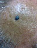
2 Light Source Source Polarizer is required Light Detector Detector Cross- Polarizing filter Only light undergoing multiple scattering events will result in sufficient change in angle of polarization (deeper, birefringence). Epidermis How can dermoscopy help? Helps differentiate benign from malignant lesions Miss less melanomas (sensitivity) Biopsy less benign lesions (specificity) Penetrating Light Superficial Light Surface Glare Dermis Improves malignant to benign biopsy ratio (accuracy) SK BCC Evidence The sensitivity of detecting melanoma went from 70% to 83% with the use of dermoscopy based on a meta-analysis of 13 studies. The specificity improved from 75% to 86%. 1 The sensitivity for melanoma diagnosis by primary care physicians using dermoscopy increased from 56% to 76%. 2 Primary care physicians using dermoscopy improved their triage of skin lesions suggestive of skin cancer by 25%. 3 Amelanotic melanoma 1. Bafounta ML, et al. Is dermoscopy (epiluminescence microscopy) useful for the diagnosis of melanoma? Results of a meta-analysis using techniques adapted to the evaluation of diagnostic tests. Arch Dermatol. Oct 2001;137(10): Westerhoff K, et al. Increase in the sensitivity for melanoma diagnosis by primary care physicians using skin surface microscopy. Br J Dermatol Nov;143(5): Argenziano G, et al. Dermoscopy improves accuracy of primary care physicians to triage lesions suggestive of skin cancer. J Clin Oncol Apr 20;24(12): Parallel pattern (volar skin = palms/soles) Benign furrows - ridges How dermoscopy helps 2

3 Dermoscopic features of melanoma Record your dermoscopy Attach your dermatoscope to your phone or a camera Photograph every lesion that you biopsy clinical and dermoscopic Download your photos to your computer When your path report comes back, look at your photos with the confirmed diagnosis Learning process Tips for storage and retrieval Label your photos with the name of the diagnosis so you can find them by dx in future Picasa is a free program from Google that is easy to search Don t put patient names or MRN s on files to avoid the HIPAA police Find your photos by date Use your image to accelerate your learning and provide images for consultations/referrals This all leads to improved learning and patient care. Features to look for in a dermatoscope Polarized and non-polarized in one package Attaches to your smartphone Second best attaches to a camera conveniently Charges easily Affordable Dermatoscopes DermLite: DL1, DL2, DL3, DL4 Attach to iphones/ipods/ipads and Androids Canfield Veos: iphone/ipod only Handiscope: iphone/ipod only Heine-Delta scopes attach to cameras with special hardware Welch Allyn scopes don t attach to anything and are not polarized Free for all Apple and Android devices Includes: Two step algorithm Useful Charts 50 Unknown cases Many images 3

 Suspicious (Bx or STMM)")

, aggregated globules,")
 parallel pattern 3.")
4 Two-step diagnostic procedure Two Step Algorithm Benign DF, SK, CCA hemangioma Malignant BCC, SCC Nevus vs : Is the lesion a melanocytic tumor? Step 2 Nevus (Reassure) Suspicious (Bx or STMM) (Biopsy) Two-step diagnostic procedure 1. Network Pigment network Negative network Polygonal lines tumors have: 1. Network (with two exceptions), aggregated globules, streaks, homogeneous blue pigment In addition: 2. On volar skin (palms & soles) parallel pattern 3. On face - Pseudo-network pattern Pigment network Nevus 4


5 Exception I: Dermatofibroma Pattern trumps structure Fine network surrounding a scar like area Exception 2: Pattern trumps structure Solar lentigo: network with fingerprinting, fine interrupted lines & moth eaten border Ink spot lentigo: uniform black network 1b. Negative network with negative network 1c. Polygonal lines Polygonal lines indicates that the lesion is melanocytic. 2. Aggregated globules 2. Globules Aggregated Peripheral rim 5


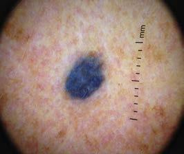
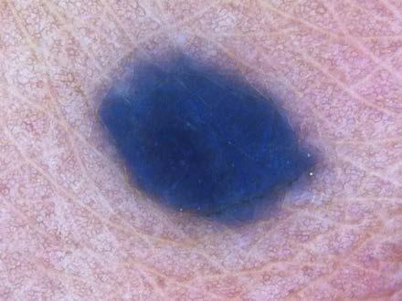
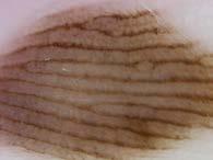
6 3. Streaks Blue nevus 4. Homogeneous blue pigmentation 4. Homogeneous blue pigmentation Parallel pattern Palms/Soles & Nails = parallel pattern (acral) Facial = pseudo-network pattern 6
 Suspicious (Bx or STMM) (Biopsy) Two-step diagnostic")

: Large grey-blue ovoid nests Multiple grey-blue globules Leaf-like")
7 Two-step diagnostic procedure Is the lesion a melanocytic tumor? If it has one of these features, then Step 2 Nevus (Reassure) Suspicious (Bx or STMM) (Biopsy) Two-step diagnostic procedure Delicate network 1. Dermatofibroma Central scar-like/crystalline Benign DF, SK, CCA hemangioma Malignant BCC, SCC Order of dx. 1. DF 2. BCC 3. SCC 4. SK 5. Hemangioma 6. Clear cell acanthoma Ring-like globules Vessels / blush in center 2. BCC Positive features (At least one present): Large grey-blue ovoid nests Multiple grey-blue globules Leaf-like areas Brown to gray-blue discrete bulbous blobs Leaf-like pattern Leaflike areas Spoke wheel areas Arborizing tree-like telangiectasia Ulceration 7


8 Arborizing vessels In-focus, red, branching vessels Hairpin vessels usually with a white halo 3. SCC Focally scaly/keratotic and rough Glomerular vessels focally present at periphery Keratin pearls & white circles Rosettes (strawberry pattern) Brown dots/globules aligned in a linear fashion at the periphery PD NB: Pigmented AK can also have structures seen in LMM! 4. SK Milia-like cyst 5. Vascular lesions hemangiomas and angiokeratomas Comedo-like opening Fissures & ridges Fingerprint-like Fat fingers Hairpin vessels Moth-eaten borders Lacunae separated by septae This is a: 1. lesion 2. melanocytic lesion 8


")
 9")

9 This is a: 1. lesion 2. melanocytic lesion This is a: 1. lesion 2. melanocytic lesion This is a: 1. lesion 2. melanocytic lesion Step 2: Nevus vs. Nevus Suspicious Step 2 (Reassure) (Bx or STMM) (Biopsy) 9

 specific structures SPECIFICITY (highest reported) ODDS RATIO (highest reported) Atypical network 77% 89% 9.")

10 The Beauty and the Beast sign * Benign nevi tend to adhere to one of ten recurrent patterns (finite #). These patterns all fit the definition of beauty, demonstrating symmetry of pattern, structure, and color. The most common benign patterns are: 6* * ü Deviate from global benign patterns ü Have at least one of the melanoma specific features listed below MM SPECIFIC STRUCTURE / FEATURE SENSITIVITY (highest reported) specific structures SPECIFICITY (highest reported) ODDS RATIO (highest reported) Atypical network 77% 89% 9.0 Streaks 23% 99% 5.8 Negative network 22% 95% 2.0 Chrysalis (Crystalline) 5% 99% 9.7 Atypical dots & globules 88% 97% 4.8 Atypical blotch 38% 88% 4.1 BWV 51% 99% 13 Regression structures 46% 94% 8.0 Atypical vessels 63% 96% 12.5 Peripheral brown structureless areas 63% 96% 28 Obieta MP, Braun RP, Scope A, Rabinovitz H, Marghoob AA. Dermoscopy of superficial spreading melanoma. G Ital Dermatol Venereol. 2009;144: Does this lesion manifest one of the 10 benign nevus patterns? 1) yes 2) no 10


 yes 2) no 1) yes 2) no 11")
11 Does this lesion manifest any MM specific structures? 1) yes 2) no Peripheral network Central hypopigmented and structureless area 2 Nevus Does this lesion manifest one of the 10 benign nevus patterns? 1) yes 2) no Does this lesion manifest any MM specific structures? 1) yes 2) no 11


12 Milky red area Atypical globules Atypical network 3 Does this lesion manifest one of the 10 benign nevus patterns? 1) yes 2) no Does this lesion manifest any MM specific structures? 1) yes 2) no 12


:441-50. Dermoscopy Courses American Dermoscopy Meeting is held yearly in the summer in a national park: http://www.")
 yearly fall FMX offers dermoscopy workshops: Orlando, FL Sept 20-24, 2016")
13 Young woman with pruritic eruption Arrowhead Jet plane Triangle on top of circle Dermoscopy Resources Scabies mite Free dermoscopy app - Dermoscopy: Two Step Algorithm. Available on itunes and for Android Authors: Marghoob AA, Usatine RP, Jaimes N. Dermoscopy. Website from Italy that includes a free dermoscopy tutorial International Dermoscopy Society, Marghoob A, Usatine R. Dermoscopy. In: Usatine R, Pfenninger J, Stulberg D, Small R, eds. Dermatologic and Cosmetic Procedures in Office Practice. Philadelphia, PA: Elsevier; Marghoob AA, Usatine RP, Jaimes N. Dermoscopy. In: Usatine R, Smith M, Mayeaux EJ, Chumley H. Color Atlas of Family Medicine,2nd Edition. New York, NY: McGraw-Hill; 2013 Marghoob AA, Usatine RP, Jaimes N. Dermoscopy for the family physician. Am Fam Physician Oct 1;88(7): Dermoscopy Courses American Dermoscopy Meeting is held yearly in the summer in a national park: Great Smokey Mountains, August 11-13, 2016 Questions? AFP 2013 American Academy of Family Physicians (AAFP) yearly fall FMX offers dermoscopy workshops: Orlando, FL Sept 20-24, 2016 Memorial Sloan-Kettering Cancer Center holds a yearly dermoscopy workshop each fall in New York City: Manhattan October 14-15,
comedo-like openings (clods, brown or orange & circles) milia-like cysts (dots or clods, white) 1/29/18 Dotted vessels are also commonly seen in SCC
 milia-like cysts (dots or clods, white) 1/29/18 Dotted vessels are also commonly seen in SCC") Brown circles Dotted vessels are also commonly seen in SCC Step1 1. Nevus (unequivocal) 2. DF/IDN 3. BCC 4. SCC Network Patchy network Peripheral network & central hypopigmentation DF: network with central
Brown circles Dotted vessels are also commonly seen in SCC Step1 1. Nevus (unequivocal) 2. DF/IDN 3. BCC 4. SCC Network Patchy network Peripheral network & central hypopigmentation DF: network with central
22/04/2015. Dermoscopy of Melanoma. Ilsphi Browne. Overview
 Dermoscopy of Melanoma Ilsphi Browne Overview The device Dermoscopic criteria (terminology) Colour Patterns Global features Local features Approach to diagnosing pigmented lesions Other uses in general
Dermoscopy: Recognizing Top Five Common In- Office Diagnoses
 Dermoscopy: Recognizing Top Five Common In- Office Diagnoses Vu A. Ngo, DO Department of Family Medicine and Dermatology Choctaw Nation Health Services Authority Learning Objectives Introduction to dermoscopy
Disclosure. Objectives. PAFP CME Conference Lou Mancano MD, FAAFP Reading Health System November 18, 2016
 PAFP CME Conference Lou Mancano MD, FAAFP Reading Health System November 18, 2016 1 Disclosure The speaker has no conflict of interest, financial agreement, or working affiliation with any group or organization.
6/17/2018. Breaking Bad (Part 1) Dermoscopy of Brown(ish) Things. Bad?
 Dermoscopy of Brown(ish) Things. Bad?") Breaking Bad (Part 1) Dermoscopy of Brown(ish) Things Jennie T. Clarke, MD ssociate Professor of Dermatology University of Utah School of Medicine Bad? 1 Brown(ish) Things Bad Melanoma Pigmented basal
Breaking Bad (Part 1) Dermoscopy of Brown(ish) Things Jennie T. Clarke, MD ssociate Professor of Dermatology University of Utah School of Medicine Bad? 1 Brown(ish) Things Bad Melanoma Pigmented basal
Malignant non-melanocytic lesions
 Malignant non-melanocytic lesions Course C023: Fundamentals of Dermoscopy March 4, 2019, 11:20 AM - 11:50 PM Room: 146B Jason B. Lee, MD Professor & Vice Chair Director of Dermatopathology & Pigmented
Key factors in successfully integrating dermoscopy into your clinical practice
 Key factors in successfully integrating dermoscopy into your clinical practice S051 Dilemmas and challenges in skin cancer therapies and management Monday, March 4 th 2019 (9AM-12PM) Room 209A 10:56-11:09AM
Melanoma and Dermoscopy. Disclosure Statement: ABCDE's of melanoma. Co-President, Usatine Media
 Melanoma and Dermoscopy Richard P. Usatine, MD, FAAFP Professor, Family and Community Medicine Professor, Dermatology and Cutaneous Surgery Medical Director, University Skin Clinic University of Texas
Dermoscopy, the use of a handheld
 ONLINE EXCLUSIVE Dermoscopy in family medicine: A primer Dermoscopy allows you to see deeper into the skin than with the naked eye. Here s how you can make use of it to spot malignant conditions sooner.
Appendix : Dermoscopy
 Go Back to the Top To Order, Visit the Purchasing Page for Details APP Appendix : Dermoscopy Dermoscopy, also known as dermatoscopy, epiluminoscopy and epiluminescent microscopy, is an effective non-invasive
Non-melanocytic Patterns
 Non-melanocytic Lesions Non-melanocytic Patterns Michelle Tarbox, MD Assistant Professor of Dermatology and Dermatopathology Texas Tech University Health Sciences Center 2018 Seborrheic keratoses Acanthotic
Introduction to Dermoscopy. Nicholas Compton, MD June 16, 2010
 Introduction to Dermoscopy Nicholas Compton, MD June 16, 2010 Overview What is dermoscopy Brief history Types of dermoscopy General approach to lesion of interest 2 step algorithm 3-point checklist Practice
Introduction to Dermoscopy. Disclosure. Introduction
 Introduction to Dermoscopy 1 Disclosure Dr. Deborah Bren has no conflict of interest, financial agreement, or working affiliation with any group or organization. 2 Introduction Deborah A. Bren, DO Family
Dermoscopy in everyday practice. What and Why? When in doubt cut it out? Trilokraj Tejasvi MD
 Dermoscopy in everyday practice Trilokraj Tejasvi MD Assistant Professor, Department of Dermatology, Director Teledermatology services, University of Michigan, Faculty Associate, GLOBAL REACH, Michigan
10/3/2018. Dermoscopy: Looking beneath the surface of the skin. Dermoscopy for Family Medicine 10/11/2018
 Dermoscopy for Family Medicine 10/11/2018 Jane M. Grant-Kels, MD, FAAD Founding Chair Emeritus, Dept of Dermatology Professor of Dermatology, Pathology & Pediatrics Director of the Cut Oncology Ctr & Melanoma
Dermoscopy. Enhanced Diagnostic Ability: Pigmented Lesions. Ted Rosen, MD Baylor College of Medicine Houston, Texas
 Dermoscopy Enhanced Diagnostic Ability: Pigmented Lesions Ted Rosen, MD Baylor College of Medicine Houston, Texas Faculty Disclosure Statement No conflicts relevant to this workshop! Sir William Osler
It can be helpful in some cases of actinic keratosis, Bowen s disease and squamous cell carcinoma
 Dermoscopy Introduction, Terminology and Structures (to be read in conjunction with the Diagnostic Dermoscopic Algorithm) Copyright to Cunliffe TP (Jan. 2017) All rights reserved Introduction Dermoscopy
Non-Melanocytic Pattern Dermoscopy
 Non-Melanocytic Pattern Dermoscopy I have no conflicts of interest to disclose Except that I LOVE dermoscopy Michelle Tarbox, MD Assistant Professor of Dermatology and Dermatopathology Texas Tech University
Clinical and Dermoscopic Features of Thin Nodular Melanoma
 Clinical and Dermoscopic Features of Thin Nodular Melanoma A study of the International Dermoscopy Society Coordinator: Dr. Alexander J. Stratigos and colleagues, alstrat2@gmail.com ** Extended to May
Basics in Dermoscopy
 Basics in Dermoscopy Manal Bosseila Professor of Dermatology, Cairo University Member of European Academy Dermatology & Venereology EADV Member of International Dermoscopy Society IDS Member of Aesthetic
Regression 2/3/18. Histologically regression is characterized: melanosis fibrosis combination of both. Distribution: partial or focal!
 Regression Margaret Oliviero MSN, ARNP Harold S. Rabinovitz MD Histologically regression is characterized: melanosis fibrosis combination of both Distribution: partial or focal! Dermatoscopic terminology
What is Dermoscopy? Early Dermoscopes. Deciphering Dermoscopy: Terminology, Features & Algorithms 6/17/2018
 Deciphering Dermoscopy: Terminology, Features & Algorithms Where did it come from and why do we use it? Jennie T. Clarke, MD Associate Professor of Dermatology University of Utah School of Medicine What
Dermoscopy. Sir William Osler. Dermoscopy. Dermoscopy. Melanoma USA Primary Care Update Faculty Disclosure Statement
 Diagnostic Ability: Pigmented Lesions Ted Rosen, MD Baylor College of Medicine Houston, Texas Enhanced 2010 Primary Care Update Faculty Disclosure Statement Ted Rosen, MD Speakers Bureau: Abbott, Amgen,
Yes. Breaking Bad II: Dermoscopy of Pink-ish Things. Does it Fit? Yes 6/17/2018. Yes. Joslyn Kirby, MD, MS, MEd
 Breaking Bad II: Dermoscopy of Pink-ish Things Joslyn Kirby, MD, MS, MEd Yes Observe Yes Step 2. Fit a Benign Nevus Pattern? Does it Fit? Step 1: Melanocytic? pigment network, globules, homogeneous? No
Fundamentals of dermoscopy
 Fundamentals of dermoscopy Learning objectives Upon completion of this session, participants should be able to: describe the basic principles of dermoscopy identify features associated with pigmented and
INTRODUCTION HOUSEKEEPING June 11 th Dr John Adams Dermatologist/Dermoscopist MOLEMAP NZ/Australia MOLESAFE USA
 INTRODUCTION HOUSEKEEPING June 11 th 2015 Dr John Adams Dermatologist/Dermoscopist MOLEMAP NZ/Australia MOLESAFE USA Program Skin cancer statistics. Dermoscopy description and usefulness. Patient /lesion
Acral and Mucosal Dermoscopy
 Acral and Mucosal Dermoscopy Caroline C. Kim, MD Assistant Professor, Department of Dermatology Harvard Medical School Director, Pigmented Lesion Clinic Associate Director, Cutaneous Oncology Program Beth
MODULE 1. LOCAL AND GENERAL CRITERIA IN PIGMENTED MELANOCYTIC LESIONS.
 DERMOSCOPY TEACHING PROGRAMME Dermoscopy Teaching Programme Module 1 MODULE 1. LOCAL AND GENERAL CRITERIA IN PIGMENTED MELANOCYTIC LESIONS. Dermoscopy is a non-invasive in vivo technique that provides
DIFFERENCES IN DERMOSCOPIC IMAGES FROM NON-POLARIZED DERMOSCOPE AND POLARIZED DERMOSCOPE INFLUENCE THE DIAGNOSTIC ACCURACY AND CONFIDENCE LEVEL.
 DIFFERENCES IN DERMOSCOPIC IMAGES FROM NON-POLARIZED DERMOSCOPE AND POLARIZED DERMOSCOPE INFLUENCE THE DIAGNOSTIC ACCURACY AND CONFIDENCE LEVEL. 1. Steven Q. Wang MD 1 (wangs@mskcc.org) 2. Stephen W. Dusza
Mole mapping and monitoring. Dr Stephen Hayes. Associate Specialist in Dermatology, University Hospital Southampton
 Mole mapping and monitoring Dr Stephen Hayes Associate Specialist in Dermatology, University Hospital Southampton Outline of presentation The melanoma epidemic Benefits of early detection Risks of the
Dermoscopy. Synonyms. Dermoscopy. Definition. Dermoscopy opens up a world of colour and structure that can t be seen with the naked eye
 Synonyms Dermoscopy Australasian College of Dermatologists G.P Training Module Dermoscopy Dermatoscopy Epiluminescence microscopy Skin surface microscopy Incident light microscopy Oil immersion microscopy
50 interactive dermoscopic case discussions Dr Stephen Hayes
 50 interactive dermoscopic case discussions Dr Stephen Hayes Annotations will be found on your memory drive, as will 100 case discussions and other learning material Melanoma 2mm thick Ugly duckling-one
The impact of GP sub-specialisation and dermatoscopy use on diagnostic accuracy for melanomas in Australia
 The impact of GP sub-specialisation and dermatoscopy use on diagnostic accuracy for melanomas in Australia Cliff Rosendahl, Gail Williams, Diann Eley, Tobias Wilson, Greg Canning, Jeffrey Keir, Ian McColl,
Benign versus Cancerous Lesions How to tell the difference FMF 2014 Christie Freeman MD, CCFP, DipPDerm, MSc
 1 Benign versus Cancerous Lesions How to tell the difference FMF 2014 Christie Freeman MD, CCFP, DipPDerm, MSc Benign lesions Seborrheic Keratoses: Warty, stuck-on Genetics and birthdays Can start in late
Dermoscopy Quiz 3-Point Checklist Algorithm
 Dermoscopy Quiz 3-Point Checklist Algorithm GLOBAL PATTERN Globular LOCAL CRITERIA Aggregated globules Milia-like cysts 3 POINT CHECK LIST Symmetrical No abnormal net Slight Blue-white veil BENIGN MELANOCYTIC
Skin Cancer A Personal Approach. Dr Matthew Strack Dunedin New Zealand
 Skin Cancer A Personal Approach Dr Matthew Strack Dunedin New Zealand Outline Dermoscopy Instruments and setup Photochemosurgery Clinical Aim: Leave with 2-3 ideas JLE Benign Junctional Nevus Management
STUDY. Scott W. Menzies, MB,BS, PhD; Karin Westerhoff, MD; Harold Rabinovitz, MD; Alfred W. Kopf, MD; William H. McCarthy, MBBS, MEd; Brian Katz
 STUDY Surface Microscopy of Pigmented Basal Cell Carcinoma Scott W. Menzies, MB,BS, PhD; Karin Westerhoff, MD; Harold Rabinovitz, MD; Alfred W. Kopf, MD; William H. McCarthy, MBBS, MEd; Brian Katz Objectives:
Prediction without Pigment: a decision algorithm for non-pigmented skin malignancy
 DERMATOLOGY PRACTICAL & CONCEPTUAL www.derm101.com Prediction without Pigment: a decision algorithm for non-pigmented skin malignancy Cliff Rosendahl 1, Alan Cameron 1, Philipp Tschandl 2, Agata Bulinska
INVESTIGATION. The relation between dermoscopy and histopathology of basal cell carcinoma *
 INVESTIGATION The relation between dermoscopy and histopathology of basal cell carcinoma * 351 Nazan Emiroglu 1 Fatma Pelin Cengiz 1 Funda Kemeriz 2 DOI: http://dx.doi.org/10.1590/abd1806-4841.20153446
Age-related prevalence of dermatoscopic patterns of acral melanocytic nevi
 DERMATOLOGY PRACTICAL & CONCEPTUAL www.derm101.com Age-related prevalence of dermatoscopic patterns of acral melanocytic nevi Reiko Suzaki 1, Sumiko Ishizaki 1, Hitoshi Iyatomi 2, Masaru Tanaka 1 1 Department
Dermoscopy-a BRIEF introduction
 Dermoscopy-a BRIEF introduction Aim of presentation -to tell you what dermoscopy is -to show some of what it can do -point the interested learner to further resources Overview of dermoscopy Dermoscopy
Revised Pattern Analysis: a method for the accurate diagnosis of pigmented skin lesions
 Dermatoscopy for Students A concise outline of: Revised Pattern Analysis: a method for the accurate diagnosis of pigmented skin lesions And Chaos and Clues: a decision algorithm for routine practice to
Abrupt Intralesional Color Change on Dermoscopy as a New Indicator of Early Superficial Spreading Melanoma in a Japanese Woman
 Published online: June 24, 2015 1662 6567/15/0072 0123$39.50/0 This is an Open Access article licensed under the terms of the Creative Commons Attribution-NonCommercial 3.0 Unported license (CC BY-NC)
Rosettes in actinic keratosis and squamous cell carcinoma: distribution, association to other dermoscopic signs and description of the rosette pattern
 DOI: 10.1111/jdv.14474 JEADV ORIGINAL ARTICLE Rosettes in actinic keratosis and squamous cell carcinoma: distribution, association to other dermoscopic signs and description of the rosette pattern B. Lozano-Masdemont,
Review of vasculature visualized on dermoscopy
 doi: 10.1111/1346-8138.13686 Journal of Dermatology 2017; 44: 525 532 REVIEW ARTICLE Review of vasculature visualized on dermoscopy Yaei TOGAWA Department of Dermatology, Chiba University Graduate School
Description of Some Dermatoscopic Features of Acral Pigmented Lesions in Iranian Patients: A Preliminary Study
 ORIGINAL REPORT Description of Some Dermatoscopic Features of Acral Pigmented Lesions in Iranian Patients: A Preliminary Study Reza Nemati Ahmadabad 1, Hayede Ghaninezhad 1, Homayoon Moslehi 2, Sahar Azizahari
Features Causing Confusion between Basal Cell Carcinoma and Squamous Cell Carcinoma in Clinical Diagnosis
 TH Ryu, et al pissn 1013-9087ㆍeISSN 2005-3894 Ann Dermatol Vol. 30, No. 1, 2018 https://doi.org/10.5021/ad.2018.30.1.64 ORIGINAL ARTICLE Features Causing Confusion between Basal Cell Carcinoma and Squamous
Triage amalgamated dermoscopic algorithm (TADA) for skin cancer screening
 for skin cancer screening") DERMATOLOGY PRACTICAL & CONCEPTUAL www.derm101.com Triage amalgamated dermoscopic algorithm (TADA) for skin cancer screening Tova Rogers 1, Maria Marino 1, Stephen W. Dusza 1, Shirin Bajaj 1, Michael A.
DERMATOLOGY PRACTICAL & CONCEPTUAL www.derm101.com Triage amalgamated dermoscopic algorithm (TADA) for skin cancer screening Tova Rogers 1, Maria Marino 1, Stephen W. Dusza 1, Shirin Bajaj 1, Michael A.
Total body photography in high risk patients
 Total body photography in high risk patients Doug Grossman, MD, PhD Department of Dermatology Huntsman Cancer Institute University of Utah Summer AAD F032 Practical Considerations for Patients with Melanoma
Diagnosis of Lentigo Maligna Melanoma. Steven Q. Wang, M.D. Memorial Sloan-Kettering Cancer Center Basking Ridge, NJ
 Diagnosis of Lentigo Maligna Melanoma Steven Q. Wang, M.D. Memorial Sloan-Kettering Cancer Center Basking Ridge, NJ Conflict of Interest: None Topics Epidemiology and Natural History Clinical and Histologic
Dermoscopy of non-pigmented skin lesions: a literature review
 Hong Kong J. Dermatol. Venereol. (2017) 25, 13-21 Review Article Dermoscopy of non-pigmented skin lesions: a literature review S Thomas, X Li, HP Soyer In this article, we will review benchmark dermoscopic
Supplementary Online Content
 Supplementary Online Content Chernoff KA, Marghoob AA, Lacouture ME, Deng L, Busam KJ, Myskowski PL. Dermoscopic findings in cutaneous metastases. JAMA Dermatol. Published online January 15, 2014. doi:10.1001/jamadermatol.2013.8502
Case Report A Case of Cystic Basal Cell Carcinoma Which Shows a Homogenous Blue/Black Area under Dermatoscopy
 Volume 20, Article ID 450472, 4 pages doi:0.55/20/450472 Case Report A Case of Cystic Basal Cell Carcinoma Which Shows a Homogenous Blue/Black Area under Dermatoscopy Akihiro Yoneta, Kohei Horimoto, Keiko
Reports on Scientific Meetings
 Hong Kong J. Dermatol. Venereol. (2016) 24, 146-153 The Hong Kong Society of Dermatology and Venereology Annual Scientific Meeting 2016 Reported by BTH Chan, CT Chau, CW Chow, CC Koh, WYK Lam, BS Tong,
BJD British Journal of Dermatology. Summary. What s already known about this topic? CLINICAL AND LABORATORY INVESTIGATIONS
 CLINICAL AND LABORATORY INVESTIGATIONS BJD British Journal of Dermatology Pigmented nodular melanoma: the predictive value of dermoscopic features using multivariate analysis M.A. Pizzichetta, 1 H. Kittler,
Phoebe Rich MD Adjunct Professor OHSU Portland, Oregon
 Nail Tips for Diagnosis and Management of Nail Disorders Winter Clinical Dermatology Conference 2017 Hawaii Phoebe Rich MD Adjunct Professor OHSU Portland, Oregon Objectives diagnostic clues for benign
Dermoscopic Features of Non-Pigmented Eccrine Poromas in. Department of Dermatology, Shinshu University School of Medicine,
 Original article Dermoscopic Features of Non-Pigmented Eccrine Poromas in Association with their Histopathological Features Akane Minagawa, Hiroshi Koga,* Masaomi Takahashi, + Kenji Sano, + Ryuhei Okuyama,
المركب النموذج--- سبيتز وحمة = Type Spitz's Nevus, Compound SPITZ NEVUS 1 / 7
 SPITZ NEVUS 1 / 7 Epidemiology An annual incidence rate of 1.4 cases of Spitz nevus per 100,000 individuals has been estimated in Australia, compared with 25.4 per 100,000 individuals for cutaneous melanoma
Toby Maurer, MD University of California, San Francisco. Lifetime risk of an American developing melanoma
 Distinguishing Pigmented Skin Lesions and Melanoma Toby Maurer, MD University of California, San Francisco Epidemiology of Melanoma Lifetime risk of an American developing melanoma 1935: 1 in 1500 1980:
Dermatoscopic features of cutaneous non-facial non-acral lentiginous growth pattern melanomas
 DERMATOLOGY PRACTICAL & CONCEPTUAL www.derm101.com Dermatoscopic features of cutaneous non-facial non-acral lentiginous growth pattern melanomas Jeff Keir 1 1 Department of Dermatology, School of Medicine,
DERMATOLOGY PRACTICAL & CONCEPTUAL. Introduction. Dermoscopy. Hiroshi Sakai 1, Kyoko Tonomura 1, Hirotsugu Shirabe 1, Masaru Tanaka 2
 DERMATOLOGY PRACTICAL & CONCEPTUAL www.derm101.com Assessment of the colors of melanin pigment in acral compound nevus by using a novel dermoscopy technique with surgical light illumination and saturation
Learning Objectives. Tanning. The Skin. Classic Features. Sun Reactive Skin Type Classification. Skin Cancers: Preventing, Screening and Treating
 Learning Objectives Skin Cancers: Preventing, Screening and Treating Robert A. Baldor, MD, FAAFP Professor, Family Medicine & Community Health University of Massachusetts Medical School Distinguish the
Aspects on in vivo imaging techniques for diagnostics of pigmented skin lesions
 Thesis for the Degree of Doctor of Philosophy Aspects on in vivo imaging techniques for diagnostics of pigmented skin lesions Karin Terstappen (Westerhoff) Department of Dermatology and Venereology Institure
Teaching point. Case 1 2/3/18. Challenging Cases. Examples of challenging cases?
 Challenging Cases Examples of challenging cases? 1. Challenge in diagnosis 2. Challenge in monitoring an off label treatment 3. Challenge where clinical diagnosis does not match the pathology diagnosis
Oncology and surgery. Dra. Irene Palacios. Clínica Universidad de Navarra
 Oncology and surgery Dra. Irene Palacios. Clínica Universidad de Navarra HOW TO MANAGE HIGH RISK SCC Yaouhi Gloria Xu Anokhi Jambusaria-Pahlajani HOW TO STAGE SCC Problem with tumor depth Often not posible
Cover Page. The handle holds various files of this Leiden University dissertation.
 Cover Page The handle http://hdl.handle.net/1887/22172 holds various files of this Leiden University dissertation. Author: Rhee, Jasper Immanuel van der Title: Clinical characteristics and management of
Common Benign Lesions and Skin Cancers. 22nd May 2015 Dr Mark Foley
 Common Benign Lesions and Skin Cancers 22nd May 2015 Dr Mark Foley Thank you for downloading this file. This intended to supplement the presentation given at the NZ Wound Care Conference, it is not intended
Multiple Primary Melanoma in a Thai Male: A Case Report
 Case Report Multiple Primary Melanoma in a Thai Male: A Case Report J Med Assoc Thai 2014; 97 (Suppl. 2): S234-S238 Full text. e-journal: http://www.jmatonline.com Kittisak Payapvipapong MD*, Pinyapat
STUDY. Dermoscopic Characteristics of Congenital Melanocytic Nevi Affecting Acral Volar Skin
 STUDY Dermoscopic Characteristics of Congenital Melanocytic Nevi Affecting Acral Volar Skin Akane Minagawa, MD; Hiroshi Koga, MD; Toshiaki Saida, MD, PhD Objective: To characterize the dermoscopic features
Dr Stephen Hayes Associate Specialist in Dermatology University Hospital Southampton
 South East Dermatology Transformation and Sustainability Network Guildford, 19 th April 2018 Dermoscopy as an effective skin lesion triage tool in GP surgeries Dr Stephen Hayes Associate Specialist in
STUDY. Dermoscopy of Squamous Cell Carcinoma and Keratoacanthoma
 ONLINE FIRST STUDY Dermoscopy of Squamous Cell Carcinoma and Keratoacanthoma Cliff Rosendahl, MBBS; Alan Cameron, MBBS; Giuseppe Argenziano, MD; Iris Zalaudek, MD; Philipp Tschandl, MD; Harald Kittler,
Apps and Telemedicine H. Peter Soyer Dermatology Research Centre
 Apps and Telemedicine H. Peter Soyer Dermatology Research Centre p.soyer@uq.edu.au https://twitter.com/hpsoyer William Gibson The future is already here it's just not very evenly distributed Vision 3D
Management of patients with melanocytic and non-melanocytic neoplasms
 Management of patients with melanocytic and non-melanocytic neoplasms Ashfaq Marghoob MD Harold Rabinovitz MD Margaret Oliviero ARNP Harald Kittler MD Jupiter Cancer Centrer Characteristic Dermoscopic
STUDY. Characteristic Epiluminescent Microscopic Features of Early Malignant Melanoma on Glabrous Skin
 Characteristic Epiluminescent Microscopic Features of Early Malignant Melanoma on Glabrous Skin A Videomicroscopic Analysis STUDY Shinji Oguchi, MD; Toshiaki Saida, MD, PhD; Yoko Koganehira, MD; Sachiko
F006 Imaging in Dermatology Melanocytic Neoplasia Clinical-Confocal-Pathological-Correlations
 F006 Imaging in Dermatology Melanocytic Neoplasia Clinical-Confocal-Pathological-Correlations Melissa Gill, MD SkinMedical Research and Diagnostics Dobbs Ferry, NY, USA Department of Pathology SUNY Downstate
Intraoperative Dermoscopy for Identification of Early Basal Cell Carcinomas in Basal Cell Nevus Syndrome
 Intraoperative Dermoscopy for Identification of Early Basal Cell Carcinomas in Basal Cell Nevus Syndrome Disclosures I have no industry related, financial, or other disclosures Goals Discuss the clinical
SCREENING FOR SKIN CANCER IN PRIMARY CARE: IMPLEMENTATION OF DERMOSCOPY
 SCREENING FOR SKIN CANCER IN PRIMARY CARE: IMPLEMENTATION OF DERMOSCOPY A Dissertation Submitted to the Graduate Faculty of the North Dakota State University of Agriculture and Applied Science By Erin
Accepted Article. Dermoscopic diagnosis of amelanotic/hypomelanotic melanoma
 Received Date : 19-May-2016 Revised Date : 01-Sep-2016 Accepted Date : 20-Sep-2016 Article type : Research Letter Dermoscopic diagnosis of amelanotic/hypomelanotic melanoma M.A. Pizzichetta, 1 H. Kittler,
Principles of Dermatoscopy of Pigmented Skin Lesions
 Principles of Dermatoscopy of Pigmented Skin Lesions Wilhelm Stolz, U. Semmelmayer, K. Johow, and Walter H. C. Burgdorf There has been a dramatic increase in the incidence of malignant melanoma in most
Finding Melanoma. Is not easy!
 Finding Melanoma Is not easy! Finding Melanoma Victoria mean depth at diagnosis is 1.5 mm. Melanoma 1.5mm Has Stage 1B Mortality 10% Melanoma Spotting a killer! Spotting a killer Visual Clues What are
Skin lesions The Good and the Bad. Dr Virginia Hubbard Ipswich Hospital NHS Trust Barts and the London School of Medicine and Dentistry
 Skin lesions The Good and the Bad Dr Virginia Hubbard Ipswich Hospital NHS Trust Barts and the London School of Medicine and Dentistry Case 1 32 year old woman Australian Lesion on back New hair growing
Clinical characteristics
 Skin Cancer Fernando Vega, MD Seattle Healing Arts Clinical characteristics Precancerous lesions Common skin cancers ACTINIC KERATOSIS Precancerous skin lesions Actinic keratoses Dysplastic melanocytic
DERMOSCOPY. October 23-24, 2015 INTERMEDIATE COURSE. 11th Annual. New York. is pleased to announce:
 is pleased to announce: October 23-24, 2015 11th Annual INTERMEDIATE COURSE COURSE LOCATION Memorial Sloan Kettering Cancer Center ZUCKERMAN RESEARCH CENTER 417 East 68th Street between First and York
Palmoplantar melanocytic nevi: dermoscopic and histopathological correlation
 Research Article Palmoplantar melanocytic nevi: dermoscopic and histopathological correlation Mónica Andrea Barengo 1, María Paula Gutiérrez 1, Enrique Valente 2, Alejandro Ruiz Lascano 3 Abstract Introduction.
Multispectral Digital Skin Lesion Analysis. Summary
 Subject: Multispectral Digital Skin Lesion Analysis Page: 1 of 8 Last Review Status/Date: March 2016 Multispectral Digital Skin Lesion Analysis Summary There is interest in noninvasive devices that will
PDF of Trial CTRI Website URL -
 Clinical Trial Details (PDF Generation Date :- Wed, 25 Jul 2018 13:50:16 GMT) CTRI Number Last Modified On 10/05/2013 Post Graduate Thesis Type of Trial Type of Study Study Design Public Title of Study
Melanocytic Global Patterns Reticular Globular Cobblestone Homogeneous Starburst Multicomponent Nonspecific
 Step 1 Step 2 DERMOSCOPIC ANALYSIS CHECKLIST Melanocytic vs Nonmelanocytic Pigment network Brown dots / globules Homogeneous blue global pattern Acral patterns By default Melanocytic Global Patterns Reticular
VACAVILLE DERMATOLOGY
 Connecting the Dots on those Spots NANDAN V. KAMATH, M.D. VACAVILLE DERMATOLOGY Sources All of the photos were taken with permission from the Dermnet NZ website - Dermnet New Zealand after communicating
Dermatopathology: The tumor is composed of keratinocytes which show atypia, increase mitoses and abnormal mitoses.
 Squamous cell carcinoma (SCC): A common malignant tumor of keratinocytes arising in the epidermis, usually from a precancerous condition: 1- UV induced actinic keratosis, usually of low grade malignancy.
Case Report Dermoscopy Clues in Pigmented Bowen s Disease
 Dermatology Research and Practice Volume 2010, Article ID 464821, 9 pages doi:10.1155/2010/464821 Case Report Dermoscopy Clues in Pigmented Bowen s Disease Daniela Gutiérrez-Mendoza, 1 Roberto Narro-Llorente,
Diagnostic Dermatology & Dermatologic Surgery Overview
 Diagnostic Dermatology & Dermatologic Surgery Overview A comprehensive range of innovative products & accessories for dermatologic diagnosis & surgery Clinical imaging The Fully integrated patient management
Management of Atypical Pigmented Lesions
 Management of Atypical Pigmented Lesions Jennifer A. Stein MD, PhD Associate Director, Pigmented Lesion Section Ronald O. Perelman Department of Dermatology NYU Langone Medical Center July 29, 2017 1-4
Usefulness of Dermatoscopy for the Preoperative Assessment of the Histopathologic Aggressiveness of Basal Cell Carcinoma
 HS Kim, et al Ann Dermatol Vol. 27, No. 6, 2015 http://dx.doi.org/10.5021/ad.2015.27.6.682 ORIGINAL ARTICLE Usefulness of Dermatoscopy for the Preoperative Assessment of the Histopathologic Aggressiveness
Dr Amanda Oakley. Dermatologist Dept of Dermatology, Health Waikato Adjunct Associate Professor, Waikato Clinical Campus
 Dr Amanda Oakley Dermatologist Dept of Dermatology, Health Waikato Adjunct Associate Professor, Waikato Clinical Campus 14:00-16:00 WS #14: Dermoscopy Part 1 Skin Lesions and Dermatoscopy 16 August 2018
IT S FUNDAMENTAL MY DEAR WATSON! A SHERLOCKIAN APPROACH TO DERMATOLOGY
 IT S FUNDAMENTAL MY DEAR WATSON! A SHERLOCKIAN APPROACH TO DERMATOLOGY Skin, Bones, and other Private Parts Symposium Dermatology Lectures by Debra Shelby, PhD, DNP, FNP-BC, FADNP, FAANP Debra Shelby,
Antonella Tosti Fredric Brandt Endowed Professor of Dermatology & Cutaneous Surgery
 Dermoscopy in the evaluation and treatment of hair loss Antonella Tosti Fredric Brandt Endowed Professor of Dermatology & Cutaneous Surgery DISCLOSURE OF RELATIONSHIPS WITH INDUSTRY Antonella Tosti, MD
HEINE Dermatoscopes. Magnification Magnification x 40x* (digital)
") [ 079 ] HEINE DELTA 20 T ic 1 NEW: NC 2 mini 3000 Magnification Magnification 10 16 x 40x* (digital) 6 10 x 30x* (digital) 10 x Illumination illumination XHL illumination Toggle function Examination mode
[ 079 ] HEINE DELTA 20 T ic 1 NEW: NC 2 mini 3000 Magnification Magnification 10 16 x 40x* (digital) 6 10 x 30x* (digital) 10 x Illumination illumination XHL illumination Toggle function Examination mode
Dermatology for the PCP Deanna G. Brown, MD, FAAD Susong Dermatology Consulting Staff at CHI Memorial
 Dermatology for the PCP Deanna G. Brown, MD, FAAD Susong Dermatology Consulting Staff at CHI Memorial Cutaneous Oncology for the PCP Deanna G. Brown, MD, FAAD Susong Dermatology Consulting Staff at CHI