INTRODUCTION HOUSEKEEPING June 11 th Dr John Adams Dermatologist/Dermoscopist MOLEMAP NZ/Australia MOLESAFE USA
|
|
|
- Alvin Stokes
- 5 years ago
- Views:
Transcription
1 INTRODUCTION HOUSEKEEPING June 11 th 2015 Dr John Adams Dermatologist/Dermoscopist MOLEMAP NZ/Australia MOLESAFE USA
2 Program Skin cancer statistics. Dermoscopy description and usefulness. Patient /lesion selection. Algorithms- The 3-point checklist Moles and melanomas. Quiz. Break tea/coffee! Dermoscopy of non-pigmented lesions. Basal Cell and Squamous cell carcinoma Observations and interesting lesions.

3 4 hour marathon
4 Skin Cancer in New Zealand Only melanoma is registered (Not SCC, BCC etc) It is estimated that 67,000 NMSCs are treated each year By far the most common cancer, costing the health system $57 million each year Melanoma in 2011 was the fourth most commonly registered cancer and the sixth most common cause of cancer death Male melanoma rates are now higher than females and the male death rates are nearly double
5 Cancer Registrations and Deaths Melanoma Male Female Total Deaths Male Female Total Deaths
6 Melanoma in New Zealand Male Female Deaths
7 DERMOSCOPY Dermatoscopy Epiluminoscopy Epiluminescence microscopy Skin surface microscopy Amplified surface microscopy
8 Combination of a high quality lens applied (or very close) to the skin surface and a lighting system to visualise subsurface structures. Modern dermatoscopes are light, handheld and battery operated. Primarily used to diagnose melanocytic lesions.
9 Two Systems: Fluid contact with oil or alcohol gel between faceplate of the instrument and the skin to eliminate surface reflection. Excellent focus but may compress vessels. Polarised lenses which do not need to be in contact and can be rapidly scanned over the body surface. May be better for observing the vasculature and do not need disinfecting between patients.

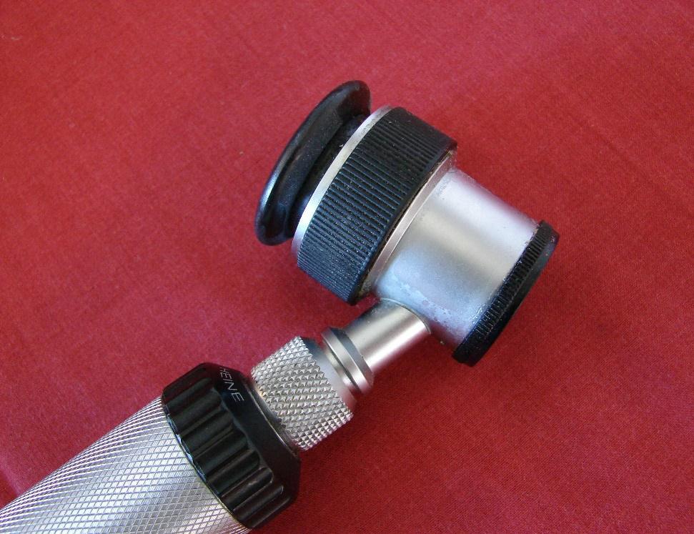
10 Heine Delta 10 Heine Delta 20

11 3GEN Dermlite
12 EFFECTIVENESS: There are several studies to demonstrate that dermoscopy is useful in the diagnosis of melanoma. It may be up to 35% more accurate than clinical diagnosis. Reduction in number of benign lesions excised. In primary care, results in referral of more suspicious lesions and less banal ones
13 CLINICAL DIAGNOSIS: Training and utilisation of dermoscopy is recommended for clinicians routinely examining pigmented lesions (Level of evidence A) Clinical Practice Guidelines for the Management of Melanoma in Australia and New Zealand November 2008

14 Naevus Pattern Previous Excisions NMSC Pregnancy Family history Patient Lesion Occupation Recreation Outdoor Sports Racial/ Country of Origin Skin type Hair/Eye UV Exposure Sunbeds Sunburns Sundamage Medical History Medications Immunosuppression
15 Which patients should be examined clinically? There is basically no agreement in the literature concerning the effectiveness of skin cancer screening. However there is at least some evidence that adults with risk factors for melanoma (personal or family history of melanoma, multiple naevi, fair skin, multiple sunburns, previous non-melanoma skin cancer) might benefit from skin examination for melanoma screening. IDS 2007
16 Patients younger than 50 years who present with more than 50 naevi in total or more than 20 naevi on the deltoid area of the upper arm. Patients older that 50 years who present with evidence of chronic solar damage. IDS 2013.
17 In high risk patients total cutaneous examination should be considered to be the standard of care. All of the skin including palms, soles and scalp should be examined clinically. Additionally self-examination of the skin on a monthly basis should be encouraged.
18 Documentation of the clinical and dermoscopic features for relevant atypical or changing lesions examined including specific anatomical site(s), is an important part of good clinical practice and should be encouraged wherever possible.
19 Which lesions deserve closer examination with dermoscopy? Lesions with reported history of change (colour, size, shape or symptom) even if the patient is not able to specify a particular concern. New or changing lesions in adults>50. A lesion different to the others (ugly duckling sign). Lesions which look all the same from a distance but differ close-up (little red riding hood sign). Lesions that look clinically like a melanoma (eccentric peripheral pigmentation or ABCD).

20
21
22
23
24 DIGITAL IMAGING Attaching a digital camera to a dermatosope or using a specially set up camera to take high quality digital images of lesions. Allows follow-up of recorded lesions. It is estimated that dermoscopy can detect 92% of melanomas immediately due to typical features and the remaining 8% because of change in atypical lesions.
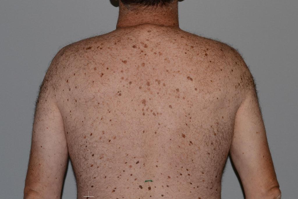
25 MOLEMAPPING: Using a combination of total body photography (TBF) with computer software to archive the images and allow remote diagnosis by dermatologist (Molemap NZ). TBF allows precise localisation of the naevi and the appearance of new lesions can be noted. (24-30 images)

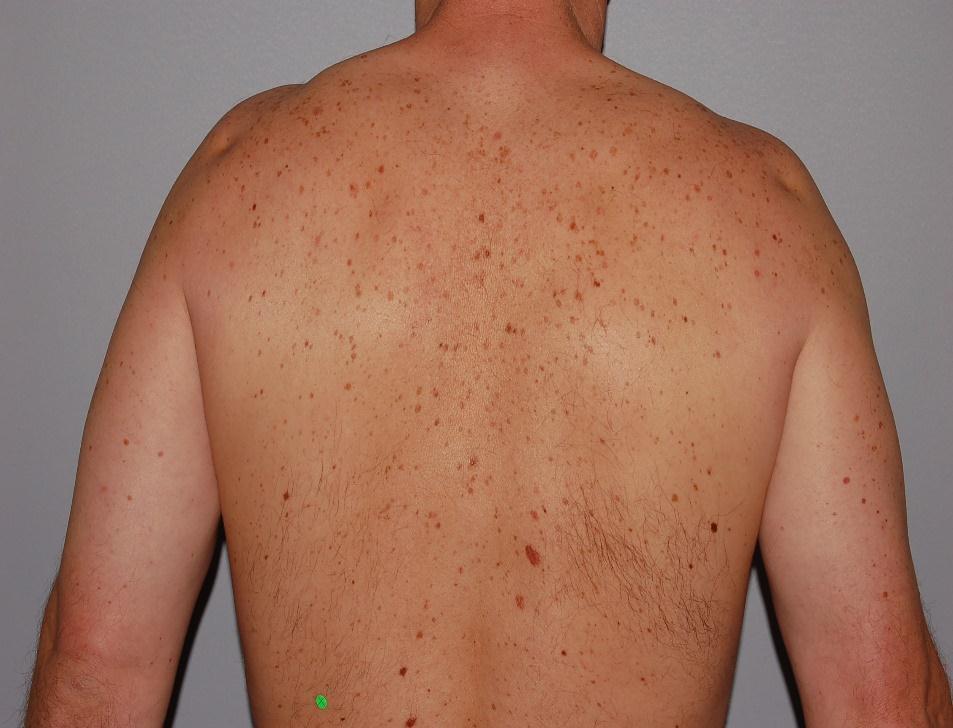
26 Total Body Photography
27
28

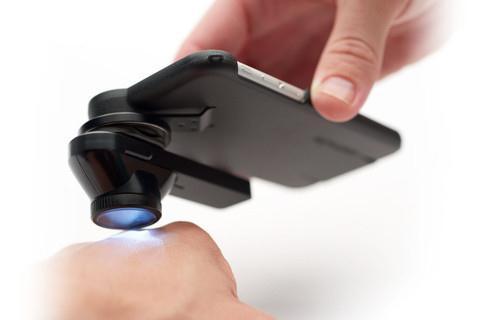
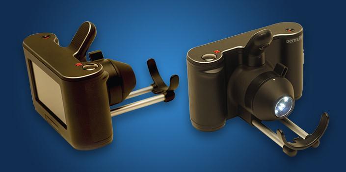
29 Dermlite Cam Unashamed Advertisement
30
31 Macroscopic Image Dermoscopic Image
32 AUTOMATIC (SMART) SYSTEMS: SIAscope, Solarscan, Melafind Using artificial intelligence (AI) and neural network methods to predict abnormalities in lesions compared to stored parameters. Not especially useful for nonpigmented lesions and cannot decide which lesions need imaging.
33 Automatic dermoscopic image analysis has three stages: 1. Image segmentation 2. Feature extraction and selection 3. Lesion classification
34

35
36
37 Compared to the histopathology results from 77 lesions: The Automatic System (Dermogenius) Sensitivity 96% Specificity 15% Diagnostic accuracy 47% Experienced Dermoscopist Sensitivity 100% Specificity 67% Diagnostic accuracy 95%
38 Dermoscopic Diagnostic Aids and Algorithms
39 The First Step Algorithm Pattern Analysis ABCD Rule Menzie s Method The Seven Point Checklist The Three Point Checklist BLINK
40 THE FIRST STEP ALGORITHM To determine whether the lesion is Melanocytic (naevus or melanoma) or Non-Melanocytic (BCC, seborrhoeic keratosis etc)
41 FIRST STEP ALGORITHM Melanocytic Non-Melanocytic Benign (naevus) or Melanoma (3 POINT CHECKLIST) Seborrhoeic keratosis BCC Haemangioma etc
42 Benign and Malignant Melanocytic lesions have one or more of the following: Pigment Network Aggregated (pigment) globules/dots Streaks Homogeneous blue pigmentation Parallel pattern

43 PIGMENT NETWORK is defined as a brown grid over a light brown background

44 AGGREGATED GLOBULES. Round to oval variable size uniformly distributed over lesion.

45 STREAKS Brown-black linear structures often with bulbous tips usually at periphery of lesion.

46 HOMOGENEOUS BLUE PIGMENTATION Hallmark of blue naevi.

47 PARALLEL PATTERN Found almost exclusively in melanocytic lesions on palm/soles
48 PATTERN ANALYSIS Is the preferred method of most experts in dermoscopy to diagnose skin lesions. The traditional ABCD and scoring methods are all still very useful but it is best to stick to one technique to avoid confusion.
49 PATTERN ANALYSIS involves describing the 1 GLOBAL FEATURES and 2 LOCAL FEATURES A simultaneous assessment of the diagnostic value of all the dermoscopic features of the lesion
50 GLOBAL FEATURES Reticular Pattern Globular Pattern Cobblestone Pattern Homogeneous Pattern Starburst Pattern Parallel Pattern Multicomponent Pattern Unspecific Pattern
51 LOCAL FEATURES Pigment network Streaks Dots and Globules Blue-white veil Regression structures Blotches Hypopigmentation Vascular structures Milia like cysts Comedo-like openings Brain-like fissures Exophytic papillary structures Red lacunas Central white patch Leaf-like areas Spoke-wheel areas Large blue-gray ovoid nests Multiple blue-gray globules
52 It is likely that many of these features(criteria) will undergo deletion or change and there are at least 50 local dermoscopic features that have been described in the last 10 years that await international recognition and agreement of definition.
53 3 rd Consensus Conference of the International Dermoscopy Society Thursday April 6th 2015 Vienna Austria Terminology in historical perspective and why we need a new consensus A plea for metaphoric terms Preliminary results of the internet survey ( not yet published)

54 GLOBAL FEATURES- RETICULAR PATTERN Characteristic of benign melanocytic lesions with a regular grid network covering most of the lesion. Fades out at edges.

55 GLOBAL FEATURES GLOBULAR PATTERN Typical for benign naevi especially in young persons.
56 COBBLESTONE PATTERN Large closely aggregated large gloular structures(papillomatous)
57 STARBURST PATTERN Pigmented streaks in a radial arrangement at the edge of the lesion- Spitz/Reed naevi

58 PARALLEL PATTERN Pigmentation on palms/soles in the furrows-acral naevi Parallel ridge pattern- melanoma

59 MULTICOMPONENT PATTERN Combination of three patternsmelanoma
60 The Dermoscopy report:proposal for Standardization (10 points) 1. Age, history(of lesion), personal/family history 2. Clinical description of the lesion 3. The 2-step method of distinguishing melanocytic/nonmelanocytic 4. Standardized terms to describe structures as defined by the Dermoscopy Consensus Report of The dermoscopic algorithm used
61 6. Information on the imaging equipment and magnification 7. Clinical and dermoscopic images of the tumour 8. Diagnosis or differential diagnosis 9. Decision concerning the management 10. Any specific comments for the pathologist if excision and histopathologic examination are recommended
22/04/2015. Dermoscopy of Melanoma. Ilsphi Browne. Overview
 Dermoscopy of Melanoma Ilsphi Browne Overview The device Dermoscopic criteria (terminology) Colour Patterns Global features Local features Approach to diagnosing pigmented lesions Other uses in general
Appendix : Dermoscopy
 Go Back to the Top To Order, Visit the Purchasing Page for Details APP Appendix : Dermoscopy Dermoscopy, also known as dermatoscopy, epiluminoscopy and epiluminescent microscopy, is an effective non-invasive
Introduction to Dermoscopy. Nicholas Compton, MD June 16, 2010
 Introduction to Dermoscopy Nicholas Compton, MD June 16, 2010 Overview What is dermoscopy Brief history Types of dermoscopy General approach to lesion of interest 2 step algorithm 3-point checklist Practice
Dermoscopy Quiz 3-Point Checklist Algorithm
 Dermoscopy Quiz 3-Point Checklist Algorithm GLOBAL PATTERN Globular LOCAL CRITERIA Aggregated globules Milia-like cysts 3 POINT CHECK LIST Symmetrical No abnormal net Slight Blue-white veil BENIGN MELANOCYTIC
It can be helpful in some cases of actinic keratosis, Bowen s disease and squamous cell carcinoma
 Dermoscopy Introduction, Terminology and Structures (to be read in conjunction with the Diagnostic Dermoscopic Algorithm) Copyright to Cunliffe TP (Jan. 2017) All rights reserved Introduction Dermoscopy
Dermoscopy: Recognizing Top Five Common In- Office Diagnoses
 Dermoscopy: Recognizing Top Five Common In- Office Diagnoses Vu A. Ngo, DO Department of Family Medicine and Dermatology Choctaw Nation Health Services Authority Learning Objectives Introduction to dermoscopy
MODULE 1. LOCAL AND GENERAL CRITERIA IN PIGMENTED MELANOCYTIC LESIONS.
 DERMOSCOPY TEACHING PROGRAMME Dermoscopy Teaching Programme Module 1 MODULE 1. LOCAL AND GENERAL CRITERIA IN PIGMENTED MELANOCYTIC LESIONS. Dermoscopy is a non-invasive in vivo technique that provides
Dermoscopy. Enhanced Diagnostic Ability: Pigmented Lesions. Ted Rosen, MD Baylor College of Medicine Houston, Texas
 Dermoscopy Enhanced Diagnostic Ability: Pigmented Lesions Ted Rosen, MD Baylor College of Medicine Houston, Texas Faculty Disclosure Statement No conflicts relevant to this workshop! Sir William Osler
Fundamentals of dermoscopy
 Fundamentals of dermoscopy Learning objectives Upon completion of this session, participants should be able to: describe the basic principles of dermoscopy identify features associated with pigmented and
Dermoscopy in everyday practice. What and Why? When in doubt cut it out? Trilokraj Tejasvi MD
 Dermoscopy in everyday practice Trilokraj Tejasvi MD Assistant Professor, Department of Dermatology, Director Teledermatology services, University of Michigan, Faculty Associate, GLOBAL REACH, Michigan
Dermoscopy. Sir William Osler. Dermoscopy. Dermoscopy. Melanoma USA Primary Care Update Faculty Disclosure Statement
 Diagnostic Ability: Pigmented Lesions Ted Rosen, MD Baylor College of Medicine Houston, Texas Enhanced 2010 Primary Care Update Faculty Disclosure Statement Ted Rosen, MD Speakers Bureau: Abbott, Amgen,
Disclosure. Objectives. PAFP CME Conference Lou Mancano MD, FAAFP Reading Health System November 18, 2016
 PAFP CME Conference Lou Mancano MD, FAAFP Reading Health System November 18, 2016 1 Disclosure The speaker has no conflict of interest, financial agreement, or working affiliation with any group or organization.
comedo-like openings (clods, brown or orange & circles) milia-like cysts (dots or clods, white) 1/29/18 Dotted vessels are also commonly seen in SCC
 milia-like cysts (dots or clods, white) 1/29/18 Dotted vessels are also commonly seen in SCC") Brown circles Dotted vessels are also commonly seen in SCC Step1 1. Nevus (unequivocal) 2. DF/IDN 3. BCC 4. SCC Network Patchy network Peripheral network & central hypopigmentation DF: network with central
Brown circles Dotted vessels are also commonly seen in SCC Step1 1. Nevus (unequivocal) 2. DF/IDN 3. BCC 4. SCC Network Patchy network Peripheral network & central hypopigmentation DF: network with central
Dermoscopy-a BRIEF introduction
 Dermoscopy-a BRIEF introduction Aim of presentation -to tell you what dermoscopy is -to show some of what it can do -point the interested learner to further resources Overview of dermoscopy Dermoscopy
6/17/2018. Breaking Bad (Part 1) Dermoscopy of Brown(ish) Things. Bad?
 Dermoscopy of Brown(ish) Things. Bad?") Breaking Bad (Part 1) Dermoscopy of Brown(ish) Things Jennie T. Clarke, MD ssociate Professor of Dermatology University of Utah School of Medicine Bad? 1 Brown(ish) Things Bad Melanoma Pigmented basal
Breaking Bad (Part 1) Dermoscopy of Brown(ish) Things Jennie T. Clarke, MD ssociate Professor of Dermatology University of Utah School of Medicine Bad? 1 Brown(ish) Things Bad Melanoma Pigmented basal
The impact of GP sub-specialisation and dermatoscopy use on diagnostic accuracy for melanomas in Australia
 The impact of GP sub-specialisation and dermatoscopy use on diagnostic accuracy for melanomas in Australia Cliff Rosendahl, Gail Williams, Diann Eley, Tobias Wilson, Greg Canning, Jeffrey Keir, Ian McColl,
Dermoscopy. Synonyms. Dermoscopy. Definition. Dermoscopy opens up a world of colour and structure that can t be seen with the naked eye
 Synonyms Dermoscopy Australasian College of Dermatologists G.P Training Module Dermoscopy Dermatoscopy Epiluminescence microscopy Skin surface microscopy Incident light microscopy Oil immersion microscopy
Key factors in successfully integrating dermoscopy into your clinical practice
 Key factors in successfully integrating dermoscopy into your clinical practice S051 Dilemmas and challenges in skin cancer therapies and management Monday, March 4 th 2019 (9AM-12PM) Room 209A 10:56-11:09AM
50 interactive dermoscopic case discussions Dr Stephen Hayes
 50 interactive dermoscopic case discussions Dr Stephen Hayes Annotations will be found on your memory drive, as will 100 case discussions and other learning material Melanoma 2mm thick Ugly duckling-one
Skin Cancer A Personal Approach. Dr Matthew Strack Dunedin New Zealand
 Skin Cancer A Personal Approach Dr Matthew Strack Dunedin New Zealand Outline Dermoscopy Instruments and setup Photochemosurgery Clinical Aim: Leave with 2-3 ideas JLE Benign Junctional Nevus Management
Dermoscopy STFM Richard Usatine, MD 5/2/16. Disclosure Statement: Some Dermatoscopes. Dermoscopy Video. Thanks to Dr.
 Disclosure Statement: Dermoscopy STFM 2016 Richard P. Usatine, MD, FAAFP Professor, Family and Community Medicine Professor, Dermatology and Cutaneous Surgery Medical Director, Clinic University of Texas
Skin lesions The Good and the Bad. Dr Virginia Hubbard Ipswich Hospital NHS Trust Barts and the London School of Medicine and Dentistry
 Skin lesions The Good and the Bad Dr Virginia Hubbard Ipswich Hospital NHS Trust Barts and the London School of Medicine and Dentistry Case 1 32 year old woman Australian Lesion on back New hair growing
Clinical and Dermoscopic Features of Thin Nodular Melanoma
 Clinical and Dermoscopic Features of Thin Nodular Melanoma A study of the International Dermoscopy Society Coordinator: Dr. Alexander J. Stratigos and colleagues, alstrat2@gmail.com ** Extended to May
What is Dermoscopy? Early Dermoscopes. Deciphering Dermoscopy: Terminology, Features & Algorithms 6/17/2018
 Deciphering Dermoscopy: Terminology, Features & Algorithms Where did it come from and why do we use it? Jennie T. Clarke, MD Associate Professor of Dermatology University of Utah School of Medicine What
Common Benign Lesions and Skin Cancers. 22nd May 2015 Dr Mark Foley
 Common Benign Lesions and Skin Cancers 22nd May 2015 Dr Mark Foley Thank you for downloading this file. This intended to supplement the presentation given at the NZ Wound Care Conference, it is not intended
Skin Cancer. Dr Elizabeth Ogden Associate Specialist in Dermatology East and North Herts Dr Elizabeth Ogden
 Skin Cancer Dr Elizabeth Ogden Associate Specialist in Dermatology East and North Herts 13.10.16 Skin Cancer Melanoma mole cancer - is a true cancer which can metastasize and kill Non Melanoma skin cancer
NEW. DELTA 20 Plus Dermatoscope Head
 04 MOre details FOR an even better diagnosis: POLARISATION, HIGH PERFORMANCE LEDs and HIGH RESOLUTION optics. While the rate of new cases of malignant melanoma is increasing worldwide, mortality rates
Mole mapping and monitoring. Dr Stephen Hayes. Associate Specialist in Dermatology, University Hospital Southampton
 Mole mapping and monitoring Dr Stephen Hayes Associate Specialist in Dermatology, University Hospital Southampton Outline of presentation The melanoma epidemic Benefits of early detection Risks of the
Basics in Dermoscopy
 Basics in Dermoscopy Manal Bosseila Professor of Dermatology, Cairo University Member of European Academy Dermatology & Venereology EADV Member of International Dermoscopy Society IDS Member of Aesthetic
Age-related prevalence of dermatoscopic patterns of acral melanocytic nevi
 DERMATOLOGY PRACTICAL & CONCEPTUAL www.derm101.com Age-related prevalence of dermatoscopic patterns of acral melanocytic nevi Reiko Suzaki 1, Sumiko Ishizaki 1, Hitoshi Iyatomi 2, Masaru Tanaka 1 1 Department
Dr Amanda Oakley. Dermatologist Dept of Dermatology, Health Waikato Adjunct Associate Professor, Waikato Clinical Campus
 Dr Amanda Oakley Dermatologist Dept of Dermatology, Health Waikato Adjunct Associate Professor, Waikato Clinical Campus 14:00-16:00 WS #14: Dermoscopy Part 1 Skin Lesions and Dermatoscopy 16 August 2018
Malignant non-melanocytic lesions
 Malignant non-melanocytic lesions Course C023: Fundamentals of Dermoscopy March 4, 2019, 11:20 AM - 11:50 PM Room: 146B Jason B. Lee, MD Professor & Vice Chair Director of Dermatopathology & Pigmented
Benign versus Cancerous Lesions How to tell the difference FMF 2014 Christie Freeman MD, CCFP, DipPDerm, MSc
 1 Benign versus Cancerous Lesions How to tell the difference FMF 2014 Christie Freeman MD, CCFP, DipPDerm, MSc Benign lesions Seborrheic Keratoses: Warty, stuck-on Genetics and birthdays Can start in late
BLINCK A diagnostic algorithm for skin cancer diagnosis combining clinical features with dermatoscopy findings
 DERMATOLOGY PRACTICAL & CONCEPTUAL www.derm101.com BLINCK A diagnostic algorithm for skin cancer diagnosis combining clinical features with dermatoscopy findings Peter Bourne, MBBS 1, Cliff Rosendahl,
Dr Stephen Hayes Associate Specialist in Dermatology University Hospital Southampton
 South East Dermatology Transformation and Sustainability Network Guildford, 19 th April 2018 Dermoscopy as an effective skin lesion triage tool in GP surgeries Dr Stephen Hayes Associate Specialist in
HEINE Dermatoscopes. Magnification Magnification x 40x* (digital)
") [ 079 ] HEINE DELTA 20 T ic 1 NEW: NC 2 mini 3000 Magnification Magnification 10 16 x 40x* (digital) 6 10 x 30x* (digital) 10 x Illumination illumination XHL illumination Toggle function Examination mode
[ 079 ] HEINE DELTA 20 T ic 1 NEW: NC 2 mini 3000 Magnification Magnification 10 16 x 40x* (digital) 6 10 x 30x* (digital) 10 x Illumination illumination XHL illumination Toggle function Examination mode
Dermoscopy, the use of a handheld
 ONLINE EXCLUSIVE Dermoscopy in family medicine: A primer Dermoscopy allows you to see deeper into the skin than with the naked eye. Here s how you can make use of it to spot malignant conditions sooner.
Histopathological and SIAscopic Correlation of Pigmented Skin Lesions
 Histopathological and SIAscopic Correlation of Pigmented Skin Lesions Professor Sujatha Fernando MBBS(Hon), MSc(London, Distinction), FRSTM&H, FRCPA, FIAC, FACTM Senior Consultant in Anatomical Pathology,
Aspects on in vivo imaging techniques for diagnostics of pigmented skin lesions
 Thesis for the Degree of Doctor of Philosophy Aspects on in vivo imaging techniques for diagnostics of pigmented skin lesions Karin Terstappen (Westerhoff) Department of Dermatology and Venereology Institure
Description of Some Dermatoscopic Features of Acral Pigmented Lesions in Iranian Patients: A Preliminary Study
 ORIGINAL REPORT Description of Some Dermatoscopic Features of Acral Pigmented Lesions in Iranian Patients: A Preliminary Study Reza Nemati Ahmadabad 1, Hayede Ghaninezhad 1, Homayoon Moslehi 2, Sahar Azizahari
10/3/2018. Dermoscopy: Looking beneath the surface of the skin. Dermoscopy for Family Medicine 10/11/2018
 Dermoscopy for Family Medicine 10/11/2018 Jane M. Grant-Kels, MD, FAAD Founding Chair Emeritus, Dept of Dermatology Professor of Dermatology, Pathology & Pediatrics Director of the Cut Oncology Ctr & Melanoma
STUDY. Dermoscopic Characteristics of Congenital Melanocytic Nevi Affecting Acral Volar Skin
 STUDY Dermoscopic Characteristics of Congenital Melanocytic Nevi Affecting Acral Volar Skin Akane Minagawa, MD; Hiroshi Koga, MD; Toshiaki Saida, MD, PhD Objective: To characterize the dermoscopic features
Acral and Mucosal Dermoscopy
 Acral and Mucosal Dermoscopy Caroline C. Kim, MD Assistant Professor, Department of Dermatology Harvard Medical School Director, Pigmented Lesion Clinic Associate Director, Cutaneous Oncology Program Beth
Revised Pattern Analysis: a method for the accurate diagnosis of pigmented skin lesions
 Dermatoscopy for Students A concise outline of: Revised Pattern Analysis: a method for the accurate diagnosis of pigmented skin lesions And Chaos and Clues: a decision algorithm for routine practice to
Regression 2/3/18. Histologically regression is characterized: melanosis fibrosis combination of both. Distribution: partial or focal!
 Regression Margaret Oliviero MSN, ARNP Harold S. Rabinovitz MD Histologically regression is characterized: melanosis fibrosis combination of both Distribution: partial or focal! Dermatoscopic terminology
Melanoma and Dermoscopy. Disclosure Statement: ABCDE's of melanoma. Co-President, Usatine Media
 Melanoma and Dermoscopy Richard P. Usatine, MD, FAAFP Professor, Family and Community Medicine Professor, Dermatology and Cutaneous Surgery Medical Director, University Skin Clinic University of Texas
Contrast with Australian Guidelines A/Pr Pascale Guitera,
 Contrast with Australian Guidelines A/Pr Pascale Guitera, Dermatologist, Sydney University NO CONFLICT OF INTEREST Sydney Melanoma Diagnostic Centre, RPAH 2011 2008 225 pages 16 pages http://www.cancer.org.au/file/healthprofessionals/clinica
Hauora Maori. Map. Non-Melanoma Skin Lesion. Clinically Seborrhoeic Keratosis. Management Options. Punch Biopsy. Refer to Public Skin Lesion Service
 Care map information Information resources for patients and carers Updates to this Care Map Hauora Maori Pacific Skin lesion history Clinical examination and diagnostic test Suspicious of Melanoma Non-Melanoma
Non-Melanocytic Pattern Dermoscopy
 Non-Melanocytic Pattern Dermoscopy I have no conflicts of interest to disclose Except that I LOVE dermoscopy Michelle Tarbox, MD Assistant Professor of Dermatology and Dermatopathology Texas Tech University
Apps and Telemedicine H. Peter Soyer Dermatology Research Centre
 Apps and Telemedicine H. Peter Soyer Dermatology Research Centre p.soyer@uq.edu.au https://twitter.com/hpsoyer William Gibson The future is already here it's just not very evenly distributed Vision 3D
Development and validation of a scoring system for SIAscopic diagnosis of pigmented skin lesions in primary care
 Development and validation of a scoring system for SIAscopic diagnosis of pigmented skin lesions in primary care J Hunter 1,2, FM Walter 3,5, M Moncrieff 1, S Cotton 4 PN Hall 1, J Emery 3,5 1 Dept of
STUDY. Scott W. Menzies, MB,BS, PhD; Karin Westerhoff, MD; Harold Rabinovitz, MD; Alfred W. Kopf, MD; William H. McCarthy, MBBS, MEd; Brian Katz
 STUDY Surface Microscopy of Pigmented Basal Cell Carcinoma Scott W. Menzies, MB,BS, PhD; Karin Westerhoff, MD; Harold Rabinovitz, MD; Alfred W. Kopf, MD; William H. McCarthy, MBBS, MEd; Brian Katz Objectives:
Multispectral Digital Skin Lesion Analysis. Summary
 Subject: Multispectral Digital Skin Lesion Analysis Page: 1 of 8 Last Review Status/Date: March 2016 Multispectral Digital Skin Lesion Analysis Summary There is interest in noninvasive devices that will
Non-melanocytic Patterns
 Non-melanocytic Lesions Non-melanocytic Patterns Michelle Tarbox, MD Assistant Professor of Dermatology and Dermatopathology Texas Tech University Health Sciences Center 2018 Seborrheic keratoses Acanthotic
:- HEINE IC1 NEW! 21st Century Digital Dermatoscopy
 :- HEINE IC1 21st Century Digital Dermatoscopy The HEINE ic1 is a new image capturing device to record clinical and dermatoscopic images for documenting, monitoring and supporting differential diagnosis
The GP s approach to the patient who is worried about sun, skin and moles. Dr Stephen Hayes
 The GP s approach to the patient who is worried about sun, skin and moles Dr Stephen Hayes Associate Specialist in Dermatology, University Hospital Southampton Dr Stephen Hayes DECLARATION OF INTERESTS
Case Report Micromelanomas: A Review of Melanomas 2mmand a Case Report
 Case Reports in Oncological Medicine, Article ID 206260, 4 pages http://dx.doi.org/10.1155/2014/206260 Case Report Micromelanomas: A Review of Melanomas 2mmand a Case Report Sharad P. Paul 1,2,3 1 Skin
Cover Page. The handle holds various files of this Leiden University dissertation.
 Cover Page The handle http://hdl.handle.net/1887/22172 holds various files of this Leiden University dissertation. Author: Rhee, Jasper Immanuel van der Title: Clinical characteristics and management of
INVESTIGATION. The relation between dermoscopy and histopathology of basal cell carcinoma *
 INVESTIGATION The relation between dermoscopy and histopathology of basal cell carcinoma * 351 Nazan Emiroglu 1 Fatma Pelin Cengiz 1 Funda Kemeriz 2 DOI: http://dx.doi.org/10.1590/abd1806-4841.20153446
Graph-based Pigment Network Detection in Skin Images
 Graph-based Pigment Network Detection in Skin Images M. Sadeghi a,b, M. Razmara a, M. Ester a, T. K. Lee a,b,c, M. S. Atkins a a Simon Fraser University, 8888 University Drive, Burnaby, BC, Canada, V5A1S6;
Decision Support System for Skin Cancer Diagnosis
 The Ninth International Symposium on Operations Research and Its Applications (ISORA 10) Chengdu-Jiuzhaigou, China, August 19 23, 2010 Copyright 2010 ORSC & APORC, pp. 406 413 Decision Support System for
Cancer Council Australia Wiki Guidelines 2017
 WHAT IS THE ROLE OF SEQUENTIAL DIGITAL DERMOSCOPY IMAGING IN MELANOMA DIAGNOSIS? Cancer Council Australia Wiki Guidelines 2017 SHORT-TERM MONITORING 3 months Any change leads to excision Any melanocytic
STUDY. Characteristic Epiluminescent Microscopic Features of Early Malignant Melanoma on Glabrous Skin
 Characteristic Epiluminescent Microscopic Features of Early Malignant Melanoma on Glabrous Skin A Videomicroscopic Analysis STUDY Shinji Oguchi, MD; Toshiaki Saida, MD, PhD; Yoko Koganehira, MD; Sachiko
Melanoma. Consultation on draft guideline - stakeholder comments. Comments to be submitted before 5pm on Friday 13 March 2015
 Please note: Please fill in both the stakeholder organisation and name of commentator fields. We cannot accept forms with attachments such as research articles, letters or leaflets. Stakeholder organisation(s)
Assessment of SIAscopy in the triage of suspicious skin tumours
 Skin Research and Technology 2014; 0: 1 5 Printed in Singapore All rights reserved doi: 10.1111/srt.12138 2014 John Wiley & Sons A/S. Published by John Wiley & Sons Ltd Skin Research and Technology Assessment
Total body photography in high risk patients
 Total body photography in high risk patients Doug Grossman, MD, PhD Department of Dermatology Huntsman Cancer Institute University of Utah Summer AAD F032 Practical Considerations for Patients with Melanoma
MELANOMA: HANDS-ON OR HANDS-OFF?
 MELANOMA: HANDS-ON OR HANDS-OFF? M SCHAMM MBChB (Pret), FCS (SA) Endocrine and Transplant Surgeon, Department of Surgery, University of the Witwatersrand; and Clinical Head Transplant Surgery, Charlotte
CANCER INSIGHT. FOR GPs. Summer 2018 WHAT YOU NEED TO KNOW ABOUT SKIN CANCER VISIT. our Skin Cancer Recognition Toolkit at
 CANCER INSIGHT FOR GPs Summer 2018 WHAT YOU NEED TO KNOW ABOUT SKIN CANCER VISIT our Skin Cancer Recognition Toolkit at www.doctors.net. uk/sct Cancer Research UK, Angel Building, 407 St John Street, London
Diagnostic Dermatology & Dermatologic Surgery Overview
 Diagnostic Dermatology & Dermatologic Surgery Overview A comprehensive range of innovative products & accessories for dermatologic diagnosis & surgery Clinical imaging The Fully integrated patient management
Prior Authorization Review Panel MCO Policy Submission
 Prior Authorization Review Panel MCO Policy Submission A separate copy of this form must accompany each policy submitted for review. Policies submitted without this form will not be considered for review.
Dermoscopic Features of Non-Pigmented Eccrine Poromas in. Department of Dermatology, Shinshu University School of Medicine,
 Original article Dermoscopic Features of Non-Pigmented Eccrine Poromas in Association with their Histopathological Features Akane Minagawa, Hiroshi Koga,* Masaomi Takahashi, + Kenji Sano, + Ryuhei Okuyama,
Diagnostics guidance Published: 11 November 2015 nice.org.uk/guidance/dg19
 VivaScope 1500 and 3000 imaging systems for detecting skin cancer lesions Diagnostics guidance Published: 11 November 2015 nice.org.uk/guidance/dg19 NICE 2018. All rights reserved. Subject to Notice of
STUDY. Identification of Clinically Featureless Incipient Melanoma Using Sequential Dermoscopy Imaging
 STUDY Identification of Clinically Featureless Incipient Melanoma Using Sequential Dermoscopy Imaging Harald Kittler, MD; Pascale Guitera, MD; Elisabeth Riedl, MD; Michelle Avramidis, MD; Ligia Teban,
Optical Diagnostic Devices for Evaluating Skin Lesions Suspected of Malignancy. Original Policy Date
 MP 2.01.29 Optical Diagnostic Devices for Evaluating Skin Lesions Suspected of Malignancy Medical Policy Section Medicine Issue 12:2013 Original Policy Date 12:2013 Last Review Status/Date Reviewed with
Page: 1 of 16. Optical Diagnostic Devices for Evaluating Skin Lesions Suspected of Malignancy
 Last Review Status/Date: December 2014 Page: 1 of 16 Skin Lesions Suspected of Malignancy Description There is interest in non-invasive devices that will improve the diagnosis of malignant skin lesions.
Features Causing Confusion between Basal Cell Carcinoma and Squamous Cell Carcinoma in Clinical Diagnosis
 TH Ryu, et al pissn 1013-9087ㆍeISSN 2005-3894 Ann Dermatol Vol. 30, No. 1, 2018 https://doi.org/10.5021/ad.2018.30.1.64 ORIGINAL ARTICLE Features Causing Confusion between Basal Cell Carcinoma and Squamous
Introduction to Dermoscopy. Disclosure. Introduction
 Introduction to Dermoscopy 1 Disclosure Dr. Deborah Bren has no conflict of interest, financial agreement, or working affiliation with any group or organization. 2 Introduction Deborah A. Bren, DO Family
Dermoscopy of non-pigmented skin lesions: a literature review
 Hong Kong J. Dermatol. Venereol. (2017) 25, 13-21 Review Article Dermoscopy of non-pigmented skin lesions: a literature review S Thomas, X Li, HP Soyer In this article, we will review benchmark dermoscopic
RCGP and Cancer Research UK Workshop. Hilton Newcastle Gateshead, Bottle Bank, Gateshead, NE8 2AR 13 th July 2017
 Hilton Newcastle Gateshead, Bottle Bank, Gateshead, NE8 2AR 13 th July 2017 Dr Richard Roope RCGP and Cancer Research UK Cancer Clinical Champion Senior Clinical Advisor Cancer Research UK How can GPs
Living Beyond Cancer Skin Cancer Detection and Prevention
 Living Beyond Cancer Skin Cancer Detection and Prevention Cutaneous Skin Cancers Identification Diagnosis Treatment options Prevention What is the most common cancer in people? What is the most common
STUDY. Epiluminescence Microscopy for the Diagnosis of Doubtful Melanocytic Skin Lesions
 STUDY Epiluminescence Microscopy for the Diagnosis of Doubtful Melanocytic Skin Lesions Comparison of the ABCD Rule of Dermatoscopy and a New 7-Point Checklist Based on Pattern Analysis Giuseppe Argenziano,
=C 0= C8E4 <4;0=><0 3806=>B8B
 4558284=C 0=3 45542C8E4 B8B Wednesday 20 th November 2002 Centenary Institute of Cancer Medicine & Cell Biology Royal Prince Alfred Hospital Missenden Road Camperdown NSW WORKSHOP SUMMARY
Palmoplantar melanocytic nevi: dermoscopic and histopathological correlation
 Research Article Palmoplantar melanocytic nevi: dermoscopic and histopathological correlation Mónica Andrea Barengo 1, María Paula Gutiérrez 1, Enrique Valente 2, Alejandro Ruiz Lascano 3 Abstract Introduction.
MELANOCYTIC LESIONS: EFFECTIVE MANAGEMENT IN PRIMARY CARE: Part 2
 MELANOCYTIC LESIONS: EFFECTIVE MANAGEMENT IN PRIMARY CARE: Part 2 In the second part of our feature on pigmented skin lesions, dermatology specialist Dr Sweta Rai describes the steps to rational decision-making
Diagnosis of Lentigo Maligna Melanoma. Steven Q. Wang, M.D. Memorial Sloan-Kettering Cancer Center Basking Ridge, NJ
 Diagnosis of Lentigo Maligna Melanoma Steven Q. Wang, M.D. Memorial Sloan-Kettering Cancer Center Basking Ridge, NJ Conflict of Interest: None Topics Epidemiology and Natural History Clinical and Histologic
DIFFERENCES IN DERMOSCOPIC IMAGES FROM NON-POLARIZED DERMOSCOPE AND POLARIZED DERMOSCOPE INFLUENCE THE DIAGNOSTIC ACCURACY AND CONFIDENCE LEVEL.
 DIFFERENCES IN DERMOSCOPIC IMAGES FROM NON-POLARIZED DERMOSCOPE AND POLARIZED DERMOSCOPE INFLUENCE THE DIAGNOSTIC ACCURACY AND CONFIDENCE LEVEL. 1. Steven Q. Wang MD 1 (wangs@mskcc.org) 2. Stephen W. Dusza
Be SunSmart Everywhere!
 Be SunSmart Everywhere! DID YOU KNOW? Sun exposure adds up day after day, and it happens every time you re in the sun. Damage is permanent and irreversible. MYTH Sunburn happens only when we go to the
The most common mistakes on dermatoscopy of melanocytic lesions
 Review paper The most common mistakes on dermatoscopy of melanocytic lesions Grażyna Kamińska-Winciorek 1, Waldemar Placek 2 1 The Center for Diagnostics and Treatment of Skin Diseases, Katowice, Poland
Case Report A Case of Cystic Basal Cell Carcinoma Which Shows a Homogenous Blue/Black Area under Dermatoscopy
 Volume 20, Article ID 450472, 4 pages doi:0.55/20/450472 Case Report A Case of Cystic Basal Cell Carcinoma Which Shows a Homogenous Blue/Black Area under Dermatoscopy Akihiro Yoneta, Kohei Horimoto, Keiko
Case Report Dermoscopy Clues in Pigmented Bowen s Disease
 Dermatology Research and Practice Volume 2010, Article ID 464821, 9 pages doi:10.1155/2010/464821 Case Report Dermoscopy Clues in Pigmented Bowen s Disease Daniela Gutiérrez-Mendoza, 1 Roberto Narro-Llorente,
BJD British Journal of Dermatology. Summary. What s already known about this topic? CLINICAL AND LABORATORY INVESTIGATIONS
 CLINICAL AND LABORATORY INVESTIGATIONS BJD British Journal of Dermatology Pigmented nodular melanoma: the predictive value of dermoscopic features using multivariate analysis M.A. Pizzichetta, 1 H. Kittler,
SCREENING FOR SKIN CANCER IN PRIMARY CARE: IMPLEMENTATION OF DERMOSCOPY
 SCREENING FOR SKIN CANCER IN PRIMARY CARE: IMPLEMENTATION OF DERMOSCOPY A Dissertation Submitted to the Graduate Faculty of the North Dakota State University of Agriculture and Applied Science By Erin
Pathology of the skin. 2nd Department of Pathology, Semmelweis University
 Pathology of the skin 2nd Department of Pathology, Semmelweis University Histology of the skin Epidermis: Stratum corneum Stratum granulosum Stratum spinosum Stratum basale Dermis: papillary and reticular
*Please see amendment for Pennsylvania Medicaid at the end
 1 of 32 Number: 0188 Policy *Please see amendment for Pennsylvania Medicaid at the end of this CPB. I. Aetna considers total body photography (TBP) and dermoscopy (also known as total body imaging, digital
Multiple Primary Melanoma in a Thai Male: A Case Report
 Case Report Multiple Primary Melanoma in a Thai Male: A Case Report J Med Assoc Thai 2014; 97 (Suppl. 2): S234-S238 Full text. e-journal: http://www.jmatonline.com Kittisak Payapvipapong MD*, Pinyapat
Optical Diagnostic Devices for Evaluating Skin Lesions Suspected of Malignancy
 2.01.42 Optical Diagnostic Devices for Evaluating Skin Lesions Suspected of Malignancy Section 2.0 Medicine Subsection Effective Date November 26, 2014 Original Policy Date November 26, 2014 Next Review
Management of Atypical Pigmented Lesions
 Management of Atypical Pigmented Lesions Jennifer A. Stein MD, PhD Associate Director, Pigmented Lesion Section Ronald O. Perelman Department of Dermatology NYU Langone Medical Center July 29, 2017 1-4
DERMATOLOGY PRACTICAL & CONCEPTUAL. Gabriel Salerni 1,2, Teresita Terán 3, Carlos Alonso 1,2, Ramón Fernández-Bussy 1 ABSTRACT
 DERMATOLOGY PRACTICAL & CONCEPTUAL www.derm101.com The role of dermoscopy and digital dermoscopy follow-up in the clinical diagnosis of melanoma: clinical and dermoscopic features of 99 consecutive primary
Finding Melanoma. Is not easy!
 Finding Melanoma Is not easy! Finding Melanoma Victoria mean depth at diagnosis is 1.5 mm. Melanoma 1.5mm Has Stage 1B Mortality 10% Melanoma Spotting a killer! Spotting a killer Visual Clues What are