Retina Grand Rounds. Stephen Huddleston MD Charles Retina Institute University of Tennessee Hamilton Eye Institute
|
|
|
- Drusilla Norman
- 5 years ago
- Views:
Transcription
1 Retina Grand Rounds Stephen Huddleston MD Charles Retina Institute University of Tennessee Hamilton Eye Institute
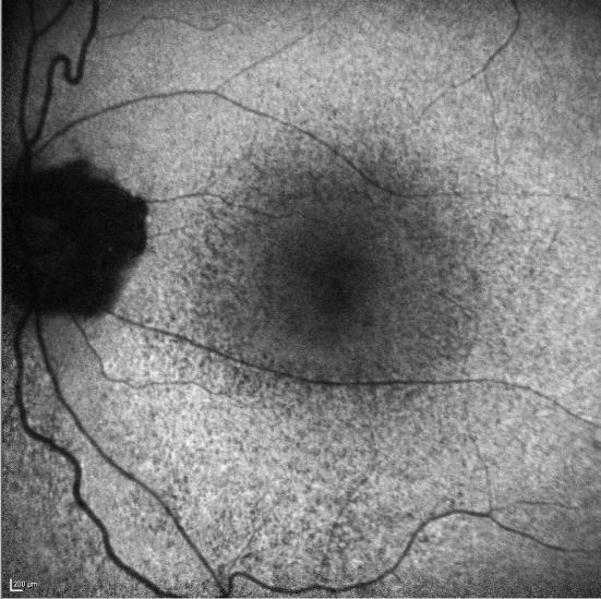
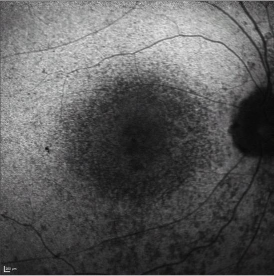
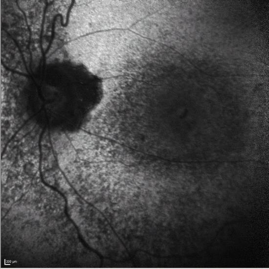
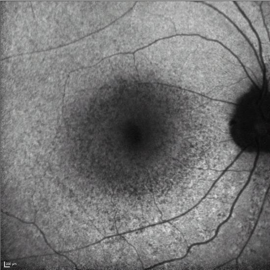
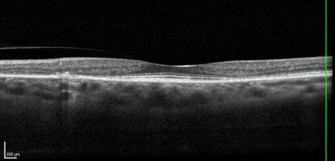
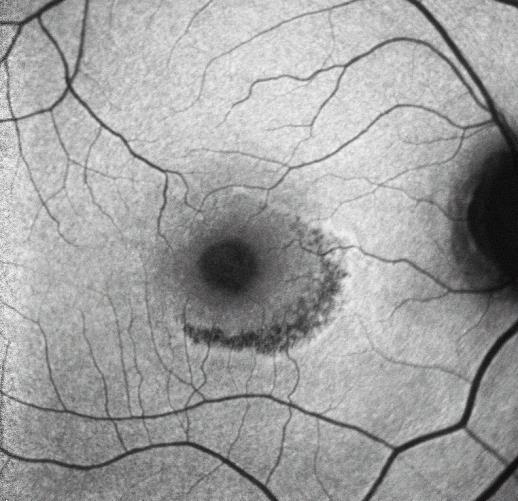
2 Fundus Autoflourescence


3 Plaquenil Toxicity Excellent treatment for a variety of Auto immune conditions Lupus, sarcoidosis, graft vs host, rheumatoid arthritis Efficacy marred by toxicity issues causing blindness Cumulative effect based on lifetime dose Cases of toxicity have decreased with lower average dosing and better screening modalities

4 Plaquenil Toxicity *Irreversible/permanent damage*


5 Plaquenil Toxicity *Key Point: Caucasian/African Americans parafoveal damage; Asian americans often extramacular* Damage worsens after stopping treatment Early detection crucial

6 Q: Traumatic dialysis with RD? 19 y/o athlete struck in eye with Baseball. Vitreous hemorrhage obscuring area of damage. Emergent surgery with buckle?

7 A: Choroidal Rupture Observe: VH clears revealing choroidal rupture without retinal tears One week apart

8 62 y/o immunocompetent Man with blurriness. Acute Retinal Necrosis?


9 Suspicion for ARN, CNS lymphoma, TB, Etc. MRI normal. Labs normal. Diagnostic PPV And treated with Ganciclovir injection. Sample obtained strongly positive for Acute Retinal Necrosis?


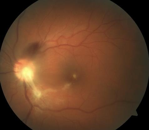
10 Toxoplasmosis Successfully treated with PO ABX and intravitreal clindamycin twice
11 Toxoplasmosis Caused by the protozoan toxoplasma gondii Single-cell obligate intracellular parasite 22.5% of US population infected 2% of US pop with signs of ocular involvement 3 major forms: Oocyst (soil form, hardy) Tachyzoite (infectious form, fast growing, vulnerable) Tissue cyst (contains infectious bradyzoites) Most common cause of infectious retinochoriditis in adults and children
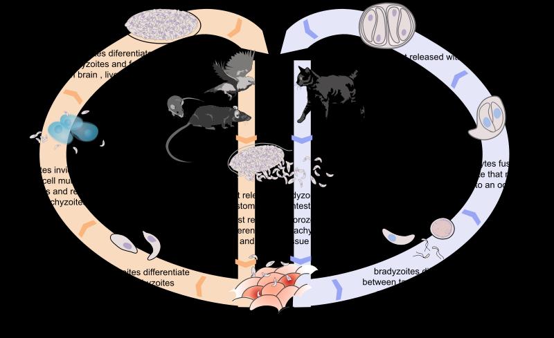
12 Toxoplasmosis Life Cycle
13 Toxoplasmosis Typical: Unilateral focal retinitis with overlying vitritis at edge of old chorioretinal scar Atypical: large areas of retinal recrosis or retinochoridits without pre-existing scar. Can be multifocal and/or bilateral with or without vasculitis Elderly and immunocompromised Rapid tachyzoite production and poor immune response Up to 20% with increased intraocular pressure Other secondary causes of vision loss: cataract, CME, serous retinal detachments, and CNV


14 TB or fungal retinitis? 22 y/o complaining of blurry vision and denying drug use
15 Atypical Toxoplasmosis PPV, EMM peel, clinda Starts to come back and he leaves the country
16 Toxoplasmosis Diagnosis primarily based on clinical signs Aqueous PCR with lower sensitivity then vitreous PCR Vitreous has closer contact to necrotic lesions with theorized higher T. Gondii DNA concentration. IgG antibodies present within the first two weeks Detectable for life, placental transfer in newborns IgM present acutely Detectable for less than one year IgA present acutely as well Disappears within 7 months
17 Toxoplasmosis Antibiotics highly effective against toxoplasma tachyzoites Transition from tachyzoite to bradyzoite takes only days Cysts form within weeks Triple therapy (gold standard, now irrelevant) Pyrimethamine mg Qday (cost impossibly high 2/2 Martin Shkreli) Needs folinic acid supplementation (5mg qday) Sulfadiazine mg Qday Sulfa allergy? Consider clindamycin 300mg QID Prednisone mg/kg/day Treatment generally lasts 4-6 weeks
18 Toxoplasmosis TMP/SMX 160/80 mg BID Azithromycin 250mg Qday (combined with pyrimethamine) Pregnancy: spiramycin 400mg TID (poor availability) Alternatives: azithromycin, clindamycin, atovaquone Sulfas can be used in the first two trimesters Intravitreal clindamycin a better option HIV: high risk of CNS and ocular recurrence. Consider long-term prophylaxis such as 1 tab TMP/SMX every third day after quiescence achieved Intravitreals: Intravitreal clinda/dex as effective as standard oral treatment with fewer side effects. No difference in recurrence rate. Give Clindamycin alone if diagnosis not confirmed.


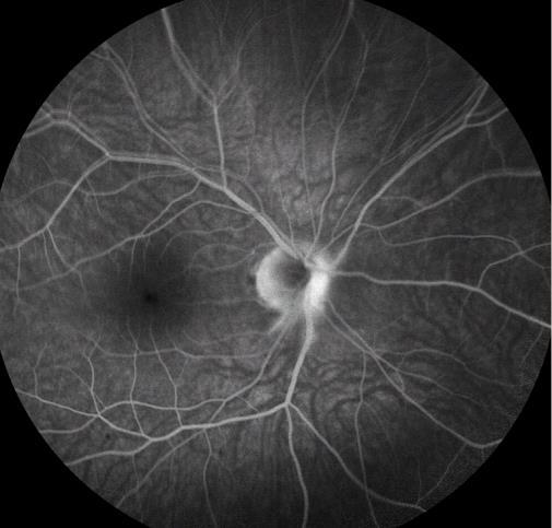
19 Toxoplasmosis causing rapidly worsening optic nerve lesion Day 1 Day 2 22 y/o with 20/70 vision, followed with optic nerve whiteneing for several weeks prior to referral


20 Final vision HM. What happened? Week one Week two Tues Oral azithromycin Mon Wed PPV clinda/dex Clinda/Dex Fri Wed

21 BRAO/BRVO Current VA: 20/400


22 Bilateral Retinal Detachment Mac on with 3 separate detachments Mac off with macular hole


23 Bilateral Retinal Detachment Same day bilateral PPV EPC FGX with ILM peel OS Tears everywhere 360 laser 20/40 vision, should keep improving


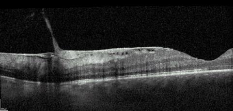
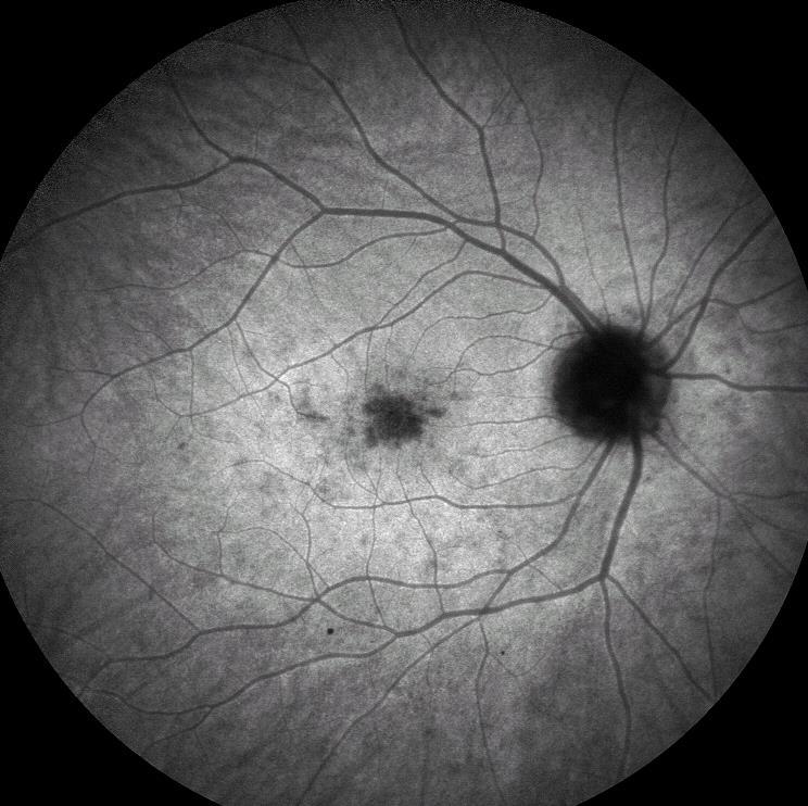
24 18 y/o Caucasian college student with 1 week of gray spot in vison OD and 20/70 vision Last Case
25 ICG late IVFA ICG early

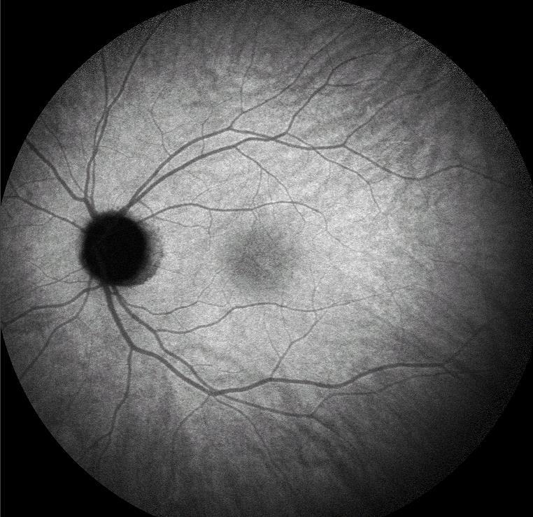
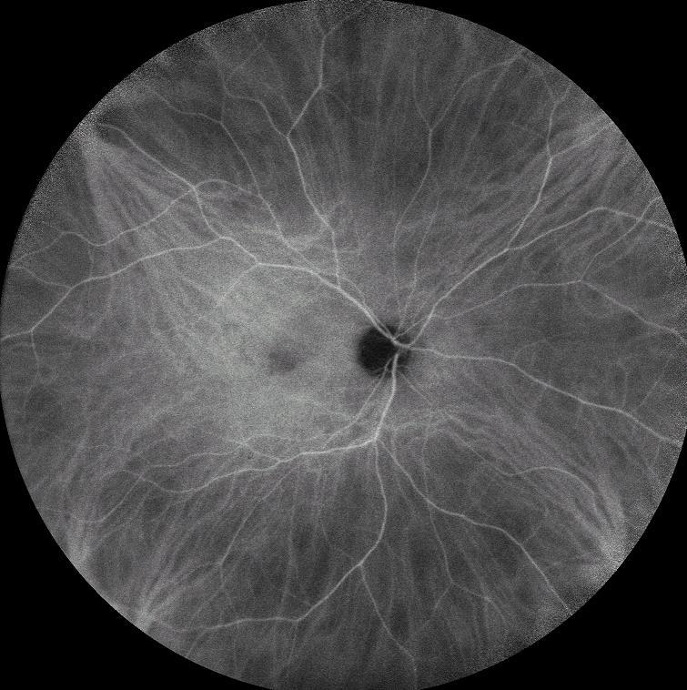

26 ICG late MEWDS: Multiple Evanescent White Dot Syndrome IVFA ICG early
27 MEWDS: Multiple Evanescent White Dot Syndrome Primarily Healthy young women (80% female) Usually with preceding mild viral illness Acute painless unilateral vision loss (rarely bilateral) Perhaps the mildest of the white dot syndromes: Acute Posterior Multifocal Placoid Pigment Epitheliopathy (APMPEE) Multifocal choroiditis and Panuveits (MCP) Punctate Inner Choroiditis (PIC) Birdshot Choroidopathy Lesions at the level of the outer retina and RPE Self limiting with excellent prognosis
28 Questions?
29 Further Reading Marmor MF, Kellner U, Lai TY, et al. Revised recommendations on screening for chloroquine and hydroxychloroquine retinopathy. Ophthalmology 2011;118: Melles RB, Marmor MF. The risk of toxic retinopathy in patients on long-term hydroxychloroquine therapy. JAMA Ophthalmol 2014;132: Marmor MF, Hu J. Effect of disease stage on progression of hydroxychloroquine retinopathy. JAMA Ophthalmol 2014;132: American Academy of Ophthalmology. Retina/Vitreous: Multiple evanescent white dot syndrome Practicing Ophthalmologists Learning System, San Francisco: American Academy of Ophthalmology, Joseph A, Rahimy E, Freund KB, et al. Fundus autofluorescence and photoreceptor bleaching in multiple evanscent white dot syndrome. Ophthalmic Surg Lasers Imaging Retina Nov 1;44(6): American Academy of Ophthalmology. Retina/Vitreous: Toxoplasmosis Practicing Ophthalmologists Learning System, San Francisco: American Academy of Ophthalmology, 2017.
Acute Retinal Necrosis Secondary to Varicella Zoster Virus in an Immunosuppressed Post-Kidney Transplant Patient
 CM&R Rapid Release. Published online ahead of print September 20, 2012 as Aperture Acute Retinal Necrosis Secondary to Varicella Zoster Virus in an Immunosuppressed Post-Kidney Transplant Patient Elizabeth
Rare Presentation of Ocular Toxoplasmosis
 Case Report Rare Presentation of Ocular Toxoplasmosis Rakhshandeh Alipanahi MD From Department of Ophthalmology, Nikookari Eye Hospital, Tabriz University of Medical Sciences, Tabriz, Iran. Correspondence:
Interesting, unusual and eclectic cases from 2017 Robert A. Mittra, MD VitreoRetinal Surgery, P.A. Minneapolis, MN
 Fundus, SG Interesting, unusual and eclectic cases from 2017 Robert A. Mittra, MD VitreoRetinal Surgery, P.A. Minneapolis, MN Which is most likely? A) Age > 65, history of HTN B) Age 40 65, history of
Interesting, unusual, eclectic cases from 2017 Robert A. Mittra, MD VitreoRetinal Surgery, P.A. Minneapolis, MN
 56 yo female, EW Presented to outside Ophthalmologist Diagnosed with viral conjunctivitis, but viral testing was negative. Also had pain around the eye and on the right side of her face Interesting, unusual,
Differential diagnosis of posterior uveitis
 Differential diagnosis of posterior uveitis Diagnostic approach 45-year old male. Floaters and decreased vision since 1 week Fever, lymphadenopathy, myalgias, night sweats, two months ago Oral ulcer sporadically
Toxoplasma gondii. Jarmila Kliescikova, MD 1. LF UK
 Toxoplasma gondii Jarmila Kliescikova, MD 1. LF UK Toxoplasma gondii Apicomplexa, Koccidia Obligate intracellular parasite Distribution: cosmopolite Transmission: alimentary transplacentary (transfusions,
CLINICALCASE PROVOST J, SEKFALI R, AMOROSO F, ZAMBROWSKI O, MIERE A
 CLINICALCASE PROVOST J, SEKFALI R, AMOROSO F, ZAMBROWSKI O, MIERE A Department of ophthalmology, Souied E. (MD,PhD) Centre Hospitalier Intercommunal de Créteil Université Paris Est HISTORY 13 years old
Cerebral Toxoplasmosis in HIV-Infected Patients. Ahmed Saad,MD,FACP
 Cerebral Toxoplasmosis in HIV-Infected Patients Ahmed Saad,MD,FACP Introduction Toxoplasmosis: Caused by the intracellular protozoan, Toxoplasma gondii. Immunocompetent persons with primary infection
Review Article Ocular Toxoplasmosis: Controversies in Primary and Secondary Prevention
 Ashdin Publishing Journal of Neuroinfectious Diseases Vol. 4 (2013), Article ID 235689, 5 pages doi:10.4303/jnd/235689 ASHDIN publishing Review Article Ocular Toxoplasmosis: Controversies in Primary and
Ocular Toxoplasmosis Uveitis course Antalya Miles Stanford Medical Eye Unit St Thomas Hospital London
 Ocular Toxoplasmosis Uveitis course Antalya 2013 Miles Stanford Medical Eye Unit St Thomas Hospital London Toxoplasma gondii Obligate, intracellular, apicomplexan protozoan Infects > 1/3 world population
11/29/2016 MACULAR MALADIES: TYPICAL & ATYPICAL CASES
 MACULAR MALADIES: TYPICAL & ATYPICAL CASES Dawn Pewitt, OD, FAAO Triad Eye Institute, Grove, OK Dpewitt@triadeye.com Disclosure Statement: No financial disclosures COPE 51218-PS Please silence all mobile
Moncef Khairallah, MD
 Moncef Khairallah, MD Department of Ophthalmology, Fattouma Bourguiba University Hospital Faculty of Medicine, University of Monastir Monastir, Tunisia INTRODUCTION IU: anatomic form of uveitis involving
Ocular Toxoplasmosis MAJOR REVIEW. Acquired toxoplasmosis. Congenital toxoplasmosis. June 2007 Kerala Journal of Ophthalmology 141
 June 2007 Kerala Journal of Ophthalmology 141 MAJOR REVIEW Ocular Toxoplasmosis Dr. Mamta Agarwal DNB, Dr. Jyotirmay Biswas MS Toxoplasmosis is the most common cause of posterior uveitis in many parts
Retinal Manifestations of Systemic Disease Part 1
 The Retina and Systemic diseases Retinal Manifestations of Systemic Disease Part 1 Sundeep Dev, MD VRSF Retinal Update 2019 VitreoRetinal Surgery, PA 1 Retinitis/Vasculitis Vitreous cells Serous detachments
Various presentations of herpes simplex retinochoroiditis A case series
 Various presentations of herpes simplex retinochoroidits 47 Various presentations of herpes simplex retinochoroiditis A case series M. T. K. Perera 1, T. S. Keragala 1, M. Gamage 2 The Journal of the College
ISPUB.COM. Photopsia post flu: A case of MEWDS. S Baisakhiya, S Dulani, S Lele INTRODUCTION CASE HISTORY
 ISPUB.COM The Internet Journal of Ophthalmology and Visual Science Volume 8 Number 1 Photopsia post flu: A case of MEWDS S Baisakhiya, S Dulani, S Lele Citation S Baisakhiya, S Dulani, S Lele. Photopsia
White-Spot Syndromes of the Retina Lee Jampol, M.D. Chicago, IL
 Objectives At the conclusion of the program, the attendees will be able to: 1. recognize the various white-spot syndromes of the retina 2. initiate appropriate diagnostic tests of patients with the white-spot
Bilateral acute retinal necrosis in a patient with multiple sclerosis on natalizumab
 Bilateral acute retinal necrosis in a patient with multiple sclerosis on natalizumab Arjun B. Sood, Emory University Gokul Kumar, Emory University Joshua Robinson, Emory University Journal Title: Journal
o White dot syndromes pattern recognition o Activity and damage o Quality of life o Key points o Idiopathic o Sarcoidosis o Multiple sclerosis
 Introduction Clinical Assessment of Posterior Uveitis Philip I. Murray Centre for Translational Inflammation Research University of Birmingham Birmingham and Midland Eye Centre o Classification of uveitis
Often asymptomatic but can cause a reduction in BCVA and distortion of vision.
 Christopher Wolfe, OD, FAAO, Dipl. ABO Epiretinal Membrane (ERM) and Vitreomacular Traction (VMT) Epiretinal membrane (macular pucker, cellophane maculopathy, premacular fibrosis) consists of a layer of
Seong Joon Ahn *, Jooyoung Joung, Sang Hyup Lee and Byung Ro Lee *
 Ahn et al. BMC Ophthalmology (2018) 18:310 https://doi.org/10.1186/s12886-018-0985-x CASE REPORT Open Access Intravitreal dexamethasone implant therapy for the treatment of cystoid macular Oedema due to
Widefield Retinal Imaging with Auto Fluorescence Technology in the Optometric Practice
 Widefield Retinal Imaging with Auto Fluorescence Technology in the Optometric Practice This course will define ultra-widefield retinal imaging and autofluorescence for the attendee. Will show how it is
Doc, I See a Donut in My Vision : An Optometrist s Guide to a Rare Cause of Choroidal Neovascular Membrane
 Doc, I See a Donut in My Vision : An Optometrist s Guide to a Rare Cause of Choroidal Neovascular Membrane Linda Pham, OD, Tobin Ansel, OD, Nancy Shenouda-Awad, OD, FAAO, West Haven VA Medical Center Abstract
Deep Trouble. Thomas Stone, MD Retina Associates of Kentucky River City Retina Conference May 15, 2014
 Deep Trouble Thomas Stone, MD Retina Associates of Kentucky River City Retina Conference May 15, 2014 History 20 yo WM Decreased vision OU, OD>OS Sudden onset blurred central vision 12 days prior 4 days
Diagnosis of uveitis, how to proceed?
 EOS meeting Cairo, 2018 Diagnosis of uveitis, how to proceed? Mohamed G.A Saleh Lecturer of Ophthalmology Assiut University Size of the problem 15/100000 in US every year. 10% of blindness Prevalence varies
Lecture-7- Hazem Al-Khafaji 2016
 TOXOPLASMOSIS Lecture-7- Hazem Al-Khafaji 2016 TOXOPLASMOSIS It is a disease caused by Toxoplasma gondii which is a protozoan parasite that is infects a variety of mammals and birds throughout the world.
Scholars Journal of Applied Medical Sciences (SJAMS)
") Scholars Journal of Applied Medical Sciences (SJAMS) Abbreviated Key Title: Sch. J. App. Med. Sci. Scholars Academic and Scientific Publisher A Unit of Scholars Academic and Scientific Society, India www.saspublisher.com
Scholars Journal of Applied Medical Sciences (SJAMS) Abbreviated Key Title: Sch. J. App. Med. Sci. Scholars Academic and Scientific Publisher A Unit of Scholars Academic and Scientific Society, India www.saspublisher.com
Toxoplasma gondii. Definitive Host adult forms sexual reproduction. Intermediate Host immature forms asexual reproduction
 Toxoplasma gondii cosmopolitan distribution seropositive prevalence rates vary generally 20-75% generally causes very benign disease in immunocompetent adults tissue cyst forming coccidia predator-prey
Management of uveitis
 Management of uveitis DR. ANUPAMA KARANTH Anti-inflammatory agents -itis = inflammation Treatment : stop inflammation Use anti-inflammatory drugs Most potent of such agents : Corticosteroids Corticosteroids
Ophthalmology. Juliette Stenz, MD
 Ophthalmology Juliette Stenz, MD Required Slide Disclosures NO SIGNIFICANT FINANCIAL, GENERAL, OR OBLIGATION INTERESTS TO REPORT Required Slide At the end of this session, students will be able to: 1.
The White Re)na. Joseph Alsberge, MD January 20, 2018
na. Joseph Alsberge, MD January 20, 2018") The White Re)na Joseph Alsberge, MD January 20, 2018 58 y/o man with floaters and pain OD x 2 weeks PMH: oral and genital herpes Va OD 20/50 Anterior OD: KP and 3+ AC cell Posterior: Vitri)s, occlusive
The White Re)na Joseph Alsberge, MD January 20, 2018 58 y/o man with floaters and pain OD x 2 weeks PMH: oral and genital herpes Va OD 20/50 Anterior OD: KP and 3+ AC cell Posterior: Vitri)s, occlusive
Fundus Autofluorescence. Jonathan A. Micieli, MD Valérie Biousse, MD
 Fundus Autofluorescence Jonathan A. Micieli, MD Valérie Biousse, MD The retinal pigment epithelium (RPE) has many important functions including phagocytosis of the photoreceptor outer segments Cone Rod
Retina Conference. Janelle Fassbender, MD, PhD University of Louisville Department of Ophthalmology and Visual Sciences 09/04/2014
 Retina Conference Janelle Fassbender, MD, PhD University of Louisville Department of Ophthalmology and Visual Sciences 09/04/2014 Subjective CC/HPI: 64 year old Caucasian female referred by outside ophthalmologist
Subject: Electroretinography
 01-92000-28 Original Effective Date: 05/15/15 Reviewed: 03/22/18 Revised: 01/01/19 Subject: Electroretinography THIS MEDICAL COVERAGE GUIDELINE IS NOT AN AUTHORIZATION, CERTIFICATION, EXPLANATION OF BENEFITS,
Ganciclovir-resistant CMV retinitis in a patient with wild-type plasma CMV. North Carolina School of Medicine, Chapel Hill, NC
 JCM Accepts, published online ahead of print on 15 February 2012 J. Clin. Microbiol. doi:10.1128/jcm.00029-12 Copyright 2012, American Society for Microbiology. All Rights Reserved. 1 Ganciclovir-resistant
Optical Coherence Tomography in Diabetic Retinopathy. Mrs Samantha Mann Consultant Ophthalmologist Clinical Lead of SEL-DESP
 Optical Coherence Tomography in Diabetic Retinopathy Mrs Samantha Mann Consultant Ophthalmologist Clinical Lead of SEL-DESP Content OCT imaging Retinal layers OCT features in Diabetes Some NON DR features
Ophthalmology: Questions and Answers. Current Topics in Ophthalmology. Disclosures: Common Questions:
 Current Topics in Ophthalmology Ophthalmology: Questions and Answers Jacque Duncan, MD Professor, Clinical Ophthalmology UCSF Primary Care Medicine: Principles and Practice October 30, 2013 With help from:
OPTIC DISC PIT Pathogenesis and Management OPTIC DISC PIT
 OPTIC DISC PIT Pathogenesis and Management Abdel-Latif Siam Ain Shams University Cairo Egypt OPTIC DISC PIT Congenital pit is an atypical coloboma usually located on the temporal edge of the disc, associated
Serpiginous choroidopathy
 PHILIPPINE JOURNL OF Ophthalmology Vol. 36 No. 2 July Dec em ber 2011 CSE REPORT Kristine Corpus, MD 1 ndrew ijasa, RT 1 Egidio Jose Fortuna, MD 1-2 1, 3, 4 Narciso tienza Jr., MD Serpiginous choroidopathy
Ocular imaging in acquired retinopathy with multiple myeloma
 Ocular imaging in acquired retinopathy with multiple myeloma ABDELRAHMAN GABER SALMAN MD- FRCS (GLASG)- MRCS (ED) ASSOCIATE PROFESSOR AIN SHAMS UNIVERSITY EVRS 2015 Immunogammopathies Immunogammopathies
Imaging in uveitis. Anthony Hall
 Imaging in uveitis Anthony Hall Causes of Vision Loss in Uveitis 1. Cystoid macular oedema 26% 2. Cataract 19% 3. Glaucoma 11% 4. Permanent macular damage 5% Rothova et al BJO 1996; 80: 332-336 Macular
Misdiagnosed Vogt-Koyanagi-Harada (VKH) disease and atypical central serous chorioretinopathy (CSC)
 disease and atypical central serous chorioretinopathy (CSC)") HPTER 12 Misdiagnosed Vogt-Koyanagi-Harada (VKH) disease and atypical central serous chorioretinopathy (S) linical Features VKH disease is a bilateral granulomatous panuveitis often associated with exudative
HPTER 12 Misdiagnosed Vogt-Koyanagi-Harada (VKH) disease and atypical central serous chorioretinopathy (S) linical Features VKH disease is a bilateral granulomatous panuveitis often associated with exudative
Clinically Significant Macular Edema (CSME)
") Clinically Significant Macular Edema (CSME) 1 Clinically Significant Macular Edema (CSME) Sadrina T. Shaw OMT I Student July 26, 2014 Advisor: Dr. Uwaydat Clinically Significant Macular Edema (CSME) 2
Clinically Significant Macular Edema (CSME) 1 Clinically Significant Macular Edema (CSME) Sadrina T. Shaw OMT I Student July 26, 2014 Advisor: Dr. Uwaydat Clinically Significant Macular Edema (CSME) 2
2009 REIMBURSEMENT GUIDE, VISUCAM and VISUCAM NM/FA
 2009 REIMBURSEMENT GUIDE FF 450 PLUS PRO NM, VISUCAM and VISUCAM NM/FA Zeiss Fundus Cameras INTRODUCTION The following guide provides an overview of billing and reimbursement for procedures performed with
Ahmed M. Bawazeer MD, FRCSC 1 Nizamuddin SHM MD, FRCS(Glasg), FMRF 2
, FMRF 2") CLINICAL STUDY Clinical Experience with Trimethoprim - Sulfamethoxazole and Prednisolone in the Treatment of Ocular Toxoplasmosis with Zone -1 Posterior Pole and Peri-papillary Lesions Ahmed M. Bawazeer
CLINICAL STUDY Clinical Experience with Trimethoprim - Sulfamethoxazole and Prednisolone in the Treatment of Ocular Toxoplasmosis with Zone -1 Posterior Pole and Peri-papillary Lesions Ahmed M. Bawazeer
Re-emerging infections: Syphilis & Tuberculosis
 Re-emerging infections: Syphilis & Tuberculosis Nicholas Jones Manchester Royal Eye Hospital Syphilis and TB - historical plagues? Syphilis incidence over 40yrs Manchester: Manchester: The Syphilis Capital
Outline. Brief history and principles of ophthalmic ultrasound. Types of ocular ultrasound. Examination techniques. Types of Ultrasound
 Ultrasound and Intraocular Tumors 2015 Ophthalmic Photographers' Society Mid-Year Program Cagri G. Besirli MD, PhD Kellogg Eye Center University of Michigan Outline Brief history and principles of ophthalmic
Retina Grandrounds. Retinal Grand Rounds Exhibition of Common Entities
 Retina Grandrounds Retinal Grand Rounds Exhibition of Common Entities Mohammad R Rafieetary, O.D. mrafieetary@charlesrteina.com Exhibitions 1) Choroidal Neovascular Membrane 2) Vitreomacular Interface
Coccidia. Eucoccidioside
 Coccidia Kingdom Sub-Kingdom Phylum Class Order Family Genus Species Protista Protozoa Apicomplexa Sporozoasida Eucoccidioside Sarcocystidae Toxoplasma gondii 1 Toxoplasma gondii (life cycle) Sexual cycle
GUIDELINE FOR THE MANAGEMENT OF TOXOPLASMOSIS ENCEPHALITIS
 GUIDELINE FOR THE MANAGEMENT OF TOXOPLASMOSIS ENCEPHALITIS Full title of guideline Guideline for the management of toxoplasmosis encephalitis Author Dr P Venkatesan (ID consultant) Division and specialty
Contractor Information. LCD Information. Local Coverage Determination (LCD): Ophthalmic Angiography (Fluorescein and Indocyanine Green) (L34426)
: Ophthalmic Angiography (Fluorescein and Indocyanine Green) (L34426)") Local Coverage Determination (LCD): Ophthalmic Angiography (Fluorescein and Indocyanine Green) (L34426) Links in PDF documents are not guaranteed to work. To follow a web link, please use the MCD Website.
Local Coverage Determination (LCD): Ophthalmic Angiography (Fluorescein and Indocyanine Green) (L34426) Links in PDF documents are not guaranteed to work. To follow a web link, please use the MCD Website.
How and Why Should we Care? Muge R. Kesen, MD Retina Specialty Institute (RSI)
") How and Why Should we Care? Muge R. Kesen, MD Retina Specialty Institute (RSI) Disclosure No financial interest or relationships to disclose. Objectives Overview of different types of uveitis Diagnostic
How and Why Should we Care? Muge R. Kesen, MD Retina Specialty Institute (RSI) Disclosure No financial interest or relationships to disclose. Objectives Overview of different types of uveitis Diagnostic
Optical Coherence Tomography: Pearls for the Anterior Segment Surgeon Basic Science Michael Stewart, M.D.
 Optical Coherence Tomography: Pearls for the Anterior Segment Surgeon Basic Science Michael Stewart, M.D. Disclosure OCT Optical Coherence Tomography No relevant financial relationships I will refer to
Contractor Information. LCD Information. Local Coverage Determination (LCD): Ophthalmic Angiography (Fluorescein and Indocyanine Green) (L34426)
: Ophthalmic Angiography (Fluorescein and Indocyanine Green) (L34426)") Local Coverage Determination (LCD): Ophthalmic Angiography (Fluorescein and Indocyanine Green) (L34426) Links in PDF documents are not guaranteed to work. To follow a web link, please use the MCD Website.
Local Coverage Determination (LCD): Ophthalmic Angiography (Fluorescein and Indocyanine Green) (L34426) Links in PDF documents are not guaranteed to work. To follow a web link, please use the MCD Website.
Ocular and Periocular Trauma. Tina Rutar, MD. Assistant Professor of Ophthalmology and Pediatrics. Director, Visual Center for the Child
 Ocular and Periocular Trauma Tina Rutar, MD Assistant Professor of Ophthalmology and Pediatrics Director, Visual Center for the Child University of California, San Francisco Phone: 415-353-2560 Fax: 415-353-2468
Diagnosis and treatment of diabetic retinopathy. Blake Cooper MD Ophthalmologist Vitreoretinal Surgeon Retina Associates Kansas City
 Diagnosis and treatment of diabetic retinopathy Blake Cooper MD Ophthalmologist Vitreoretinal Surgeon Retina Associates Kansas City Disclosures Consulted for Novo Nordisk 2017,2018. Will be discussing
Acute Central Vision Loss in an IV Drug User
 CASE REPORT Acute Central Vision Loss in an IV Drug User Brittany A. Bettendorf, MD; Mary Thomson, MD; David Reichstein, MD; Jaren Thomas, MD ABSTRACT This report describes the case of a 21-year-old heroin
Eye screening for patients taking hydroxychloroquine (Plaquenil )
") Eye screening for patients taking hydroxychloroquine (Plaquenil ) This leaflet contains important information for people taking hydroxychloroquine. This leaflet is available on audio CD. No one need face
Eye screening for patients taking hydroxychloroquine (Plaquenil ) This leaflet contains important information for people taking hydroxychloroquine. This leaflet is available on audio CD. No one need face
Eye screening for patients taking hydroxychloroquine (Plaquenil )
") Eye screening for patients taking hydroxychloroquine (Plaquenil ) Support throughout central vision loss This leaflet contains important information about eye health checks for people taking hydroxychloroquine.
Eye screening for patients taking hydroxychloroquine (Plaquenil ) Support throughout central vision loss This leaflet contains important information about eye health checks for people taking hydroxychloroquine.
RETINA 2018 OBJECTIVES OCT VERY USEFUL INFORMATION SAFE AND FRIENDLY 1/11/2018 KELLY MITCHELL
 RETINA 2018 KELLY MITCHELL OBJECTIVES HIGHLIGHT NEW DIAGNOSTIC & TREATMENT OPTIONS REVIEW DIAGNOSTIC KEYS OF SELECT RETINAL DISEASES DISCUSS USE OF IMAGING AND REFERRAL RECOURSES FOR PATIENT BENEFIT OCT
Sudden Vision Loss. Brendan Girschek, MD, FRCSC, FACS Vitreoretinal Surgery Cedar Valley Medical Specialists
 Sudden Vision Loss Brendan Girschek, MD, FRCSC, FACS Vitreoretinal Surgery Cedar Valley Medical Specialists My Credentials -Residency in Ophthalmology at the LSU Eye Center in New Orleans, LA -Fellowship
Patient AB. Born in 1961 PED
 Clinical Atlas Patient AB Born in 1961 PED Autofluorescence Dilated 45 EasyScan Zero-dilation IR 45 Fundus Dilated 45 In the fundus photos (Canon CX1) the PED is not able to be seen. However, the extent
8/6/17. Disclosures Aerie Pharmaceuticals Alcon BioTissue Diopsys Optovue Shire
 Nathan Lighthizer, O.D., F.A.A.O. Associate Professor Assistant Dean for Clinical Care Director of Continuing Education Chief of Specialty Care Clinics Oklahoma College of Optometry Tahlequah, OK lighthiz@nsuok.edu
Top Pediatric Retinal Diseases you don t want to miss! Retinopathy of Prematurity (ROP) Aggressive, Posterior ROP (AP ROP)
 Aggressive, Posterior ROP (AP ROP)") Top 10 10 Pediatric Retinal Diseases you don t want to miss! Polly Quiram MD, PhD Vitreoretinal Surgery, PA Retinal Update Jan 26th, 2018 ROP Retinoblastoma Coats disease Persistent fetal vasculature Familial
Top 10 10 Pediatric Retinal Diseases you don t want to miss! Polly Quiram MD, PhD Vitreoretinal Surgery, PA Retinal Update Jan 26th, 2018 ROP Retinoblastoma Coats disease Persistent fetal vasculature Familial
Disclosures. How small?
 Disclosures Still have not changed. Surgical Panel Sunil K. Srivastava, MD Cleveland, OH Lets get these few comments out of the way How small? Do you stain for ILM peels? And if so what do you use Do you
White Dot Syndromes Noninfectious Chorioretinopathies Update 2019
 White Dot Syndromes Noninfectious Chorioretinopathies Update 2019 Kelly T. Mitchell, MD Retina Service TTUHSC Definition Noninfectious disease Inflammation of choroid, choriocapillaris, RPE, and Retina
Panretinal acute multifocal placoid pigment epitheliopathy: a novel posterior uveitis syndrome with HLA-A3 and HLA-C7 association
 BRIEF REPORT Open Access Panretinal acute multifocal placoid pigment epitheliopathy: a novel posterior uveitis syndrome with HLA-A3 and HLA-C7 association Kevin R Baxter 1,2* and E Mitchel Opremcak 3 Abstract
Patient 1. Grand Rounds. Medical History. Ocular History. Medications. Exam 10/28/ year old African American male. Blur OD x 3 months
 Patient 1 Grand Rounds Anthony DeWilde, OD 72 year old African American male Blur OD x 3 months Last eye exam 10 years ago Ocular History Medical History Mixed Mechanism Glaucoma S/P LPI OU Was on Xalatan
Slide 4. Slide 5. Slide 6
 Slide 1 Slide 4 Demographics El Paso Eye Care Border Healthcare-Based Grand Rounds Derek N. Cunningham, O.D. 80-90% Mexican-Americans Diabetes Hypertension Hyperlipidemia Obesity 70% uninsured High poverty
Course # Flashes and Floaters and Curtains, Oh My!
 Course # 132 Flashes and Floaters and Curtains, Oh My! FLASHES and FLOATERS and CURTAINS, OH MY!!! FLASHES OF LIGHT Vitreous is the villain Retinal traction Retinal hole Retinal tear Migraine Classic migraine
Course # Flashes and Floaters and Curtains, Oh My!
 Course # 132 Flashes and Floaters and Curtains, Oh My! FLASHES and FLOATERS and CURTAINS, OH MY!!! FLASHES OF LIGHT Vitreous is the villain Retinal traction Retinal hole Retinal tear Migraine Classic migraine
EFFICACY OF ANTI-VASCULAR ENDOTHELIAL GROWTH FACTOR AGENTS IN RETINAL DISORDER FOR BETTER VISUAL ACUITY
 EFFICACY OF ANTI-VASCULAR ENDOTHELIAL GROWTH FACTOR AGENTS IN RETINAL DISORDER FOR BETTER VISUAL ACUITY Diwakar chaudhary *1, 2, Hu shuqiong, Long Yuan and Xiong kun 1 Yangtze University, 1 Nanhuan Road
연령연관황반변성에서망막혈관종성증식과동반된망막색소상피박리의임상양상과일차적인광역학치료의결과
 연령연관황반변성에서망막혈관종성증식과동반된망막색소상피박리의임상양상과일차적인광역학치료의결과 40 Table. Clinical characteristics and results of patients undergoing photodynamic therapy for retinal angiomatous proliferation Patients No. Age/ sex Eye
Test Bank for Medical Surgical Nursing An Integrated Approach 3rd Edition by White
 Test Bank for Medical Surgical Nursing An Integrated Approach 3rd Edition by White Link full download : http://testbankair.com/download/test-bank-for-medical-surgical-nursing-anintegrated-approach-3rd-edition-by-white/
Multimodal imaging of retinal metastasis masquerading as an acute retinal necrosis
 https://doi.org/10.1186/s40942-018-0149-4 International Journal of Retina and Vitreous CASE REPORT Multimodal imaging of retinal metastasis masquerading as an acute retinal necrosis Fukutaro Mano 1*, Stephen
Case : The glaucoma consult Case: The Glaucoma Consult Case: The Glaucoma Consult Case : The Glaucoma Consult Case : The weekend call you don t want
 1 2 3 4 5 6 Case : The glaucoma consult CC: second opinion on glaucoma HPI: OU/ 1 mos/optometrist thinks I have glaucoma/ no Rx yet / no POAG family hx 62 BM, VA OD = 20/40, VA OS = 20/30 NS +1 IOP: 20
improvement. In these cases, meticulous rinsing of the stromal interface after flap creation in the eye banks or before injection into the anterior
 ase 2. n 88-year-old man with Fuchs endothelial dystrophy had SK in the left eye for bullous keratopathy after cataract surgery and trabeculectomy in 1997. Preoperatively, the SV in this eye was 20/200.
Uveitis. What is Uveitis?
 Uveitis What is Uveitis? Uveitis [u-vee-i-tis] is a term for inflammation of the eye. It can occur in one eye or both eyes and affects the layer of the eye called the uvea [u-vee-uh]. It also can be associated
What You Should Know About Acute Macular Neuroretinopathy
 What You Should Know About Acute Macular Neuroretinopathy David J. Browning MD, PhD Chong Lee BS Acute macular neuroretinopathy is a condition characterized by the sudden, painless onset of paracentral
Recurrent intraocular hemorrhage secondary to cataract wound neovascularization (Swan Syndrome)
") Recurrent intraocular hemorrhage secondary to cataract wound neovascularization (Swan Syndrome) John J. Chen MD, PhD; Young H. Kwon MD, PhD August 6, 2012 Chief complaint: Recurrent vitreous hemorrhage,
Recurrent intraocular hemorrhage secondary to cataract wound neovascularization (Swan Syndrome) John J. Chen MD, PhD; Young H. Kwon MD, PhD August 6, 2012 Chief complaint: Recurrent vitreous hemorrhage,
EyePACS Grading System (Part 3): Detecting Proliferative (Neovascular) Diabetic Retinopathy. George Bresnick MD MPA Jorge Cuadros OD PhD
: Detecting Proliferative (Neovascular) Diabetic Retinopathy. George Bresnick MD MPA Jorge Cuadros OD PhD") EyePACS Grading System (Part 3): Detecting Proliferative (Neovascular) Diabetic Retinopathy George Bresnick MD MPA Jorge Cuadros OD PhD Anatomy of the eye: 3 Normal Retina Retinal Arcades Macula Optic
EyePACS Grading System (Part 3): Detecting Proliferative (Neovascular) Diabetic Retinopathy George Bresnick MD MPA Jorge Cuadros OD PhD Anatomy of the eye: 3 Normal Retina Retinal Arcades Macula Optic
Acute zonal occult outer retinopathy: A case report
 Optometry (2010) 81, 22-27 Acute zonal occult outer retinopathy: A case report Josephine O. Ibironke, O.D., a and Andrew S. Gurwood, O.D. b a The Wilmer Eye Institute, Johns Hopkins University School of
Grand Rounds. Eddie Apenbrinck M.D. University of Louisville School of Medicine Department of Ophthalmology & Visual Sciences 6/20/2014
 Grand Rounds Eddie Apenbrinck M.D. University of Louisville School of Medicine Department of Ophthalmology & Visual Sciences 6/20/2014 Subjective CC: sudden painless loss of vision OD HPI: 75 year old
Incorporating OCT Angiography Into Patient Care
 Incorporating OCT Angiography Into Patient Care Beth A. Steele, OD, FAAO OCT A: Introduction Isolates microvascular circulation from OCT image data Axial resolution = 5 microns (i.e. fine capillaries visible)
Applying structure-function to solve clinical cases
 Applying structure-function to solve clinical cases Professor Michael Kalloniatis Centre for Eye Health, and, School of Optometry and Vision Science Acknowledgements Some material prepared by Nayuta Yoshioka
UVEITIS. Dr. Yılmaz ÖZYAZGAN
 UVEITIS Dr. Yılmaz ÖZYAZGAN UVEITIS DEFINITION BY STRICT DEFINITION, UVEITIS IS AN INFLAMMATION OF UVEAL TRACT. BUT IN PRACTICAL, IT IS GENERALLY NOT RESTRICTED TO THE UVEA AND INVOLVES OTHER ADJACENT
Chapter 2 Indocyanine Green Angiography in Uveitis
 Chapter 2 Indocyanine Green Angiography in Uveitis Shilpa Kodati, Samuel P. Burke, and Thomas A. Albini Introduction Indocyanine green angiography (ICGA) became available in the early 1990s and has since
Why Is Imaging Critical in My Uveitis Practice?
 Why Is Imaging Critical in My Uveitis Practice? Dilraj S. Grewal, MD Developed in collaboration Imaging Is the Backbone of Uveitis Workup and Monitoring Treatment Response FP FAF B- scan Multimodal Imaging
Recalcitrant Diabetic Macular Oedema: Therapeutic Options
 December 2007 A. Giridhar et al. - Recalcitrant DME 451 CONSULTATION S E C T I O N Recalcitrant Diabetic Macular Oedema: Therapeutic Options Dr. Cyrus M Shroff 1, Dr. N S Muralidhar 2, Dr. R Narayanan
Five Retinal Findings Not to Miss 2019 Susan M. Malinowski, MD, FACS Retina Consultants of Michigan
 Five Retinal Findings Not to Miss 2019 Retina Consultants of Michigan I. UGH Plus Syndrome associated with PCIOL A. Definition 1. Uveitis-glaucoma-hyphema (UGH) syndrome was first described by Ellingson
Champlain LHIN. Estimated that 55,563 people over age 18 live with diabetes
 Champlain LHIN Estimated that 55,563 people over age 18 live with diabetes Healthy, caring communities supported by health services of choice that achieve results- today and for the future Impact of Diabetes
Local Coverage Determination (LCD): Scanning Computerized Ophthalmic Diagnostic Imaging (SCODI) (L34431)
: Scanning Computerized Ophthalmic Diagnostic Imaging (SCODI) (L34431)") Local Coverage Determination (LCD): Scanning Computerized Ophthalmic Diagnostic Imaging (SCODI) (L34431) Links in PDF documents are not guaranteed to work. To follow a web link, please use the MCD Website.
Local Coverage Determination (LCD): Scanning Computerized Ophthalmic Diagnostic Imaging (SCODI) (L34431) Links in PDF documents are not guaranteed to work. To follow a web link, please use the MCD Website.
Mycobacterial Ocular Inflammation. Akbar Shakoor, M.D. John A. Moran Eye Center, University of Utah
 Mycobacterial Ocular Inflammation Akbar Shakoor, M.D. John A. Moran Eye Center, University of Utah Financial Disclosure I have no financial interests or relationships to disclose. Applied anatomy What
Royal Berkshire Hospital Dunedin Hospital. Prince Charles Eye Unit Pi Princess Margaret Hospital
 Vitreoretinal Surgery Mr Vaughan Tanner www.tanner-eyes.co.uk eyes Reading Royal Berkshire Hospital Dunedin Hospital Windsor Prince Charles Eye Unit Pi Princess Margaret Hospital Success rates VR surgery
Study of clinical significance of optical coherence tomography in diagnosis & management of diabetic macular edema
 Original Research Article Study of clinical significance of optical coherence tomography in diagnosis & management of diabetic macular edema Neha Kantilal Desai 1,*, Somesh Vedprakash Aggarwal 2, Sonali
OCT and muti-focal ERG findings in spontaneous closure of bilateral traumatic macular holes
 Doc Ophthalmol (2008) 116:159 164 DOI 10.1007/s10633-008-9113-1 CASE REPORT OCT and muti-focal ERG findings in spontaneous closure of bilateral traumatic macular holes Hongling Chen Æ Mingzhi Zhang Æ Shizhou
! Honoraria. " Kemin " Nicox " Review of Optometry " Optometric Management " VSP. ! Scientific Advisory Boards
 Financial Disclosure: JP Choroid Chronicles Joseph J. Pizzimenti, OD, FAAO pizzimen@nova.edu! Honoraria " Kemin " Nicox " Review of Optometry " Optometric Management " VSP! Scientific Advisory Boards "
Interventions for Toxoplasma Retinochoroiditis
 Ophthalmic Technology Assessment Interventions for Toxoplasma Retinochoroiditis A Report by the American Academy of Ophthalmology Stephen J. Kim, MD, 1 Ingrid U. Scott, MD, MPH, 2 Gary C. Brown, MD, MBA,
OCCLUSIVE VASCULAR DISORDERS OF THE RETINA
 OCCLUSIVE VASCULAR DISORDERS OF THE RETINA Learning outcomes By the end of this lecture the students would be able to Classify occlusive vascular disorders (OVD) of the retina. Correlate the clinical features