Imaging in uveitis. Anthony Hall
|
|
|
- Dina Hubbard
- 5 years ago
- Views:
Transcription
1 Imaging in uveitis Anthony Hall

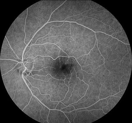
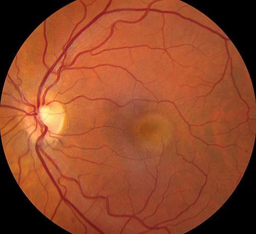
2 Causes of Vision Loss in Uveitis 1. Cystoid macular oedema 26% 2. Cataract 19% 3. Glaucoma 11% 4. Permanent macular damage 5% Rothova et al BJO 1996; 80:
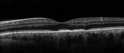
3 Macular OCT in uveitis CMO 2/3 of uveitis patients The leading cause of visual loss Drives treatment decisions Others Permanent outer retinal damage ERM CNVM

4 Patterns of OCT thickening in uveitis A. Cystic B. Diffuse C. SRF

5 OCT markers of visual prognosis Loss of outer retinal structures EZ Outer limiting membrane

6 Non CMO causes of visual loss ERM CNVM Retinitis
7 CNVM

8 ERM ERM PTMH FTMH
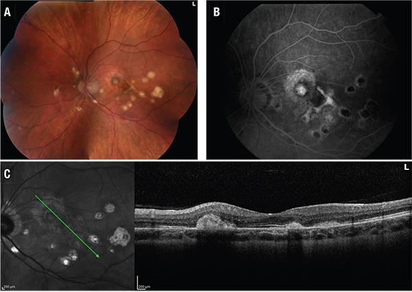
9 Syphilitic placoid

10 Ms GH 26 yr old 6 days of pain redness and photophobia. OH unremarkable GH good OE Left AAU with normal fundus
11 Treatment Investigation
12 Treatment Intensive topical steroids Investigation U&E LFT ACE VDRL HLA B27

13 Review at three weeks Pain and redness resolved but VA 6/12

14
15 Mr DF 21 yr old man JIA and ongoing uveitis Prednisolone, Humira and methotrexate Sec cataract and glaucoma
16 O/E: OD OS 6/9 VA 6/36 18 IOP cells AC ++ cells ++ cells Vitreous ++ cells No MO DFE Apparent MO R>L OCT
17
18 Managed with left intravitreal TA 4 mg/0.1 ml

19
20
21
22

23 RNFL thickness uveitis pts without glaucoma
24 RNFL thickness uveitis pts with glaucoma

25

26 Syphilitic placoid

27 Pre and post treatment OCT ASPPE
28 Mr EW 65M admitted under ID for non-tuberculous mycobacterial (Mycobacterium chimera) sternal wound + aortic graft infection on b/g of sternotomy for Type A aortic dissection in March 2016 Treated with imipenem, clarythromycin, moxifloxacin ID suggest antimicrobial plan: Clarithromycin 500mg BD, moxifloxacin 400mg nocte, rifabutin 300mg mane, Ethambutol 1.2g mane Referred to Ophthalmology OPC with mild floaters in R) eye Nil pain/ LOV
29 O/E: OD OS 6/5 VA 6/4 9 IOP 12 17/17 Ishihara 17/17 D+Q AC D+Q 1+ cells Vitreous Nil cells multifocal deep white lesions in choroid on posterior pole, not affecting macula/disc DFE R>L multifocal deep white lesions in choroid on posterior pole, not affecting macula/disc


30

31
32

33 45 yo man with three days of central visual loss GH URTI 3 weeks prior 6/6 6/60 RE all clear LE vit cells +/- Yellow mac lesion with haemorrhage

34 FA

35
36 1 week
37 6 weeks
38 Mrs FI 59 yr old woman 2 yrs progressive visual loss with floaters OH unremarkable GH good
39 O/E: OD OS 6/18 VA 6/18 9 IOP 12 17/17 Ishihara 17/ cells AC 1 + cells ++ cells Vitreous ++ cells Multiple pale outer retinal choroidal lesions with pigmentation DFE Multiple pale outer retinal choroidal lesions with pigmentation CT

40
41 Mr NK 59 yr old man 10 yrs progressive uveitis and MFC OH unremarkable GH good

42

43
44 Ms FW 28 yr old woman URTI followed by painless paracentral scotoma
45 O/E: OD OS 6/7.5 VA 6/7.5 9 IOP 12 17/17 Ishihara 17/17 + cells AC D+Q 1+ cells Vitreous + cells Confluent pale outer retinal lesions with early pigmentation DFE Confluent pale outer retinal lesions with early pigmentation OCT

46
47 Mr TN 38 yr old Vietnamese man 2 weeks of headache 1 Week bilateral central visual loss
48 O/E: OD OS 6/36 VA 6/60 7 IOP 5 17/17 Ishihara 17/17 ++ cells AC ++ cells ++ cells Vitreous ++ cells Disc swelling Multifocal cloudy serous detachment DFE Disc swelling Multifocal serous detachment OCT

49

50
51 VKH Acute phase Prodrome with HA, meningism, hearing changes then Acute bilat uveitis Serous detachment Disc swelling Acutely steroid responsive Convalescent phase Vitiligo and poliosis Retinal depigmentation Relapsing remitting or chronic uveitis
52 Mrs VH 85 yr old woman presents with 1 week of right sided orbital ache and mild visual loss OH bilat cat ext GH severe RA, treated with MTX and ritux Recent RA related pneumonitis treated with short course of steroids
53 O/E: OD OS 6/7.5 VA 6/7.5 9 IOP 12 17/17 Ishihara 17/17 D+Q AC D+Q 1+ cells Vitreous Nil cells Large single raised mass inferiorly DFE NAD CT

54
55

56
57 Causes of Scleritis (n = 188, Foster et al, 1994) 12% 3% 6% 8% 9% 19% 43% idiopathic RA infections GPA Rel Pol PAN/Hep B Others
58 Scleritis DDx Mild Moderate Episcleritis, atopy (esp limbal), inflamed pingueculum or pterygium HZO, iritis, sinus disease Severe ACG, HZO, uveitis Necrotizing SCC, radionecrosis (+/- infection) Posterior CSR, VKH, uveal effusion syndrome, choroidal primary or secondary, IOI, TED, optic neuritis
59 Mrs CA 31 yr old woman presented with unilateral central distortion
60 O/E: OD OS 6/5 VA 6/18 19 IOP 19 D+Q AC D+Q quiet Vitreous Occ cell only NAD DFE Multiple quiet flat white lesions around posterior pole Single raised lesion with surrounding pigment

61

62
63

64
65 PIC Essex et al 136 patients with PIC 93% female Ave -4 dioptres 46% unilateral 12% recurrent PIC lesions 22% new CNVM Median final VA 6/24
66 Mrs RW 45 yr old woman Referred with long standing R>L intermediate uveitis No longer responding to rpt orbital steroids GH good

67
68 Thank you
Misdiagnosed Vogt-Koyanagi-Harada (VKH) disease and atypical central serous chorioretinopathy (CSC)
 disease and atypical central serous chorioretinopathy (CSC)") HPTER 12 Misdiagnosed Vogt-Koyanagi-Harada (VKH) disease and atypical central serous chorioretinopathy (S) linical Features VKH disease is a bilateral granulomatous panuveitis often associated with exudative
HPTER 12 Misdiagnosed Vogt-Koyanagi-Harada (VKH) disease and atypical central serous chorioretinopathy (S) linical Features VKH disease is a bilateral granulomatous panuveitis often associated with exudative
Clinical Case Presentation. Branch Retinal Vein Occlusion. Sarita M. Registered Nurse Whangarei Base Hospital
 Clinical Case Presentation on Branch Retinal Vein Occlusion Sarita M. Registered Nurse Whangarei Base Hospital Introduction Case Study Pathogenesis Clinical Features Investigations Treatment Follow-up
Surgery in patients with uveitis. Lyndell Lim and Anthony Hall
 Surgery in patients with uveitis Lyndell Lim and Anthony Hall Disclosures Off label treatments Paid advisory board Bayer Paid research support Allergan (makers of Ozurdex) Paid research support B and L
Moncef Khairallah, MD
 Moncef Khairallah, MD Department of Ophthalmology, Fattouma Bourguiba University Hospital Faculty of Medicine, University of Monastir Monastir, Tunisia INTRODUCTION IU: anatomic form of uveitis involving
A Curious Case of Bilateral Optic Disc Edema Brittney Dautremont, DO, MPH
 A Curious Case of Bilateral Optic Disc Edema Brittney Dautremont, DO, MPH PGY2 Ophthalmology Resident Grandview Medical Center Dayton, OH CASE PRESENTATION 51 year old white female presenting with blurred
Deep Trouble. Thomas Stone, MD Retina Associates of Kentucky River City Retina Conference May 15, 2014
 Deep Trouble Thomas Stone, MD Retina Associates of Kentucky River City Retina Conference May 15, 2014 History 20 yo WM Decreased vision OU, OD>OS Sudden onset blurred central vision 12 days prior 4 days
Macular Hole Associated with Vogt-Koyanagi-Harada Disease at the Acute Uveitic Stage
 Published online: September 15, 2015 2015 The Author(s) Published by S. Karger AG, Basel 1663 2699/15/0063 0328$39.50/0 This article is licensed under the Creative Commons Attribution-NonCommercial 4.0
Slide 4. Slide 5. Slide 6
 Slide 1 Slide 4 Demographics El Paso Eye Care Border Healthcare-Based Grand Rounds Derek N. Cunningham, O.D. 80-90% Mexican-Americans Diabetes Hypertension Hyperlipidemia Obesity 70% uninsured High poverty
You can C-ME after Uveitis
 You can C-ME after Uveitis Abstract: Approximately 50% of uveitis patients will present with vision loss secondary to cystoid macular edema[1]. Two patients with uveitis present with a constant decrease
Update on management of Anterior Uveitis
 Update on management of Anterior Uveitis Parthopratim Dutta Majumder Senior Consultant, Department of Uvea & Intraocular Inflammation Medical Research Foundation, Sankara Nethralaya ABCD of Treating a
What do you need to know about posterior uveitis
 What do you need to know about posterior uveitis Dr. Anthony Hall MD FRANZCO Director of Ophthalmology Alfred Hospital, Melbourne, Australia Alfred Hospital Disclosures Off label treatments Paid advisory
Case report 12/10/2014. Delphine Lam ; Dr Mayer Srour Service d ophtalmologie Professeur E.Souied Université Paris Est
 Case report 12/10/2014 Delphine Lam ; Dr Mayer Srour Service d ophtalmologie Professeur E.Souied Medical history Man, 75 years old Complaint: Vision loss in left eye in June 2014 Past ophthalmologic history:
History/principles of the OCT What does the normal retinal OCT look like Vitreal disorders Retinal/RPE disorders Choroidal disorders
 Nathan Lighthizer, O.D., F.A.A.O. Assistant Professor Assistant Dean for Clinical Care Director of Continuing Education Chief of Specialty Care Clinics Chief of Electrodiagnostics Clinic Oklahoma College
Differential diagnosis of the red eye. Carol Slight Nurse Practitioner Ophthalmology
 Differential diagnosis of the red eye Carol Slight Nurse Practitioner Ophthalmology The red eye Conjunctivitis HSV Keratitis Acute angle closure glaucoma Anterior Uveitis Red eye Scleritis Subconjunctival
Optical coherence tomography findings in a child with posterior scleritis
 European Journal of Ophthalmology / Vol. 18 no. 6, 2008 / pp. 1007-1010 SHORT OMMUNITIONS & SE REPORTS Optical coherence tomography findings in a child with posterior scleritis H. ERDÖL, M. KOL,. TÜRK
J of Evolution of Med and Dent Sci/ eissn , pissn / Vol. 4/ Issue 55/ July 09, 2015 Page 9665
 RARE PRESENTATION OF BILATERAL CHOROIDAL METASTASIS FROM PRIMARY MUCO-EPIDERMOID CARCINOMA OF THE PAROTID GLAND: A G. Premalatha 1, Ramya Seetamraju 2 HOW TO CITE THIS ARTICLE: G. Premalatha, Ramya Seetamraju.
8/6/17. Disclosures Aerie Pharmaceuticals Alcon BioTissue Diopsys Optovue Shire
 Nathan Lighthizer, O.D., F.A.A.O. Associate Professor Assistant Dean for Clinical Care Director of Continuing Education Chief of Specialty Care Clinics Oklahoma College of Optometry Tahlequah, OK lighthiz@nsuok.edu
HLA-B27-related anterior Uveitis
 HLA-B27-related anterior Uveitis Nicholas Jones Manchester Uveitis Clinic The Royal Eye Hospital Manchester Anterior means anterior only IUSG classification: Anterior uveitis = Iris & pars plicata AU
o White dot syndromes pattern recognition o Activity and damage o Quality of life o Key points o Idiopathic o Sarcoidosis o Multiple sclerosis
 Introduction Clinical Assessment of Posterior Uveitis Philip I. Murray Centre for Translational Inflammation Research University of Birmingham Birmingham and Midland Eye Centre o Classification of uveitis
Glaucoma & Inflammation. Jorge L. Fernandez Bahamonde, MD.
 Glaucoma & Inflammation. Jorge L. Fernandez Bahamonde, MD. Definition. Inflammatory ocular conditions compromise outflow of aqueous humor. Keratitis Episcleritis. Scleritis. Uveitis Glaucoma & Keratitis.
Ocular side effects of biologic cancer therapy. Miss Stella Hornby Oxford Eye Hospital
 Ocular side effects of biologic cancer therapy Miss Stella Hornby Oxford Eye Hospital Overview Ocular toxicity from anti-cancer therapy Not uncommon but underestimated and underreported Vision related
Pearls, Pitfalls and Advances in Neuro-Ophthalmology
 Pearls, Pitfalls and Advances in Neuro-Ophthalmology Nancy J. Newman, MD Emory University Atlanta, GA Consultant for Gensight Biologics, Santhera Data Safety Monitoring Board for Quark AION Study Medical-legal
Rafik Girgis. Consultant Ophthalmic Surgeon ( Cataract & Primary Care)
") Rafik Girgis Consultant Ophthalmic Surgeon ( Cataract & Primary Care) Blepharitis Is a very common condition which usually bilateral & symmetrical. The main types are: Anterior, posterior or mixed Complications:
Rafik Girgis Consultant Ophthalmic Surgeon ( Cataract & Primary Care) Blepharitis Is a very common condition which usually bilateral & symmetrical. The main types are: Anterior, posterior or mixed Complications:
Neuro-Ophthalmic Masqueraders
 Neuro-Ophthalmic Masqueraders Leonid Skorin, Jr., OD, DO, MS, FAAO, FAOCO Mayo Clinic Health System in Albert Lea Denise Goodwin, OD, FAAO Pacific University College of Optometry Please silence all mobile
Dr Jo-Anne Pon. Dr Sean Every. 8:30-9:25 WS #70: Eye Essentials for GPs 9:35-10:30 WS #80: Eye Essentials for GPs (Repeated)
") Dr Sean Every Ophthalmologist Southern Eye Specialists Christchurch Dr Jo-Anne Pon Ophthalmologist Southern Eye Specialists, Christchurch Hospital, Christchurch 8:30-9:25 WS #70: Eye Essentials for GPs
Dr Sean Every Ophthalmologist Southern Eye Specialists Christchurch Dr Jo-Anne Pon Ophthalmologist Southern Eye Specialists, Christchurch Hospital, Christchurch 8:30-9:25 WS #70: Eye Essentials for GPs
5/2/2016 EYE EMERGENCIES. Nathaniel Pelsor, O.D., FAAO Talley Medical-Surgical Eye Care Associates. Anatomy. Tools
 EYE EMERGENCIES Nathaniel Pelsor, O.D., FAAO Talley Medical-Surgical Eye Care Associates Anatomy Tools 1 Contact dermatitis Blepharitis HSV Preseptal Cellulitis Anterior Chamber Subconjunctival hemorrhage
Principle of OCT. Reading Between the Lines: OCT Interpretation. Initial Concept. Advantage: High Resolution Cross Section Images
 Principle of OCT Reading Between the Lines: OCT Interpretation Mohammad Rafieetary, OD, FAAO mrafieetary@charlesretina.com Introduction Optical Biopsy Morphologic Evaluation of Live Tissue Measurements
Applying structure-function to solve clinical cases
 Applying structure-function to solve clinical cases Professor Michael Kalloniatis Centre for Eye Health, and, School of Optometry and Vision Science Acknowledgements Some material prepared by Nayuta Yoshioka
Uveitis. Pt Info Brochure. Q: What is Uvea?
 Pt Info Brochure Uveitis Q: What is Uvea? A: Uvea is the middle layer of the eye. It is the most vascular structure of the eye. It provides nutrition to the other parts of the eye. The uvea is made up
Interesting, unusual and eclectic cases from 2017 Robert A. Mittra, MD VitreoRetinal Surgery, P.A. Minneapolis, MN
 Fundus, SG Interesting, unusual and eclectic cases from 2017 Robert A. Mittra, MD VitreoRetinal Surgery, P.A. Minneapolis, MN Which is most likely? A) Age > 65, history of HTN B) Age 40 65, history of
CONSENT FOR CATARACT SURGERY REQUEST FOR SURGICAL OPERATION / PROCEDURE AND ANAESTHETIC
 CONSENT FOR CATARACT SURGERY REQUEST FOR SURGICAL OPERATION / PROCEDURE AND ANAESTHETIC Your doctor has indicated that the condition of your eye appears stable and your cataract surgery and/or implantation
Interesting, unusual, eclectic cases from 2017 Robert A. Mittra, MD VitreoRetinal Surgery, P.A. Minneapolis, MN
 56 yo female, EW Presented to outside Ophthalmologist Diagnosed with viral conjunctivitis, but viral testing was negative. Also had pain around the eye and on the right side of her face Interesting, unusual,
Case #1: 68 M with floaters OS
 Case #1: 68 M with floaters OS Point-of-Care Ocular Sonography for the Emergency Department Nate Teismann MD Dept of Emergency Medicine, UCSF Topics in EM 2012 Acute onset of dark spots in L eye 2 days
CLINICALCASE PROVOST J, SEKFALI R, AMOROSO F, ZAMBROWSKI O, MIERE A
 CLINICALCASE PROVOST J, SEKFALI R, AMOROSO F, ZAMBROWSKI O, MIERE A Department of ophthalmology, Souied E. (MD,PhD) Centre Hospitalier Intercommunal de Créteil Université Paris Est HISTORY 13 years old
UVEITIS IN GENERAL. Information for patients UVEITIS CLINIC WHAT IS UVEITIS? MAIN CATEGORIES OF UVEITIS
 Information for patients UVEITIS CLINIC UVEITIS IN GENERAL WHAT IS UVEITIS? The uvea is a name given to the pigmented layer of tissue inside the eye. When all or part of the uvea becomes inflamed, the
Rare Presentation of Ocular Toxoplasmosis
 Case Report Rare Presentation of Ocular Toxoplasmosis Rakhshandeh Alipanahi MD From Department of Ophthalmology, Nikookari Eye Hospital, Tabriz University of Medical Sciences, Tabriz, Iran. Correspondence:
White-Spot Syndromes of the Retina Lee Jampol, M.D. Chicago, IL
 Objectives At the conclusion of the program, the attendees will be able to: 1. recognize the various white-spot syndromes of the retina 2. initiate appropriate diagnostic tests of patients with the white-spot
ISPUB.COM. An Atypical Presentation of Posterior Scleritis. A Ramanathan, A Gaur CASE REPORT
 ISPUB.COM The Internet Journal of Ophthalmology and Visual Science Volume 8 Number 2 A Ramanathan, A Gaur Citation A Ramanathan, A Gaur.. The Internet Journal of Ophthalmology and Visual Science. 2009
epitheliopathy associated with diffuse retinal vasculitis
 British Journal of Ophthalmology, 1986, 70, 255-259 Acute posterior multifocal placoid pigment epitheliopathy associated with diffuse retinal vasculitis and late haemorrhagic macular detachment MAKOTO
11/29/2016 MACULAR MALADIES: TYPICAL & ATYPICAL CASES
 MACULAR MALADIES: TYPICAL & ATYPICAL CASES Dawn Pewitt, OD, FAAO Triad Eye Institute, Grove, OK Dpewitt@triadeye.com Disclosure Statement: No financial disclosures COPE 51218-PS Please silence all mobile
OCT Interpretation in Retinal Disease
 OCT Interpretation in Retinal Disease Jay M. Haynie, OD, FAAO Financial Disclosure I have received honoraria or am on the advisory board for the following companies: Carl Zeiss Meditec Advanced Ocular
Fundus Autofluorescence. Jonathan A. Micieli, MD Valérie Biousse, MD
 Fundus Autofluorescence Jonathan A. Micieli, MD Valérie Biousse, MD The retinal pigment epithelium (RPE) has many important functions including phagocytosis of the photoreceptor outer segments Cone Rod
Mild NPDR. Moderate NPDR. Severe NPDR
 Diabetic retinopathy Diabetic retinopathy is the most common cause of blindness in adults aged 35-65 years-old. Hyperglycaemia is thought to cause increased retinal blood flow and abnormal metabolism in
ISPUB.COM. Photopsia post flu: A case of MEWDS. S Baisakhiya, S Dulani, S Lele INTRODUCTION CASE HISTORY
 ISPUB.COM The Internet Journal of Ophthalmology and Visual Science Volume 8 Number 1 Photopsia post flu: A case of MEWDS S Baisakhiya, S Dulani, S Lele Citation S Baisakhiya, S Dulani, S Lele. Photopsia
Tuberous sclerosis presenting as atypical aggressive retinal astrocytoma with proliferative retinopathy and vitreous haemorrhage
 Case Report Brunei Int Med J. 2015; 11 (1): 49-53 Tuberous sclerosis presenting as atypical aggressive retinal astrocytoma with proliferative retinopathy and vitreous haemorrhage Pui Ling TANG and Mae-Lynn
Case : The glaucoma consult Case: The Glaucoma Consult Case: The Glaucoma Consult Case : The Glaucoma Consult Case : The weekend call you don t want
 1 2 3 4 5 6 Case : The glaucoma consult CC: second opinion on glaucoma HPI: OU/ 1 mos/optometrist thinks I have glaucoma/ no Rx yet / no POAG family hx 62 BM, VA OD = 20/40, VA OS = 20/30 NS +1 IOP: 20
Nausheen Khuddus, MD Melissa Elder, MD, PhD
 Nausheen Khuddus, MD Melissa Elder, MD, PhD Nausheen Khuddus, MD Pediatric Ophthalmologist and Strabismus Specialist Accent Physicians Gainesville, Florida What Is Uveitis? Uveitis is caused by inflammatory
Clinically Significant Macular Edema (CSME)
") Clinically Significant Macular Edema (CSME) 1 Clinically Significant Macular Edema (CSME) Sadrina T. Shaw OMT I Student July 26, 2014 Advisor: Dr. Uwaydat Clinically Significant Macular Edema (CSME) 2
Clinically Significant Macular Edema (CSME) 1 Clinically Significant Macular Edema (CSME) Sadrina T. Shaw OMT I Student July 26, 2014 Advisor: Dr. Uwaydat Clinically Significant Macular Edema (CSME) 2
WHAT IS YOUR DIAGNOSIS? By ADREA R. BENKOFF M.D.
 WHAT IS YOUR DIAGNOSIS? By ADREA R. BENKOFF M.D. Anterior Chamber Inflammation and Iris Depigmentation Noted 25 Years After Cataract Extraction Decreasing Vision Over a 5- Year Period 64 year old white
UNDERSTAND MORE ABOUT UVEITIS UVEITIS
 UNDERSTAND MORE ABOUT UVEITIS UVEITIS Uveitis What is uveitis? Uveitis is inflammation of the uvea, the middle layer of your eye. The eye is shaped much like a tennis ball, with three different layers
Uveitis unplugged: systemic therapy
 Uveitis unplugged: systemic therapy Hobart 2017 Peter McCluskey Save Sight Institute Sydney Eye Hospital Sydney Medical School University of Sydney Sydney Australia No financial or proprietary interest
Neuro-Ocular Grand Rounds Anthony B. Litwak,OD, FAAO VA Medical Center Baltimore, Maryland
 Neuro-Ocular Grand Rounds Anthony B. Litwak,OD, FAAO VA Medical Center Baltimore, Maryland Dr. Litwak is on the speaker and advisory boards for Alcon and Zeiss Meditek COMMON OPTIC NEUROPATHIES THAT CAN
White Dot Syndromes Noninfectious Chorioretinopathies Update 2019
 White Dot Syndromes Noninfectious Chorioretinopathies Update 2019 Kelly T. Mitchell, MD Retina Service TTUHSC Definition Noninfectious disease Inflammation of choroid, choriocapillaris, RPE, and Retina
Retinal Manifestations of Systemic Disease Part 1
 The Retina and Systemic diseases Retinal Manifestations of Systemic Disease Part 1 Sundeep Dev, MD VRSF Retinal Update 2019 VitreoRetinal Surgery, PA 1 Retinitis/Vasculitis Vitreous cells Serous detachments
Differential diagnosis of posterior uveitis
 Differential diagnosis of posterior uveitis Diagnostic approach 45-year old male. Floaters and decreased vision since 1 week Fever, lymphadenopathy, myalgias, night sweats, two months ago Oral ulcer sporadically
Mark Dunbar: Disclosure
 Important Things to Understand About OCT Mark T. Dunbar, O.D., F.A.A.O. Bascom Palmer Eye Institute University of Miami, School of Medicine Mark Dunbar: Disclosure Optometry Advisory Board for: Allergan
Ophthalmology Unit Referral Guidelines
 Ophthalmology Unit Referral Guidelines Austin Health Ophthalmology Unit holds sub-specialty sessions to discuss and plan the treatment of patients with specific ocular conditions. General including cataract
What you can expect with OZURDEX
 Important Information About Noninfectious Uveitis Affecting the Back Segment of the Eye and Treatment What you can expect with OZURDEX Approved Use OZURDEX (dexamethasone intravitreal implant) is a prescription
Ophthalmology. Juliette Stenz, MD
 Ophthalmology Juliette Stenz, MD Required Slide Disclosures NO SIGNIFICANT FINANCIAL, GENERAL, OR OBLIGATION INTERESTS TO REPORT Required Slide At the end of this session, students will be able to: 1.
Neuro-Ocular Grand Rounds
 Neuro-Ocular Grand Rounds Anthony B. Litwak,OD, FAAO VA Medical Center Baltimore, Maryland Dr. Litwak is on the speaker and advisory boards for Alcon and Zeiss Meditek COMMON OPTIC NEUROPATHIES THAT CAN
The effect of a single intravitreal implantation of dexamethasone on the fellow eye in bilateral non-infectious uveitis case report
 European Review for Medical and Pharmacological Sciences The effect of a single intravitreal implantation of dexamethasone on the fellow eye in bilateral non-infectious uveitis case report J. CISZEWSKA,
OCT Interpretation. Financial Disclosure. Jay M. Haynie, OD, FAAO. OCT Image Layers 7/21/2014
 OCT Interpretation Jay M. Haynie, OD, FAAO Financial Disclosure I have received honoraria or am on the advisory board for the following companies: Olympia Tacoma Renton Kennewick - Washington Carl Zeiss
Quantitative Evaluation of Sunset Glow Fundus in Vogt Koyanagi Harada Disease
 Quantitative Evaluation of Sunset Glow Fundus in Vogt Koyanagi Harada Disease Saburosuke Suzuki Department of Ophthalmology, Keio University, School of Medicine, Shinjuku, Tokyo, Japan Purpose: To evaluate
CHAPTER 13 CLINICAL CASES INTRODUCTION
 2 CHAPTER 3 CLINICAL CASES INTRODUCTION The previous chapters of this book have systematically presented various aspects of visual field testing and is now put into a clinical context. In this chapter,
D JO. Bilateral Shallow Anterior Chamber And Transient Myopia As A Presenting Feature Of Vogt Koyanagi Harada Syndrome
 46 Bilateral Shallow Anterior Chamber And Transient Myopia As A Presenting Feature Of Vogt Koyanagi Harada Syndrome Abstract Rahul Kumar Sharma, Abhishek Dagar, Vivek Kumar Vitreo-Retina Department, Venu
Posterior Segment Disease: Case Challenges
 Disclosures Posterior Segment Disease: Case Challenges Steven Ferrucci, OD, FAAO Chief, Optometry Sepulveda VA Professor, SCCO/MBKU Speakers bureau and/or Advisory Board for: Alcon Allergan Macula Risk
Unexplained visual loss in seven easy steps
 Unexplained visual loss in seven easy steps Andrew G. Lee, MD Chair Ophthalmology, Houston Methodist Hospital, Professor, Weill Cornell MC; Adjunct Professor, Baylor COM, U Iowa, UTMB Galveston, UT MD
Cases CFEH. CFEH Facebook Case #4
 CFEH Cases CFEH Facebook Case #4 A 42 year old female has noticed a floater in her left eye for many years but no flashes. She also reports hazy vision in this eye that has been present all her life. She
Retina Grand Rounds. Stephen Huddleston MD Charles Retina Institute University of Tennessee Hamilton Eye Institute
 Retina Grand Rounds Stephen Huddleston MD Charles Retina Institute University of Tennessee Hamilton Eye Institute Fundus Autoflourescence 2013 2016 Plaquenil Toxicity Excellent treatment for a variety
Learn Connect Succeed. JCAHPO Regional Meetings 2017
 Learn Connect Succeed JCAHPO Regional Meetings 2017 How Retinal Imaging Guides Treatment Odette Margit Houghton MD Question 1 Which OCT has the highest resolution? A: Swept source OCT B: Spectral domain
CLINICAL PEARLS IN OCULAR ONCOLOGY
 CLINICAL PEARLS IN OCULAR ONCOLOGY IRIS NEVUS - Two kinds circumscribed and diffuse - Photodocumentation important to monitor growth - Risk Factors for iris nevus growth to melanoma (ABCDEF) A Age (young),
Course # Getting to Know Your OCT
 Course # 140 Getting to Know Your OCT Course Title: Lecturer: Getting to Know Your OCT Brad Sutton, OD, FAAO IU School of Optometry Financial Disclosures No financial disclosures Optical Coherence Tomography-OCT
Juvenile Idiopathic Arthritis with Associated Bilateral Anterior Uveitis in a Four Year- Old Girl
 Juvenile Idiopathic Arthritis with Associated Bilateral Anterior Uveitis in a Four Year- Old Girl Pavlina S. Kemp, MD, Susannah Q. Longmuir, MD August 14, 2012 Chief complaint: Central posterior synechiae
Case History. Legends of the Posterior Segment CASE 1 4/22/2018
 Legends of the Posterior Segment Blair Lonsberry, MS, OD, MEd., FAAO Professor of Optometry Pacific University College of Optometry blonsberry@pacificu.edu CASE 1 Case History 38 black male, complaining
Double trouble: a patient with both HLA-B27 anterior uveitis and HLA-A29 birdshot chorioretinitis
 Haddad and Reddy Journal of Ophthalmic Inflammation and Infection 2014, 4:28 BRIEF REPORT Open Access Double trouble: a patient with both HLA-B27 anterior uveitis and HLA-A29 birdshot chorioretinitis Zeina
30 Years of Clinical Challenges
 Case RM 30 Years of Clinical Challenges Anthony B. Litwak, OD, FAAO VA Medical Center Baltimore, Maryland 62 yowm PMH: HTN POH unremarkable -FOH c/o eyes are scratchy, uses OTC zaditor BVA 20/20 OD 20/30
Note: This is an outcome measure and can be calculated solely using registry data.
 Measure #191 (NQF 0565): Cataracts: 20/40 or Better Visual Acuity within 90 Days Following Cataract Surgery -- National Quality Strategy Domain: Effective Clinical Care DESCRIPTION: Percentage of patients
ZEISS AngioPlex OCT Angiography. Clinical Case Reports
 Clinical Case Reports Proliferative Diabetic Retinopathy (PDR) Case Report 969 PROLIFERATIVE DIABETIC RETINOPATHY 1 1-year-old diabetic female presents for follow-up of proliferative diabetic retinopathy
Objectives. Unexplained Vision Loss: Where Do I Go From Here. History. History. Drug Induced Vision Loss
 Objectives Unexplained Vision Loss: Where Do I Go From Here Denise Goodwin, OD, FAAO Coordinator, Neuro-ophthalmic Disease Clinic Pacific University College of Optometry goodwin@pacificu.edu Know the importance
Progressive Symptomatic Retinal Detachment Complicating Retinoschisis. Initial Reporting Questionnaire
 Progressive Symptomatic Retinal Detachment Complicating Retinoschisis In association with the British Ophthalmological Surveillance Unit Ethics ref: 13/NW/0037 Initial Reporting Questionnaire Case Definition:
Recalcitrant Diabetic Macular Oedema: Therapeutic Options
 December 2007 A. Giridhar et al. - Recalcitrant DME 451 CONSULTATION S E C T I O N Recalcitrant Diabetic Macular Oedema: Therapeutic Options Dr. Cyrus M Shroff 1, Dr. N S Muralidhar 2, Dr. R Narayanan
EPIRETINAL MEMBRANE & VITREOMACULAR TRACTION
 EPIRETINAL MEMBRANE & VITREOMACULAR TRACTION Management of ERM and VMT K.V.Chalam,MD,PhD,MBA,FACS Professor and Director of Retina Loma Linda Eye Institute Los Angeles, USA REVIEW ANATOMY The vitreous
Course # Flashes and Floaters and Curtains, Oh My!
 Course # 132 Flashes and Floaters and Curtains, Oh My! FLASHES and FLOATERS and CURTAINS, OH MY!!! FLASHES OF LIGHT Vitreous is the villain Retinal traction Retinal hole Retinal tear Migraine Classic migraine
Course # Flashes and Floaters and Curtains, Oh My!
 Course # 132 Flashes and Floaters and Curtains, Oh My! FLASHES and FLOATERS and CURTAINS, OH MY!!! FLASHES OF LIGHT Vitreous is the villain Retinal traction Retinal hole Retinal tear Migraine Classic migraine
Methotrexate for uveitis associated with juvenile idiopathic arthritis: Value and requirement for additional anti-inflammatory medication
 European Journal of Ophthalmology / Vol. 17 no. 5, 2007 / pp. 743-748 Methotrexate for uveitis associated with juvenile idiopathic arthritis: Value and requirement for additional anti-inflammatory medication
Ganglion cell analysis by optical coherence tomography (OCT) Jonathan A. Micieli, MD Valérie Biousse, MD
 Jonathan A. Micieli, MD Valérie Biousse, MD") Ganglion cell analysis by optical coherence tomography (OCT) Jonathan A. Micieli, MD Valérie Biousse, MD Figure 1. Normal OCT of the macula (cross section through the line indicated on the fundus photo)
Ganglion cell analysis by optical coherence tomography (OCT) Jonathan A. Micieli, MD Valérie Biousse, MD Figure 1. Normal OCT of the macula (cross section through the line indicated on the fundus photo)
Ocular Lecture. Sue Bednar NP Ali Atwater PA-C
 Ocular Lecture Sue Bednar NP Ali Atwater PA-C Triaging Ocular Complaints Painful Eye/Red eye +/-blurry vision +/-visual loss +/-floaters +/-fevers If any of the above findings exist, pt is likely to have
Patient 1. Grand Rounds. Medical History. Ocular History. Medications. Exam 10/28/ year old African American male. Blur OD x 3 months
 Patient 1 Grand Rounds Anthony DeWilde, OD 72 year old African American male Blur OD x 3 months Last eye exam 10 years ago Ocular History Medical History Mixed Mechanism Glaucoma S/P LPI OU Was on Xalatan
OPTIC DISC PIT Pathogenesis and Management OPTIC DISC PIT
 OPTIC DISC PIT Pathogenesis and Management Abdel-Latif Siam Ain Shams University Cairo Egypt OPTIC DISC PIT Congenital pit is an atypical coloboma usually located on the temporal edge of the disc, associated
Optic Nerve Disorders: Structure and Function and Causes
 Optic Nerve Disorders: Structure and Function and Causes Using Visual Fields, OCT and B-scan Ultrasound to Diagnose and Follow Optic Nerve Visual Losses Ohio Ophthalmological Society and Ophthalmic Tech
Vanderbilt Eye Institute Clinical Trials
 April, 2010 Vanderbilt Eye Institute Clinical Trials Ophthalmology Actively Recruiting Studies For information on our clinical trials and other studies, please contact: Sandy Owings, COA, CCRP Clinic Director
New Drugs for Uveitis. Medical Eye Unit St Thomas Hospital
 New Drugs for Uveitis Miles Stanford Medical Eye Unit St Thomas Hospital x Epithelium x x Antigen Y Y Y Y IgG m cd4 IL-2 Y m + IL-12 Cytotoxic T B pmn Ig s PG s. LTB4 O - IL-6 TNFα IFNγγ IL-2 Th1 IL-10
ACTIVATED OR NOT? RETINAL CASE PRESENTATION Shorye Payne, MD Medical Retinal Specialist Robley Rex VA Eye Clinic
 ACTIVATED OR NOT? RETINAL CASE PRESENTATION Shorye Payne, MD Medical Retinal Specialist Robley Rex VA Eye Clinic C We anticipate that the future management of posterior uveal melanoma (PUM) will focus
Intravitreal Triamcinolone Acetonide for Macular Edema in HLA-B27 Negative Ankylosing Spondylitis
 105 This is an Open Access article licensed under the terms of the Creative Commons Attribution- NonCommercial-NoDerivs 3.0 License (www.karger.com/oa-license), applicable to the online version of the
ADULT-ONSET FOVEOMACULAR VITELLIFORM DYSTROPHY. By: Chris Munnerlyn, OMT Student University of Arkansas for Medical Sciences
 ADULT-ONSET FOVEOMACULAR VITELLIFORM DYSTROPHY By: Chris Munnerlyn, OMT Student University of Arkansas for Medical Sciences ADULT-ONSET FOVEOMACULAR VITELLIFORM DYSTROPHY (AOFVD) AOFVD is a condition that
Acute Eyes for ED. Enis Kocak. The Alfred Ophthalmology
 Acute Eyes for ED Enis Kocak The Alfred Ophthalmology The problem with eyes Things to cover Ocular anatomy Basic assessment Common presentations Eye first aid and procedures Ophthalmic emergencies What
Evolving therapies for posterior uveitis. Infliximab (Remicade) Infliximab: pharmacology. FDA-approved monoclonal antibody therapy Target
 Infliximab: pharmacology. FDA-approved monoclonal antibody therapy Target") Evolving therapies for posterior uveitis Sam Dahr, M.D. September 17, 2005 Midwest Ophthalmology Conference Infliximab (Remicade) FDA approved for Crohn s disease, rheumatoid arthritis, and psoriatic arthritis
Evolving therapies for posterior uveitis Sam Dahr, M.D. September 17, 2005 Midwest Ophthalmology Conference Infliximab (Remicade) FDA approved for Crohn s disease, rheumatoid arthritis, and psoriatic arthritis
Outline. Brief history and principles of ophthalmic ultrasound. Types of ocular ultrasound. Examination techniques. Types of Ultrasound
 Ultrasound and Intraocular Tumors 2015 Ophthalmic Photographers' Society Mid-Year Program Cagri G. Besirli MD, PhD Kellogg Eye Center University of Michigan Outline Brief history and principles of ophthalmic
Characterization of serous retinal detachments in uveitis patients with optical coherence tomography
 Characterization of serous retinal detachments in uveitis patients with optical coherence tomography Annamieka Simmons-Rear, Oregon Health and Science University Steven Yeh, Emory University Brian T. Chan-Kai,
Screening for Uveitis in Children
 Information for patients and parents Manchester Royal Eye Hospital Paediatric Uveitis Service Screening for Uveitis in Children What is uveitis? Uveitis is inflammation of a layer of the eye, called the