Interesting, unusual, eclectic cases from 2017 Robert A. Mittra, MD VitreoRetinal Surgery, P.A. Minneapolis, MN
|
|
|
- Alice Franklin
- 5 years ago
- Views:
Transcription


1 56 yo female, EW Presented to outside Ophthalmologist Diagnosed with viral conjunctivitis, but viral testing was negative. Also had pain around the eye and on the right side of her face Interesting, unusual, eclectic cases from 2017 Robert A. Mittra, MD VitreoRetinal Surgery, P.A. Minneapolis, MN Improved after a week or so, but the vision dropped VA 20/70 OD, 20/25 OS IOP 17 OD, 15 OS PMHx External Photo on presentation to outside Ophthalmologist Hyperlipidemia, GERD, Osteoarthritis NO MEDS OCT OD Fundus Exam OD Retinal detachment inferiorly OD, looked exudative. NO breaks seen and marked vitreous debris 1


2 BScan OD Work Up Patient placed on oral steroids for inflammatory component, medical work up initiated CBC, metabolic profile CT Scan Chest and Abdomen, R/O metastatic tumor Concern for melanoma with possible overlying bleed and adjacent exudative detachment 2 months later OCT OD VA 20/25+ OD, 20/20 OS Off steroids, were tapered slowly Exudative detachment almost completely gone on exam BScan Further work up Full body PET SCAN, MRI of the head 2


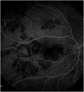
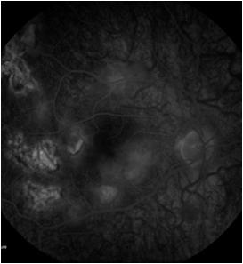
3 64 yo female, LJ Fundus Sent in for macular edema, possible hypertensive retinopathy OS Patient is a nurse, history of Mild BP elevation 162/62 on presentation VA 20/50 OD, 20/60 OS IOP 18 OD, 19 OS SLE 1 2+ NS OU FA OCT Diagnosis? 3 weeks later VA worse 20/80 Atypical Ischemic Optic Neuropathy CRVO Leber s Hereditary Optic Neuropathy Infectious Bartonella Medical Work Up CBC, ESR, CRP VDRL, FTA ABS ANA Toxo IgG, IgM Toxocara Lyme Titer B. Henselae titer TB testing 3


4 OCT OS VA worse on follow up, edema worse ESR and CRP testing was high, Temporal Artery biopsy donenegative Toxoplasma IgG 81.5, IgM negative Other labs negative Avastin injection given Started on Bactrim DS BID 3 months later, VA 20/600 OS 3 months after presentation, Fundus OS 8 months after presentation VA 20/400 OS 10 months after initial presentation Emergency triage call from patient VA worse OD VA 20/80 OD, 20/300 OS IOP 14, 13 SLE 2+ NS OU 4


 Does not take any medications VA 20/80 OD, 20/20 OS IOP")
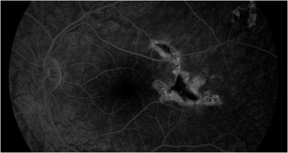
5 Fundus OD FA OCT OD Repeat Labs CBC, ESR, QuantiFERON Gold, Bartonella, Lyme, Toxocara, Toxoplasma Patient started empirically on Azithromycin 19 yo healthy female, A.S. Exam 2 days of vision loss OD, no complaints OS Central vision affected, thought it was a damaged contact lens, ( 1.50 OD, 1.25 OS) Does not take any medications VA 20/80 OD, 20/20 OS IOP 17, 18 SLE: Normal OU 5


6 Fundus FA OD FA OS OCT OD OCT OD OCT OD 6


7 OCT OS 7 days after symptoms onset, VA 20/150 OD, 20/20 OS 7 days after symptom onset 2 weeks after symptoms, VA 20/150+ OD, 20/20 OS 2 weeks after symptoms 2 weeks after symptoms 7

8 6 weeks after presentation 6 weeks after presentation, VA 20/40 Acute Posterior Multifocal Placoid Pigment Epitheliopathy (APMPPE) Acute Posterior Multifocal Placoid Pigment Epitheliopathy (APMPPE) Healthy young patients, average age of 25 Rapid loss of vision of one or both eyes Multiple post equatorial gray white lesions at the level of the RPE 50% have inflammatory cells in the vitreous Can be seen after flu like illness, flu or varicella vaccine, can be associated with systemic and cerebral vasculitis FA block early, stain late ICG dark spots corresponding to acute lesions without late staining OCT classically RPE and adjacent photoreceptors during acute phase and recovery when the lesions heal Rarely unilateral, one eye follows other usually days or weeks after the first Differential Diagnosis Lab Work Up Serpiginous Choroidopathy lesions similar acutely, but resolve more slowly and lesion marked atrophy of the choriocapillaris More chance of CNVM development Persistent Placoid maculopathy Relentless Placoid Choroidopathy QuantiFERON TB Gold negative Treponema Pallidum negative Toxoplasma AB IgG, IgM negative Lyme negative ANA negative CBC/diff negative ESR negative MRI Head/Orbits negative 8


 Age 40 65, history of")
9 OCT findings Fundus, SG Gass' Atlas RPE and adjacent photoreceptors Occasional SRF over placid lesions Later studies have shown that acute lesions affect the outer retina Which is most likely? A) Age > 65, history of HTN B) Age 40 65, history of HTN C) Less than 40, healthy 9


10 18 yo female 4 months later, VA 20/25 History of Asthma controlled on Singulair and Loratadine Lab work was extensive FBS, HbA1c, CBC with diff, PT/PTT, ESR, lipid profile, homocysteine, ANA, Hemoglobin electrophoresis, VDRL, Cryoglobulins, antiphospholipid antibodies, Lupus anticoagulant, serum protein electrophoresis, blood viscosity Options discussed: observation vs treatment After 3 Avastin injections 71 yo female, CC Decrease in vision OD VA 20/70 OD, 20/25 OS Diagnosis AMD Neovascular Given Lucentis, after one injection improved to 20/40 Clinical Trial of Implant Device OD Inserted surgically after two treatments, can be refilled in the clinic Trial is still ongoing, results will be available soon and further study is planned 10


11 10 yo boy presents 3 days after July 4th What is the most likely problem? Direct hit OS from a firecracker 1 week later, VA worse 8/200 VA OD 20/20, OS 3/200 IOP 12 OD, 16 OS SLE OD NL, OS Lid hemorrhage, 2+ cell/flare Fundus VH B Scan VH no RD, no FB OCT shows edema consistent with commotion in the fovea, but also a lamellar macular hole appearance Fundus OS What Now? Surgery? 11


12 2 months after initial injury OCT OS, observed for 6 months prior to intervention 1 week after PPV/MS, VA 20/ YO female with pseudoexfoliation glaucoma Sudden vision loss 10 years after CE/IOL S/P large functioning superior trab 24 yo Male, TR Sent for emergency Retinal Detachment OS, Macula ON Not a high myope History, VA down for 1 month Diabetes Type 1, on Insulin, Atorvastatin, Lisinopril VA 20/30 OD, 20/100 OS IOP 18 OD, 16 OS SLE NL OU 12


13 Fundus FA OD FA OS OCT 2 months later, VA 20/20 OD, 20/40 OS Severe PDR OU Need anti VEGF and PRP in the right eye Needs surgery OS 13
 Age > 65, history of HTN B)")


14 Fundus ES Most Likely? A) Age > 65, history of HTN B) Age 40 65, history of HTN C) Less than 40, healthy Over one year later, VA 20/20 Patient is 22 yo Runs track for a big ten school Noticed decrease in vision after an intensive work out Extensive medical work up negative. Initially observed, but vision dropped to 20/400 over several weeks and patient opted for treatment 14


15 72 yo Male, sent for choroidal nevus OS, LM Fundus VA 20/25 OD, 20/40 OS IOP 15 OD, 16 OS SLE PCIOL OU FA OCT OS B Scan OS, Height 1.69mm, Width 6.83, high internal reflectivity Choroidal Metastasis History of throat cancer biopsy reveal Squamous Cell Carcinoma, primary lung. Initial MRI of the Head showed no CNS involvement Treated with radiation and chemo Currently on chemo infusion every two weeks Discussed external beam radiation if systemic chemo unsuccessful in shrinking the tumor 15


16 45 yo male, JB Fundus OS Sent by ER for retinal detachment Sudden loss of vision superior visual field OS PMH HTN, but untreated BP 124/90 VA 20/20 OU, IOP 18,19 SLE NL OU OCT OS BRAO OS in a 45 YO patient Medical work up Carotid Doppler, Echo Lipid Panel 25 yo female, KH Fundus Mild central vision loss OU, OD > OS No PMH, no medications Color vision NORMAL VA 20/40 OD, 20/30 OS, IOP 18 OD, 17 OS SLE NORMAL 16


17 FA 4 years later, VA 20/60 OD, 20/100 OS CONE Dystrophy Genetic Testing 4 years later, the patient now has dramatically decreased color vision OU Differential Diagnosis Can be AD, AR or X linked Associated with many gene defects, genetic testing done, awaiting results 41 yo female, TU Fundus OD Noted to have unusual lesion OD 18 years prior VA 20/400 OD, 20/20 OS IOP 14 OD, 16 OS SLE NORMAL OU 17


18 FA OD OCT OD B Scan OD Thank You Did not show shadowing 18
Interesting, unusual and eclectic cases from 2017 Robert A. Mittra, MD VitreoRetinal Surgery, P.A. Minneapolis, MN
 Fundus, SG Interesting, unusual and eclectic cases from 2017 Robert A. Mittra, MD VitreoRetinal Surgery, P.A. Minneapolis, MN Which is most likely? A) Age > 65, history of HTN B) Age 40 65, history of
Deep Trouble. Thomas Stone, MD Retina Associates of Kentucky River City Retina Conference May 15, 2014
 Deep Trouble Thomas Stone, MD Retina Associates of Kentucky River City Retina Conference May 15, 2014 History 20 yo WM Decreased vision OU, OD>OS Sudden onset blurred central vision 12 days prior 4 days
11/29/2016 MACULAR MALADIES: TYPICAL & ATYPICAL CASES
 MACULAR MALADIES: TYPICAL & ATYPICAL CASES Dawn Pewitt, OD, FAAO Triad Eye Institute, Grove, OK Dpewitt@triadeye.com Disclosure Statement: No financial disclosures COPE 51218-PS Please silence all mobile
OCT Angiography The Next Frontier
 Choroid Retina avascular 5/13/2017 OCT Angiography The Next Frontier Pierce Kenworthy OD, FAAO June 9, 2017 OCT Angiography (OCTA) 2016 Non-invasive, motion contrast imaging Represents erythrocyte movement
Patient AB. Born in 1961 PED
 Clinical Atlas Patient AB Born in 1961 PED Autofluorescence Dilated 45 EasyScan Zero-dilation IR 45 Fundus Dilated 45 In the fundus photos (Canon CX1) the PED is not able to be seen. However, the extent
OCT Interpretation in Retinal Disease
 OCT Interpretation in Retinal Disease Jay M. Haynie, OD, FAAO Financial Disclosure I have received honoraria or am on the advisory board for the following companies: Carl Zeiss Meditec Advanced Ocular
OCT Interpretation. Financial Disclosure. Jay M. Haynie, OD, FAAO. OCT Image Layers 7/21/2014
 OCT Interpretation Jay M. Haynie, OD, FAAO Financial Disclosure I have received honoraria or am on the advisory board for the following companies: Olympia Tacoma Renton Kennewick - Washington Carl Zeiss
Clinically Significant Macular Edema (CSME)
") Clinically Significant Macular Edema (CSME) 1 Clinically Significant Macular Edema (CSME) Sadrina T. Shaw OMT I Student July 26, 2014 Advisor: Dr. Uwaydat Clinically Significant Macular Edema (CSME) 2
Clinically Significant Macular Edema (CSME) 1 Clinically Significant Macular Edema (CSME) Sadrina T. Shaw OMT I Student July 26, 2014 Advisor: Dr. Uwaydat Clinically Significant Macular Edema (CSME) 2
ADULT-ONSET FOVEOMACULAR VITELLIFORM DYSTROPHY. By: Chris Munnerlyn, OMT Student University of Arkansas for Medical Sciences
 ADULT-ONSET FOVEOMACULAR VITELLIFORM DYSTROPHY By: Chris Munnerlyn, OMT Student University of Arkansas for Medical Sciences ADULT-ONSET FOVEOMACULAR VITELLIFORM DYSTROPHY (AOFVD) AOFVD is a condition that
Optical Coherence Tomography: Pearls for the Anterior Segment Surgeon Basic Science Michael Stewart, M.D.
 Optical Coherence Tomography: Pearls for the Anterior Segment Surgeon Basic Science Michael Stewart, M.D. Disclosure OCT Optical Coherence Tomography No relevant financial relationships I will refer to
ACTIVATED OR NOT? RETINAL CASE PRESENTATION Shorye Payne, MD Medical Retinal Specialist Robley Rex VA Eye Clinic
 ACTIVATED OR NOT? RETINAL CASE PRESENTATION Shorye Payne, MD Medical Retinal Specialist Robley Rex VA Eye Clinic C We anticipate that the future management of posterior uveal melanoma (PUM) will focus
ZEISS AngioPlex OCT Angiography. Clinical Case Reports
 Clinical Case Reports Proliferative Diabetic Retinopathy (PDR) Case Report 969 PROLIFERATIVE DIABETIC RETINOPATHY 1 1-year-old diabetic female presents for follow-up of proliferative diabetic retinopathy
Past ocular history. DME Case 1. Patient presents blurred VA. Hemoglobin A1c 11.5% -- patient states sugars have not been in good control
 Past ocular history Patient presents blurred VA DME Case 1 Hemoglobin A1c 11.5% -- patient states sugars have not been in good control PDR with macular edema OU Rishi Singh MD Cleveland Clinic OD OS 1
Case : The glaucoma consult Case: The Glaucoma Consult Case: The Glaucoma Consult Case : The Glaucoma Consult Case : The weekend call you don t want
 1 2 3 4 5 6 Case : The glaucoma consult CC: second opinion on glaucoma HPI: OU/ 1 mos/optometrist thinks I have glaucoma/ no Rx yet / no POAG family hx 62 BM, VA OD = 20/40, VA OS = 20/30 NS +1 IOP: 20
Retina Grand Rounds. Stephen Huddleston MD Charles Retina Institute University of Tennessee Hamilton Eye Institute
 Retina Grand Rounds Stephen Huddleston MD Charles Retina Institute University of Tennessee Hamilton Eye Institute Fundus Autoflourescence 2013 2016 Plaquenil Toxicity Excellent treatment for a variety
CLINICALCASE PROVOST J, SEKFALI R, AMOROSO F, ZAMBROWSKI O, MIERE A
 CLINICALCASE PROVOST J, SEKFALI R, AMOROSO F, ZAMBROWSKI O, MIERE A Department of ophthalmology, Souied E. (MD,PhD) Centre Hospitalier Intercommunal de Créteil Université Paris Est HISTORY 13 years old
Fundus Autofluorescence. Jonathan A. Micieli, MD Valérie Biousse, MD
 Fundus Autofluorescence Jonathan A. Micieli, MD Valérie Biousse, MD The retinal pigment epithelium (RPE) has many important functions including phagocytosis of the photoreceptor outer segments Cone Rod
Case Follow Up. Sepi Jooniani PGY-1
 Case Follow Up Sepi Jooniani PGY-1 Triage 54 year old M Pt presents to prelim states noticed today he had reddness to eyes, states worse in R eye. Pt denies any pain or itching. No further complaints.
The Human Eye. Cornea Iris. Pupil. Lens. Retina
 The Retina Thin layer of light-sensitive tissue at the back of the eye (the film of the camera). Light rays are focused on the retina then transmitted to the brain. The macula is the very small area in
Slide 4. Slide 5. Slide 6
 Slide 1 Slide 4 Demographics El Paso Eye Care Border Healthcare-Based Grand Rounds Derek N. Cunningham, O.D. 80-90% Mexican-Americans Diabetes Hypertension Hyperlipidemia Obesity 70% uninsured High poverty
Retina Conference. Janelle Fassbender, MD, PhD University of Louisville Department of Ophthalmology and Visual Sciences 09/04/2014
 Retina Conference Janelle Fassbender, MD, PhD University of Louisville Department of Ophthalmology and Visual Sciences 09/04/2014 Subjective CC/HPI: 64 year old Caucasian female referred by outside ophthalmologist
Posterior Segment Update
 Posterior Segment Update Featured Speaker: Dr. Kyle Cheatham, FAAO, DIP ABO DISCLOSURE STATEMENT We have no direct financial or proprietary interest in any companies, products or services mentioned in
Principle of OCT. Reading Between the Lines: OCT Interpretation. Initial Concept. Advantage: High Resolution Cross Section Images
 Principle of OCT Reading Between the Lines: OCT Interpretation Mohammad Rafieetary, OD, FAAO mrafieetary@charlesretina.com Introduction Optical Biopsy Morphologic Evaluation of Live Tissue Measurements
Neuro-Ocular Grand Rounds Anthony B. Litwak,OD, FAAO VA Medical Center Baltimore, Maryland
 Neuro-Ocular Grand Rounds Anthony B. Litwak,OD, FAAO VA Medical Center Baltimore, Maryland Dr. Litwak is on the speaker and advisory boards for Alcon and Zeiss Meditek COMMON OPTIC NEUROPATHIES THAT CAN
Sudden Vision Loss. Brendan Girschek, MD, FRCSC, FACS Vitreoretinal Surgery Cedar Valley Medical Specialists
 Sudden Vision Loss Brendan Girschek, MD, FRCSC, FACS Vitreoretinal Surgery Cedar Valley Medical Specialists My Credentials -Residency in Ophthalmology at the LSU Eye Center in New Orleans, LA -Fellowship
Incorporating OCT Angiography Into Patient Care
 Incorporating OCT Angiography Into Patient Care Beth A. Steele, OD, FAAO OCT A: Introduction Isolates microvascular circulation from OCT image data Axial resolution = 5 microns (i.e. fine capillaries visible)
Neuro-Ocular Grand Rounds
 Neuro-Ocular Grand Rounds Anthony B. Litwak,OD, FAAO VA Medical Center Baltimore, Maryland Dr. Litwak is on the speaker and advisory boards for Alcon and Zeiss Meditek COMMON OPTIC NEUROPATHIES THAT CAN
Diagnosis and treatment of diabetic retinopathy. Blake Cooper MD Ophthalmologist Vitreoretinal Surgeon Retina Associates Kansas City
 Diagnosis and treatment of diabetic retinopathy Blake Cooper MD Ophthalmologist Vitreoretinal Surgeon Retina Associates Kansas City Disclosures Consulted for Novo Nordisk 2017,2018. Will be discussing
Grand Rounds Clinical Cases from Alex D. Gibberman, O.D. Harpers Point Eye Associates
 Grand Rounds Clinical Cases from 2016 Alex D. Gibberman, O.D. Harpers Point Eye Associates Relevant Financial Interests -none Case 1: 54 year old African American Female CC: Noticed a green line in
ISPUB.COM. Photopsia post flu: A case of MEWDS. S Baisakhiya, S Dulani, S Lele INTRODUCTION CASE HISTORY
 ISPUB.COM The Internet Journal of Ophthalmology and Visual Science Volume 8 Number 1 Photopsia post flu: A case of MEWDS S Baisakhiya, S Dulani, S Lele Citation S Baisakhiya, S Dulani, S Lele. Photopsia
Course # Flashes and Floaters and Curtains, Oh My!
 Course # 132 Flashes and Floaters and Curtains, Oh My! FLASHES and FLOATERS and CURTAINS, OH MY!!! FLASHES OF LIGHT Vitreous is the villain Retinal traction Retinal hole Retinal tear Migraine Classic migraine
Course # Flashes and Floaters and Curtains, Oh My!
 Course # 132 Flashes and Floaters and Curtains, Oh My! FLASHES and FLOATERS and CURTAINS, OH MY!!! FLASHES OF LIGHT Vitreous is the villain Retinal traction Retinal hole Retinal tear Migraine Classic migraine
12/2/16. Ways to differentiate:
 Nate Lighthizer, O.D., F.A.A.O. Assistant Dean for Clinical Care Services Director of CE Chief of Specialty Care Clinics Chief of Electrodiagnostics Clinic Oklahoma College of Optometry lighthiz@nsuok.edu
Diabetic Retinopathy
 Diabetic Retinopathy Diabetes mellitus is one of the leading causes of irreversible blindness worldwide. In the United States, it is the most common cause of blindness in people younger than 65 years.
A Patient s Guide to Diabetic Retinopathy
 Diabetic Retinopathy A Patient s Guide to Diabetic Retinopathy 840 Walnut Street, Philadelphia PA 19107 www.willseye.org Diabetic Retinopathy 1. Definition Diabetic retinopathy is a complication of diabetes
Optical Coherence Tomography in Diabetic Retinopathy. Mrs Samantha Mann Consultant Ophthalmologist Clinical Lead of SEL-DESP
 Optical Coherence Tomography in Diabetic Retinopathy Mrs Samantha Mann Consultant Ophthalmologist Clinical Lead of SEL-DESP Content OCT imaging Retinal layers OCT features in Diabetes Some NON DR features
FROM OUTDATED TO UPDATED Eminence-Based Medicine
 FROM OUTDATED TO UPDATED Eminence-Based Medicine Evidence-Based Medicine A REVIEW OF KEY CLINICAL TRIALS Anthony DeWilde, OD FAAO 1 EMINENCE BASED MEDICINE 2 EVIDENCE BASED MEDICINE 3 4 CLINICAL TRIALS
Diabetic Retinopathy: Managing the Extremes. J. Michael Jumper, MD West Coast Retina
 Diabetic Retinopathy: Managing the Extremes J. Michael Jumper, MD West Coast Retina Case 1: EC 65 y.o. HM No vision complaints Meds: Glyburide Metformin Pioglitazone Va: 20/20 OU 20/20 Case 2: HS 68 y.o.
Retinal Vein Occlusion
 Retinal Update 2018 Retinal Vein Occlusion Case Presentations to Myself Branch Vein Occlusion What medical evaluation do you recommend for this 72 year old patient? Is there anything you ask of your medical
Clinical Case Presentation. Branch Retinal Vein Occlusion. Sarita M. Registered Nurse Whangarei Base Hospital
 Clinical Case Presentation on Branch Retinal Vein Occlusion Sarita M. Registered Nurse Whangarei Base Hospital Introduction Case Study Pathogenesis Clinical Features Investigations Treatment Follow-up
Learn Connect Succeed. JCAHPO Regional Meetings 2015
 Learn Connect Succeed JCAHPO Regional Meetings 2015 OPTIC NEUROPATHY AS EASY AS 1,2,3,4 OPTIC NERVE ANATOMY M. Tariq Bhatti, MD Departments of Ophthalmology and Neurology Duke Eye Center and Duke University
Retinal Manifestations of Systemic Disease Part 1
 The Retina and Systemic diseases Retinal Manifestations of Systemic Disease Part 1 Sundeep Dev, MD VRSF Retinal Update 2019 VitreoRetinal Surgery, PA 1 Retinitis/Vasculitis Vitreous cells Serous detachments
EFFICACY OF ANTI-VASCULAR ENDOTHELIAL GROWTH FACTOR AGENTS IN RETINAL DISORDER FOR BETTER VISUAL ACUITY
 EFFICACY OF ANTI-VASCULAR ENDOTHELIAL GROWTH FACTOR AGENTS IN RETINAL DISORDER FOR BETTER VISUAL ACUITY Diwakar chaudhary *1, 2, Hu shuqiong, Long Yuan and Xiong kun 1 Yangtze University, 1 Nanhuan Road
Learn Connect Succeed. JCAHPO Regional Meetings 2017
 Learn Connect Succeed JCAHPO Regional Meetings 2017 How Retinal Imaging Guides Treatment Odette Margit Houghton MD Question 1 Which OCT has the highest resolution? A: Swept source OCT B: Spectral domain
FA Conference. Lara Rosenwasser Newman, M.D. 10/2/14 University of Louisville Department of Ophthalmology and Visual Sciences
 FA Conference Lara Rosenwasser Newman, M.D. 10/2/14 University of Louisville Department of Ophthalmology and Visual Sciences Patient Presentation CC: (sent by optometrist) Blurry/foggy vision HPI: 62 yo
Doc, I See a Donut in My Vision : An Optometrist s Guide to a Rare Cause of Choroidal Neovascular Membrane
 Doc, I See a Donut in My Vision : An Optometrist s Guide to a Rare Cause of Choroidal Neovascular Membrane Linda Pham, OD, Tobin Ansel, OD, Nancy Shenouda-Awad, OD, FAAO, West Haven VA Medical Center Abstract
OPTIC DISC PIT Pathogenesis and Management OPTIC DISC PIT
 OPTIC DISC PIT Pathogenesis and Management Abdel-Latif Siam Ain Shams University Cairo Egypt OPTIC DISC PIT Congenital pit is an atypical coloboma usually located on the temporal edge of the disc, associated
Differential diagnosis of posterior uveitis
 Differential diagnosis of posterior uveitis Diagnostic approach 45-year old male. Floaters and decreased vision since 1 week Fever, lymphadenopathy, myalgias, night sweats, two months ago Oral ulcer sporadically
Venous Occlusive Diseases
 Venous Occlusive Diseases Bruce R. Saran, MD Adjunct Assistant Clinical Professor of Medicine Scheie Eye Institute University of Pennsylvania School of Medicine Philadelphia, PA -a division of: RVO Demographics
Five steps: Overview
 Optic atrophy is not a diagnosis Andrew G. Lee, MD Professor of Ophthalmology, Neurology and Neurosurgery, Weill Cornell Medical College Chair, Department of Ophthalmology, Houston Methodist Hospital,
8/6/17. Disclosures Aerie Pharmaceuticals Alcon BioTissue Diopsys Optovue Shire
 Nathan Lighthizer, O.D., F.A.A.O. Associate Professor Assistant Dean for Clinical Care Director of Continuing Education Chief of Specialty Care Clinics Oklahoma College of Optometry Tahlequah, OK lighthiz@nsuok.edu
 measure of your overall performance. An isolated glucose test is helpful to let you know what your sugar level is at one moment, but it doesn t tell you whether or not your diabetes is under adequate control
What Is O.C.T. and Why Should I Give A Rip? OCT & Me How Optical Coherence Tomography Changed the Life of a Small Town Optometrist 5/19/2014
 OCT & Me How Optical Coherence Tomography Changed the Life of a Small Town Optometrist Email: myoder@wcoil.com Mark A. Yoder, O.D. 107 N. Main Street PO Box 123 Bluffton, OH 45817 @yoderod 115.02 Histoplasma
White-Spot Syndromes of the Retina Lee Jampol, M.D. Chicago, IL
 Objectives At the conclusion of the program, the attendees will be able to: 1. recognize the various white-spot syndromes of the retina 2. initiate appropriate diagnostic tests of patients with the white-spot
A Curious Case of Bilateral Optic Disc Edema Brittney Dautremont, DO, MPH
 A Curious Case of Bilateral Optic Disc Edema Brittney Dautremont, DO, MPH PGY2 Ophthalmology Resident Grandview Medical Center Dayton, OH CASE PRESENTATION 51 year old white female presenting with blurred
Brampton Hurontario Street Brampton, ON L6Y 0P6
 Diabetic Retinopathy What is Diabetic Retinopathy Diabetic retinopathy is one of the leading causes of blindness world-wide. Diabetes damages blood vessels in many organs of the body including the eyes.
Clinical Features of Bilateral Acute Idiopathic Maculopathy
 Clinical Features of Bilateral Acute Idiopathic Maculopathy Toru Nakazawa,, Katsuhiro Yamaguchi, Masahiko Shimura, Madoka Yoshida, Yuki Yoshioka and Makoto Tamai Department of Ophthalmology, Katta General
Objectives. Unexplained Vision Loss: Where Do I Go From Here. History. History. Drug Induced Vision Loss
 Objectives Unexplained Vision Loss: Where Do I Go From Here Denise Goodwin, OD, FAAO Coordinator, Neuro-ophthalmic Disease Clinic Pacific University College of Optometry goodwin@pacificu.edu Know the importance
PART 1: GENERAL RETINAL ANATOMY
 PART 1: GENERAL RETINAL ANATOMY General Anatomy At Ora Serrata At Optic Nerve Head Fundoscopic View Of Normal Retina What Is So Special About Diabetic Retinopathy? The WHO definition of blindness is
Goals/Objectives. Disclosures. Risk Factors RAO and RVO. Risk Factors. Retinal Artery Occlusions Branch and Central
 Jeffrey D. Perotti, OD, MS Indiana University School of Optometry Goals/Objectives RETINAL VASCULAR OCCLUSIONS FOR THE PRIMARY CARE CLINICIAN Using cases as a framework, review current evaluation and management
Mark Dunbar: Disclosure
 Important Things to Understand About OCT Mark T. Dunbar, O.D., F.A.A.O. Bascom Palmer Eye Institute University of Miami, School of Medicine Mark Dunbar: Disclosure Optometry Advisory Board for: Allergan
Why Is Imaging Critical in My Uveitis Practice?
 Why Is Imaging Critical in My Uveitis Practice? Dilraj S. Grewal, MD Developed in collaboration Imaging Is the Backbone of Uveitis Workup and Monitoring Treatment Response FP FAF B- scan Multimodal Imaging
Retina Grandrounds. Retinal Grand Rounds Exhibition of Common Entities
 Retina Grandrounds Retinal Grand Rounds Exhibition of Common Entities Mohammad R Rafieetary, O.D. mrafieetary@charlesrteina.com Exhibitions 1) Choroidal Neovascular Membrane 2) Vitreomacular Interface
Overview INTRODUCTION 3/15/2018. Headache Emergencies. Other way to differentiate between them? Is there an easy way to differentiate between them?
 Overview Headache Emergencies Primary versus Secondary headache disorder Red flags 4 cases of unusual headache emergencies Disclaimer: we will not talk about brain bleed as patients usually go the ED.
Diabetic Retinopatathy
 Diabetic Retinopatathy Jay M. Haynie, OD, FAAO Financial Disclosure I have received honoraria or am on the advisory board for the following companies: Carl Zeiss Meditec Arctic DX Macula Risk Advanced
How Strongly Do You Feel That This Patient Has Glaucoma? % % % % %
 My Favorite Cases Anthony B. Litwak, OD, FAAO VA Medical Center Baltimore, Maryland Dr. Litwak is a speaker and on advisory boards for Alcon and Zeiss Meditek CASE CR 35 yohf Neg PMH +FOH mother and grandmother
My Favourite Cases Anthony B. Litwak, OD, FAAO VA Medical Center Baltimore, MD
 My Favourite Cases Anthony B. Litwak, OD, FAAO VA Medical Center Baltimore, MD Dr. Litwak is a speaker and on advisory boards for Alcon and Zeiss Meditek CASE CR! 35 YOHF! Neg PMH! +FOH mother and grandmother
Neuro Ocular Grand Rounds Anthony B. Litwak, OD, FAAO VA Medical Center Baltimore, MD
 Neuro Ocular Grand Rounds Anthony B. Litwak, OD, FAAO VA Medical Center Baltimore, MD 58 YOWM! C/O I think there is something wrong with my vision, but I m not sure what it is.! +PMH for HTN, atrial fibrillation,
Central Serous Chorioretinopathy Associated with Parvovirus B-19 Infection
 Central Serous Chorioretinopathy Associated with Parvovirus B-19 Infection AVRAHAM DISHY, DE PERON RICCARDO Servizio di Oftalmologia e Oftalmochirurgia Ospedale Regionale di Lugano Anamnese: A 24-year-old
Imaging in uveitis. Anthony Hall
 Imaging in uveitis Anthony Hall Causes of Vision Loss in Uveitis 1. Cystoid macular oedema 26% 2. Cataract 19% 3. Glaucoma 11% 4. Permanent macular damage 5% Rothova et al BJO 1996; 80: 332-336 Macular
OCT Angiography in Primary Eye Care
 OCT Angiography in Primary Eye Care An Image Interpretation Primer Julie Rodman, OD, MS, FAAO and Nadia Waheed, MD, MPH Table of Contents Diabetic Retinopathy 3-6 Choroidal Neovascularization 7-9 Central
Diabetic Retinopathy A Presentation for the Public
 Diabetic Retinopathy A Presentation for the Public Ray M. Balyeat, MD The Eye Institute Tulsa, Oklahoma The Healthy Eye Light rays enter the eye through the cornea, pupil and lens. These light rays are
Retinal Tear and Detachment
 Retinal Tear and Detachment Introduction The retina is the layer of tissue in the back of the eye that is responsible for vision. It is attached to the choroid tissue, which supplies the retina with blood.
Retinal Complications of Obstructive Sleep Apnea A Growing Concern!
 Retinal Complications of Obstructive Sleep Apnea A Growing Concern! Jay M. Haynie, OD, FAAO Financial Disclosure I have received honoraria or am on the advisory board for the following companies: Carl
Splitting of Differentials: a New Type of Foveal Retinoschisis
 Splitting of Differentials: a New Type of Foveal Retinoschisis Tobin Ansel, OD, MS Belinda Weinberg, OD Nancy Shenouda-Awad, OD, FAAO Abstract West Haven VA Medical Center Stellate Nonhereditary Idiopathic
ISPUB.COM. An Atypical Presentation of Posterior Scleritis. A Ramanathan, A Gaur CASE REPORT
 ISPUB.COM The Internet Journal of Ophthalmology and Visual Science Volume 8 Number 2 A Ramanathan, A Gaur Citation A Ramanathan, A Gaur.. The Internet Journal of Ophthalmology and Visual Science. 2009
Diabetic Retinopathy. Barry Emara MD FRCS(C) Giovanni Caboto Club October 3, 2012
 Giovanni Caboto Club October 3, 2012") Diabetic Retinopathy Barry Emara MD FRCS(C) Giovanni Caboto Club October 3, 2012 Outline Statistics Anatomy Categories Assessment Management Risk factors What do you need to do? Objectives Summarize the
Diabetic Retinopathy Barry Emara MD FRCS(C) Giovanni Caboto Club October 3, 2012 Outline Statistics Anatomy Categories Assessment Management Risk factors What do you need to do? Objectives Summarize the
INFORMED CONSENT FOR AVASTIN TM (BEVACIZUMAB) INTRAVITREAL INJECTION
 INTRAVITREAL INJECTION") INFORMED CONSENT FOR AVASTIN TM (BEVACIZUMAB) INTRAVITREAL INJECTION INDICATIONS: Age-related macular degeneration (AMD) is the leading cause of blindness in people over 50 years of age. It is caused by
INFORMED CONSENT FOR AVASTIN TM (BEVACIZUMAB) INTRAVITREAL INJECTION INDICATIONS: Age-related macular degeneration (AMD) is the leading cause of blindness in people over 50 years of age. It is caused by
Vascular Disease Ocular Manifestations of Systemic Hypertension
 Vascular Disease Ocular Manifestations of Systemic Hypertension Maynard L. Pohl, OD, FAAO Pacific Cataract & Laser Institute 10500 NE 8 th Street, Suite 1650 Bellevue, WA 98004 USA 425-462-7664 Cerebrovascular
Facts About Diabetic Eye Disease
 Facts About Diabetic Eye Disease Points to Remember 1. Diabetic eye disease comprises a group of eye conditions that affect people with diabetes. These conditions include diabetic retinopathy, diabetic
Patient 1. Grand Rounds. Medical History. Ocular History. Medications. Exam 10/28/ year old African American male. Blur OD x 3 months
 Patient 1 Grand Rounds Anthony DeWilde, OD 72 year old African American male Blur OD x 3 months Last eye exam 10 years ago Ocular History Medical History Mixed Mechanism Glaucoma S/P LPI OU Was on Xalatan
Misdiagnosed Vogt-Koyanagi-Harada (VKH) disease and atypical central serous chorioretinopathy (CSC)
 disease and atypical central serous chorioretinopathy (CSC)") HPTER 12 Misdiagnosed Vogt-Koyanagi-Harada (VKH) disease and atypical central serous chorioretinopathy (S) linical Features VKH disease is a bilateral granulomatous panuveitis often associated with exudative
HPTER 12 Misdiagnosed Vogt-Koyanagi-Harada (VKH) disease and atypical central serous chorioretinopathy (S) linical Features VKH disease is a bilateral granulomatous panuveitis often associated with exudative
Intravitreal Injection
 for patients Eye Clinic Ipswich Hospital Tel: 01473 703230 Intravitreal Injection What is an intravitreal injection? An intravitreal injection is the injection of a drug into the vitreous body (the jelly
You can C-ME after Uveitis
 You can C-ME after Uveitis Abstract: Approximately 50% of uveitis patients will present with vision loss secondary to cystoid macular edema[1]. Two patients with uveitis present with a constant decrease
04/11/2014. Retina Coding and Reimbursement 101. Financial Disclosure. Chief Complaint
 Retina Coding and Reimbursement 101 William T. Koch, COA, COE, CPC Administrative Director Director of Billing Operations The Retina Institute St. Louis, Missouri Advisory Boards Allergan Genentech Regeneron
Mark T. Dunbar Disclosures. Hot Topics in Retina. The Challenge. Where is the Fluid? 4/12/2016
 A PANEL DISCUSSION: RAPID-FIRE POSTERIOR SEGMENT UPDATE! Steve Ferrucci, OD Chief, Optometry Sepulveda VA 16111 Plummer St #112e Sepulveda CA, 91343 Mark T. Dunbar, OD Bascom Palmer Eye Institute Miami,
Local Coverage Determination (LCD): Scanning Computerized Ophthalmic Diagnostic Imaging (SCODI) (L34431)
: Scanning Computerized Ophthalmic Diagnostic Imaging (SCODI) (L34431)") Local Coverage Determination (LCD): Scanning Computerized Ophthalmic Diagnostic Imaging (SCODI) (L34431) Links in PDF documents are not guaranteed to work. To follow a web link, please use the MCD Website.
Local Coverage Determination (LCD): Scanning Computerized Ophthalmic Diagnostic Imaging (SCODI) (L34431) Links in PDF documents are not guaranteed to work. To follow a web link, please use the MCD Website.
Various presentations of herpes simplex retinochoroiditis A case series
 Various presentations of herpes simplex retinochoroidits 47 Various presentations of herpes simplex retinochoroiditis A case series M. T. K. Perera 1, T. S. Keragala 1, M. Gamage 2 The Journal of the College
Rare Presentation of Ocular Toxoplasmosis
 Case Report Rare Presentation of Ocular Toxoplasmosis Rakhshandeh Alipanahi MD From Department of Ophthalmology, Nikookari Eye Hospital, Tabriz University of Medical Sciences, Tabriz, Iran. Correspondence:
CENTRAL MERSEY LOCAL OPTICAL COMMITTEE
 CENTRAL MERSEY LOCAL OPTICAL COMMITTEE OPTOMETRIC REFERRAL GUIDELINES The ocular conditions listed in this document are intended to reflect those that might be encountered in optometric practice and this
The College of Optometrists - Learning outcomes for the Professional Certificate in Medical Retina
 Learning outcomes for the Professional Certificate in Medical Retina, incorporating diabetic retinopathy screening and age related macular degeneration The professional certificate is a prerequisite to
Top Pediatric Retinal Diseases you don t want to miss! Retinopathy of Prematurity (ROP) Aggressive, Posterior ROP (AP ROP)
 Aggressive, Posterior ROP (AP ROP)") Top 10 10 Pediatric Retinal Diseases you don t want to miss! Polly Quiram MD, PhD Vitreoretinal Surgery, PA Retinal Update Jan 26th, 2018 ROP Retinoblastoma Coats disease Persistent fetal vasculature Familial
Top 10 10 Pediatric Retinal Diseases you don t want to miss! Polly Quiram MD, PhD Vitreoretinal Surgery, PA Retinal Update Jan 26th, 2018 ROP Retinoblastoma Coats disease Persistent fetal vasculature Familial
FA vs. OCTA? The status of OCTA, today. Fukuoka, JSOS 2016 Gerd Klose. Korobelnik J Fr Ophthalmol (2015)
") FA vs. OCTA? The status of OCTA, today Korobelnik J Fr Ophthalmol (2015) Fukuoka, JSOS 2016 Gerd Klose 1 2 FA / ICGA a well-founded Gold standard! Benefits Useful for many pathologies High contrast, detailed
FA vs. OCTA? The status of OCTA, today Korobelnik J Fr Ophthalmol (2015) Fukuoka, JSOS 2016 Gerd Klose 1 2 FA / ICGA a well-founded Gold standard! Benefits Useful for many pathologies High contrast, detailed
Dr/ Marwa Abdellah EOS /16/2018. Dr/ Marwa Abdellah EOS When do you ask Fluorescein angiography for optic disc diseases???
 When do you ask Fluorescein angiography for optic disc diseases??? 1 NORMAL OPTIC DISC The normal optic disc on fluorescein angiography is fluorescent due to filling of vessels arising from the posterior
OCCLUSIVE VASCULAR DISORDERS OF THE RETINA
 OCCLUSIVE VASCULAR DISORDERS OF THE RETINA Learning outcomes By the end of this lecture the students would be able to Classify occlusive vascular disorders (OVD) of the retina. Correlate the clinical features
Course # Getting to Know Your OCT
 Course # 140 Getting to Know Your OCT Course Title: Lecturer: Getting to Know Your OCT Brad Sutton, OD, FAAO IU School of Optometry Financial Disclosures No financial disclosures Optical Coherence Tomography-OCT
FRANZCO, MD, MBBS. Royal Darwin Hospital
 Diabetes and Eye By Dr. Nishantha Wijesinghe FRANZCO, MD, MBBS Consultant Ophthalmologist Royal Darwin Hospital 98% of Diabetics do not need to suffer from severe visual loss Yet Diabetic eye disease is
Leo Semes, OD, FAAO UAB Optometry
 Leo Semes, OD, FAAO UAB Optometry Safe; inert Has long track record - over 45 years Mixes with plasma and highlights blood vessel compromise Using specific exciting (490 nm)and absorption (510 nm) filters
Macular Hole. Helpline
 Macular Hole The retina is a light-sensitive layer of tissue lining the back of the eye. The macula is a small area at the centre of the retina responsible for all of our central vision, most of our colour
Diabetic Retinopathy WHAT IS DIABETIC RETINOPATHY? WHAT CAUSES DIABETIC RETINOPATHY? WHAT ARE THE STAGES OF DIABETIC RETINOPATHY?
 Diabetic Retinopathy WHAT IS DIABETIC RETINOPATHY? Diabetic retinopathy affects 8 million Americans with diabetes. A leading cause of blindness in American adults, it is caused by damage to the small blood