and Western Infirmary, Glasgow
|
|
|
- Isabel Blankenship
- 5 years ago
- Views:
Transcription

1 233 J. Physiol. (I944) 103, I FURTHER OBSERVATIONS ON THE EFFECTS OF ALLOXAN ON THE PANCREATIC ISLETS BY J. SHAW DUNN, E. DUFFY, M. K. GILMOUR, J. KIRKPATRICK AND N. G. B. McLETCHIE From the Pathological Department of the University and Western Infirmary, Glasgow (Received 13 January 1944) In the accounts which have been given [Dunn, McLetchie & Sheehan, 1943; Dunn, Kirkpatrick, McLetchie & Telfer, 1943] of the finding of necrosis of the pancreatic islets after intravenous injection of alloxan in rabbits, it was pointed out that this lesion came to light only because of the unexpected deaths at hr. of certain of the animals so treated. Later it was recognized that such deaths were preceded by hypoglyeaemia and hypothermia, and sometimes by convulsions as had been observed by Jacobs [1937]. The doses which gave fatal results were usually relatively large, 200- mg./kg. body weight, but it was known, from experiments done prior to finding the islet lesions, that similar deaths might occur, though rarely, after a much smaller dose, in one case 25 mg./kg. On the other hand, somne rabbits had survived for more than 48 hr. doses of and even 500 mg./kg., given on 1 day, without it being realized from ordinary observation that they had pfassed through a critical phase. As the pancreas was not examined in these animals the condition of the islets was not ascertained, nor was any alteration of the blood sugar suspected. Once the lesion of the islets had been found it appeared desirable that further information should be obtained as to the conditions of its occurrence and its pathogenesis, and it has been possible to carry out some experiments with this in view. These experiments have combined general observations on the animals and estimations of blood sugar with histological examination of the pancreas, and have been intended to find answers to three main questions: (1) the frequency of occurrence of islet necrosis after single intravenous doses of alloxan such as have been known to produce it; (2) the rate of development of necrosis from the earliest stages, and (3) the effects, if any, of smaller doses. EXPERIMENTS The rabbits used were bf various breeds. They were kept in metabolism cages and their usual daily food was 150 g. each of oats and bran, mixed with 200 c.c. water. About 150 g. cabbage was also given daily with rare lapses due PH. CIII. 16

2 234 J. S. DUNN AND OTHERS to difficulties of supply. The output of urine was measured: in testing for sugar Benedict's reagent was used. The blood sugar was estimated by the Folin and Wu method. For fixation of all tissues, except the pancreas, Bouin's solution gave satisfactory results. For the pancreas the best results were obtained with Helly's formol-zenker solution (5-8 hr.), but even with this fixative it proved more difficult to secure standard fixation of the soft and rather loose tissue of the rabbit pancreas, especially from freshly killed animals, than of any of the other organs. Under- and over-fixation are both undesirable and the differing thickness of the various parts of the pancreas of different rabbits may render it difficult to avoid either of these. Stains used have been Mayer's acid haemalum and eosin, methylene-blue eosin, and modifications of Bensley's fuchsin-aniline-blue-orange method [Warren, 1938] as follows: 1. Acid fuchsin 20 g. Aniline oil 5 c.c. Distilled water 100 c.c. 10 min. 2. Wash rapidly in distilled water 3. 1 % phosphomolybdic acid 10 min. 4. Stain for j-1 hr. or more in: Soluble blue 0.5 g. Orange G 2-0 g. Oxalic acid 2-0 g.* Distilled water 100 c.c. 5. Differentiate in methylated spirit 2-4 min. 6. Dehydrate and mount * Or S c.c. glacial acetic acid. Stage 4 should be continued until the acinar tissue is blue and only the ac-cells and red corpuscles are stiongly red. Differentiation renders normal f-cells blue to low-power examination. The special feature of this method which has been relied upon for the present work, is its strong selective staining of oc cells in orange-red or red. This is so distinctive, even in acutely damaged islets, that cells not so stained can clearly be placed in another category and we have reckoned the latter as fl-cells. In live-fixed tissu; the stainable material in the oc-cells may only rarely appear as distinct.granules. The method renders the granules of normal fl-cells blue or blue-violet, but so far this result has not been obtained with certainty in acutely damaged cells, which tend to take the orange. (1) Effects of single large doses of alloxaon. Exps. 1-9 The pancreas was examined in nine rabbits after single large doses of alloxan alone. During the first few hours these animals were quiet and inclined to be stationary, but were quite strong when handled and some would eat cabbage or carrot. No. 6 developed hypoglycaemic convulsions at 51 hr. and no. 7 at 6- hr. and had to be resuscitated by glucose per os. Fig. 1 shows the movements
3 ALLOXAN AND PANCREATIC ISLETS 235 of blood sugar in nos. 4-9 for 4-61 hr. After the last recorded reading, glucose was given by stomach tube to all except no. 5 in order to prevent death from hypoglycaemia during the night; and this was successful except in no. 6. No. 5, which received no glucose, appeared perfectly normal until it was killed Ca ci p I Time in hours Fig. 1. at 24 hr. Nos. 4, 6 and 8 had.been given water only on the day before injection; the others were fed normally. Nos. 4 and 6, which were starved, had comparatively slight initial rises of blood sugar and developed hypoglycaetia early: no. 8 had a high initial rise but was hypoglycaemic at 6 hr. Against this, No Alloxan mg./kg TABLE 1 Result Killed at 9 hr. Killed at 12 hr. Died at 18 hr. Died at 33 hr. Killed at 24 hr. Died at 15 hr. Glycosuria: killed at 25 days Glycosuria: died at 10 days Glycosuria 20 days: killed at 50 days State of islets Necrosis in al Necrosis in all Necrosis in all Necrosis in all Necrosis in all Necrosis in all Almost complete disappearance Almost complete disappearance Loss of some islets and disorganization of others with regeneration no. 7, which was fed normally, had no initial ris& of blood sugar and developed hypoglycaemic convulsions at 61 hr. Other significant data are given in Table 1. In four other rabbits which had doses of 200 mg./kg. there was glycosuria continuing for 18 days up to at least 3 months. From these results it appears justifiable to conclude that destruction of islet cells, which may be 16-2

4 236 J. S. DUNN AND OTHERS very extensive or almost complete, always results from intravenous injection of at least 200 mg./kg. of alloxan, but unless hypoglycaemic convulsions develop it is possible to have little indication from general observation that an animal is ill. The histological appearances of damaged islet cells in the acute stages of such experiments from 9 hr. onwards are those of coagulative necrosis and have already been described. By the modified Bensley method the majority of the necrosed cells stain pale orange, though fine blue granules are sometimes seen in them. They are evidently,8-cells and these appear to be universally affected, though it would be impossible to say that no cell of this type survives in any islet. The strongly eosinophile cells which may be observed in many islets, usually at the periphery, stain characteristically as ao-cells. Many of these exhibit the features of live cells with normal nuclei but with swollen bodies packed with the specific granules. Some identifiable ac-cells are obviously necrosed with shrunken pyknotic nuclei so that their immunity to the action of the agent is not absolute but relative. (2) Early stages of development of the islet lesions. Exps Six rabbits received mg./kg. of alloxan intravenously and were killed for examination at 1j-5 hr. The movements of blood sugar in five of these are shown in Fig. 2. In the accounts of these experiments only the state of the islets is described. In all cases the acinar tissue of the pancreas was normal, but eosinophile material of colloid appearance was sometimes seen in the smaller ducts. The thyroids and suprarenals showed no chan-ges. Exp. 10. Wt g., killed at 1; hr. On low-power examination the islets had an open cribriform appearance in excess of anything seen normally: with the high power this was confirmed in that the cell ribbons were separated from one another by a space, fully the width of a cell: scanty fine basophile particles were frequently present in this space (P1. 1, fig. 4). With haemalum and eosin the cell bodies were of normal appearance, but in the majority of the central cells, no doubt all fl-cells, the nuclei were definitely hyperchromatic though retaining their normal size and outline and the appearance of a reticulum. With Bensley's stain the ac-cells, which normally are relatively scanty and may be from 0 to 8 in any islet section, were of normal appearance and had normal nuclei. The fl-cells showed no depletion of blue-stained material in their cytoplasm but distinct granules were not identified. Exp. 11. Wt g., killei at 14 hr. The islets showed less degrees of the changes seen in Exp. 10: hyperchromasia of nuclei was less and affected fewer cells. Exp. 12. Wt g., killed at 2 hr. The appearances in the islets were as in Exp. 10 and not more advanced.
5 ALLOXAN AND PANCREATIC ISLETS 237 Exp. 13. Wt g., killed at 3 hr. The islets showed well-marked changes: hyperchromasia of nuclei in the central cells was intense, sometimes obliterating the appearance of a reticulum, but there was no karyorrhexis. The cytoplasm of these cells stained more homogeneously with eosin than at the earlier stages or in normal cells. In addition, some cells were dislocated from attachment to their fellows, and while some of these retained their quadrate X Time in hours Fig. 2. or polygonal shape a few had become rounded off and had paler stained cytoplasm. The strongly eosinophile ac-cells were quite unusually prominent, most being peripheral but some central in the islets. In the majority of th,e ac-cells the nuclei were normal, but in a few, with swollen rounded bodies, the nuclei were shrunken and pyknotic. Bensley's stain gave much more definitive appearances. The brightly stained ac-cells were swollen to twice their normal size or more, and had usually become rounded in shape. They appeared more numerous than normally and sometimes formed an almost continuous ring of 16-3

6 238 J. S. DUNN AND OTHERS up to a dozen cells at the periphery of an islet. The appearance of increase in number was difficult to account for except. by the actual increase in size: no mitoses were seen in them. The bodies of the f-cells mostly stained heavily and tended to retain some of the orange; granules could not be seen. Some of the dislocated cells had a washed-out appearance with some granular substance remaining, but it could not be decided that this was specific granulation. Exp. 14. Wt g., killed at 3 hr. 40 min. The islet changes were of the same kind as in Exp. 13 but less severe. In the majority of the cells the nuclei were hyperchromatic but with recognizable reticulum: there was no karyorrhexis. Only a few cells were dislocated. The cytoplasm of the fl-cells stained rather more homogeneously than normally and only a few had a washed-out appearance. With Bensley's stain the a-cells were not enlarged or conspicuous. Exp. 15. Wt g., killed at 5 hr. The fl-cells here, as in Exp. 13, showed intense nuclear hyperchromasia and homogeneous staining of their cytoplasm with loss of texture suggesting early necrosis. Only a few cells were dislocated (P1. 1, fig. 5). By Bensley's stain the ax-cells were enlarged and rounded as in Exp. 13, but they appeared only slightly increased in number. None of the oc-cells had hyperchromatic nuclei nor were mitoses seen in them. (3) Effects of smaller doses. Exps The dose given to these animals was 50 mg./kg., and this appeared to have little or no effect on their well-being, as they moved about and fed normally afterwards. The movements of blood sugar in nos are shown in Fig. 3. The initial rise was not very high in any animal and no hypoglycaemic phase was noted. In nos. 18, 19 and 21 the blood-sugar level remained unaltered throughout. Nos were killed at 24 hr.; nos at 48 hr. No. 24 had a second dose at 24 hr. Exp. 16. Wt. 0 g., diet ordinary. Sugar was present in the urine next morning. Killed at 24 hr. Though apparently in perfect health and eating full rations, this animal was found to have an early infection of pseudo-tuberculosis in the lymphoid tissue of the intestine. All the islets were markedly changed. In almost all there was an interrupted ring of swollen, brightly eosinophile cells at the periphery with sometimes a few centrally. These were identifiable as o-cells by Bensley's stain. In the centres the fl-cells were almost all rounded off and slightly swollen: their nuclei were pyknotic, yet few showed karyorrhexis or karyolysis: their bodies generally stained pale with eosin and in some the stainable substance appeared washed-out (P1. 1, fig. 6). With Bensley's stain fine granules could be identified in the fl-cells, usually numerous, but in some, corresponding to those described as washed out, they were very scanty, giving the cell bodies an empty appearance. Exp. 17. Wt. 2 g., water only was given for 24 hr. before injection. The blood sugar rose to 230 mg./100 c.c. at 4- hr. Glucose 5 g./kg. was given by
7 ALLOXAN AND PANCREATIC ISLETS 239 stomach tube at 4- and at 61 hr. There was slight glycosuria in' the morning. Killed at 24 hr. All the islets showed changes of the same kind as in Exp. 16, though somewhat less in degree. Exp. 18. Wt g., received water only for 24 hr. before injection. There was no rise of blood sugar at 2 or at 4 hr., and no glycosuria appeared. Killed at 24 hr. Many of the islets here retained normal appearances. It was only on systematic examination with the high power that any changes were recognized. These affected only about a tenth of the islets and were restricted to dislocation and rounding off of a few,b-cells with a washed-out appearance in some of these. 250 b4 150 o _, O~~ ~~~~~~. ~~~~Fi. 3.. ~~ ~~. ~~~~~~~~~~~~~~~~~~~~~ Time in hours Fig. 3. Exp. 19. Wt g., ordinary diet. There was no rise of blood sugar at 2 or at 4 hr. Killed at 24 hr. The islets appeared practically normal. One or two hyperchromatic nuclei were seen in a few, but it could not be decided that this constituted a definite abnormality. Exp. 20. Wt g., water only was given for 24 hr. before injection. The blood sugar rose to 165 mg./100 c.c. at 2 hr. Glucose 3 g./kg. was given by stomach tube at 61 hr. and 2 g./kg. at 11 hr. Sugar was present in the urine next morning and the blood sugar was 320 mg./100 c.c. Killed at 24 hr. Bouin fixation only was available. The majority of the islets appeared normal but in a few there was a washed-out appearance of some of the central cells. Exp. 21. Wt g., water only was given for 24 hr. before injection. The blood sugar rose from 99 to 147 mg./100 c.c. at 2 hr. No glycosuria. Killed at 48 hr. The majority of the islets appeared quite normal even on close inspection, but in one or two there was a group of about a dozen washed-out and crumpled cells with pyknotic nuclei in the centre. Similar appearances affecting fewer cells could be recognized in other islets. There was only a slight degree of swelling and prominence of ac-cells in some islets.

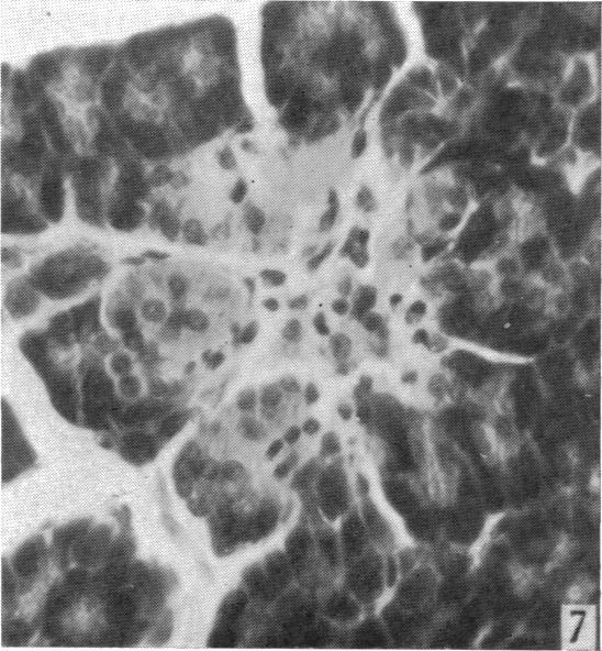
8 240 J. S. DUNN AND OTHERS Exp. 22. Wt g., ordinary diet. There was no rise of blood sugar at 2 hr. Killed at 48 hr. The islets generally appeared normal but on close examination with the high power one or two dislocated cells with shrunken bodies and pyknotic nuclei were seen in a few of them. No mitoses were found. Exp. 23. Wt g., ordinary diet. The blood sugar rose from 98 to 170 mg./100 c.c. at I hr. and remained at about that level for 51 hr., falling to 110 mg. at 10 hr. Next morning it was 120 mg. The animal was killed at 48 hr. On low-power examination the islets were notably altered, the majority being reduced in size and having very irregular ill-defined outlines (P1. 1, fig. 7). With the high power it was clear that this deformation had resulted from collapse of the centres of the islets due to shrinkage or disappearance of cells, whilst groups of well-preserved cells at the periphery had been pushed inwards. In the centres the remaining cells were variable in appearance: some showed shrunken pyknotic nuclei and greatly shrunken bodies: a few were necrotic with karyorrhexis or karyolysis: others showed a washed-out appearance of their cytoplasm. The majority of the well-preserved peripheral cells were a-cells and these had swollen eosinophile bodies which took the orange-red stain by Bensley's method. Mitoses were seen in peripheral cells in some islets but rarely more than one in any islet (P1. 1, fig. 8): the category of the dividing cells could not be determined as their cytoplasm was bloated and lacking in specific granulation. Exp. 24. Wt. 2 g., full diet with carrots. Doses of 50 mg./kg. given on first and second days: there was practically no rise of blood sugar after the first dose. Killed 48 hr. after first dose. With low power most islets appeared normal but the high power revealed various degrees of damage in.about one-fifth: the commonest change was dislocation and rounding off of fl-cells; their nuclei were hyperchromatic and the cytoplasm had a washed-out appearance (P1. 1, fig. 9). In one or two islets there was definite necrosis of a central mass of cells. The cx-cells in damaged islets were generally swollen and prominent. No mitoses were found on careful search either in the damaged islets or in the more normal ones. DISCUSSION The results of the first nine experiments, and of four others referred to, show that intravenous doses of alloxan of at least 200 mg./kg. in rabbits always cause severe damage to the islets of Langerhans. While this lesion is developing most animals are somewhat depressed and may not eat, and some pass into hypoglycaemic convulsions after 4-24 hr.: death then occurs unless glucose is administered [cf. Jacobs, 1937]. It is possible, however, for a rabbit to survive a dose of 200 mg./kg. for 24 hr. with necrosis in the whole of its islets and without showing very definite outward signs of illness (Exp. 5). This fact, which could hardly have been expected where such a specialized and important tissue was concerned, helps, to account for the apparent capriciousness in effect even of

9 ALLOXAN AND PANCREATIC ISLETS 241 large doses of alloxan: it may also have some significance in relation to the possible occurrence of acute damage of the islets from any cause in the human subject. It has already been shown that with large doses of the above order the islet cells exhibit the features of coagulative necrosis at nine hours, and the view that by that time they are dead is confirmed by the progress of nucleolysis in animals examined at later periods. In Exps the doses were mg./kg., and it is considered certain that frank necrosis would have been recognizable in the islets had the animals been allowed to live for 9 hr. or longer. Histological examination revealed certain changes from 1 hr. onwards: the earliest and most definite was hyperchromasia of s-cell nuclei, which preceded any recognizable cytoplasmic change. The hyperchromasia was increased in intensity at 3 and at 5 hr. and was then associated with altered staining of the cytoplasm and with dislocation of some cells. It is impossible to tell by histological examination the earliest stage at which cells are dead, but it seems probable that death had occurred in some cells at 3 hr. when they were separating from their attachments: it may, of course, have occurred earlier. The opening up of channels between the cell ribbons (P1. 1, figs. 4, 5) at these early stages cannot be interpreted with certainty. Some degree of this may be seen at times in normal islets, though usually the ribbons are closely packed together. Here the condition appeared to be in excess of normal and the spaces contained scanty granular debris suggesting that they had had some fluid content. The appearance is thought to be compatible with a temporary increase in functional activity of the islets which might be in operation immediately after dosage. From observation of the general behaviour only of rabbits which received 50 mg./kg. of alloxan (Exps ), it would not have been suspected that any serious internal lesion had occurred. Examination of the pancreas in these animals gave variable results. In one (no. 19) all the islets appeared normal; in four, nos. 18 and 20 killed at 24 hr., and nos. 21 and 22 killed at 48 hr., the majority were normal and only slight changes were present in the remaining few: these comprised nuclear pyknosis and depletion of cytoplasmic substance in some cells, resulting in a washed-out appearance by ordinary staining. The focal distribution of these slight lesions, notable also in Exp. 24, may possibly be related to the state of activity of individual islets during the period when the agent was having effect. In contrast with these slight changes, there were quite pronounced alterations in the islets in nos. 16 and 17,-examined at 24 hr., and in no. 23, examined at 48 hr. In these the fl-cells were much changed: they were rarely frankly necrotic but their outlines were rounded off and their nuclei hyperchromatic, while the granular substance in their cytoplasm was much depleted. With the special stain great diminution of the fine fl-granules was -notable in many cells. By 48 hr. most of these cells had shrivelled or

10 242 J. S. DUNN AND OTHERS collapsed, taking up little space, so that the islets were small with deformed outlines. These cellular changes were markedly in contrast with those of coagulative necrosis and karyolysis seen at the same periods after large doses, and as they had taken so long to develop it appeared to be a reasonable interpretation that while essentially necrobiotic in character, they were the result of overactivity and exhaustion of the cells after stimulation, rather than of a directly lethal toxic effect. A further interesting and highly suggestive feature in the histological appearances in the islets is the differential effect on ac- and fl-cells. It had already been noted that in islets necrosed after large doses any surviving cells were situated marginally, and that their bodies were strongly eosinophile. It is now confirmed by special staining that these are ac-cells and that their bodies are enlarged to two or three times normal, with evident increase in amount of the specific granule substance. This change may be well established 3 hr. after a large dose and is also quite pronounced 24 hr. after a smaller dose wherever the fl-cells are much altered. With this change, which appears to be an active and progressive one, the majority of the ac-cells have the appearance of living cells, but some are necrosed. It seems likely that this striking difference in their behaviour is significant of some considerable difference in the respective functions of ac- and fl-cells, which by this method may become accessible to investigation. Although the movements of the blood sugar were observed only over the first few hours, a correlation of them with histological changes provides some interesting features. In eleven'animals which received large doses (Figs. 1, 2) there was a rise of blood sugar in 2 hr. to mg./100 c.c. in seven; and to mg. in three, two of which had been starved for 24 hr. In one only, no. 7, which was fed normally, the blood sugar did not rise to 100 mg./ 100 c.c., but in this animal the initial reading was unusually low and hypoglyeaemia set in rapidly. After doses of 50 mg./kg. the blood sugar rose in only five out of eight and the elevation was less than with the large doses: of these five, three were those with well-marked changes in the islets. From these observations it may be concluded that a definite initial rise of blood sugar after alloxan probably indicates that damage of islets is occurring. Absence of a rise may mean that damage is slight or nil, but may also be noted even if this is severe. Jacobs, in referring to the transient hyperglyeaemia, did not conclude that it was a specific effect of alloxan. The hypoglyeaemia which may be evident in 4 hr. appears to be a constant and specii,effect if the dose of alloxan is sufficient. Jacobs observed it invariably with doses of 70 mg./kg. and upwards, and we can confirm this from all experiments where the dose was sufficient and suitable examination was made. It will be noted that no hypoglycaemia was observed after doses of 50 mg./kg. (Exps ).


11 THE JOURNAL OF PHYSIOLOGY, VOL. 103, No. 2 PLATE 1 To face p. 243

12 ALLOXAN AND PANCREATIC ISLETS 243 SUMMARY 1. From observations on fourteen rabbits it has been confirmed that single intravenous doses of alloxan of at least 200 mg./kg. always cause serious damage of the islets of Langerhans, though there may, during the first day, be little outward indication of serious illness. After these large doses definite histological changes can be recognized in the islets after about an hour, and these may be very pronounced at 3 hr. when some fl-cells appear to be necrosed. 2. With doses of 50 mg./kg. the fl-cells sometimes show degranulation, but little necrosis, at 24 hr.: the appearances are consistent with abnormal increase of function. After these smaller doses islet changes may be only slight and focal in distribution. 3. oc-cells are less prone to the necrobiotic changes than ft-cells and, from early stages, may be enlarged with apparent increase of granules when the fl-cells are necrosed or exhausted: this observation suggests some difference in function of the two types of cell. 4. A temporary hyperglycaemia almost always occurs in the first 2 hr. where there is any damage to the islets. We acknowledge with thanks the support given to this work by grants from the Rankin Medical Research Fund of the University of Glasgow and from the Medical Research Council. We have been indebted to Mr Wm. Carson for technical assistance with special reference to the specific granule staining. REFERENCES Dunn, J. S., Kirkpatrick, J., McLetchie, N. G. B. & Telfer, S. V. [1943]. J. Path. Bact. 55, 245. Dunn, J. S., MoLetchie, N. G. B. & Sheehan, H. L. [1943]. Lancet, 1, 484. Jacobs, H. R. [193.7]. Proc. Soc. Exp. Biol. N.Y., 37, 407. Warren, S. [1938]. The Pathology of Diabente Mellitu8, 2nd. ed., Philadelphia, p. 22. EXPLANATION OF PLATE 1 Fig. 4. Exp. 10. Hyperchromasia of nuclei and opening of spaces between cell ribbons. Dose mg./kg.: time 1 hr. H. and E. x 400. Fig. 5. Exp. 15. Mfore intense nuclear hyperehromasia, dislocation of some cells, and spaces formed. Dose mg./kg. --time 5 hr. H. and E. x 480. Fig. 6. Exp. 16. Rounding off of,-cells with nuclear hyperchromasia and pale staining of cytoplasm: washed-out appearance in some cells (b). A border of enlarged a-cells above with normal nuclei and strongly stained bodies (a). Dose 50 mg./kg.: time 24 hr. H. and E. x 480. Fig. 7. Exp. 23. Collapsed and deformed islet with disappearance of most of the central,-cells: clumps of large a-cells peripherally. Dose 50 mg./kg.: time 48 hr. H. and E. x 280. Fig. 8. Exp. 23. Deformed islet, mainly a-cells. Mitosis is seen in a cell at the upper edge. H. and E. x280. Fig. 9. Exp. 24. Nuclear pyknosis and pale cytoplasmic staining of s-cells in lower part of islet, with washed-out appearlnce in some. Normal cells in upper two-thirds. Dose 50 mg./kg. first and second days: killed at 48 hr. H. and E. x 280.
J. Physiol. (I938) 94, I2.352.i2:635.34
 94, I2.352.i2:635.34") 249 J. Physiol. (I938) 94, 249-255 6I2.352.i2:635.34 EFFECTS OF CABBAGE EXTRACTS ON CARBOHYDRATE METABOLISM BY A. D. MACDONALD AND LEO WISLICKI From the Department of Pharmacology, The University of Manchester
249 J. Physiol. (I938) 94, 249-255 6I2.352.i2:635.34 EFFECTS OF CABBAGE EXTRACTS ON CARBOHYDRATE METABOLISM BY A. D. MACDONALD AND LEO WISLICKI From the Department of Pharmacology, The University of Manchester
Experiments were carried out then with the object of producing complete disappearance of the A
 Relation of Glucagon to A Cells of the Pancreas*. (22339) SERGIO A. BENCOSME AND J. FREI. (Introduced by J.S.L. Browne Departament of pathology, Queen`s University, Kingston, Ontario, Canada. In spite
PREPARED BY P.DHARANI PRASAD II YEAR B.PHARM II SEM SUB:PATHOPHYSIOLOGY
 CELL INJURY UNIT I PREPARED BY P.DHARANI PRASAD II YEAR B.PHARM II SEM SUB:PATHOPHYSIOLOGY DETECTION OF CELLULAR CHANGES AFTER INJURY BY: LIGHT MICROSCOPY OR GROSS EXAMINATION DETECT CHANGES HOURS TO DAYS
6I I:6I hypophysectomy. This diminution of diabetes is shown particularly as. hypophysectomized or totally decerebrated [Houssay and
 6I2.466.6I:6I2.492.5 KETOSIS IN THE PANCREATIC AND PHLORRHIZIN DIABETES OF HYPOPHYSECTOMIZED DOGS. BY CIRO T. RIETTI. (Institute of Physiology, Faculty of Medicine, Buenos Ayres.) IN the hypophysectomized
number Done by Corrected by Doctor Heyam Awad
 number 4 Done by Waseem Abu Obeida Corrected by Saad Al-Hayek Doctor Heyam Awad Cell injury -in the previous lectures we talked about the causes (etiology) and the mechanism (pathogenesis) of cell injury.
NUCLEAR ABNORMALITIES RESULTING FROM INHIBITION OF MITOSIS BY COLCHICINE AND OTHER SUBSTANCES '
 NUCLEAR ABNORMALITIES RESULTING FROM INHIBITION OF MITOSIS BY COLCHICINE AND OTHER SUBSTANCES ' AUSTIN M. BRUES AND ELIZABETH B. JACKSON (From the Medicul Lnborutorif~ of the Collis P. Iiz4ntingtolz M~moricil
: : (From the Department of Physiology, University of Toronto.)
") 94 612.352.2:547.435:612.34.089.87 CHOLINE AND LIVER FAT IN DIABETIC DOGS. BY C. H. BEST, G. C. FERGUSON AND J. M. HERSHEY. (From the Department of Physiology, University of Toronto.) IN the first completely
94 612.352.2:547.435:612.34.089.87 CHOLINE AND LIVER FAT IN DIABETIC DOGS. BY C. H. BEST, G. C. FERGUSON AND J. M. HERSHEY. (From the Department of Physiology, University of Toronto.) IN the first completely
H. M. Carleton, Lecturer in Histology, University of Oxford. (From the Department of Physiology.) INTRODUCTORY.
 INTRODUCTORY.") Note on the Comparative Effects on Tissues of Isotonic Saline and Distilled Water when used as Solvents for Mercuric Chloride and Formol in Histological Fixation. By H. M. Carleton, Lecturer in Histology,
Note on the Comparative Effects on Tissues of Isotonic Saline and Distilled Water when used as Solvents for Mercuric Chloride and Formol in Histological Fixation. By H. M. Carleton, Lecturer in Histology,
PLATES 24 TO 26. (Received for publication, December 4, 1935)
") Published Online: 1 March, 1936 Supp Info: http://doi.org/10.1084/jem.63.3.303 Downloaded from jem.rupress.org on January 19, 2019 THE VISCERAL LESIONS PRODUCED IN MICE BY THE SALIVARY GLAND VIRUS OF MICE*
Published Online: 1 March, 1936 Supp Info: http://doi.org/10.1084/jem.63.3.303 Downloaded from jem.rupress.org on January 19, 2019 THE VISCERAL LESIONS PRODUCED IN MICE BY THE SALIVARY GLAND VIRUS OF MICE*
ULAR LESIONS CAUSED BY CROTALUS VENOM.*
 AN EXPERIMENTAL STUDY OF THE LATE GLOMER- ULAR LESIONS CAUSED BY CROTALUS VENOM.* BY RICHARD M. PEARCE, M.D. (From the John Herr Musser Department of Research Medicine of the University of Pennsylvania,
PATHOLOGY Intracellular Degeneration LAB 1
 PATHOLOGY Intracellular Degeneration LAB 1 Cellular swelling Liver Organ :- Liver Lesion :- 1. Narrowing of hepatic sinusoids due to the swelling of hepatocyte. 2. The cytoplasm of affected hepatocyte
PITUITARY HYPERPLASIA IN A MALE MOUSE AFTER THE ADMINISTRATION OF OESTRIN
 PITUITARY HYPERPLASIA IN A MALE MOUSE AFTER THE ADMINISTRATION OF OESTRIN HAROLD BURROWS (From The Research Institute of the Royal Cancer Hospital (Free), London, S.W.3) Several observers have noticed
IT is generally recognized that glycogen is a very labile substance which
 Ester Wax as a Medium for Embedding Tissue for the Histological Demonstration of Glycogen BY. J. D. SMYTH AND C. A. HOPKINS {From the Department of Zoology, Trinity College, Dublin) INTRODUCTION IT is
TOXICITY OF MESOXALATES AND HISTOLOGICAL OBSERVATIONS AFTER CONTINUOUS ADMINISTRATION
 TOXICITY OF MESOXALATES AND HISTOLOGICAL OBSERVATIONS AFTER CONTINUOUS ADMINISTRATION YOSHITO KOBAYASHI, SHIGERU OHASHI AND SETSUYA TAKEUCHI Department of Pharmacology, Faculty of Medicine, University
Occurrence of Ciliated Vesicle-Containing Reticular Cells in the Mouse Thymus
 Occurrence of Ciliated Vesicle-Containing Reticular Cells in the Mouse Thymus By Takeshi Hoshino Department of Anatomy, Hokkaido University School of Medicine, Sapporo, Japan (Director : Prof. Takashi
(From The Rockefeller Institute) Materials and Methods. Observations with the Electron Microscope
 Materials and Methods. Observations with the Electron Microscope") ELECTRON MICROSCOPE STUDY OF THE DEVELOPMENT OF THE PAPILLOMA VIRUS IN THE SKIN OF THE RABBIT* BY ROBERT S. STONE,~ M.D., RICHARD E. SHOPE, M.D., DAN H. MOORE, P,~.D. (From The Rockefeller Institute) PLATES
ELECTRON MICROSCOPE STUDY OF THE DEVELOPMENT OF THE PAPILLOMA VIRUS IN THE SKIN OF THE RABBIT* BY ROBERT S. STONE,~ M.D., RICHARD E. SHOPE, M.D., DAN H. MOORE, P,~.D. (From The Rockefeller Institute) PLATES
destroyed, or removed from the body. The experiments to be described intravenously injected insulin was observed in normal animals, in
 417 J. Physiol. (I940) 97, 4I7-428 6I5.361.37:6I2.I29.I ON THE DISAPPEARANCE FROM THE BLOOD OF INTRAVENOUSLY INJECTED INSULIN BY H. K. GOADBY1 AND J. S. RICHARDSON From the Medical Unit Laboratories, St
investigated. According to the current hypothesis fat is completely hydrolysed
 306 J. Physiol. (I943) I02, 3o6-3I2 6i2. 322 73 DIFFERENTIATION IN THE ABSORPTION OF OLIVE OIL *0 AND OLEIC ACID IN THE. RAT By A. C. FRAZER,* From the Physiology Department, St Mary's Hospital Medical
The Chromosomes of Pour Species of Marsupials.
 The Chromosomes of Pour Species of Marsupials. By Stella C. A. Altmann, B.Sc, and Mavis E. W. Ellery, B.Sc, University of Melbourne. With Plates 37 and 38. THIS is a joint paper only in so far as the results
metabolism, as in Fr6hlich's syndrome; and by physiological facts, such Coope and Mottram (1914).
.") THE EFFECT OF PITUITRIN ON THE FATTY ACID OF THE LIVER. By R. COOPE AND E. N. CHAMBERLAIN'. (From the Department of Biochemistry, University of Liverpool.) THE work described in this paper was undertaken
THE EFFECT OF PITUITRIN ON THE FATTY ACID OF THE LIVER. By R. COOPE AND E. N. CHAMBERLAIN'. (From the Department of Biochemistry, University of Liverpool.) THE work described in this paper was undertaken
PHOSPHORUS CONTENT OF THE BLOOD IN DIABETES
 10 BLOOD PHOSPHORUS IN HEALTH AND DISEASE: IV-THE PHOSPHORUS CONTENT OF THE BLOOD IN DIABETES MELLITUS F B BYROM From the Dunn Laboratories, London Hospital Received for publication November 24th, 1928
STIRLING, M.D., SC.D., F.R.S.R, Regius Professor of the Institutes
 ON THE EPITHELIUM OF THE CORNEA. By WM. STIRLING, M.D., SC.D., F.R.S.R, Regius Professor of the Institutes of Medicine, and D AVID SKINNER, M.A., Student of Medicine in the Universty of Aberdeen. (PI.
COMMUNICATIONS PHOTOCOAGULATION OF THE RETINA* OPHTHALMOSCOPIC AND HISTOLOGICAL FINDINGS. photocoagulation of the rabbit's retina.
 Brit. J. Ophthal. (1963) 47, 577. COMMUNICATIONS PHOTOCOAGULATION OF THE RETINA* OPHTHALMOSCOPIC AND HISTOLOGICAL FINDINGS BY A. LAVYEL Haifa, Israel SINCE the introduction of the photocoagulator by Meyer-Schwickerath
THE EFFECT OF ROENTGEN RADIATION ON SPINAL GANGLIA OF ALBINO RATS
 THE EFFECT OF ROENTGEN RADIATION ON SPINAL GANGLIA OF ALBINO RATS W. C. MA AND CHIEN-LIANG HSU (From the Departmt-nts of Anatomy and Radiology, Peiping Union M~dical College, Peiping, China) From the extensive
IN a previous publication (Hewitt, 1954) a description was given of the
 a description was given of the") i 9 9 Further Observations on the Histochemistry of Fat Absorption in the Small Intestine of the Rat By W. HEWITT, M.B., B.S. (From the Department of Anatomy, St. Thomas' Hospital Medical School, London,
i 9 9 Further Observations on the Histochemistry of Fat Absorption in the Small Intestine of the Rat By W. HEWITT, M.B., B.S. (From the Department of Anatomy, St. Thomas' Hospital Medical School, London,
College of Medicine, Newcastle-on-Tyne.)
") INTERRELATION OF PARATHYROIDS, SUPRA- RENALS AND PANCREAS. BY G. A. CLARK. (From the Physiological Laboratory, Durham University College of Medicine, Newcastle-on-Tyne.) THAT the parathyroid glands have
INTERRELATION OF PARATHYROIDS, SUPRA- RENALS AND PANCREAS. BY G. A. CLARK. (From the Physiological Laboratory, Durham University College of Medicine, Newcastle-on-Tyne.) THAT the parathyroid glands have
hypoglycoemic reaction to insulin is greater in sheep after thyroidectomy, BY J. (The National Institute for Medical Research, London.
 THE RELATION OF THE THYROID GLAND TO THE ACTION OF INSULIN. BY J. H. BURN AND H. P. MARKS. (The National Institute for Medical Research, London.) AN experimental investigation of the connection between
LGM International, Inc.
 Liqui-PREP TM Cytology Atlas Preface The following pictures are examples with descriptions of cytology slides processed with the Liqui-PREP TM System.. The descriptions are reviewed by Pathologists. It
Histopathology: Cell necrosis and cytoplasmic accumulations
 Histopathology: Cell necrosis and cytoplasmic accumulations These presentations are to help you identify basic histopathological features. They do not contain the additional factual information that you
SECTION 2 CELL INJURY
 Adapted myocyte Normal myocyte Reversibly-injured myocyte SECTION 2 CELL INJURY Cell death 5/4/2014 1 5/4/2014 2 Reversible Degeneration Irreversible Cellular Swelling Fatty Change Hyaline Change Amyloid
Effects of alcoholic extract of Momordica charantia (Linn.) whole fruit powder on the pancreatic islets of alloxan diabetic albino rats
 whole fruit powder on the pancreatic islets of alloxan diabetic albino rats") Journal of Environmental Biology January 28, 29(1) 11-16 (28) Triveni Enterprises, Lucknow (India) For personal use only Free paper downloaded from: www. jeb.co.in Commercial distribution of this copy
Journal of Environmental Biology January 28, 29(1) 11-16 (28) Triveni Enterprises, Lucknow (India) For personal use only Free paper downloaded from: www. jeb.co.in Commercial distribution of this copy
GLUCOSE is the most important diffusible substance in the blood which
 ON THE ACTION OF PHLORHIZIN ON THE KIDNEY. By E. B. MAYRS. (From the Department of Pharmacology, Edinburgh.) GLUCOSE is the most important diffusible substance in the blood which is completely held back
Prepared By Jocelyn Palao and Layla Faqih
 Prepared By Jocelyn Palao and Layla Faqih The structure of the suspected atypical cell should always be compared to the structure of other similar, benign, cells which are present in the smears. The diagnosis
The Endocrine System Pituitary
 The Endocrine System Pituitary Look at your slide of the human pituitary with your naked eye. You should see a cellular region and a more fibrous region. Then view each region with your microscope under
HISTOPATHOLOGY. Shannon Martinson
 HISTOPATHOLOGY Shannon Martinson March 2013 Case #1 History: 8 year old beagle Neck pain for the past couple of weeks Paresis, followed by paralysis developed over the past few days Gross Description courtesy
'the perfusion of the cat's lung a cannula was tied into the left auricle and :547.78I.5
 280 576.809.73:547.78I.5 LIBERATION OF HISTAMINE FROM THE PERFUSED LUNG BY STAPHYLOCOCCAL TOXIN BY W. FELDBERG AND E. V. KEOGH1 From The Walter and Eliza Hall Institute, Melbourne (Received 5 March 1937)
: /18
 612.461.23: 616-001.17/18 SOME OBSERVATIONS ON THE COMPARATIVE EFFECTS OF COLD AND BURNS ON PROTEIN METABOLISM IN RATS. By G. H. LATHE 1 and R. A. PETERS. From the Department of Biochemistry, Oxford. (Received
COLOUR REACTIONS ATTRIBUTED
 LXIV. COLOUR REACTIONS ATTRIBUTED TO VITAMIN A. BY FRANCIS HOWARD CARR AND ERNEST ARTHUR PRICE. From the Laboratories of The British Drug Houses, Ltd., Graham Street, City Road, N. 1. (Received March 17th,
Edinburgh.) IN a previous paper, I recorded observations on rabbits and dogs which
 IN a previous paper, I recorded observations on rabbits and dogs which") THE EFFECT OF ADRENALIN ON SUGAR AND NITROGEN EXCRETION IN THE URINE OF BIRDS. BY D. NOEL PATON.. (From the Research Laboratory of the Royal College of Physicians, Edinburgh.) IN a previous paper, I recorded
THE EFFECT OF ADRENALIN ON SUGAR AND NITROGEN EXCRETION IN THE URINE OF BIRDS. BY D. NOEL PATON.. (From the Research Laboratory of the Royal College of Physicians, Edinburgh.) IN a previous paper, I recorded
Studies of differential staining with acid dyes in the human adenohypophysis
 467 Studies of differential staining with acid dyes in the human adenohypophysis By A. W. F. FISHER and D. BULMER (From the Anatomy Department, University of Manchester) Summary With Mallory techniques
SOME OBSERVATIONS UPON SODIUM ALGINATE. By 0. M. SOLANDT. From the Physiological Laboratory, Cambridge.
 582.6 SOME OBSERVATIONS UPON SODIUM ALGINATE. By 0. M. SOLANDT. From the Physiological Laboratory, Cambridge. (Received for publication 13th December 1940.) ALGINIC acid was discovered by Stanford in 1883
Department of Anatomy, Glasgow University
 THE UPTAKE OF LABELLED SULPHATE INJECTED INTO THE HOST ANIMAL BY CARTILAGE HOMOGRAFTS By G. M. WYBURN, D.Sc., F.R.F.P.S.G., and P. BACSICH, D.Sc., M.D. Department of Anatomy, Glasgow University INTRODUCTION
STUDIES IN BLOOD DIASTASE. FACTORS WHICH CAUSE. The effects of the following procedures on the blood diastase have
 STUDIES IN BLOOD DIASTASE. FACTORS WHICH CAUSE VARIATIONS IN THE AMOUNT OF DIASTASE IN THE BLOOD. By CHARLES REID and B. NARAYANA. From the Department of Physiology, Prince of Wales Medical College, Patna.
CHRONIC RENAL DISEASE IN RATS FOLLOWING A TEMPORARY
 40 CHRONIC RENAL DISEASE IN RATS FOLLOWING A TEMPORARY DEFICIENCY OF POTASSIUM P. FOURMAN, R. A. McCANCE AND R. A. PARKER From the Departments of Experimental Medicine and Pathology, University of Cambridge
Notes on the Effects of Pretreatment of Spores on the Iodinophile Vacuole of Henneguya exilisl
 Proceedings of the Iowa Academy of Science Volume 76 Annual Issue Article 57 1969 Notes on the Effects of Pretreatment of Spores on the Iodinophile Vacuole of Henneguya exilisl Dennis Galinsky Drake University
Necrosis is death of cells and tissues in the living animal. Focal/ Multifocal necrosis- terms used for one
 Necrosis Necrosis Necrosis is death of cells and tissues in the living animal. Focal/ Multifocal necrosis- terms used for one or more, small, clearly defined areas of necrosis. Diffuse necrosis- term used
Exercise 6. Procedure
 Exercise 6 Procedure Growing of root tips Select a few medium-sized onion bulbs. Carefully remove the dry roots present. Grow root tips by placing the bulbs on glass tubes (of about 3 4 cm. diameter) filled
The basis of Disease
 General Curriculum The basis of Disease ZHOU REN 周韧 Prof., M.D., Ph.D. Institute of Pathology & Forensic Medicine Department of Pathology & Patho-physiology Zhenjiang University Judicial Evidence & Evaluation
Cell Adaptation, Cell Injury and Cell Death
 Cell Adaptation, Cell Injury and Cell Death Pathology:- is the study of structural and functional abnormalities that are expressed as diseases of organs and systems. Modern pathology, proposed that injury
Spermatogenesis Following Experimental Testicular Ischemia
 Spermatogenesis Following Experimental Testicular Ischemia Frank Hinman, Jr, MD, and Gilbert I Smith, MD REGENERATION of the spermatogenic elements of the testis after depression by testosterone and by
DURING the past ten years it has been suggested that the classical form
 163 The Golgi Apparatus and Lipoidal Bodies in Exocrine and Endocrine Cells in the Pancreas of Man By DENNIS LACY (From the Department of Zoology and Comparative Anatomy, St. Bartholomew's Medical College)
Evaluation of Breast Specimens Removed by Needle Localization Technique
 Evaluation of Breast Specimens Removed by Needle Localization Technique Specimen Handling: The breast specimen when received should be measured and grossly inspected for any orientation designated by the
My Journey into the World of Salivary Gland Sebaceous Neoplasms
 My Journey into the World of Salivary Gland Sebaceous Neoplasms Douglas R. Gnepp Warren Alpert Medical School at Brown University Rhode Island Hospital Pathology Department Providence RI Asked to present
PRACTICAL ROADMAP. GLANDS AFFECTING LIFESTYLE WJ van der Spuy & T Tshabalala
 PRACTICAL ROADMAP GLANDS AFFECTING LIFESTYLE WJ van der Spuy & T Tshabalala GLANDS AFFECTING LIFESTYLE Submandibular gland (salivary gland) Liver Pancreas Hypophysis (pituitary gland) Thyroid Suprarenal
Avian Pathology. Bacterial diseases: histo slides. ECVP-ESVP Summer School 2012 Frédérique NGUYEN
 Avian Pathology Bacterial diseases: histo slides ECVP-ESVP Summer School 2012 Frédérique NGUYEN Bacterial diseases: histo slides B1. Turkey. Organs? Morphologic diagnosis? Special procedure? B2. Hen. Organ?
Exfoliative cytology of diffuse mesothelioma
 Exfoliative cytology of diffuse mesothelioma G. HEFIN ROBERTS AND G. M. CAMPBELL From the Pathology Department, Southern General Hospital, Glasgow J. clin. Path., 1972, 25, 577-582 SYNOPSIS The exfoliative
Mechanisms of Cell Injury
 Causes of Cell Injury 1- oxygen deprivation (anoxia) 2- physical agents 3- chemical agents 4- infections agents 5- immunologic reactions 6- genetic defects 7- nutritional imbalances Mechanisms of Cell
Core Lab #3 Investigating Diabetes Mellitus
 Name: Introduction Diabetes is a malfunction of one of the major homeostatic mechanisms in the body the endocrine system. Two hormones, insulin and glucagon control the level of sugar in the blood. The
A SIMPLE METHOD OF REMOVING LEUCOCYTES FROM BLOOD. by virtue of their fluid content, but also by conferring on the patient's blood
 METHOD OF REMOVING LEUCOCYTES FROM BLOOD. 281 the salt content falls below or rises above these limits the leucocytes do not emigrate and do not phagocyte or destroy bacteria. It has been shown also that
![[ 3<M ] STUDIES ON TAPEWORM PHYSIOLOGY](/thumbs/78/78631839.jpg "[ 3<M ] STUDIES ON TAPEWORM PHYSIOLOGY") [ 3
[ 3 CONTRIBUTION TO THE HISTOPATHOLOGY OF FILARIASIS
 CONTRIBUTION TO THE HISTOPATHOLOGY OF FILARIASIS PHILIP H. HARTZ Public Health Service, Curacao, N.W.I. The histologic changes caused by filariasis (Wucheria Bancrofti) are considered to be non-specific
Since, for many months after section of the right vagus in the neck,
 THE INFLUENCE OF THE VAGUS ON THE ISLETS OF LANGERHANS. Part II. The effect of cutting the vagus upon sugar tolerance. BY G. A. CLARK. (From the Physiological Laboratory, Sheffield University.) IN Part
[1920], in studies on the human pleural membrane, pointed out the
![[1920], in studies on the human pleural membrane, pointed out the](/thumbs/84/89127006.jpg "[1920], in studies on the human pleural membrane, pointed out the") 'ca -.101 6II.25:6II.OI8.86 NERVES AND NERVE ENDINGS IN THE VISCERAL PLEURA OF THE CAT. BY A. I. G. McLAUGHLIN. (From the Unit Laboratories, University College Hospital Medical School.) (Received September
'ca -.101 6II.25:6II.OI8.86 NERVES AND NERVE ENDINGS IN THE VISCERAL PLEURA OF THE CAT. BY A. I. G. McLAUGHLIN. (From the Unit Laboratories, University College Hospital Medical School.) (Received September
THE RELATION OF HYPERGLYCEMIA TO THE RELATIVE BLOOD VOLUME, CHLORINE CONCENTRATION, AND CHLORINE DISTRIBUTION IN THE BLOOD OF DOGS.
 Published Online: 1 July, 1925 Supp Info: http://doi.org/10.1084/jem.42.1.89 Downloaded from jem.rupress.org on September 28, 2018 THE RELATION OF HYPERGLYCEMIA TO THE RELATIVE BLOOD VOLUME, CHLORINE CONCENTRATION,
Epithelia will be discussed according to the following scheme: Type Number of layers Shape Line drawing. Squamous Cuboidal Columnar
 Epithelia Epithelia will be discussed according to the following scheme: Type Number of layers Shape Line drawing Simple Squamous Cuboidal Columnar Covering and Lining epithelium Pseudostratified Stratified
the Action o f Radium Rays upon the Cells o f Jensen's Rat Sarcoma.
 Downloaded from http:rspb.royalsocietypublishing.org on May 3, 2018 482 On the Action o f Radium Rays upon the Cells o f Jensen's Rat Sarcoma. By S. Buss, D.Sc.,* and H elen Chambers, M.D. (Communicated
EXPERIMENTAL THERMAL BURNS I. A study of the immediate and delayed histopathological changes of the skin.
 EXPERIMENTAL THERMAL BURNS I A study of the immediate and delayed histopathological changes of the skin. RJ Brennan, M.D. and B. Rovatti M.D. The purpose of this study was to determine the progressive
Cellular Pathology. Histopathology Lab #2 (web) Paul Hanna Jan 2018
 Paul Hanna Jan 2018") Cellular Pathology Histopathology Lab #2 (web) Paul Hanna Jan 2018 Slide #91 Clinical History: a necropsy was performed on an aged cat the gross pathological changes included: widespread subcutaneous edema
Cellular Pathology Histopathology Lab #2 (web) Paul Hanna Jan 2018 Slide #91 Clinical History: a necropsy was performed on an aged cat the gross pathological changes included: widespread subcutaneous edema
Human Saliva as a Convenient Source of Ribonuclease. By S. BRADBURY
 Human Saliva as a Convenient Source of Ribonuclease 323 By S. BRADBURY (From the Cytological Laboratory, Department of Zoology, University Museum, Oxford) SUMMARY Saliva, heated to 80 C for 10 minutes
University College, London.)
") 62.45:577.74.5 THE REPLACEMENT OF DEPLETED ADRENALNE N THE SUPRARENALS. BY G. P. CROWDEN. (From the Department of Physiology and Biochemistry, University College, London.) THE physiological control of
62.45:577.74.5 THE REPLACEMENT OF DEPLETED ADRENALNE N THE SUPRARENALS. BY G. P. CROWDEN. (From the Department of Physiology and Biochemistry, University College, London.) THE physiological control of
Electron Microscope Studies of HeLa Cells Infected with Herpes Virus
 244 STOKER, M. G. P., SMITH, K. M. & Ross, R. W. (1958). J. gen. Microbiol. 19,244-249 Electron Microscope Studies of HeLa Cells Infected with Herpes Virus BY M: G. P. STOKER, K. M. SMITH AND R. W. ROSS
When, however, the adreno-cortical extract was given before the test a
 CORTISONE INDUCED IMPAIRMENT OF GLUCOSE TOLERANCE IN THE DETECTION OF THE DIABETIC DIATHESIS. BY L. J. P. DUNCAN. From the Department of Therapeutics, University of Edinburgh. (Received for publication
PANCREATITIS IN YOUNG CHILDREN
 PANCREATITIS IN YOUNG CHILDREN BY J. COLLINS From the Roan Antelope Copper Mines, Luanshya, N. Rhodesia Pancreatitis is rare in very young children and the medical literature contains comparatively few
[485] THE BREAKDOWN OF THE THORACIC GLAND IN THE ADULT INSECT, RHODNIUS PROLIXUS
![[485] THE BREAKDOWN OF THE THORACIC GLAND IN THE ADULT INSECT, RHODNIUS PROLIXUS](/thumbs/90/101209894.jpg "[485] THE BREAKDOWN OF THE THORACIC GLAND IN THE ADULT INSECT, RHODNIUS PROLIXUS") [485] THE BREAKDOWN OF THE THORACIC GLAND IN THE ADULT INSECT, RHODNIUS PROLIXUS BY V. B. WIGGLESWORTH Department of Zoology, University of Cambridge (Received 3 November 1954) The thoracic or ' prothoracic'
[485] THE BREAKDOWN OF THE THORACIC GLAND IN THE ADULT INSECT, RHODNIUS PROLIXUS BY V. B. WIGGLESWORTH Department of Zoology, University of Cambridge (Received 3 November 1954) The thoracic or ' prothoracic'
PATHOLOGY OF LIVER TUMORS
 PATHOLOGY OF LIVER TUMORS Pathobasic, 31.05.2016 WHO Classification Approach to a Liver Mass Lesion in a patient with chronic liver disease? Lesion in a patient without chronic liver disease? Malignant
CINtec p16 INK4a Staining Atlas
 CINtec p16 INK4a Staining Atlas Rating Rating Positive The rating positive will be assigned if the p16 INK4a -stained slide shows a continuous staining of cells of the basal and parabasal cell layers of
CHANGES IN BUCCAL CELLS IN THE ANAEMIAS
 J. clin. Path. (1959), 12, 222. CHANGES IN BUCCAL CELLS IN THE ANAEMIAS BY M. M. BODDINGTON From the Department of Pathology, Churchill Hospital, Headington, Oxford (RECEIVED FOR PUBLICATION DECEMBER 19,
Johannesburg.) Johannesburg (6000 feet) it is customary to regard the normal red cell
 Johannesburg (6000 feet) it is customary to regard the normal red cell") THE BLOOD COUNT AND BODY TEMPERATURE IN NORMAL RATS. By ARTHUR DIGHTON STAMMERS. (From the Department of Physiology, University of the Witwatersrand, Johannesburg.) IN connection with the maintenance of
THE BLOOD COUNT AND BODY TEMPERATURE IN NORMAL RATS. By ARTHUR DIGHTON STAMMERS. (From the Department of Physiology, University of the Witwatersrand, Johannesburg.) IN connection with the maintenance of
PHENANTHRIDINE COMPOUNDS
 Brit. J. Pharmacol. (195), 5, 398. THE CHEMOTHERAPEUTIC ACTION OF PHENANTHRIDINE COMPOUNDS PART IV ACTIVITY IN VITRO BY J. A. LOCK From the Wellcome Laboratories of Tropical Medicine, N.W.1 183, Euston
Biologie, and in the Revista de la Sociedad Argentina de Biologia under
 193 6I2.492:6I2.352.I2 RELATION OF THE PITUITARY GLAND TO THE ACTION OF INSULIN AND ADRENALINE. BY A. B. CORKILL, H. P. MARKS AND W. E. WHITE". (From the National Institute for Medical Research, Hampstead,
PROBABLE HODGKIN'S DISEASE IN A DOG: REPORT OF A CASE 1
 PROBABLE HODGKIN'S DISEASE IN A DOG: REPORT OF A CASE 1 LEONARD K. STALKER, M.D. Fellow in Surgery, The Mayo Foundation CARL F. SCHLOTTHAUER, D.V.M. AND WILLIAM H. FELDMAN, D.V.M., M.S. Division of Experimental
Histopathology: skin pathology
 Histopathology: skin pathology These presentations are to help you identify, and to test yourself on identifying, basic histopathological features. They do not contain the additional factual information
AUTOIMMUNE RESPONSES TO HUMAN TUMOUR ANTIGENS
 510 AUTOIMMUNE RESPONSES TO HUMAN TUMOUR ANTIGENS MADELINE HODKINSON* AND G. TAYLOR From the Immunology Department, Royal Infirmary, Manchester Received for publication May 14, 1969 THE most convincing
VETERINARY HEMATOLOGY ATLAS OF COMMON DOMESTIC AND NON-DOMESTIC SPECIES COPYRIGHTED MATERIAL SECOND EDITION
 VETERINARY HEMATOLOGY ATLAS OF COMMON DOMESTIC AND NON-DOMESTIC SPECIES SECOND EDITION COPYRIGHTED MATERIAL CHAPTER ONE HEMATOPOIESIS GENERAL FEATURES All blood cells have a finite life span, but in normal
(From the Physiotogicat Laboratory, Cambridge.)
") THE OXYGEN EXCHANGE OF THE SUPRARENAL GLAND. BY K. 0. NEUMAN. (From the Physiotogicat Laboratory, Cambridge.) THIS paper deals with the question of the amount of oxygen taken in by a unit weight of the
THE OXYGEN EXCHANGE OF THE SUPRARENAL GLAND. BY K. 0. NEUMAN. (From the Physiotogicat Laboratory, Cambridge.) THIS paper deals with the question of the amount of oxygen taken in by a unit weight of the
IN normal male fowls, four developmental stages of spermatogenetic activity
 Development of the Testis Tubule in the Fowl By GAMAL A. R. KAMAR (From the Animal Production Department, Faculty of Agriculture, Cairo University, Giza, Egypt) With three plates (figs. 1-3) SUMMARY Three
PANCREATIC PATHOLOGY IN DASYURID MARSUPIALS
 PANCREATIC PATHOLOGY IN DASYURID MARSUPIALS Authors: H. D. ATTWOOD, and P. A. WOOLLEY Source: Journal of Wildlife Diseases, 16(2) : 245-25 Published By: Wildlife Disease Association URL: https://doi.org/1.7589/9-558-16.2.245
Overton,1 who has worked exhaustively at the subject, looked upon. considered by some to be due to the state of the fluid originally in the
 THE EFFECTS OF TEMPERATURE ON THE OSMOTIC PROPER- TIES OF MUSCLE. By D. H. DE SOUZA. (From the Physiological Laboratory, University of Sheffield.) (With six diagrams in the text.) (Received for publication
EDUCATIONAL COMMENTARY MORPHOLOGIC CHANGES IN PERIPHERAL BLOOD CELLS
 EDUCATIONAL COMMENTARY MORPHOLOGIC CHANGES IN PERIPHERAL BLOOD CELLS Educational commentary is provided through our affiliation with the American Society for Clinical Pathology (ASCP). To obtain FREE CME/CMLE
Diabetes in the Real World
 Diabetes in the Real World Diabetes Mellitus Common hormonal disorder of dogs and cats due to insufficient insulin being produced or cells not being able to respond to insulin Results in hyperglycaemia
OF FATTY LIVERS. XLII. PROTEIN AND THE DIETARY PRODUCTION. On a number of occasions however use of this diet has failed to produce in our
 XLII. PROTEIN AND THE DIETARY PRODUCTION OF FATTY LIVERS. BY HAROLD JOHN CHANNON AND HARRY WILKINSON. From the Department of Biochemistry, The University, Liverpool. (Received December 20th, 1934.) THE
Client Information Sheet Copyright Bilton Veterinary Centre All rights Reserved. Diabetes Mellitus
 What is Diabetes Mellitus? Diabetes Mellitus Diabetes mellitus (DM) is often called sugar diabetes in order to distinguish it from a condition called Diabetes insipidus, which is a totally separate condition.
T shrinkage which Burkitt s lymphoma undergoes
 MORPHOLOGY OF BURKITT S LYMPHOMA DURING REGRESSION INDUCED BY CYCLOPHOSPHAMIDE B.. OSUNKOYA, MB, BS, PHD, V. A. NGU, MS, FRCS,? AND FRANCES C. MOTTRAM, BSc* The histologic and cytologic eatures o Burkitt
WSC , Conference 9, Case 1. Tissue from a nyala.
 WSC 2009-2010, Conference 9, Case 1. Tissue from a nyala. MICROSCOPIC DESCRIPTION: Heart, atrium (1 pt.): Approximately 40% of the atrial myocardium is replaced by areas of fibrous connective tissue (1
THE INFLUENCE OF TEMPERATURE ON HEMOLYSIS IN HYPOTONIC SOLUTIONS.'
 THE INFLUENCE OF TEMPERATURE ON HEMOLYSIS IN HYPOTONIC SOLUTIONS.' BY PAUL A. LEWIS, M.D. (From the Antitoxin Laboratory of the Massachusetts State Board of Health.) During the year I905-I906 it was my
Fixation... Questions 1 Answers 16. Processing... Questions 25 Answers 36. Safety... Questions 67 Answers 73
 Table of Contents Fixation... Questions 1 Answers 16 Processing... Questions 25 Answers 36 Instrumentation... Questions 43 Answers 58 Safety... Questions 67 Answers 73 Laboratory Mathematics & Solution
Prelab #4 BLOOD; BONE MARROW; RESPIRATORY; INTEGUEMENT Page 1
 Prelab #4 BLOOD; BONE MARROW; RESPIRATORY; INTEGUEMENT Page 1 Blood Slide 101 This a classic slide of blood cells using a Wright stain. Inspect red blood cells and their appearance. Note the approximate
Microscopic Examination of Urine
 Download http://www.vetlab.com/kova.htm Definition of urine sediment: all solid materials suspended in the urine - a semiquantative evaluation of the urine sediment Significance of formed elements in the
HISTOLOGY VIRTUAL LABORATORY BLOOD AND LYMPHATICS SYSTEM
 HISTOLOGY VIRTUAL LABORATORY BLOOD AND LYMPHATICS SYSTEM Login: http://histopath.westernu.edu Histology Atlas AND Virtual Histology links. I. HEMATOLOGY - PERIPHERAL BLOOD Purpose: To be able to identify
Cytoplasmic changes Nuclear changes
 The presence of infection in the female genital tract may procure certain cellular changes in the epithelium. Such changes are seen in nucleus and cytoplasm surrounding the nucleus. Cytoplasmic changes