ANA Diagnostics Using Indirect Immunofluorescence
|
|
|
- Stella Johnson
- 6 years ago
- Views:
Transcription

1 ANA Diagnostics Using Indirect Immunofluorescence EUROIMMUN AG Seekamp Lübeck (Germany) Tel / Fax
2 Table of contents Autoantibodies against cell nuclei (ANA) Autoantibodies negative (AC-0) Autoantibodies against cell nuclei, homogeneous (AC-1) Autoantibodies against dsdna Autoantibodies against cell nuclei, DFS pattern (AC-2) Autoantibodies against centromeres (AC-3) Autoantibodies against nucleoplasm, fine speckled (AC-4) Autoantibodies against Ku (AC-4) Autoantibodies against Mi-2 (AC-4) Autoantibodies against TIF1-gamma (AC-4) Autoantibodies against nucleoplasm, coarse speckled (AC-5) Autoantibodies against nuclear dots (AC-6) Autoantibodies against few nuclear dots (AC-7) Autoantibodies against PM-Scl (AC-8) Autoantibodies against U3-nRNP / fibrillarin (AC-9) Autoantibodies against RNA polymerase I (AC-10) Autoantibodies against NOR-90 (AC-10) Autoantibodies against nucl. membrane (AC-11 / AC-12) Autoantibodies against PCNA (AC-13) Autoantibodies against CENP-F (AC-14) Autoantibodies against F-actin (AC-15) Autoantibodies against tropomyosin (AC-16) Autoantibodies against vimentin (AC-16) Autoantibodies against vinculin (AC-17) Autoantibodies against lysosomes (AC-18) Autoantibodies against PL-7 and PL-12 (AC-19) Autoantibodies against ribosomal P proteins (AC-19) Autoantibodies against SRP (AC-19) Autoantibodies against Jo-1 (AC-20) Autoantibodies against mitochondria (AC-21) Autoantibodies against Golgi apparatus (AC-22) Cytoplasmic rods and rings (AC-23) Autoantibodies against centrosomes (AC-24) Autoantibodies against spindle fibres (AC-25) Autoantikörper gegen NuMA (AC-26) Autoantibodies against midbody (AC-27) Autoantibodies against MCA (AC-28) Autoantibodies against Topoisomerase I (Scl-70) (AC-29) Dilution scheme for immunofluorescence EUROPattern: Automated evaluation of IIFT
3 Autoantibodies against cell nuclei (ANA) Definition Anti-nuclear autoantibodies are directed against antigens of the cell nucleus. These autoantigens are named after their biochemical characteristics (DNA, histones, ribonucleoproteins: RNP), the disease associated with the corresponding autoantibody (SS-A, SS-B: Sjögren s syndrome, antigens A and B; PM-Scl: polymyositis, progressive systemic sclerosis) or, occasionally, after the patient in whom the corresponding antibody was first detected (Sm, Ro, La). More than 100 autoantigens are presented in. The most important among them are: Polynucleotides Double-stranded DNA, single-stranded DNA, RNA Histones H1, H2A, H2B, H3, H4, H2A-H2B complex Ribonucleoproteins U1-(n)RNP, Sm, SS-A (Ro), SS-B (La) Nucleolar antigens U3-(n)RNP/fibrillarin, RNA polymerase I, PM-Scl (PM-1), 7-2-RNP (To), 4-6-S-RNA, NOR-90 (nucle olar organiser) Centromeres Kinetochore proteins Other proteins Topoisomerase I (Scl-70), PCNA (cyclin I), nuclear granules, Ku, Mi-2, lamins, lamin receptors Analytics Due to its high sensitivity and specificity, the indirect immunofluorescence test (IIFT) using human epithelial cells (HEp-2) and primate liver is the gold standard for the detection of anti-nuclear autoantibodies (ANA). The signal intensities of a positive and a negative sample differ significantly and microscopic evaluation allows an exact determination of how the indicator dye (usually fluorescein) is spread in the tissue or cells. Each bound autoantibody causes a typical fluorescence pattern, depending on the location of the corresponding autoantigen. If the analysis result is positive, test substrates with defined single antigens (ELISA, western blot, line blot) are used for further differentiation. Using monospecific test methods alone is not sufficient for the determination of antinuclear autoantibodies since not all relevant antigens are available in their purified form. For verification of analysis results, monospecific tests should always be accompanied by IIFT. 3
4 Evaluation Anti-nuclear autoantibodies (ANA) in patient serum are a characteristic finding in many diseases, in particular, but not exclusively, rheumatic diseases. In the foreground are the following: Autoimmune disease ANA prevalence (%) Systemic lupus erythematosus (SLE, active) Drug-induced lupus erythematosus 100 Mixed connective tissue disease (MCTD, Sharp syndr.) 100 Rheumatoid arthritis Other rheumatic diseases Progressive systemic sclerosis Polymyositis / dermatomyositis Sjögren s syndrome Autoimmune hepatitis (AIH) Ulcerative colitis 26 The detection of autoantibodies against cell nuclei is an important diagnostic indicator in many autoimmune diseases. Antibodies against nuclear antigens are directed against various cell nuclear components (biochemical substances in the cell nucleus). These encompass nucleic acids, cell nuclear proteins and ribonucleoproteins. They are a characteristic finding in many diseases, in particular rheumatic diseases. The frequency (prevalence) of anti-nuclear antibodies in inflammatory rheumatic diseases is between 20 % and 100 %, and it is lowest in rheumatoid arthritis at between 20 % and 40 %. Therefore, differential antibody diagnostics against nuclear antigens is indispensible for the diagnosis of individual rheumatic diseases and their differentiation from other autoimmune diseases. Systemic lupus erythematosus The determination of antibodies against double-stranded DNA (dsdna) is considered the most important criterion for the diagnosis of systemic lupus erythematosus (SLE), also referred to as lupus erythematosus disseminatus (LED). Immune complexes consisting of dsdna and corresponding autoantibodies cause tissue damage in the subcutis, kidneys and other organs. The antibody titer correlates with the activity of the disease. Antibodies against nucleosomes 4
5 and Sm are also considered to be pathognomonic for SLE. Antibodies against other polynucleotides, ribonucleotides, histones and further nuclear antigens can also be detected in this disease. In drug-induced lupus erythematosus with manifestations such as arthralgia, arthritis, exanthema, serositis, myalgia, heptomegalia and splenomegalia, antibodies against histones are constantly observed. This reversible form of SLE can be induced by antibiotics (e.g. penicillin, streptomycin, tetracyclines), chemotherapeutic agents (e.g. INH, sulfonamides), anticonvulsants (e.g. phenytoin, hydantoines), antiarrythmics (e.g. procainamide, practolol), antihypertensives (e.g. reserpine, hydralazine), psychotropics (e.g. chlorpromazine), anti-thyroid drugs (e.g. thiouracil derivatives), anti-rheumatoid basis therapeutics (e.g. gold, D penicillamine) and other drugs such as contraceptives and allopurinol. Autoantibodies in systemic lupus erythematosus (SLE) Antigen Prevalence (%) Double-stranded DNA Single-stranded DNA Nucleosomes RNA 50 RNA helicase A 6 Histones U1-nRNP Sm 5 40 SS-A (Ro) SS-B (La) PCNA-like 3 Ku 10 Ribosomal P proteins 10 Mixed connective tissue disease High autoantibody titers against U1-nRNP are characteristic for mixed connective tissue disease. The antibody titer correlates with the activity of the disease. 5
6 Autoantibodies in mixed connective tissue disease (MCTD, Sharp syndr.) Antigen Prevalence (%) U1-nRNP Single-stranded DNA Rheumatoid arthritis In rheumatoid arthritis (RA), antibodies against histones can be observed in up to half of all cases, whereas antibodies against U1-nRNP are found more rarely. Antibodies against RANA ( rheumatoid arthritis nuclear antigen ) cannot be detected using. Autoantibodies in rheumatoid arthritis Antigen Prevalence (%) Histones Single-stranded DNA 8 U1-nRNP 3 Progressive systemic sclerosis Progressive systemic sclerosis ( PSS, scleroderma) can manifest itself in two forms, which cannot always be clearly differentiated. Until now, antibodies against fibrillarin, RNA polymerase I and topoisomerase I (Scl-70) have only been observed in the diffuse form of the disease. Autoantibodies against centromeres are associated with the limited form of PSS. Autoantibodies in progressive systemic sclerosis (limited form) Antigen Prevalence (%) Centromeres Autoantibodies in progressive systemic sclerosis (diffuse form) Antigen Prevalence (%) Fibrillarin 5 10 PM-Scl (PM-1): (75 kda / 100 kda main antigen) 13 (10 / 7) 6
7 Autoantibodies in progressive systemic sclerosis (diffuse form) Antigen Prevalence (%) Topoisomerase I (Scl-70) RNA polymerase I 4 Ku, incl. overlap syndrome with PM/DM RNP (To) rare NOR-90 (nucleolar organiser region) rare Polymyositis / dermatomyositis Autoantibodies against PM-Scl occur in polymyositis and dermatomyositis. Other anti-nuclear antibodies (Mi-1, Mi-2 and Ku) and antibodies against Jo-1 can also be found in these diseases. Autoantibodies in polymyositis and dermatomyositis Antigen Prevalence (%) PM-Scl (PM-1), incl. overlap syndrome with PSS Jo-1 (histidyl-trna synthetase) Mi-1 10 Mi Ku, incl. overlap syndrome with PSS Single-stranded DNA SRP 5 TIF1-gamma 5 PL-7, PL-12 (aminoacyl-trna synthetases) 3 4 Sjögren s syndrome In (primary) Sjögren s syndrome, antibodies against SS-A and SS-B are present, mainly in combination with one another. In addition, autoantibodies against the salivary secretory ducts are found in 40 to 60 % of cases. Autoantibodies in primary Sjögren s syndrome Antigen Prevalence (%) SS-A (Ro) SS-B (La)
8 Autoantibodies in primary Sjögren s syndrome Antigen Prevalence (%) Single-stranded DNA 13 (Salivary excretory ducts 40 60) Primary biliary cholangitis (formerly: primary biliary cirrhosis) In addition to antibodies against mitochondria, various autoantibodies against cell nuclei are associated with primary biliary cholangitis. Some of them can be considered pathognomonic. Furthermore, antibodies against SS-A and centromeres can also be frequently found in PBC. The presence of these two antibodies or antibodies against gp210 indicate an unfavourable prognosis. Autoantibodies in primary biliary cholangitis Antigen Prevalence (%) AMA-M2 95 Nuclear dots Nuclear membrane SS-A 20 Centromeres At times, antibodies against nuclear antigens are detectable in subjectively healthy individuals, with a prevalence of 5 % and usually at a low titer (different immunoglobulin classes, but mainly IgM). Anti-nuclear autoantibodies: The most important associated diseases Antigen Disease Prevalence (%) dsdna Systemic lupus erythematosus (SLE) ssdna RNA SLE Drug-induced SLE Mixed connective tissue disease Polymyositis / dermatomyositis Progressive systemic sclerosis (PSS), Sjögren s syndrome, rheum. arthritis SLE PSS, Sjögren s syndrome
9 Anti-nuclear autoantibodies: The most important associated diseases Antigen Disease Prevalence (%) Histones Drug-induced SLE SLE RA U1-nRNP MCTD (Sharp syndrome) SLE RA Sm SLE 5 40 SS-A (Ro) SS-B (La) Sjögren s syndrome SLE Neonatal lupus syndrome Sjögren s syndrome SLE Fibrillarin PSS, diffuse 5 10 RNA polymerase I PSS, diffuse 4 RNA helicase A SLE 6 PM-Scl (PM-1) Poly- / dermatomyositis / overlap syndr PSS, diffuse 13 Centromeres PSS, limited Topoisomerase I PSS, diffuse PCNA-like SLE 3 SLE 10 Ku Poly- / dermatomyositis, PSS Mi-1, Mi-2 Dermatomyositis 5 30 Antibodies against cytoplasmic components of cannot always be clearly differentiated by their immunofluorescence pattern. Only a few cytoplasm-reactive antibodies can be assigned to a particular disease, e.g. antibodies against mitochondria in primary biliary cholangitis and antibodies against the proteins Jo-1, PL-7 and PL-12 in polymyositis and dermatomyositis. Further rare antibodies found in polymyositis are those directed against OJ, EJ and signal recognition particles (SRP). Other cytoplasmic antibodies against ribosomes, Golgi apparatus, lysosomes and cytoskeletal components such as vimentin and cytokeratins are of minor clinical significance. The diagnostic value of mitosis-associated antigens has also not yet been finally clarified. When all these arguments are considered, the high immunological relevance and the resulting diagnostic value of anti-nuclear autoantibodies become evident. 9

10 Autoantibodies negative (AC-0) show no specific fluorescence of the cell nuclei. 10


11 Autoantibodies against cell nuclei, homogeneous (AC-1) show a homogeneous fluorescence of the cell nuclei. The condensed chromosomes of mitotic cells are positive. The area surrounding the chromosomes is dark. On the substrate primate liver a homogeneous, partly coarse to fine clumpy fluorescence of the cell nuclei can be observed. Known target antigens: dsdna, ssdna, nucleosomes and histones. Clinical association: SLE, drug-induced SLE and juvenile idiopathic arthritis. 11
 The standard substrate for the immunofluorescence test is the haemoflagellate Crithidia luciliae.")


12 Autoantibodies against dsdna AAb against dsdna pos. (kinetoplast) AAb against dsdna neg. (cell nucleus) AAb against dsdna neg. (basal body) The standard substrate for the immunofluorescence test is the haemoflagellate Crithidia luciliae. It possesses a dsdna-containing giant mitochondrion (kinetoplast) which, apart from dsdna, essentially displays no antigens which occur also in the cell nucleus. Antibodies which react with the kinetoplast are therefore directed exclusively against dsdna. With C. luciliae they produce a homogenous, partly edge-accentuated fluorescence of the kinetoplast. Any reaction in the cell nucleus is not evaluated; fluorescence in the basal body of the flagellum is without significance. Antibodies to ssdna cannot stain the kinetoplast. Clinical association: Autoantibodies against dsdna are found exclusively in SLE and in % of cases, depending on the method of investigation and the disease activity. 12

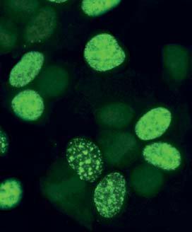
13 Autoantibodies against cell nuclei, DFS pattern (AC-2) On the substrate autoantibodies against the DFS70 antigen (and possibly other antigens) depict a uniformly distributed dense fine speckled fluorescence with granular staining of the condensed chromosomes. Known target antigen: DFS70. Clinical association: Autoantibodies against DFS70 have been found in patients with different diseases (amongst others atopic dermatitis, asthma and interstitial cystitis) and in healthy blood donors. Due to their low prevalence in systemic autoimmune rheumatic diseases it had been discussed whether the detection of these autoantibodies can be used as an exclusion criterion. It has recently been shown, however, that anti-dfs70 antibodies also occur in autoimmune rheumatic diseases with a prevalence of up to 11 %. The clinical association remains unclear. 13
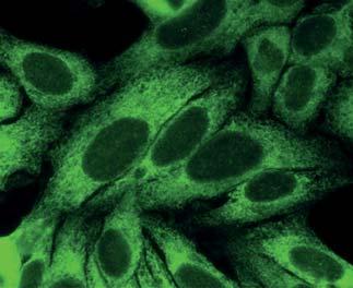
 or in two parallel ribbons")

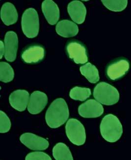
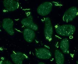
14 Autoantibodies against centromeres (AC-3) show a very specific fluorescence pattern, which is characterised by fine, evenly sized granules (generally 46 or 92 centromeres per cell nucleus). The granules in interphase cells are spread evenly over the nucleus, while in mitotic cells they are arranged either ribbon-like on the equatorial plane (metaphase) or in two parallel ribbons approaching the centrioles (anaphase). On tissue sections of primate liver 10 to 20 granules, which are spread over the cell nucleus, can be seen. The fluorescence of these granules is significantly weaker than the HEp-2 cell staining and is therefore easy to miss. Mitotic cells are only rarely detected on liver substrate. Known target antigens: CENP-A and -B. Clinical association: With a high specificity and a prevalence of %, antibodies against centromeres are pathognomonic for the limited form of progressive systemic sclerosis. In the limited form the extremities are favoured and the inner organs less affected. 14


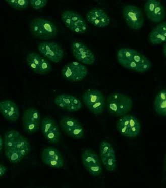
15 Autoantibodies against nucleoplasm, fine speckled (AC-4) show a fine speckled fluorescence of the cell nuclei in the interphase. The nucleoli are also reactive, but they are slightly silhouetted against the nucleoplasm. In some samples they do not react at all. Mitotic cells show a speckled fluorescence, with the chromosomes excluded. On tissue sections of primate liver there is no speckled reaction in the hepatocyte nuclei, but the nucleoli show a smooth fluorescence in samples with a high antibody titer. Known target antigens: SS-A and SS-B. Clinical association: Sjögren s syndrome, SLE and neonatal LE. 15


16 Autoantibodies against Ku (AC-4) In the indirect immunofluorescence test with, antibodies against Ku exhibit a fine speckled fluorescence of the cell nuclei and the nucleoli are positive in parts. There is hardly any difference noticeable to antibodies against SS-A, SS-B, Sm and RNP. However, if primate liver sections are incubated in parallel, possibly in the same field, a typical clumpy-speckled staining of the cell nuclei is found, which is an almost certain proof of antibodies to Ku. Clinical association: Autoantibodies against Ku occur with the following prevalences: % in overlap syndrome of poly-/dermatomyositis and progressive systemic sclerosis (often accompanied by primary pulmonary hypertension), 5 10 % in various forms of myositis, 10 % in systemic lupus erythematosus and up to 5 % in progressive systemic sclerosis. 16


17 Autoantibodies against Mi-2 (AC-4) Autoantibodies to Mi-2 show a fine-speckled fluorescence of the cell nuclei in the indirect immunofluorescence test with. The nucleoli are partly unaffected. With primate liver, autoantibodies against Mi-2 depict a fine speckled fluorescence of the hepatocyte nuclei. Clinical association: Antibodies against Mi-2 are highly specific markers for dermatomyositis with nail fold hypertrophy. They are found in 5 30 % of patients with dermatomyositis and in 8 12 % of patients with idiopathic myositis. 17


18 Autoantibodies against TIF1-gamma (AC-4) TIF1-gamma-transfected cells Autoantibodies against TIF1-gamma cause a fine speckled fluorescence on, which is distributed over the whole cell nucleus but leaves the nucleoli free. Mitotic cells also exhibit a fine speckled fluorescence, but the chromosomes are spared. Antibodies against TIF1-gamma react with the transfected cells of the test substrate. They produce a smooth to fine speckled fluorescence in the cytoplasm. The cell nuclei are generally only slightly stained. Clinical association: Antibodies against TIF1-gamma can be detected with a prevalence of 5 % in patients with dermatomyositis. In particular, they are specific for cancer-associated (paraneoplastic) (dermato)myositis (CAM). 18


19 Autoantibodies against nucleoplasm, coarse speckled (AC-5) generally show a coarse speckled, sometimes medium to fine speckled fluorescence, which is spread over the entire cell nucleus, leaving the nucleoli free. In mitotic cells the condensed chromosomes are dark, while the periphery shows an almost homogeneous, smooth fluorescence. Tissue sections of primate liver also show a speckled fluorescence. The nucleoli do not react. The antibodies react with primate liver to the same extent as with. Known target antigens: hnrnp, U1-nRNP, Sm and RNA polymerase III. Clinical association: SLE and mixed connective tissue disease. 19


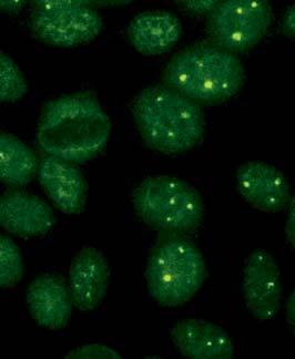
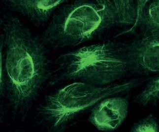
20 Autoantibodies against nuclear dots (AC-6) In immunofluorescence using, 6 20 differently sized granules which are spread over the cell nucleus (nuclear dots) can be seen in the nuclei during interphase. The cytoplasm is dark if antibodies against mitochondria, which are associated with primary biliary cholangitis, are not present at the same time. In mitotic cells the nuclear dots are dissolved. Outside the (unstained) chromosomes only isolated granules fluoresce. Antibodies against nuclear dots react with primate liver to the same extent as with. If both substrates are used in parallel, these antibodies can even be identified if antibodies against centromeres are present at the same time. This can occasionally be observed in cases of primary biliary cholangitis. Known target antigens: Sp100, Sp140, PML, SUMO and MJ/NXP-2. Clinical association: Autoantibodies against nuclear dots occur in % of patients with primary biliary cholangitis. The pattern is also an indicator for rheumatic diseases. 20
. The metaphase chromatin is usually negative.")

21 Autoantibodies against few nuclear dots (AC-7) In the immunofluorescence on, the pattern few nuclear dots merely shows 1 6 dots per cell nucleus frequently near the nucleoli. In the late S / G2 phase of the cell cycle, the cells show relatively many dots (4 6). The metaphase chromatin is usually negative. These nucleolar dots are denominated Cajal bodies (formerly: coiled bodies). On the primate liver tissue section the dots manifest slightly augmented in comparison with the. The liver may, however, also show a negative reaction. Known target antigens: p80-coilin and SMN (survival of motor neuron). Clinical association: Sjögren s syndrome and SLE. 21


22 Autoantibodies against PM-Scl (AC-8) In the immunofluorescence test with, autoantibodies against PM-Scl exhibit a homogeneous fluorescence of the nucleoli with a simultaneous weaker, fine-speckled reaction of the nucleoplasm. The condensed chromosomes of the mitotic cells are unaffected; a fine, speckled fluorescence is shown outside of the chromosomes. A homogeneous fluorescence of the nucleoli also appears on frozen sections of primate liver, as well as a very weak, fine-speckled to reticular staining of the cell nucleus. Clinical association: PM-Scl antibodies can be detected in % of patients with polymyositis/systemic sclerosis overlap syndrome. Here, the autoantibodies are usually directed against both main antigens: PM-Scl75 and PM- Scl100. If progressive systemic sclerosis is exclusively present, antibodies to PM-Scl75 show a prevalence of 10 %, and antibodies to PM-Scl100 a prevalence of 7 %. Using test systems which detect only anti-pm-scl100, some patients with progressive systemic sclerosis remain unidentified. 22


23 Autoantibodies against U3-nRNP / fibrillarin (AC-9) On interphase cells show a speckled fluorescence of the nucleoli. Mitotic cells show a coronary perichromosomal fluorescence. The substrate primate liver depicts a homogeneous fluorescence of the cell nuclei. Clinical association: Antibodies against fibrillarin have so far been observed only in progressive systemic sclerosis (diffuse form). The prevalence is 5 10 %. 23


24 Autoantibodies against RNA polymerase I (AC-10) show a granular fluorescence of the nucleoli. The nucleoplasm is almost dark. In mitotic cells the region of condensed chromosomes is not stained. Outside of the chromosomes a fine granular to smooth fluorescence can be seen. If autoantibodies against NOR-90 occur in parallel, one to several dots fluoresce on mitotic cells. On primate liver tissue sections, the nucleoli show a positive reaction. Clinical association: Antibodies against RNA polymerase I have so far only been detected in progressive systemic sclerosis (diffuse form). The prevalence amounts to 4 %. 24
.")
25 Autoantibodies against NOR-90 (AC-10) On in the metaphase, one to few little dots fluoresce within the condensed chromosome material. They correspond to the nucleolus organisator (NOR). The cytoplasm of mitotic cells may be weakly positive. Interphase cells show a granular fluorescence of the nuceloli. Clinical association: Progressive systemic sclerosis (diffuse form). 25
 On the interphase cells show a homogeneous fluorescence of the cell nuclei, with the rims of the")

26 Autoantibodies against nucl. membrane (AC-11 / AC-12) On the interphase cells show a homogeneous fluorescence of the cell nuclei, with the rims of the nuclei accentuated. The chromosomes of mitotic cells are dark. On tissue sections of primate liver a characteristic linear fluorescence of the nuclear membrane can be seen. Known target antigens: gp210, lamin A, lamin B and C, and lamin B receptor. Clinical association: Antibodies against nuclear membrane occur in primary biliary cholangitis (PBC). 26


27 Autoantibodies against PCNA (AC-13) Autoantibodies against PCNA show a cell cycle-dependent fluorescence pattern with. Half of the cell nuclei of all interphase cells exhibit a bright, fine speckled basic fluorescence with the nucleoli being unaffected. The same fluorescence pattern is found with the other half, but the intensity is lower by a factor of 10. The area of the condensed chromosomes is not stained in the mitosis; the surrounding area of the chromosomes shows only a weak, fine speckled fluorescence, corresponding to the darker nuclei of the interphase cells in pattern and intensity. The reaction with primate liver is largely negative. Clinical association: PCNA antibodies are specific for SLE. The prevalence, however, is only 3 %. 27

28 Autoantibodies against CENP-F (AC-14) On, autoantibodies against CENP-F show a fine to coarse speckled fluorescence of the cell nuclei. The staining intensity varies strongly, G2-phase nuclei show the strongest fluorescence while G1-phase nuclei react with much weaker intensity or not at all. Apart from this, the mitotic cells fluoresce especially strongly (with the exception of the chromosome region), smooth to fine speckled. The centromeres are exclusively positive in the prometa- and metaphase and then show many small and mat aligned dots. In prometaphase cells, the nuclear membrane is often slightly stained. During ana- and telophase, sometimes an intensive fluorescence of the midbody occurs. The cytoplasm of mitotic cells is diffusely stained. The primate liver does not show any specific reaction. Clinical association: In 50 % of the patients who display antibodies against CENP-F, a malign underlying disease is present. Different tumours must be taken into consideration. 28
. On individual or several bunched fibre structures fluoresce.")

, the exclusion of a combined liver disease (overlap syndrome) and for delimitation of AIH against alcohol-")
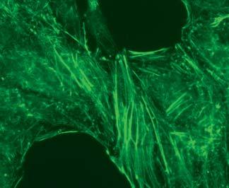
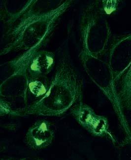
29 Autoantibodies against F-actin (AC-15) VSM47 Autoantibodies against F-actin cause a microfilamentous fluorescence pattern using the cell line VSM47 (vascular smooth muscle). On individual or several bunched fibre structures fluoresce. Theyare located primarily in the cytoplasm, but can also stretch over the cell nuclei. On tissue sections of primate liver there is a strong reaction of the bile canaliculi. Clinical association: The determination of autoantibodies against F-actin is of particular significance for the diagnosis of AIH (prevalence around %), the exclusion of a combined liver disease (overlap syndrome) and for delimitation of AIH against alcohol- or drug-induced cirrhosis and other forms of chronic liver inflammation, such as virus-induced hepatitis and primary sclerosing cholangitis (PBC). 29


30 Autoantibodies against tropomyosin (AC-16) On, autoantibodies against tropomyosin cause a pattern of fibre slings. The primate liver shows a fibrillar pattern in the parenchyma. Clinical association: Myastenia gravis and ulcerative colitis, in rare cases Crohn s disease. 30


31 Autoantibodies against vimentin (AC-16) Antibodies against vimentin cause staining of a fine net of fibres in the cytoplasm of. The net is particularly dense near the cell nuclei. In mitotic cells numerous round fluorescing droplets can be seen outside the dark chromosomes. These are probably condensed vimentin. On tissue sections of primate liver there is an unspecific fluorescence. Clinical association: The diagnostic relevance of autoantibodies against vimentan remains as unclear as that of the much rarer autoantibodies against cytokeratin, tropomyosin, vinculin, etc. They are considered to be associated with different inflammatory reactions and infections. 31


32 Autoantibodies against vinculin (AC-17) Rat kidney In the cytoplasm of, antibodies against vinculin lead to an increased staining of short sections regularly spread along the stress fibers of the cytoskeleton. The primate liver shows a fluorescence of the basal boundary surface of endothelium and stroma in the sinusoids. The glomeruli and tubuli of rat kidney show a filamentous fluorescence. Clinical association: Autoantibodies agianst vinculin are very rare and are associated with Myasthenia gravis, ulcerative colitis and Crohn s disease. 32

.")
33 Autoantibodies against lysosomes (AC-18) On antibodies against lysosomes show a fine to medium or coarse droplet-shaped fluorescence of the cytoplasm. On frozen tissue sections of primate liver there is an unspecific fluorescence. Known target antigens: GWB proteins (e.g. GW182, Su / Ago2). Clinical association: PBC and neurological diseases. Autoantibodies against lysosomes are sometimes also detected in healthy persons. 33


34 Autoantibodies against PL-7 and PL-12 (AC-19) Autoantibodies against PL-7 and PL-12 show a fine speckled to homogenous cytoplasmic fluorescence with. The cell nuclei also show distinct clear dots in many cases. According to recent findings, these enzymes are not solely localised in the cytoplasm, but are also found in the cell nucleus in some species. On frozen tissue sections of primate liver there is an unspecific fluorescence. Clinical association: Antibodies against PL-7 and PL-12 occur in myositis with a prevalence of up to 4 %. 34


35 Autoantibodies against ribosomal P proteins (AC-19) Autoantibodies against ribosomal P proteins cause a smooth to fine speckled staining of the cytoplasm when using as the substrate. Hepatocytes of the primate liver show a cytoplasmic fluorescence of the entire surface with patchy accentuation. There is no reaction with low-titer samples. Clinical association: Autoantibodies against ribosomal P proteins are a characteristic marker for SLE. The prevalence is around 10 %. 35


36 Autoantibodies against SRP (AC-19) Autoantibodies against SRP produce a mainly cytoplasmic, smooth to fine speckled fluorescence on. In mitotic cells the fluorescence is perichromosomally intensified, the chromosomes are unaffected. Hepatocytes of the primate liver generally show a fine speckled fluorescence distributed over the whole organ. Clinical association: Antibodies against SRP can be found in polymyositis and dermatomyositis in approx. 5 % of cases. They are also markers for necrotising myopathy, an autoimmune myopathy that differs from polymyositis, but can manifest with skin changes typical for dermatomyositis. 36


37 Autoantibodies against Jo-1 (AC-20) Antibodies against Jo-1 show a fine speckled to homogenous cytoplasmic fluorescence on. The cell nuclei also show distinct sharp dots in many cases. According to recent findings, these enzymes are not solely localised in the cytoplasm, but are also found in the cell nucleus in some species. On frozen tissue sections of primate liver the cytoplasm is only slightly stained. The fluorescence cannot be used for diagnostics. Clinical association: Antibodies against Jo-1 can be detected in polymyositis with a prevalence of %. They are often associated with other concurrent autoimmune diseases such as SLE, systemic sclerosis, interstitial lung fibrosis, Raynaud s syndrome and polysynovitis. 37
. The primate liver shows a speckled fluorescence of the cytoplasm.")

38 Autoantibodies against mitochondria (AC-21) HEp-2-cells contain the antigens M2, M3, M5 and M9; here the antibodies produce a coarse speckled fluorescence of the cytoplasm which does not include the nucleus (previously, the likewise PBC-relevant nuclear dots also reacting were wrongly suspected of being stray mitochondria). The primate liver shows a speckled fluorescence of the cytoplasm. The cell nuclei are dark. The reaction of the tissue is generally weaker than that of HEp-2 cells. Clinical association: Autoantibodies against mitochondria can be detected in various diseases. They often occur together with other autoantibodies, e.g. with autoantibodies against cell nuclei. Antibodies to mitochondria are of particular significance for the diagnosis of primary biliary cholangitis (PBC). The prevalence is up to 95 %. 38


39 Autoantibodies against Golgi apparatus (AC-22) Autoantibodies against Golgi apparatus present in the indirect immunofluorescence on as reticular-granular structures which are in contact with the cell nucleus on one side. In cells which are in the mitosis, the Golgi apparatus is to a large extent dispersed. Here the antibodies show no reaction. On primate liver the cytoplasm of hepatocytes is also stained. Known target antigens: Giantin / macrogolgin, golgin-95 / GM130, golgin-160, golgin-97 and golgin-245. Clinical association: Autoantibodies against Golgi apparatus occur in different autoimmune diseases, particularly in SLE and Sjögren s syndrome and rheumatoid arthritis. Detection of these antibodies has little relevance due to their low disease specificity. 39

40 Cytoplasmic rods and rings (AC-23) Rods and rings are a cytoplasmic pattern on that has been described only recently. These filamentous structures, which are expressed in all stages of the cell cycle, present themselves as rings, rods or loops. Known target antigens: It is assumed that the reaction is directed against the autoantigen inosine monophosphate dehydrogenase 2 (IMPDH2). Clinical association: The depicted pattern was observed mainly in patients with hepatitis C infections, particularly after treatment with interferon-alpha or ribavirin (prevalence 35 %). 40


41 Autoantibodies against centrosomes (AC-24) A typical positive result is characterised by fluorescing centrosomes in the cytoplasm of, namely one or two centrioles per cell. In mitotic cells the centrioles are located at two opposing poles. On primate liver, high-titer samples produce small fluorescing dots in the cytoplasm of hepatocytes. Known target antigens: Pericentrin, ninein, Cep250 and Cep110. Clinical association: A high titer (> 1:1,000) indicates progressive systemic sclerosis or Raynaud s syndrome, the prevalence however, only amounts to a few percent. The pattern was also observed in infections. 41

42 Autoantibodies against spindle fibres (AC-25) Using, antibodies against the spindle fibre antigen MSA-2 (HsEG5) can be detected. In the presence of these antibodies only the spindle fibres of the mitotic cells, but not the cell nuclei of the interphase cells are stained. On tissue sections of primate liver a speckled fluorescence of the cell nuclei can be observed. Known target antigen: HsEG5. Clinical association: Autoantibodies against spindle fibres (MSA-2) occur predominantly in SLE. 42

 may occur, amongst other diseases, in Sjögren s syndrome and different forms of arthritis, sometimes")
43 Autoantikörper gegen NuMA (AC-26) On, antibodies against NuMA (MSA-1) in the interphase show a fine speckled to reticular fluorescence of the nuclear matrix, with the exception of the nucleoli. In mitotic cells in the metaphase, the spindle fibres manifest as two opposing fans. The staining is most intense in the direction of the centrioles. The primate liver shows a granular fluorescence. Clinical association: Antibodies against NuMA (MSA-1) may occur, amongst other diseases, in Sjögren s syndrome and different forms of arthritis, sometimes also in anti-phospholipid syndrome (APS) and in systemic lupus erythematosus (SLE). 43

44 Autoantibodies against midbody (AC-27) In the indirect immunofluorescence test, in the metaphase of mitosis show a fine speckled fluorescence of the equatorial plane in the presence of midbody antibodies. In contrast to the pattern found with antibodies against centromeres, this fluorescing line remains in the middle until the end of mitosis. Their length corresponds to the whole cell width in the separation zone, and the line increasingly shortens until only a fluorescing dot is seen in the telophase, binding the daughter cells together ( goodbye kiss ). Clinical association: Raynaud syndrome, malignoma and progressive systemic sclerosis. 44

45 Autoantibodies against MCA (AC-28) On Hep-2 cells, the chromosomes in the pro- and methaphase show a dotted fluorescence. Interphase nuclei do not present any staining. Known target antigens: Modified histon H3, MCA-1. Clinical association: Polymyalgia rheumatica, discoid lupus erythematosus, Sjögren s syndrome and chronic lymphatic leukaemia. 45


46 Autoantibodies against Topoisomerase I (Scl-70) (AC-29) Rat kidney show a fine granular nuclear fluorescence in interphase cells. The nucleoli are positive, showing a homogeneous fluorescence. The cytoplasm is dark. In mitotic cells the border area of condensed chromosomes fluoresces, sometimes the entire chromosomal region is positive. The liver exhibits a predominantly homogeneous fluorescence of the cell nuclei. On rat kidney tissue sections, the cell nuclei of the tubular epithelium fluoresce. Clinical association: Topoisomerase I antibodies are detected in 25 75% of patients with progessive systemic sclerosis (diffuse form), depending on the analysis method and the activity of the disease. 46
47 Dilution scheme for immunofluorescence Titration of serum samples in steps from 1:10 or 1:3.2 (square root of 10). is recommended for all EUROIMMUN immunofluorescence test systems. The individual dilutions can be easily set up without the need for numerical acrobatics (1:10, 1:32, 1:100, 1:320, 1:1,000, etc.). Previously the precision was exaggerated by using quadratic dilution steps. On the other hand, titrating by a factor of 4 results in too rough a framework. For every test parameter there is a suitable starting dilution. In order to simplify the test procedure and evaluation of results, two antibody cate go ri es are differentiated at EUROIMMUN: Anti bodies of group I are already diagnostically relevant at at titer of 1:10, while those of group II first at 1:100. The titers determined for each sample are classified using the symbols (+) to The differing clinical significance of antibody titers for the two groups is already incorporated into this scheme. Serum dilution Evaluation, group I Evaluation, group II 1:10 1:32 1:100 1:320 1:1,000 1:3,200 1:10, = suitable starting dilutions + = weak positive, ++ = positive, +++ = strong positive, ++++ = very strong positive Group I: most organ-specific autoantibodies (AAb), ANCA, AAb against dsdna Group II: ANA, AMA, ASMA, AAb against skeletal muscle In order to make optimal use of the great potential of indirect immunofluorescence, experts in this area always test for the majority of autoantibodies using two dilutions, for the following reasons: 47


 1:10 1:100")


48 Blocking effect: In two out of every 100 high-titer sera an untypical result is seen with the starting dilution. Some strongly positive sera even react as false negative if they are not sufficiently diluted. 1:100 1:1,000 AMA (kidney) 1:10 1:100 Anti-Yo (cerebellum) 1:10 1:100 Anti-epid. basal membrane (tongue) 1:1 1:10 panca (granulocytes) Autoantibody masking: If unspecific antibodies or additional, visually dominant autoantibodies are present in too high a concentration, they can mask a relevant antibody. 1:100 1:1,000 1:100 1:1,000 Anti-ribosomes and anti-nucleoli ANA, homog. and anti-centromeres 48
.")


49 Titer estimation: When two dilutions with an interval of a factor of 10 are used, a titer can be determined for most positive results without further incubations (see ta ble). Results are obtained one day earlier than with stepwise titrations. Fluorescence at 1:100 1:1,000 AAb titer weak negative 1:100 strong negative 1:320 strong weak 1:1,000 strong moderate 1:3,200 strong strong 1:10,000 In contrast, it is not possible to quantify a positive result from a single dilution: AAb show very different abating behaviour depending on the avidity. This is detected by the parallel analysis. Photometric systems based on cytochemical ELISA or fluorescence are hence obsolete. 1:100 1:1,000 1:100 1:1,000 ANA, homogeneous (titer 1:1,000) ANA, homogeneous (titer 1:10,000) 49


 for automated image recording.")
50 EUROPattern: Automated evaluation of IIFT EUROPattern Microscope In order to provide diagnostic laboratories with maximum support in ANA diagnostics, EUROIMMUN has developed EUROPattern, a system for automated recording of immunofluorescence images and computer-aided evaluation of a continuously increasing range of substrates. The EUROPattern Microscope can automatically process up to 500 incubation fields in less than 2 hours. For this, the mechanical stage moves into the magazine and picks up one carrier plate with slides, which are securely identified by means of a data matrix code. All fields on the slides are brought into view one by one in a precise manner. The substrates are focussed without causing fading of the fluorescence and high-quality fluorescence images are taken. Besides cell substrates, EUROIMMUN also provides tissues and purified antigens (EUROPLUS) for automated image recording. The fluorescence images are automatically archived and are available for interpretation of the recent and any subsequent analysis. Based on the recorded immunofluorescence images, EUROPattern fully automatically generates a diagnosis suggestion for all kinds of substrates. At present, this includes HEp-2/HEp cells, granulocytes (various fixing meth- 50


51 ods), Crithidia luciliae, EUROPLUS and recombinant cells (e. g. aquaporin-4, PLA2R, DPPX). EUROPattern classifies the fluorescence images into positive, negative or borderline using modern mathematical procedures and identifies the patterns. For each pattern a titer is automatically calculated from the fluorescence intensities of the incubated dilutions, which ensures reproducible quantification. The automatically generated diagnosis suggestion for each patient, including titers and confidence value, is displayed on the screen together with the fluorescence images. The diagnostician can verify the final result with one mouse click, taking into account the detailed patient history. Furthermore, batch processing of negative samples is supported. Thus, EUROPattern ensures a quick and secure processing of IIFT in laboratory diagnostics. EUROPattern is an extension module for the laboratory management software EUROLabOffice. It can be easily integrated into existing work processes and automation solutions. Presentation of results in EUROPattern 51

 CENP-F (cyclin II")
 Histones PCNA (cyclin I) RNA helicase A MSA-1")
 RNA polymerases I, II, III NOR-90,")
 Jo-1 TIF1-gamma MDA5, NXP2,")


 Ribosomal P proteins ASMA")
 Sa Filaggrin, RA keratin Citrullinated enolase")


 Sjögren s syndrome")

52 Autoantibodies against SYSTEMIC AUTOANTIBODIES AND ASSOCIATED AUTOIMMUNE DISEASES Domains of immunofluorescence Diagnostically essential relevant Dated July 2017 Cell nuclei (ANA) CENP-F (cyclin II mitosin) dsdna Nucleosomes Sm U1-RNP SS-A (Ro, 60 kda) SS-B (La) Histones PCNA (cyclin I) RNA helicase A MSA-1 (NuMA) MSA-2 (HsEg5) Topoisomerase I (Scl-70) Fibrillarin (U3-RNP) RNA polymerases I, II, III NOR-90, PDGF-R Centromeres (CENP-A, CENP-B) PM-Scl (1, 75, 100) Ku Mi-2 SRP cn-1a (Mup44) Jo-1 TIF1-gamma MDA5, NXP2, SAE1 PL-7, PL-12, OJ, EJ, SC, KS Nuclear dots Sp100, Sp140, SUMO, PML Nuclear membrane, lamin B recept. GP210, NUP62 Cardiolipin Beta-2 glycoprotein Phosphatidylserine Coagulation factor (lupus anticoag.) Prothrombin, annexin A5 C1q Mitochondria (AMA) Mitochondria (AMA M2-3E / BPO) Ribosomal P proteins ASMA F-actin IgG (rheumatoid factors) Citrullinated peptides (CCP) Sa Filaggrin, RA keratin Citrullinated enolase peptide (CEP-1) Anti-phospholipid syndrome Habitual abortions Drug.-induced lupus erythematosus Neonatal lupus erythematosus Polymyositis / Dermatomyositis Inclusion body myosits Progressive systemic sclerosis Rheumatoid arthritis Sharp syndrome (MCTD) Sjögren s syndrome Systemic lupus erythematosus Autoimmune hepatitis Primary biliary cirrhosis Paraneoplastic autoimmunity EUROIMMUN AG Seekamp Lübeck (Germany) Tel / Fax info@euroimmun.de FA_1510_I_UK_B08, 11 / 2017
ANA Diagnostics Using Indirect Immunofluorescence
 ANA Diagnostics Using Indirect Immunofluorescence EUROIMMUN D-23560 Luebeck (Germany) Seekamp 31 Tel +49 45158550 Fax 5855591 E-mail euroimmun@euroimmun.de Table of Contents Autoantibodies against cell
Clinical Laboratory. 14:42:00 SSA-52 (Ro52) (ENA) Antibody, IgG 1 AU/mL [0-40] Oct-18
![Clinical Laboratory. 14:42:00 SSA-52 (Ro52) (ENA) Antibody, IgG 1 AU/mL [0-40] Oct-18](/thumbs/95/125595183.jpg "Clinical Laboratory. 14:42:00 SSA-52 (Ro52) (ENA) Antibody, IgG 1 AU/mL [0-40] Oct-18") Clinical Laboratory Procedure Result Units Ref Interval Accession Collected Received Rheumatoid Factor
Clinical Laboratory Procedure Result Units Ref Interval Accession Collected Received Rheumatoid Factor
Clinical Laboratory. [None
 Clinical Laboratory Procedure Result Units Ref Interval Accession Collected Received Double-Stranded DNA (dsdna) Ab IgG ELISA Detected * [None 18-289-900151 Detected] Double-Stranded DNA (dsdna) Ab IgG
Clinical Laboratory. 14:41:00 Complement Component 3 50 mg/dl Oct-18
 Clinical Laboratory Procedure Result Units Ref Interval Accession Collected Received Thyroid Peroxidase (TPO) Antibody 5.0 IU/mL [0.0-9.0] 18-289-900139 16-Oct-18 Complement Component 3 50 mg/dl 18-289-900139
Autoantibodies panel ANA
 Autoantibodies panel ANA Anti-nuclear antibodies, ANA screening General: Anti-nuclear antibodies (ANA) contain all kinds of autoantibodies against nuclear antigens. Their targets are cell components in
Autoantibodies giving rise to cytoplasmic IIF staining using HEp-2 cell substrate
 Autoantibodies giving rise to cytoplasmic IIF staining using HEp-2 cell substrate Some associations of anticytoplasmic antibodies with clinical diagnoses and features HEp-2 IIF: the gold standard for ANA
Budsakorn Darawankul, MD. Maharat Nakhon Ratchasima Hospital
 Budsakorn Darawankul, MD. Maharat Nakhon Ratchasima Hospital Outline What is ANA? How to detect ANA? Clinical application Common autoantibody in ANA diseases Outline What is ANA? How to detect ANA? Clinical
Atlas of Antinuclear Antibodies
 Fluorescence patterns of cytoplasmic autoantigens There are many important organelles in the cytoplasm which fulfill various functions of the cell as described in section 1. Various autoantibodies, known
Autoantibodies in the Idiopathic Inflammatory Myopathies
 Autoantibodies in the Idiopathic Inflammatory Myopathies Steven R. Ytterberg, M.D. Division of Rheumatology Mayo Clinic Rochester, MN The Myositis Association Annual Conference St. Louis, MO Sept. 25,
Is it Autoimmune or NOT! Presented to AONP! October 2015!
 Is it Autoimmune or NOT! Presented to AONP! October 2015! Four main jobs of immune system Detects Contains and eliminates Self regulates Protects Innate Immune System! Epithelial cells, phagocytic cells
Test Name Results Units Bio. Ref. Interval
 LL - LL-ROHINI (NATIONAL REFERENCE 135091593 Age 25 Years Gender Male 30/8/2017 91600AM 30/8/2017 93946AM 31/8/2017 84826AM Ref By Final COLLAGEN DISEASES ANTIBODY ANEL ANTI NUCLEAR ANTIBODY / FACTOR (ANA/ANF),
This month, we are very pleased to introduce some new tests for Scleroderma as well as some test changes to our existing scleroderma tests/panels.
 February 20, 2017 Client Letter Test Update February 2017 Dear Colleague: This month, we are very pleased to introduce some new tests for Scleroderma as well as some test changes to our existing scleroderma
Assays. New. New. Combinations. Possibilities. Patents: EP , AU
 Assays Patents: EP 2362222, AU 2011217190 New Combinations New Possibilities Technology Classical Handling of Autoimmune Diagnostics 2-Step Diagnostics 1 st Screening 2 nd Confirmation Cell based IFA ELISA
University of Pretoria
 University of Pretoria Serodiagnostic Procedures Performed in the Department of Immunology Dr Pieter WA Meyer 1.Autoimmune Diseases Automated Anti-nuclear antibodies Anti-gliadin/ tissue transglutaminase
VASCULITIS PRODUCT HIGHLIGHTS
 VASCULITIS PRODUCT HIGHLIGHTS AESKU.DIAGNOSTICS offers a comprehensive and complete diagnostic portfolio in the field of vasculitis diagnostics. Not only are screening and profiling s available but also
Tools to Aid in the Accurate Diagnosis of. Connective Tissue Disease
 Connective Tissue Disease Tools to Aid in the Accurate Diagnosis of Connective Tissue Disease Connective Tissue Disease High quality assays and novel tests Inova offers a complete array of assay methods,
Comparison of indirect immunofluorescence and line immunoassay for autoantibody detection
 Comparison of indirect immunofluorescence and line immunoassay for autoantibody detection Y.L. Jeon, M.H. Kim, W.I. Lee, S.Y. Kang Department of Laboratory Medicine, KyungHee University School of Medicine,
Autoimmune diagnostics. A comprehensive product line for the detection of autoantibodies
 Autoimmune diagnostics A comprehensive product line for the detection of autoantibodies Autoimmune diagnostics Autoimmune diseases are chronic inflammatory processes with an indeterminate etiology. They
What will we discuss today?
 Autoimmune diseases What will we discuss today? Introduction to autoimmune diseases Some examples Introduction to autoimmune diseases Chronic Sometimes relapsing Progressive damage Epitope spreading more
Test Name Results Units Bio. Ref. Interval
 135091660 Age 44 Years Gender Male 29/8/2017 120000AM 29/8/2017 100219AM 29/8/2017 105510AM Ref By Final EXTRACTABLENUCLEAR ANTIGENS (ENA), QUANTITATIVE ROFILE CENTROMERE ANTIBODY, SERUM 20-30 Weak ositive
The Power of the ANA. April 2018 Emily Littlejohn, DO MPH
 Emergent Rheumatologic Diseases and Disorders for Primary Care. The Power of the ANA April 2018 Emily Littlejohn, DO MPH Question 1: the ANA test is: A) A screening test with high specificity to diagnose
What your autoantibodies tell us about your disease. Mark Gourley, MD
 What your autoantibodies tell us about your disease Mark Gourley, MD The Importance of the Immune System Defends us against foreign invaders Self (cancer) and Nonself (virus, bacteria, etc.) But, if the
CELL CYCLE INTRODUCTION PART I ANIMAL CELL CYCLE INTERPHASE
 CELL CYCLE INTRODUCTION The nuclei in cells of eukaryotic organisms contain chromosomes with clusters of genes, discrete units of hereditary information consisting of double-stranded DNA. Structural proteins
Pitfalls of doing ANA immunofluorescence Can we define false positive? Can we define false negative? Can results be compared between labs?
 Pitfalls of doing ANA immunofluorescence Can we define false positive? Can we define false negative? Can results be compared between labs? Amsterdam March 2011 Background: HEp-2 IIF Current literature
Rheumatologic Testing in Primary Care
 Rheumatologic Testing in Primary Care Fernando Vega, MD October 4, 2008 To help establish a diagnosis in pt with clinical features suggestive of an autoimmune disorder To exclude such disorders in pt with
APGRU4L1 Chap 12 Extra Reading Cell Cycle and Mitosis
 APGRU4L1 Chap 12 Extra Reading Cell Cycle and Mitosis Dr. Ramesh Biology is the only subject in which multiplication is the same thing as division 2007-2008 The Cell Cycle: Cell Growth, Cell Division 2007-2008
Autoimmune (AI) Disorders
 Disorders") Autoimmune (AI) Disorders Affect up to 50 million people in the U.S. 80 100 types, dozens more suspected #2 cause of chronic illness Women are more likely to be affected than men Symptoms overlap and are
Autoimmune (AI) Disorders Affect up to 50 million people in the U.S. 80 100 types, dozens more suspected #2 cause of chronic illness Women are more likely to be affected than men Symptoms overlap and are
Biology is the only subject in which multiplication is the same thing as division
 Biology is the only subject in which multiplication is the same thing as division The Cell Cycle: Cell Growth, Cell Division 2007-2008 2007-2008 Getting from there to here Going from egg to baby. the original
Biology is the only subject in which multiplication is the same thing as division
 Biology is the only subject in which multiplication is the same thing as division 2007-2008 The Cell Cycle: Cell Growth, Cell Division 2007-2008 Where it all began You started as a cell smaller than a
Autoimmune diseases. SLIDE 3: Introduction to autoimmune diseases Chronic
 SLIDE 3: Introduction to autoimmune diseases Chronic Autoimmune diseases Sometimes relapsing : and remitting. which means that they present as attacks Progressive damage Epitope spreading more and more
Alida R Harahap & Farida Oesman Department of Clinical Pathology Faculty of Medicine, University of Indonesia
 Alida R Harahap & Farida Oesman Department of Clinical Pathology Faculty of Medicine, University of Indonesia Foreign molecules = antigens Immune response Immune system non-specific specific cellular humoral
Chapter 8 The Cell Cycle
 What molecule stores your genetic information or determines everything about you? DNA a nucleic acid How are DNA molecules arranged in the nucleus? As you can see DNA is: Chapter 8 The Cell Cycle 1. Arranged
Detection of serum antinuclear antibodies in lymphoma patients
 Detection of serum antinuclear antibodies in lymphoma patients H.Y. Zou 1 *, X. Gu 2 *, W.Z. Yu 1, Z. Wang 1 and M. Jiao 1 1 Institute of Clinical Medicine, Urumqi General Hospital of Lanzhou Military
Biology is the only subject in which multiplication is the same thing as division
 Biology is the only subject in which multiplication is the same thing as division 2007-2008 The Cell Cycle: Cell Growth, Cell Division 2007-2008 Where it all began You started as a cell smaller than a
Advances in Autoantibody Testing & Clinical Applications
 Advances in Autoantibody Testing & Clinical Applications Marvin J. Fritzler PhD MD Member: IUIS-WHO-AF-CDC Serology Committee Director: Advanced Diagnostics Laboratory University of Calgary Introduction
Biology is the only subject in which multiplication is the same thing as division. AP Biology
 Biology is the only subject in which multiplication is the same thing as division Chapter 12. The Cell Cycle: Cell Growth, Cell Division Where it all began You started as a cell smaller than a period at
Interpreting Rheumatologic Lab Tests
 The black hole of medical knowledge: An internist s view of rheumatologic lab tests Interpreting Rheumatologic Lab Tests Jonathan Graf, M.D. Associate Professor of Clinical Medicine University of California,
IMTEC-ANA-LIA MAXX. Design Verification
 Design Verification IMTEC-ANA-LIA MAXX CONTENTS 1 Intended Use... 2 2 Diagnostic Sensitivity and Specificity... 2 3 Interferences... 4 4 Imprecision... 6 4.1 Within-Run Imprecision... 6 4.2 Between-Run
Cell Theory. Passive Transport
 Cell Theory 4 basic concepts of cell theory are: Cells are the units of structure (building blocks) of all animals and plants. Cells are the smallest unit of function in all animals and plants. Cells originate
Comparison of Performance of ELISA with Indirect Immunofluoresence for the Testing of Antinuclear Antibodies
 International Journal of Current Microbiology and Applied Sciences ISSN: 2319-7706 Volume 5 Number 12 (2016) pp. 423-427 Journal homepage: http://www.ijcmas.com Original Research Article http://dx.doi.org/10.20546/ijcmas.2016.512.046
Disclosures. Rheumatological Approaches to Differential Diagnosis, Physical Examination, and Interpretation of Studies. None
 Rheumatological Approaches to Differential Diagnosis, Physical Examination, and Interpretation of Studies Sarah Goglin MD Assistant Professor of Medicine Division of Rheumatology Disclosures None 1 [footer
Biology is the only subject in which multiplication is the same thing as division
 Biology is the only subject in which multiplication is the same thing as division 2007-2008 The Cell Cycle: Cell Growth, Cell Division 2007-2008 Getting from there to here Going from egg to baby. the original
Cells and Tissues. Lesson 2.1: Molecules of Life Lesson 2.2: Cells Lesson 2.3: Tissues
 2 Cells and Tissues Lesson 2.1: Molecules of Life Lesson 2.2: Cells Lesson 2.3: Tissues Chapter 2: Cells and Tissues Lesson 2.1 Molecules of Life Molecules of Life carbohydrates proteins lipids nucleic
Test Name Results Units Bio. Ref. Interval
 135091662 Age 45 Years Gender Male 29/8/2017 120000AM 29/8/2017 100215AM 29/8/2017 110825AM Ref By Final RHEUMATOID AUTOIMMUNE COMREHENSIVE ANEL ANTI NUCLEAR ANTIBODY / FACTOR (ANA/ANF), SERUM ----- 20-60
The Cell Cycle. Packet #9. Thursday, August 20, 2015
 1 The Cell Cycle Packet #9 2 Introduction Cell Cycle An ordered sequence of events in the life of a dividing eukaryotic cell and is a cellular asexual reproduction. The contents of the parent s cell nucleus
Screening of Auto Antibodies using Indirect Immunofluorescence in Auto Immune Disease Patients
 International Journal of Current Microbiology and Applied Sciences ISSN: 2319-7706 Volume 7 Number 02 (2018) Journal homepage: http://www.ijcmas.com Original Research Article https://doi.org/10.20546/ijcmas.2018.702.386
Chapter 12 The Cell Cycle: Cell Growth, Cell Division
 Chapter 12 The Cell Cycle: Cell Growth, Cell Division 2007-2008 Where it all began You started as a cell smaller than a period at the end of a sentence And now look at you How did you get from there to
BIOLOGY - CLUTCH CH.12 - CELL DIVISION.
 !! www.clutchprep.com CONCEPT: CELL DIVISION Cell division is the process by which one cell splits into two or more daughter cells. Cell division generally requires that cells produce enough materials,
CELL CYCLE INTRODUCTION PART I ANIMAL CELL CYCLE INTERPHASE EVOLUTION/HEREDITY UNIT. Activity #3
 AP BIOLOGY EVOLUTION/HEREDITY UNIT Unit 1 Part 3 Chapter 12 Activity #3 INTRODUCTION CELL CYCLE NAME DATE PERIOD The nuclei in cells of eukaryotic organisms contain chromosomes with clusters of genes,
Ch. 3 CELLS AND TISSUES. Copyright 2010 Pearson Education, Inc.
 Ch. 3 CELLS AND TISSUES Generalized Cell All cells: Human cells have three basic parts: Plasma membrane flexible outer boundary Cytoplasm intracellular fluid containing organelles Nucleus control center
Genetics and Cellular Function
 Genetics and Cellular Function DNA replication and the cell cycle Mitosis Mitosis Mitosis: division of cells that results in daughter cells with the same the genetic information that the original cell
SHORT ANSWER. Write the word or phrase that best completes each statement or answers the question.
 SHORT ANSWER. Write the word or phrase that best completes each statement or answers the question. Figure 2.1 Using Figure 2.1, match the following: 1) Rough endoplasmic reticulum 1) 2) Nucleolus 2) 3)
Mitosis. AND Cell DiVISION
 Mitosis AND Cell DiVISION Cell Division Characteristic of living things: ability to reproduce their own kind. Cell division purpose: When unicellular organisms such as amoeba divide to form offspring reproduction
Why do cells divide? Cells divide in order to make more cells they multiply in order to create a larger surface to volume ratio!!!
 Why do cells divide? Cells divide in order to make more cells they multiply in order to create a larger surface to volume ratio!!! Chromosomes Are made of chromatin: a mass of genetic material composed
Autoantibodies in the Diagnosis of Systemic Rheumatic Diseases
 I Autoantibodies in the Diagnosis of Systemic Rheumatic Diseases Carlos A, von Mfihlen and Eng M. Tan Distinct profiles of autoantibodies directed to intracellular antigens can be detected in the systemic
We also assessed the diagnostic significance of the SUBJECTS
 Annals of the Rheumatic Diseases, 1982, 41, 382-387 Antinuclear antibodies in patients with Raynaud's phenomenon: clinical significance of anticentromere antibodies C. G. M. KALLENBERG, G. W. PASTOOR,
INTERPRETATION OF LABORATORY TESTS IN RHEUMATIC DISEASE
 INTERPRETATION OF LABORATORY TESTS IN RHEUMATIC DISEASE Laboratory tests are an important adjunct in the clinical diagnosis of rheumatic diseases and are sometimes helpful in monitoring the activity of
8.4 The cell cycle multiplies cells. 8.4 The cell cycle multiplies cells
 8.4 The cell cycle multiplies cells! Cell division is a highly orchestrated process! The cell cycle is an ordered sequence of events that extends from the time a cell is first formed from a dividing parent
Cell Division. Learning Objectives: Introduction. Revised Fall 2018
 Revised Fall 2018 Cell Division Learning Objectives: 1. Define cell cycle and the ordered sequence of events in the cell cycle (Interphase and The divisional phase or M phase) 2. Explain the stages in
Overview of Diagnostic Autoantibodies in Inflammatory Myopathy
 Overview of Diagnostic Autoantibodies in Inflammatory Myopathy Minoru Satoh, M.D., Ph.D. Research Associate Professor of Medicine Division of Rheumatology and Clinical Immunology University of Florida
Molecular Cell Biology - Problem Drill 22: The Mechanics of Cell Division
 Molecular Cell Biology - Problem Drill 22: The Mechanics of Cell Division Question No. 1 of 10 1. Which of the following statements about mitosis is correct? Question #1 (A) Mitosis involves the dividing
Measurement of Antinuclear Antibodies: Assessment of Different Test Systems
 CLINICAL AND DIAGNOSTIC LABORATORY IMMUNOLOGY, Jan. 2000, p. 72 78 Vol. 7, No. 1 1071-412X/00/$04.00 0 Copyright 2000, American Society for Microbiology. All Rights Reserved. Measurement of Antinuclear
The Process of Cell Division
 Lesson Overview 10.2 The Process of Cell Division THINK ABOUT IT What role does cell division play in your life? Does cell division stop when you are finished growing? Chromosomes What is the role of chromosomes
The Cell Cycle MITOSIS
 The Cell Cycle MITOSIS Outcomes 1. Explain the events of the cell cycle Interphase Mitosis Prophase Metaphase Anaphase Telophase Cytokinesis 2. Use a simulation to demonstrate the behaviour of chromosomes
Mitosis Flap Book Excludes Prometaphase
 Mitosis Flap Book Excludes Prometaphase TEACHER S INSTRUCTIONS 1) Choose one of the foldables from the choices below. Three Color Choices Black & White Cells without Chromosomes Choose this option if you
Outline Interphase Mitotic Stage Cell Cycle Control Apoptosis Mitosis Mitosis in Animal Cells Cytokinesis Cancer Prokaryotic Cell Division
 The Cell Cycle and Cellular Reproduction Chapter 9 Outline Interphase Mitotic Stage Cell Cycle Control Apoptosis Mitosis Mitosis in Animal Cells Cytokinesis Cancer Prokaryotic Cell Division 1 2 Interphase
How Cells Divide. Chapter 10
 How Cells Divide Chapter 10 Bacterial Cell Division Bacteria divide by binary fission. -the single, circular bacterial chromosome is replicated -replication begins at the origin of replication and proceeds
Unit 4: Cell Division Guided Notes
 Unit 4: Cell Division Guided Notes 1 Chromosomes are structures that contain material When Eukaryotes are not dividing, DNA and Proteins are in a mass called: When the cell divides, it condenses and becomes
Myositis and Your Lungs
 Myositis and Your Lungs 2013 TMA Annual Patient Meeting Louisville, Kentucky Chester V. Oddis, MD University of Pittsburgh Director, Myositis Center Myositis Heterogeneous group of autoimmune syndromes
IdentRA test panel with eta. A clinically proven biomarker for earlier, accurate RA diagnosis and now, prognosis and monitoring
 IdentRA test panel with 14-3-3eta A clinically proven biomarker for earlier, accurate RA diagnosis and now, prognosis and monitoring Did you know there are more than 100 forms of arthritis? Every type
Autoantibodies andprognosis. Jiří Vencovský Institute of Rheumatology, Prague, Czech Republic
 Autoantibodies andprognosis Jiří Vencovský Institute of Rheumatology, Prague, Czech Republic HeterogeneityofIIMs Diagnosis Polymyositis Dermatomyositis IBM Necrotising myopathy Paraneoplastic Amyopathic
U3.2.3: Eukaryotic chromosomes are linear DNA molecules associated with histone proteins. (Oxford Biology Course Companion page 151).
.") Cell Division Study Guide U3.2.3: Eukaryotic chromosomes are linear DNA molecules associated with histone proteins. (Oxford Biology Course Companion page 151). 1. Describe the structure of eukaryotic DNA
Cell Division Study Guide U3.2.3: Eukaryotic chromosomes are linear DNA molecules associated with histone proteins. (Oxford Biology Course Companion page 151). 1. Describe the structure of eukaryotic DNA
The Cell Cycle. Dr. SARRAY Sameh, Ph.D
 The Cell Cycle Dr. SARRAY Sameh, Ph.D Overview When an organism requires additional cells (either for growth or replacement of lost cells), new cells are produced by cell division (mitosis) Somatic cells
Biology is the only subject in which multiplication is the same thing as division
 Biology is the only subject in which multiplication is the same thing as division 2007-2008 The Cell Cycle: Cell Growth, Cell Division Ch. 10 Where it all began You started as a cell smaller than a period
Chapter 2 Cell. Zhou Li Prof. Dept. of Histology and Embryology
 Chapter 2 Cell Zhou Li Prof. Dept. of Histology and Embryology The inner life of the cell Ⅰ. Plasma membrane (Plasmalemma) 1.1 The structure Unit membrane: inner layer 3-layered structure outer layer mediat
Cellular Reproduction, Part 1: Mitosis Lecture 10 Fall 2008
 Cell Theory 1 Cellular Reproduction, Part 1: Mitosis Lecture 10 Fall 2008 Cell theory: All organisms are made of cells All cells arise from preexisting cells How do new cells arise? Cell division the reproduction
What are Autoantibodies and how do they work in Myositis?
 What are Autoantibodies and how do they work in Myositis? Neil McHugh, University of Bath and Royal National Hospital for Rheumatic Diseases Orlando September 2015 Royal National Hospital for Rheumatic
NATIONAL LABORATORY HANDBOOK. Laboratory Testing for Antinuclear antibodies
 NATIONAL LABORATORY HANDBOOK Laboratory Testing for Antinuclear antibodies Document reference number CSPD013/2018 Document developed by National Clinical Programme for Pathology Revision number Version
Supplementary Figure Legends
 Supplementary Figure Legends Supplementary Figure 1. Comparison of RNP IC-mediated NET formation. Quantification of DNA release induced by ICs consisting of SmRNP combined with SLE IgG 961 (n = 10), 1032
Fig. 1. Simple columnar epithelial cells lining the small intestine.
 Mitosis Prelab Reading Fig. 1. Simple columnar epithelial cells lining the small intestine. The tall cells pictured in Fig. 1 form the lining of the small intestine in humans and other animals. These cells
The Cell Cycle CHAPTER 12
 The Cell Cycle CHAPTER 12 The Key Roles of Cell Division cell division = reproduction of cells All cells come from pre-exisiting cells Omnis cellula e cellula Unicellular organisms division of 1 cell reproduces
Autoantibody Standardizing Committee
 Autoantibody Standardizing Committee Subcommittee for the IUIS Quality Assessment and Standardization Committee Milan, August 25, 2013 Luís Eduardo Coelho Andrade, M.D., Ph.D. Associate Professor Rheumatology
Breaking Up is Hard to Do (At Least in Eukaryotes) Mitosis
 Mitosis") Breaking Up is Hard to Do (At Least in Eukaryotes) Mitosis Chromosomes Chromosomes were first observed by the German embryologist Walther Fleming in 1882. Chromosome number varies among organisms most
Breaking Up is Hard to Do (At Least in Eukaryotes) Mitosis Chromosomes Chromosomes were first observed by the German embryologist Walther Fleming in 1882. Chromosome number varies among organisms most
-The cell s hereditary endowment of DNA -Usually packaged into chromosomes for manageability
 Binary Fission-Bacterial Cell Division -Asexual reproduction of prokaryotes -No mitosis -Circular DNA and organelles replicate, the copies migrate to opposite sides of the elongating cell, and the cell
Chapter 3: Cells 3-1
 Chapter 3: Cells 3-1 Introduction: A. Human body consists of 75 trillion cells B. About 260 types of cells that vary in shape & size yet have much in common B. Differences in cell shape make different
Mitosis and the Cell Cycle
 Mitosis and the Cell Cycle Chapter 12 The Cell Cycle: Cell Growth & Cell Division Where it all began You started as a cell smaller than a period at the end of a sentence Getting from there to here Cell
Anti-Nuclear Antibodies (ANA). (Incorporating Anti-double stranded DNA (dsdna) and Anti-Extractable Nuclear Antigen (ENA) Antibodies)
. (Incorporating Anti-double stranded DNA (dsdna) and Anti-Extractable Nuclear Antigen (ENA) Antibodies)") Autoimmune Antibody Testing Points of Note: The interpretation of all autoantibody tests is highly dependent on the likelihood of disease in the patient. The results should always be interpreted with the
Autoimmune Antibody Testing Points of Note: The interpretation of all autoantibody tests is highly dependent on the likelihood of disease in the patient. The results should always be interpreted with the
Overview of the Cellular Basis of Life. Copyright 2009 Pearson Education, Inc., publishing as Benjamin Cummings
 Overview of the Cellular Basis of Life Cells and Tissues Cells: Carry out all chemical activities needed to sustain life Cells are the building blocks of all living things Tissues Cells vary in length,
Cell Cycle and Cell Division
 122 Cell Cycle and Cell Division 1. Meiosis I is reductional division. Meiosis II is equational division due to [1988] (a) pairing of homologous chromosomes (b) crossing over (c) separation of chromatids
Rhematologic serum testing is: Before request serologic tests, ANCA associated antigens c-anca: most commonly against the proteinase 3 (PR-3)
") thorough P/E. Rhematologic serum testing is: Useful to confirm a clinical impression or sort out a differential diagnosis Not useful as a screening test A positive test may or may not be associated with
thorough P/E. Rhematologic serum testing is: Useful to confirm a clinical impression or sort out a differential diagnosis Not useful as a screening test A positive test may or may not be associated with
Unduplicated. Chromosomes. Telophase
 10-2 Cell Division The Cell Cycle Interphase Mitosis Prophase Cytokinesis G 1 S G 2 Chromatin in Parent Nucleus & Daughter Cells Chromatin Daughter Nuclei Telophase Mitotic Anaphase Metaphase Use what
Exercise 6. Procedure
 Exercise 6 Procedure Growing of root tips Select a few medium-sized onion bulbs. Carefully remove the dry roots present. Grow root tips by placing the bulbs on glass tubes (of about 3 4 cm. diameter) filled
Goals. Cells. Cells: The Living Units. By the end of this lecture you should be able to describe.
 C H A P T E R 2 Cells: The Living Units Goals By the end of this lecture you should be able to describe. Similarities and differences between cells Why cells look and function differently The function
Insights into the DX of Pediatric SLE
 Insights into the DX of Pediatric SLE Dr. John H. Yost Pediatric Rheumatology Children s Hospital at Dartmouth Assistant Professor of Medicine Geisel School of Medicine at Dartmouth john.h.yost@hitchcock.org
Mitosis: Cell Division
 Do Now: What process do you think this cartoon is describing? Mitosis: Cell Division Key Points On Cell Division Species must reproduce in order to survive from generation to generation. All living things
Part I: The Cell Cycle
 Cellular Differentiation Part I: The Cell Cycle During your lifetime, trillions of your cells will undergo the cell cycle. This process allows you to grow, heal, and maintain your vital tissues and organs.
Chapter 10. Cell Cycle - Mitosis
 Chapter 10 Cell Cycle - Mitosis WHAT CELL REPRODUCTION ACCOMPLISHES Cell division plays important roles in the lives of organisms. Cell division replaces damaged or lost cells permits growth allows for
Bacterial cell. Origin of replication. Septum
 Bacterial cell Bacterial chromosome: Double-stranded DNA Origin of replication Septum 1 2 3 Chromosome Rosettes of Chromatin Loops Scaffold protein Chromatin Loop Solenoid Scaffold protein Chromatin loop
BIOLOGY LTF DIAGNOSTIC TEST CELL CYCLE & MITOSIS
 Biology Multiple Choice 016044 BIOLOGY LTF DIAGNOSTIC TEST CELL CYCLE & MITOSIS TEST CODE: 016044 Directions: Each of the questions or incomplete statements below is followed by five suggested answers
Organization of genetic material in eukaryotes
 Organization of genetic material in eukaryotes biologiemoleculara.usmf.md pass.: bmgu e.usmf.md 1 DNA in eukaryotes Location: In nucleus In mitochondria biologiemoleculara.usmf.md e.usmf.md pass.: bmgu