A Systematic Approach to Diabetic Photo Reading
|
|
|
- Bethany Singleton
- 5 years ago
- Views:
Transcription
1 A Systematic Approach to Diabetic Photo Reading Jacqueline Theis, OD, FAAO Please silence all mobile devices and remove items from chairs so others can sit. Unauthorized recording of this session is prohibited.
2 Disclosure Statement: C. Light Technologies Clinical Research Consultant, NIH SBIR
3 Objectives 1. Understand the pathophysiology of diabetic retinopathy to understand pattern recognition in grading diabetic screening photos 2. Describe the clinical practice guidelines for monitoring versus referral of diabetic retinopathy when it is and is not vision threatening 3. Increase the comfort level of practitioners in reading diabetic photos by reviewing numerous photos of various diabetic retinopathy levels in an interactive format
4 The Statistics 30.3 million US adults have Diabetes (25% don t know they have it) In the last 20 years, the number of adults diagnosed with DM has more than tripled (due to population aging and obesity) Diabetes is the #1 cause of adult-onset blindness in the US Types Type 1 5% of diabetes Cellular-mediated autoimmune destruction of beta cells in the pancreas à severe/absolute insulin deficiency Type 2 95% of diabetes cases Range of disease from insulin resistance with relative insulin deficiency Gestational Prediabetes 84.1 million adults (90% don t know they have it) Centers for Disease Control and Prevention. National Diabetes Statistics Report, Atlanta, GO: Centers for Disease Control and Prevention, US Department of Health and Human Services;2017.
5 Age-adjusted Prevalence of Obesity and Diagnosed Diabetes Among US Adults Obesity (BMI 30 kg/m 2 ) No Data <14.0% 14.0% 17.9% 18.0% 21.9% 22.0% 25.9% > 26.0% Diabetes No Data <4.5% 4.5% 5.9% 6.0% 7.4% 7.5% 8.9% >9.0% CDC s Division of Diabetes Translation. United States Surveillance System available at
6 Number and Percentage of U.S. Population with Diagnosed Diabetes, Percentage with Diabetes Percentage with Diabetes Number with Diabetes Number with Diabetes (Millions) Year 0 CDC s Division of Diabetes Translation. United States Diabetes Surveillance System available at
7 Epidemiology of Diabetic Retinopathy For all adults with diabetes aged % (4.2 million people) in the US have diabetic retinopathy 4.4% (700,000 people) have vision-threatening diabetic retinopathy (VTDR) Projected prevalence by million people in the US will have diabetic retinopathy 1.34 million people with VTDR Klein BE. Overview of epidemiologic studies of diabetic retinopathy. Ophthalmic Epidemiol. 2007;14: Kempen JH, O Colmain BJ, Leske MC, et al. The prevalence of diabetic retinopathy among adults in the United States. Arch Ophthalmol. 2004;122: Zhang X, Saaddine JB, Chou CF, et al. Prevalence of diabetic retinopathy in the United states, JAMA 2010;304:

8 Retinal Anatomy

9 Macular OCT
10 MAC OCT + Retinal Anatomy Photoreceptor synapses Photoreceptor cell bodies Inner Photoreceptor Segment Inner/Outer Photoreceptor Segments Outer Photoreceptor Segments PR interdigitation with RPE

11 Image from Kur J, Newman EA, Chan-Ling T. Cellular and physiological mechanisms underlying blood flow regulation in the retina and choroid in health and disease. Progress in Retinal and Eye Research. 2012;31: Drawngs by Dave Schumick from Anand-Apte and Hollyfield 2009.

12 Image from Kur J, Newman EA, Chan-Ling T. Cellular and physiological mechanisms underlying blood flow regulation in the retina and choroid in health and disease. Progress in Retinal and Eye Research. 2012;31: Drawngs by Dave Schumick from Anand-Apte and Hollyfield 2009.


13 Retinal Vasculature Dot Hemes Flame Hemes Superficial capillary Superficial capillary Deep Capillary Deep Capillary Choroid Choroid
14 Anatomy of retinal capillaries Non-fenestrated endothelial cells Cells linked by tight junctions and adherens junctions = inner Blood Retinal Barrier (ibrb) Protects retina from toxic molecules in blood circulation Pericytes Provide structural support Regulates expression of tight junction proteins Modulates endothelial cell function Glia (Muller cells, astrocytes) Provide metabolic support to neurons Maintain ibrb, needed for retinal homeostasis Immune Cells (microglia) Lechner J, O Leary OE, Stitt AW. The pathology associated with diabetic retinopathy. Vision Research. 139 (2017): 7-14

15 Vascular Complications in Diabetes Atherosclerosisà cardiovascular disease and impaired life expectancy Diabetic nephropathyà endstage renal disease Diabetic retinopathyà blindness Picture from Rask-Madsen C, King GL. Vascular complications of diabetes: mechanisms of injury and protective factors. Cell Metabolism. 2013;17:20-33
16 Pathophysiology of Diabetic Retinopathy High glucose à increased pericyte apoptosis (lose vascular autoregulation) à nutrient/oxygen deprivation to inner retina (poor blood flow) Can occur in retinopathy-free clinical appearance àmicrovascular damage ibrb breakdown à increased inflammation and oxiddationà MORE vascular dysfunction àretinal capillary non-perfusion (vasodegeneration)à MORE retinal ischemia and hypoxiaà Excessive vasopermeability in the retina à retinal thickening/edema Upregulation of VEGFà neovascularization of disc, retina, iris and angle
 à vitreous hemorrhage Repeat vitreous hemorrhage à gliosis and fibrovascular scar formation Fibrous")
17 Pathophysiology of Neovascularization (PDR) Capillary non-perfusion àretinal ischemiaà VEGFà NV NV occurs at the interface between perfused and non-perfused retina NV often grows on the surface of the retina and penetrates the ILM into the vitreous NV vessels are fenestrated (brittle, leaky) à vitreous hemorrhage Repeat vitreous hemorrhage à gliosis and fibrovascular scar formation Fibrous tissue contractionàtractional RD
18 What Causes Visual Loss in Diabetic Retinopathy? Macular edema Ischemic maculopathy Vitreous hemorrhage Retinal detachment Neovascular glaucoma
19 Clinical Findings in Diabetic Retinopathy

20 DR: Microaneurysms (MA) Loss of pericytes and/or glial attachments on capillary blood vessel wall à ballooning of capillary vessel wall Red dot <125um (1/12 the diameter of an average optic disc OR approx. the width of an average major vein at the disc margin) and has sharp margins ETDRS#10: Grading diabetic retinopathy from stereoscopic color fundus photographs an extension of the modified Airlie House Classification. Ophthalmol. 1991;98:
 Blot Hemorrhages Leakage of fluid from clusters of occluded capillaries Often occur with cotton wool")
21 DR: Hemorrhages Any spot larger than a MA Red spot of MA size with irregular margins Flame hemorrhages Leakage of fluid from MA in superficial capillary bed (localized edema) Dot hemorrhages Leakage of fluid from MA in deep capillary bed (localized edema) Blot Hemorrhages Leakage of fluid from clusters of occluded capillaries Often occur with cotton wool spots

22 DR: Cotton Wool Spots Disturbed axoplasmic flow due to an obstruction of neuronal blood flow (infarction) causes local death of the tissue and a localized superficial swelling in the retinal nerve fiber layer Round or oval, white, pale yellow-white or greyish-white, with feathery edges, frequently with striations parallel to the nerve fibers

23 DR: Intraretinal Microvascular Anomalies (IRMA) Local ischemia leads to capillary/ vascular remodeling causing large tortuous vessels NOT NEW VESSELS Anomaly of innate vasculature
 Localized")

24 Venous Abnormalities Venous Beading (VB) Localized increases in venous caliber caused by ischemia Venous Narrowing Venous Loops

25 DR: Hard Exudates (HE) Hard lipoprotein deposits that remain when blood gets reabsorbed in the retina Small white or yellowish-white deposits with sharp margins. Often have a slightly waxy or glistening appearance Usually located in the outer retina, may be more superficial
26 Drusen vs Hard Exudate Drusen Deposits at or anterior to Bruch s membrane associated with thinning or hypopigmentation of the overlying RPE Appear as deep, yellowish-white dots, sometimes circumscribed by a thin line of pigment Location Drusen Deep in the retina at level of RPE Scattered diffusely or clustered near the macula Hard Exudates Superficial, in outer or middle layers of the retina Near MA or edge of retinal edema Shape Round Irregular Appearance Dull Waxy, shiny Halo/border of pigment

27 DR: Neovascularization New vessels grown in response to ischemia/vegf NVD- neovascularization on or within 1DD of optic disc margin NVE new vessels on the surface of the retina or in the vitreous cavity NVI new vessels on the iris NVA new vessels on the angle

28 PDR + Fibrous proliferation Fibrous tissue strands or sheets of thickened posterior hyaloid surface FPE (elsewhere) or FPD (disc) Preretinal hemorrhage Boat-shaped hemorrhage and/or round, oval, or linear patches of hemorrhage just anterior to the retina or under its internal limiting member Vitreous hemorrhage Hemorrhage further forward in the vitreous cavity than PRH Retinal elevation Retinal detachment, retinoschisis, or elevation of large retinal vessel Prior laser scars Once PDR, always PDR Diabetic papillitis
29 Clinically Significant Macular Edema (CSME) Retinal thickening that involves or threatens the center of the macula (even if visual acuity is not yet reduced) According to ETDRS Thickening of the retina at or within 500um of the center of the macula (1DD) Hard exudates at or within 500um of the center of the macula, if associated with thickening of adjacent retina (not residual hard exudates remaining after disappearance of retinal thickening) A zone or zones of retinal thickening 1 disc area or larger, and part of which is within 1 DD of the center of the macula Defined because CSME responds to treatment significantly, non- CSME does not Keep in mind: ETDRS was published before OCT was around
30 International Clinical Disease Severity Scales Diabetic Retinopathy Disease Severity Scale Disease Severity Level No DR Mild NPDR Moderate NPDR Severe NPDR PDR Findings No abnormalities MAs only More than mild, less than severe. MAs + Hemes/CWS/IRMA<8A/ VB<6A 4:2:1 Rule (any of): Hemes>2A in 4 quadrants VB> 6A in 2 quadrants IRMA>8A in 1 quadrant One or more of the following: NV, vitreous/preretinal heme, tractional RD Diabetic Macular Edema Disease Severity Scale Disease Severity Level Mild DME Moderate DME Severe DME Findings Retinal thickening or HE in posterior pole but distant from center of the macula Retinal thickening or HE approaching the center of macula but not involving the center Retinal thickening or HE involving the center of the macula Wilkinson CP, Ferris FL, Klein RE, Lee PP, Agardh CD, Davis M, Dills D, Kampik A, Pararajasegaram R, Verdaguer JT. Proposed international clinical diabetic retinopathy and diabetic macular edema disease severity scales. Ophthalmol. 2003:110:

31 Standard Photograph 2A

32 Standard Photograph 8A

33 Standard Photograph 6B
34 Ophthalmological Treatment of DR Treatment Modality Indications Evidence Anti-VEGF injections Center-involving DME PDR Diabetic Retinopathy Clinical Research Network (DRCR.net) Ranibizumab for Edema of the Macula in Diabetes Study (READ-2) Study of Ranibizumab Injection in Subjects with CSDME with Central Involvement Secondary to Diabetes Mellitus (RISE and RIDE) Bevacizumab or Laser Therapy (BOLT) Study DME and VEGF-Trap-Eye: Investigation of clinical impact (DA VINCI) Study Focal laser photocoagulation Panretinal photocoagulation Vitrectomy Non-center involving DME PDR -Severe PDR -Severe vitreous hemorrhage -Tractional RD Early Treatment of Diabetic Retinopathy Study (ETDRS) Diabetic Retinopathy Study (DRS) Diabetic Retinopathy Vitrectomy Study (DRVS)
35 Optometric Treatment of DR Communicate findings and level of retinopathy to PCP Intensive treatment of glucose (Hba1c,6.0%) delays onset and slows progression of DR (but does not prevent DR completely) DCCT After 6 years Intensive blood glucose control slowed microvascular complications of Retinopathy by 76% (if no retinopathy present at baseline) Retinopathy by 54% (if some retinopathy at baseline) Neuropathy by 60% Nephropathy by 50% Takes at least 36 months for significant difference Normoglycemic re-entry (slight worsening of retinopathy) for first 18 months possible Intensive blood pressure control reduces the risk of DR by 34% - UKPDS Once retinopathy is present, glycemic control is best predictor of progression od retinopathy Screen/diagnose level of diabetic retinopathy to refer for prompt ophthalmological treatment
36 Risk Factors for DR/Progression of DR Duration of DM Severity of hyperglycemia Hypertension Current Smoker
37 Screening Guidelines for Non-Proliferative Diabetic Retinopathy Level of NPDR No NPDR Type 1 No NPDR Type 2 Mild NPDR Moderate NPDR no DME Moderate NPDR with DME but not CSME Severe NPDR Follow Up Annually beginning 5 years after onset of the disease Prompt exam at diagnosis then every 1-3 years after Every 1-3 years Every 6 months Every 3-4 months Every 3 months Gestational DM DM1/2 that becomes pregnant Do not require eye exam during pregnancy Do not appear to be at an increased risk of developing DR during pregnancy Exam early in the course of pregnancy The DCCT/EDIC Research Group. Frequency of evidence-based screening for retinopathy in type 1 diabetes. N Engl J Med. 2017;676(16): AAO 2014 preferred practice pattern

38

39
40 Pearl 1: You need to Zoom You need good screen resolution You may need to turn off your room lights
41 Case A - OD 56yo DM2 x 10 years HbA1c 7.3 Full View

42 Case A - OD 56yo DM2 x 10 years HbA1c 7.3 Zoom ONH +/- NVD +/-NVE +/-Other optic neuropathy

43 Case A - OD 56yo DM2 x 10 years HbA1c 7.3 Zoom Macula +/-MA or Hemes +/- HEX +/- CWS +/- thickening +/- irma +/- VB +/-NVE

44 Case A - OD 56yo DM2 x 10 years HbA1c 7.3 Zoom Sup. Arcades +/-MA or Hemes +/- HEX +/- CWS +/- thickening +/- irma +/- VB +/-NVE

45 Case A - OD 56yo DM2 x 10 years HbA1c 7.3 Zoom Sup. Arcades +/-MA or Hemes +/- HEX +/- CWS +/- thickening +/- irma +/- VB +/-NVE

46 Case A - OD 56yo DM2 x 10 years HbA1c 7.3 Zoom Inf. Arcades +/-MA or Hemes +/- HEX +/- CWS +/- thickening +/- irma +/- VB +/-NVE

47 Case A - OD 56yo DM2 x 10 years HbA1c 7.3 Zoom Inf. Arcades +/-MA or Hemes +/- HEX +/- CWS +/- thickening +/- irma +/- VB +/-NVE
48 Case A - OD 56yo DM2 x 10 years HbA1c 7.3 Severe? 4 Hemes/Mas 2 VB 1 IRMA Summary: +Hemes>10 +Hex Dx: Moderate NPDR
49 Why Photos are important
50 Case A - OD 56yo DM2 x 10 years HbA1c 7.3 Full View

51 Case A - OD 56yo DM2 x 10 years HbA1c 7.3 Full View
52 Case A - OS 56yo DM2 x 10 years HbA1c 7.3 Full View

53 Case A - OS 56yo DM2 x 10 years HbA1c 7.3 Zoom ONH +/- NVD +/-NVE +/-Other optic neuropathy

54 Case A - OS 56yo DM2 x 10 years HbA1c 7.3 Zoom Macula +/-MA or Hemes +/- HEX +/- CWS +/- thickening +/- irma +/- VB +/-NVE

55 Case A - OS 56yo DM2 x 10 years HbA1c 7.3 Zoom Sup. Arcades +/-MA or Hemes +/- HEX +/- CWS +/- thickening +/- irma +/- VB +/-NVE

56 Case A - OS 56yo DM2 x 10 years HbA1c 7.3 Zoom Sup. Arcades +/-MA or Hemes +/- HEX +/- CWS +/- thickening +/- irma +/- VB +/-NVE

57 Case A - OS 56yo DM2 x 10 years HbA1c 7.3 Zoom Sup. Arcades +/-MA or Hemes +/- HEX +/- CWS +/- thickening +/- irma +/- VB +/-NVE

58 Case A - OS 56yo DM2 x 10 years HbA1c 7.3 Zoom Inf. Arcades +/-MA or Hemes +/- HEX +/- CWS +/- thickening +/- irma +/- VB +/-NVE Summary: +Hemes>10 +Hex +CWS Dx: Moderate NPDR
59 Pearl 2: You need to adapt a rote approach to evaluating the photo

60 Case B OD 35yo DM1 x 17 years HbA1c 9.0 VA: 20/20-2 Full View +/-MA or Hemes +/- HEX +/- CWS +/- thickening +/- irma +/- VB +/-NVE

61 Case B OD 35yo DM1 x 17 years HbA1c 9.0 VA: 20/20-2 Zoom Macula +/-MA or Hemes +/- HEX +/- CWS +/- thickening +/- irma +/- VB +/-NVE

62 Case B OS 35yo DM1 x 17 years HbA1c 9.0 VA: 20/20-2 Full View +/-MA or Hemes +/- HEX +/- CWS +/- thickening +/- irma +/- VB +/-NVE
63 Case B OD 35yo DM1 x 17 years HbA1c 9.0 VA: 20/20-2 Zoom Macula +/-MA or Hemes +/- HEX +/- CWS +/- thickening +/- irma +/- VB +/-NVE

64
65 Case C OD 35yo DM1 x 17 years HbA1c 9.0 VA: 20/20-2

66 Case C OD 35yo DM1 x 17 years HbA1c 9.0 VA: 20/20-2
67 Case C OD 35yo DM1 x 17 years HbA1c 9.0 VA: 20/20-2
68 Case C OS 35yo DM1 x 17 years HbA1c 9.0 VA: 20/20-2
69 Case C OS 35yo DM1 x 17 years HbA1c 9.0 VA: 20/20-2
70 Case C OS 35yo DM1 x 17 years HbA1c 9.0 VA: 20/20-2
71 Case C OS 35yo DM1 x 17 years HbA1c 9.0 VA: 20/20-2
72 Case C OS 35yo DM1 x 17 years HbA1c 9.0 VA: 20/20-2
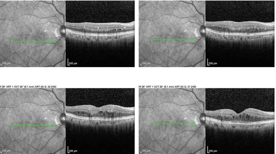
73 Pearl 3: Always assume that there is edema when you see hard exudates Pearl 4: Make sure you scroll through the entire macular scan as edema may not be captured in the global summary scan

74 Case C OD 61yo DM2 x 8 years HbA1c Full View +/-MA or Hemes +/- HEX +/- CWS +/- thickening +/- irma +/- VB +/-NVE

75 Case C OD 61yo DM2 x 8 years HbA1c Full View +/-MA or Hemes +/- HEX +/- CWS +/- thickening +/- irma +/- VB +/-NVE
76 Case C OD 61yo DM2 x 8 years HbA1c 8.3 VA: 20/ Full View +/-MA or Hemes +/- HEX +/- CWS +/- thickening +/- irma +/- VB +/-NVE

77 Case C OS 61yo DM2 x 8 years HbA1c Full View +/-MA or Hemes +/- HEX +/- CWS +/- thickening +/- irma +/- VB +/-NVE

78 Case C OS 61yo DM2 x 8 years HbA1c Full View +/-MA or Hemes +/- HEX +/- CWS +/- thickening +/- irma +/- VB +/-NVE
79 Case C OS 61yo DM2 x 8 years HbA1c 8.3 VA: 20/ Full View +/-MA or Hemes +/- HEX +/- CWS +/- thickening +/- irma +/- VB +/-NVE

80 Case C OD 61yo DM2 x 8 years HbA1c 8.3 VA: 20/ Full View +/-MA or Hemes +/- HEX +/- CWS +/- thickening +/- irma +/- VB +/-NVE

81
82

83 Case C OS 61yo DM2 x 8 years HbA1c 8.3 VA: 20/ Full View +/-MA or Hemes +/- HEX +/- CWS +/- thickening +/- irma +/- VB +/-NVE
84

85
86 Pearl 5: Exudates and surrounding edema can be reabsorbed so while you assume edema when you see exudates on the photo sometimes you will find exudates WITHOUT edema

87 Case D OD 67yo DM2 w CKD stage 3 HbA1c 8.3 VA: 20/25-2 Full View +/-MA or Hemes +/- HEX +/- CWS +/- thickening +/- irma +/- VB +/-NVE

88

89 Never underestimate the presence of hemorrhages near the macula!

90
91
92 Case D OS 67yo DM2 w CKD stage 3 HbA1c 8.3 VA: 20/50 Full View +/-MA or Hemes +/- HEX +/- CWS +/- thickening +/- irma +/- VB +/-NVE
93

94 ERM with Pseudohole

95
96
97
98
99

100

101

102 NVE
103 Pearl 6: Use the OCT and the photo to better understand the pathology. On dilated exam with a 3D view you would see the elevation missed on the fundus photo

104 Case E OD 67yo DM2 x 8 years HbA1c 7.3 VA: 20/20-2 Full View +/-MA or Hemes +/- HEX +/- CWS +/- thickening +/- irma +/- VB +/-NVE

105 Case E OD 67yo DM2 x 8 years HbA1c 7.3 VA: 20/20-2 Full View +/-MA or Hemes +/- HEX +/- CWS +/- thickening +/- irma +/- VB +/-NVE

106 Case F OD 67yo DM2 x 8 years HbA1c 7.3 VA: 20/20-2 Full View +/-MA or Hemes +/- HEX +/- CWS +/- thickening +/- irma +/- VB +/-NVE

107

108 Case F OD 65yo DM2 x 12+ years HbA1c 9.4 VA:? Full View +/-MA or Hemes +/- HEX +/- CWS +/- thickening +/- irma +/- VB +/-NVE

109 Case G OD 65yo DM2 x 12+ years HbA1c 9.4 VA:? Full View +/-MA or Hemes +/- HEX +/- CWS +/- thickening +/- irma +/- VB +/-NVE

110
111
112
113
114

115
116
117
118
119
120

121
122 Case G OD 65yo DM2 x? years HbA1c 9.3 VA: 20/30 Full View +/-MA or Hemes +/- HEX +/- CWS +/- thickening +/- irma +/- VB +/-NVE

123 Case H OD 65yo DM2 x? years HbA1c 9.3 VA: 20/30 Full View +/-MA or Hemes +/- HEX +/- CWS +/- thickening +/- irma +/- VB +/-NVE

124 Case H OD 65yo DM2 x? years HbA1c 9.3 VA: 20/30 Full View +/-MA or Hemes +/- HEX +/- CWS +/- thickening +/- irma +/- VB +/-NVE Severe? 4 Hemes 2 VB 1 IRMA

125 Case H OD 65yo DM2 x? years HbA1c 9.3 VA: 20/30 Full View +/-MA or Hemes +/- HEX +/- CWS +/- thickening +/- irma +/- VB +/-NVE

126 Case H OD 65yo DM2 x? years HbA1c 9.3 VA: 20/30 Full View +/-MA or Hemes +/- HEX +/- CWS +/- thickening +/- irma +/- VB +/-NVE

127 Case H OD 65yo DM2 x? years HbA1c 9.3 VA: 20/30 Full View +/-MA or Hemes +/- HEX +/- CWS +/- thickening +/- irma +/- VB +/-NVE
128 Case H OD 65yo DM2 x? years HbA1c 9.3 VA: 20/30 Full View +/-MA or Hemes +/- HEX +/- CWS +/- thickening +/- irma +/- VB +/-NVE
129 Pearl 7: Some retinas can burn out and not look severe- but actually be very ischemic and have NV Be ware of the retinopathy that lies outside the single fundus view.

130 Case I OS 65yo DM2 x? years HbA1c 9.3 VA: 20/30 Full View +/-MA or Hemes +/- HEX +/- CWS +/- thickening +/- irma +/- VB +/-NVE
131 Case I OS 65yo DM2 x? years HbA1c 9.3 VA: 20/30 Full View +/-MA or Hemes +/- HEX +/- CWS +/- thickening +/- irma +/- VB +/-NVE
132
133 Please remember to complete your session evaluations on the Academy.18 meeting app Tweet about this session using the official meeting hashtag #Academy18
Diabetic Retinopathy. Barry Emara MD FRCS(C) Giovanni Caboto Club October 3, 2012
 Giovanni Caboto Club October 3, 2012") Diabetic Retinopathy Barry Emara MD FRCS(C) Giovanni Caboto Club October 3, 2012 Outline Statistics Anatomy Categories Assessment Management Risk factors What do you need to do? Objectives Summarize the
Diabetic Retinopathy Barry Emara MD FRCS(C) Giovanni Caboto Club October 3, 2012 Outline Statistics Anatomy Categories Assessment Management Risk factors What do you need to do? Objectives Summarize the
PART 1: GENERAL RETINAL ANATOMY
 PART 1: GENERAL RETINAL ANATOMY General Anatomy At Ora Serrata At Optic Nerve Head Fundoscopic View Of Normal Retina What Is So Special About Diabetic Retinopathy? The WHO definition of blindness is
EyePACS Grading System (Part 3): Detecting Proliferative (Neovascular) Diabetic Retinopathy. George Bresnick MD MPA Jorge Cuadros OD PhD
: Detecting Proliferative (Neovascular) Diabetic Retinopathy. George Bresnick MD MPA Jorge Cuadros OD PhD") EyePACS Grading System (Part 3): Detecting Proliferative (Neovascular) Diabetic Retinopathy George Bresnick MD MPA Jorge Cuadros OD PhD Anatomy of the eye: 3 Normal Retina Retinal Arcades Macula Optic
EyePACS Grading System (Part 3): Detecting Proliferative (Neovascular) Diabetic Retinopathy George Bresnick MD MPA Jorge Cuadros OD PhD Anatomy of the eye: 3 Normal Retina Retinal Arcades Macula Optic
Marcus Gonzales, OD, FAAO Cedar Springs Eye Clinic
 Marcus Gonzales, OD, FAAO Cedar Springs Eye Clinic 25.6 million adults 11.3% of the adult population 10.9 million adults 65 years and older 26.9% of this age population 79 million people are Pre-diabetic!!
Diagnosis and treatment of diabetic retinopathy. Blake Cooper MD Ophthalmologist Vitreoretinal Surgeon Retina Associates Kansas City
 Diagnosis and treatment of diabetic retinopathy Blake Cooper MD Ophthalmologist Vitreoretinal Surgeon Retina Associates Kansas City Disclosures Consulted for Novo Nordisk 2017,2018. Will be discussing
Diabetic Retinopathy
 Diabetic Retinopathy Diabetes can be classified into type 1 diabetes mellitus and type 2 diabetes mellitus, formerly known as insulin-dependent diabetes mellitus, and non-insulin diabetes mellitus, respectively.
Diabetic Retinopatathy
 Diabetic Retinopatathy Jay M. Haynie, OD, FAAO Financial Disclosure I have received honoraria or am on the advisory board for the following companies: Carl Zeiss Meditec Arctic DX Macula Risk Advanced
Eyes on Diabetics: How to Avoid Blindness in Diabetic Patient
 Eyes on Diabetics: How to Avoid Blindness in Diabetic Patient Rova Virgana FK Unpad Pusat Mata Nasional RS Mata Cicendo Bandung Eye Center (Hospital and Clinic) PIT IDI Jabar 2018 Keys Facts from WHO
Clinically Significant Macular Edema (CSME)
") Clinically Significant Macular Edema (CSME) 1 Clinically Significant Macular Edema (CSME) Sadrina T. Shaw OMT I Student July 26, 2014 Advisor: Dr. Uwaydat Clinically Significant Macular Edema (CSME) 2
Clinically Significant Macular Edema (CSME) 1 Clinically Significant Macular Edema (CSME) Sadrina T. Shaw OMT I Student July 26, 2014 Advisor: Dr. Uwaydat Clinically Significant Macular Edema (CSME) 2
INTRODUCTION AND SYMPTOMS
 CHAPTER 1 INTRODUCTION AND SYMPTOMS Introduction of Diabetic Retinopathy Diabetic retinopathy (DR) is a potentially blinding complication of diabetes. It is defined as presence of one or more definite
EyePACS Grading System (Part 2): Detecting Presence and Severity of Background (Non-Proliferative) Diabetic Retinopathy Lesion
: Detecting Presence and Severity of Background (Non-Proliferative) Diabetic Retinopathy Lesion") EyePACS Grading System (Part 2): Detecting Presence and Severity of Background (Non-Proliferative) Diabetic Retinopathy Lesion George Bresnick MD MPA Jorge Cuadros OD PhD Anatomy of the eye: 3 Normal Retina
EyePACS Grading System (Part 2): Detecting Presence and Severity of Background (Non-Proliferative) Diabetic Retinopathy Lesion George Bresnick MD MPA Jorge Cuadros OD PhD Anatomy of the eye: 3 Normal Retina
Jay M. Haynie, O.D.; F.A.A.O. Olympia Tacoma Renton Kennewick Washington
 Jay M. Haynie, O.D.; F.A.A.O. Olympia Tacoma Renton Kennewick Washington I Jay M. Haynie, OD, FAAO have received honoraria from the following companies: Reichert Technologies Notal Vision Carl Zeiss Meditec
The Natural History of Diabetic Retinopathy and How Primary Care Makes A Difference
 The Natural History of Diabetic Retinopathy and How Primary Care Makes A Difference We will discuss - How exactly does blood sugar control affect retinopathy? - What are other factors that we measure in
DIABETIC RETINOPATHY
 DIABETIC RETINOPATHY C. L. B. Canny, MD FRCSC Diabetic retinopathy is the most serious eye manifestation of diabetes and is responsible for most of the blindness caused by diabetes. Diabetic retinopathy
The Human Eye. Cornea Iris. Pupil. Lens. Retina
 The Retina Thin layer of light-sensitive tissue at the back of the eye (the film of the camera). Light rays are focused on the retina then transmitted to the brain. The macula is the very small area in
Diabetic Management beyond traditional risk factors and LDL-C control: Can we improve macro and microvascular risks?
 Retinopathy Diabetes has a negative effect on eyes in many ways, increasing the risk of cataracts for example, but the most common and serious ocular complication of diabetes is retinopathy. Diabetic retinopathy
Epidemiology and Pathophysiology of Diabetic Retinopathy
 Epidemiology and Pathophysiology of Diabetic Retinopathy Vincent Reppucci, MD Director, Retina Service Mt. Sinai St. Luke s-roosevelt Hospital Attending Physician, Retina Service New York Eye and Ear Infirmary
Diabetic Retinopathy Screening in Hong Kong. Dr. Rita Gangwani M.S, FRCS (Ophth), FCOphth(HK), FHKAM Eye Institute, The University of Hong Kong
, FCOphth(HK), FHKAM Eye Institute, The University of Hong Kong") Diabetic Retinopathy Screening in Hong Kong Dr. Rita Gangwani M.S, FRCS (Ophth), FCOphth(HK), FHKAM Eye Institute, The University of Hong Kong Co-Investigators Prof. David Wong Prof. Sarah McGhee Dr. Wico
Diabetic Retinopathy Screening in Hong Kong Dr. Rita Gangwani M.S, FRCS (Ophth), FCOphth(HK), FHKAM Eye Institute, The University of Hong Kong Co-Investigators Prof. David Wong Prof. Sarah McGhee Dr. Wico
Diabetic Retinopathy: Managing the Extremes. J. Michael Jumper, MD West Coast Retina
 Diabetic Retinopathy: Managing the Extremes J. Michael Jumper, MD West Coast Retina Case 1: EC 65 y.o. HM No vision complaints Meds: Glyburide Metformin Pioglitazone Va: 20/20 OU 20/20 Case 2: HS 68 y.o.
Facts About Diabetic Eye Disease
 Facts About Diabetic Eye Disease Points to Remember 1. Diabetic eye disease comprises a group of eye conditions that affect people with diabetes. These conditions include diabetic retinopathy, diabetic
Diabetic and the Eye: An Introduction
 Diabetic and the Eye: An Introduction Lawrence Iu FRCSEd (Ophth), FCOphthHK, FHKAM (Ophthalmology) Department of Ophthalmology, Grantham Hospital & Queen Mary Hospital Background Diabetes mellitus (DM)
8/8/17. Objectives. Proliferative Diabetic Retinopathy (PDR) Examining the Diabetic Patient: What Matters Most
 Examining the Diabetic Patient: What Matters Most") Objectives Examining the Diabetic Patient: What Matters Most Jordan Keith, OD, FAAO Minneapolis, MN Simplify strategy for evaluating the eyes of diabetics Identify the threats to vision and treatments
Objectives Examining the Diabetic Patient: What Matters Most Jordan Keith, OD, FAAO Minneapolis, MN Simplify strategy for evaluating the eyes of diabetics Identify the threats to vision and treatments
RANZCO Screening and Referral Pathway for Diabetic Retinopathy #
 RANZCO Screening and Referral Pathway for Diabetic Retinopathy # Patient Presents a. Screen for Diabetic Retinopathy every 2 years b. Begin screening at diagnosis of Diabetes * Clinical Modifi ers Yearly
Guidelines for the Management of Diabetic Retinopathy for the Internist
 Visual Disorder Guidelines for the Management of Diabetic Retinopathy for the Internist JMAJ 45(1): 1 7, 2002 Sadao HORI Professor, Department of Ophthalmology, Tokyo Women s Medical University Abstract:
Diabesity A Public Health Crisis: AOA Evidence Based Translation to Care Series
 Diabesity A Public Health Crisis: AOA Evidence Based Translation to Care Series Joseph J. Pizzimenti, OD, FAAO Associate Professor Nova Southeastern University The Eye Care Institute pizzimen@nova.edu
 measure of your overall performance. An isolated glucose test is helpful to let you know what your sugar level is at one moment, but it doesn t tell you whether or not your diabetes is under adequate control
FRANZCO, MD, MBBS. Royal Darwin Hospital
 Diabetes and Eye By Dr. Nishantha Wijesinghe FRANZCO, MD, MBBS Consultant Ophthalmologist Royal Darwin Hospital 98% of Diabetics do not need to suffer from severe visual loss Yet Diabetic eye disease is
THE ROLE OF anti-vegf IN DIABETIC RETINOPATHY AND AGE RELATED MACULAR DEGENERATION
 THE ROLE OF anti-vegf IN DIABETIC RETINOPATHY AND AGE RELATED MACULAR DEGENERATION MOESTIDJAB DEPARTMENT OF OPHTHALMOLOGY SCHOOL OF MEDICINE AIRLANGGA UNIVERSITY DR SOETOMO HOSPITAL SURABAYA INTRODUCTION
Medical Retina 2011 Nicholas Lee
 Medical Retina 2011 Nicholas Lee 1 Diabetic Retinopathy Epidemiology 1000 registered blind each year 2% diabetics registered as blind (8% of all Blind Registrations) 42% with Mild Background DR will progress
OCT Angiography The Next Frontier
 Choroid Retina avascular 5/13/2017 OCT Angiography The Next Frontier Pierce Kenworthy OD, FAAO June 9, 2017 OCT Angiography (OCTA) 2016 Non-invasive, motion contrast imaging Represents erythrocyte movement
OCT Angiography in Primary Eye Care
 OCT Angiography in Primary Eye Care An Image Interpretation Primer Julie Rodman, OD, MS, FAAO and Nadia Waheed, MD, MPH Table of Contents Diabetic Retinopathy 3-6 Choroidal Neovascularization 7-9 Central
Diabetic Retinopathy A Presentation for the Public
 Diabetic Retinopathy A Presentation for the Public Ray M. Balyeat, MD The Eye Institute Tulsa, Oklahoma The Healthy Eye Light rays enter the eye through the cornea, pupil and lens. These light rays are
Optical Coherence Tomography in Diabetic Retinopathy. Mrs Samantha Mann Consultant Ophthalmologist Clinical Lead of SEL-DESP
 Optical Coherence Tomography in Diabetic Retinopathy Mrs Samantha Mann Consultant Ophthalmologist Clinical Lead of SEL-DESP Content OCT imaging Retinal layers OCT features in Diabetes Some NON DR features
Central Mersey Diabetic Retinopathy Screening Programme. Referring patients for Diabetic Retinopathy Screening
 Central Mersey Diabetic Retinopathy Screening Programme Referring patients for Diabetic Retinopathy Screening Information for GPs in Halton & St Helens, Knowsley and Warrington PCT Version: June 2008 Review
Brampton Hurontario Street Brampton, ON L6Y 0P6
 Diabetic Retinopathy What is Diabetic Retinopathy Diabetic retinopathy is one of the leading causes of blindness world-wide. Diabetes damages blood vessels in many organs of the body including the eyes.
ZEISS AngioPlex OCT Angiography. Clinical Case Reports
 Clinical Case Reports Proliferative Diabetic Retinopathy (PDR) Case Report 969 PROLIFERATIVE DIABETIC RETINOPATHY 1 1-year-old diabetic female presents for follow-up of proliferative diabetic retinopathy
New Developments in the treatment of Diabetic Retinopathy
 New Developments in the treatment of Diabetic Retinopathy B. Jeroen Klevering University Medical Centre Nijmegen - The Netherlands Topics Management of diabetic retinopathy Interventions a. primary (prevention)
Mild NPDR. Moderate NPDR. Severe NPDR
 Diabetic retinopathy Diabetic retinopathy is the most common cause of blindness in adults aged 35-65 years-old. Hyperglycaemia is thought to cause increased retinal blood flow and abnormal metabolism in
Diabetic Retinopathy
 Diabetic Retinopathy Introduction People with diabetes are more likely to have eye problems that can lead to blindness. Diabetic retinopathy is a disease of the eye s retina that is caused by diabetes.
Diabetes & Your Eyes
 Diabetes & Your Eyes Diabetes is a disease that occurs when the pancreas does not secrete enough insulin or the body is unable to process it properly. Insulin is the hormone that regulates the level of
Diabetic retinopathy damage to the blood vessels in the retina. Cataract clouding of the eye s lens. Cataracts develop at an earlier age in people
 Diabetic Retinopathy What is diabetic eye disease? Diabetic eye disease refers to a group of eye problems that people with diabetes may face as a complication of diabetes. All can cause severe vision loss
X-Plain Diabetic Retinopathy Reference Summary
 X-Plain Diabetic Retinopathy Reference Summary Introduction Patients with diabetes are more likely to have eye problems that can lead to blindness. Diabetic retinopathy is a disease of the eye s retina
Amber Priority. Image Library
 Amber Priority Image Library Amber flag Diabetic Maculopathy (M1) Pre-proliferative Diabetic Retinopathy (R2) Old, treated and now inactive DR (R1/M0/P1or R0/M0/P1) Where only partial or incomplete images
DIABETES AND YOUR EYES. Presented by Dr. Andrea Hagler
 DIABETES AND YOUR EYES Presented by Dr. Andrea Hagler Tahlequah, OK Forest Grove, OR Brief Review of Diabetes The body s endocrine system is responsible for regulating growth, reproduction, and tissue
Dr/ Marwa Abdellah EOS /16/2018. Dr/ Marwa Abdellah EOS When do you ask Fluorescein angiography for optic disc diseases???
 When do you ask Fluorescein angiography for optic disc diseases??? 1 NORMAL OPTIC DISC The normal optic disc on fluorescein angiography is fluorescent due to filling of vessels arising from the posterior
The Quick Guide to OCT Mastery 50 Real Cases with Expert Analysis
 OPTICAL COHERENCE TOMOGRAPHY The Quick Guide to OCT Mastery 50 Real Cases with Expert Analysis VOL 1 Sanjay Sharma, MD, FRCS, MSc (Epid), MBA Ophthalmologist, Epidemiologist Queen s University, Canada
Measures have been taken, by the Utah Department of Health, Bureau of Health Promotions, to ensure no conflict of interest in this activity.
 Measures have been taken, by the Utah Department of Health, Bureau of Health Promotions, to ensure no conflict of interest in this activity. CNE/CPE/CEU s are available for this live webinar. You must
OCT Assessment of the Vitreoretinal Relationship in CSME
 December 2007 Sonia Rani John et al. - IFIS 375 ORIGINAL ARTICLE OCT Assessment of the Vitreoretinal Relationship in CSME Dr. Manoj S. DNB FRCS, Dr. Unnikrishnan Nair MS DO FRCS, Dr. Gargi Sathish MS Introduction
FA Conference. Lara Rosenwasser Newman, M.D. 10/2/14 University of Louisville Department of Ophthalmology and Visual Sciences
 FA Conference Lara Rosenwasser Newman, M.D. 10/2/14 University of Louisville Department of Ophthalmology and Visual Sciences Patient Presentation CC: (sent by optometrist) Blurry/foggy vision HPI: 62 yo
Leo Semes, OD, FAAO UAB Optometry
 Leo Semes, OD, FAAO UAB Optometry Safe; inert Has long track record - over 45 years Mixes with plasma and highlights blood vessel compromise Using specific exciting (490 nm)and absorption (510 nm) filters
Clinical Trials in Diabetic Retinopathy. Harry W. Flynn Jr., M.D. Nidhi Relhan Batra, M.D.
 1 Clinical Trials in Diabetic Retinopathy 2018 Harry W. Flynn Jr., M.D. Nidhi Relhan Batra, M.D. Bascom Palmer Eye Institute 900 N.W. 17th Street Miami, FL 33136 Phone: (305) 326-6118 Fax: (305) 326-6417
Diabetic Retinopathy
 Diabetic Retinopathy Overview This presentation covers the following topics: Definitions Epidemiology of diabetic retinopathy Evidence for public health approaches Screening for diabetic retinopathy Health
Is OCT-A Needed As An Investigative Tool During The Management Of Diabetic Macular Edema
 Is OCT-A Needed As An Investigative Tool During The Management Of Diabetic Macular Edema Ayman M Khattab MD, FRCS Professor of Ophthalmology Cairo University Diabetic Macular Edema (DME) Diabetic macular
Diabetic Retinopathy
 Diabetic Retinopathy Secretary for Quality of Care Anne L. Coleman, MD, PhD Academy Staff Nicholas P. Emptage, MAE Doris Mizuiri Shannon Kealey, MLS Flora C. Lum, MD Medical Editor: Design: Approved by:
OCCLUSIVE VASCULAR DISORDERS OF THE RETINA
 OCCLUSIVE VASCULAR DISORDERS OF THE RETINA Learning outcomes By the end of this lecture the students would be able to Classify occlusive vascular disorders (OVD) of the retina. Correlate the clinical features
DIABETIC RETINOPATHY (DR) PREFERRED PRACTICE PATTERNS (PPP) Philippines 2016
 PREFERRED PRACTICE PATTERNS (PPP) Philippines 2016") DIABETIC RETINOPATHY (DR) PREFERRED PRACTICE PATTERNS (PPP) Philippines 2016 The Diabetic Retinopathy (DR) Preferred Practice Patterns (PPP) Philippines: 2016 was prepared by the VitreoRetina Society of
DIABETIC RETINOPATHY (DR) PREFERRED PRACTICE PATTERNS (PPP) Philippines 2016 The Diabetic Retinopathy (DR) Preferred Practice Patterns (PPP) Philippines: 2016 was prepared by the VitreoRetina Society of
Documentation, Codebook, and Frequencies
 Documentation, Codebook, and Frequencies Ophthalmology Retinal Imaging Examination Survey Years: 2005 to 2006 SAS Transport File: OPXRET_D.XPT December 2008 NHANES 2005 2006 Data Documentation Exam Component:
Grand Rounds: Interesting and Exemplary Cases From Guanajuato and Djibouti
 Learning Community: January 25, 2015 Grand Rounds: Interesting and Exemplary Cases From Guanajuato and Djibouti JORGE CUADROS, OD, PHD EyePACS In Guanajuato Program started in 2007 Cameras go from clinic
Clinical Case Presentation. Branch Retinal Vein Occlusion. Sarita M. Registered Nurse Whangarei Base Hospital
 Clinical Case Presentation on Branch Retinal Vein Occlusion Sarita M. Registered Nurse Whangarei Base Hospital Introduction Case Study Pathogenesis Clinical Features Investigations Treatment Follow-up
What is diabetes? Ocolusystemic Disease Essen6als. Statistics, cont. Statistics. Statistics. The Diabetes Epidemic 9/5/12
 What is diabetes? Ocolusystemic Disease Essen6als Steven Ferrucci, OD, FAAO Chief, Optometry Sepulveda VA Associate Professor, SCCO DM is a chronic disorder characterized by a lack of insulin or increased
10/8/13. the diabetes epidemic: strategies for saving sight. financial disclo$ure. unlabeled-investigative use disclosure
 10/8/13 ASORN 2013 Annual Meeting New Orleans, LA the diabetes epidemic: strategies for saving sight Kate Goldblum, CNP financial disclo$ure I have no financial interests relevant to my presentation. unlabeled-investigative
Diabetic Retinopathy What You Should Know. U.S. DEPARTMENT OF HEALTH AND HUMAN SERVICES National Institutes of Health National Eye Institute
 Diabetic Retinopathy What You Should Know U.S. DEPARTMENT OF HEALTH AND HUMAN SERVICES National Institutes of Health National Eye Institute The National Eye Institute (NEI) conducts and supports research
Diabetic Retinopathy WHAT IS DIABETIC RETINOPATHY? WHAT CAUSES DIABETIC RETINOPATHY? WHAT ARE THE STAGES OF DIABETIC RETINOPATHY?
 Diabetic Retinopathy WHAT IS DIABETIC RETINOPATHY? Diabetic retinopathy affects 8 million Americans with diabetes. A leading cause of blindness in American adults, it is caused by damage to the small blood
Diabetes and Eye Health more than meets the eye Vision Initiative - in association with PSA
 Diabetes and Eye Health more than meets the eye Vision Initiative - in association with PSA Vision 2020 Australia Vision Initiative RANZCO & OAA (Vic) Proud members of Vision 2020 Australia Outline Vision
PROGRESSION OF DIABETIC RETINOPATHY FOLLOWING CATARACT SURGERY
 PROGRESSION OF DIABETIC RETINOPATHY FOLLOWING CATARACT SURGERY Yayan Heryanto, Iwan Sovani, Arief Kartasasmita, Erwin Iskandar, Djonggi Panggabean. Dept. of Ophthalmology Medical Faculty Unpad, Cicendo
Diabetic Retinopathy
 Diabetic Retinopathy Diabetes mellitus is one of the leading causes of irreversible blindness worldwide. In the United States, it is the most common cause of blindness in people younger than 65 years.
Understanding Diabetic Retinopathy
 Understanding Diabetic Retinopathy What Is Diabetic Retinopathy? Diabetes damages blood vessels in the rear of the eye. This condition is called diabetic retinopathy. It can lead to vision loss or blindness.
Year 2 MBChB Clinical Skills Session Ophthalmoscopy. Reviewed & ratified by: Mr M Batterbury Consultant Ophthalmologist
 Year 2 MBChB Clinical Skills Session Ophthalmoscopy Reviewed & ratified by: o Mr M Batterbury Consultant Ophthalmologist Learning objectives o To understand the anatomy and physiology of the external and
Diabetic retinopathy (DR) was first PROCEEDINGS DIABETIC RETINOPATHY * Ronald Klein, MD, MPH ABSTRACT
 was first PROCEEDINGS DIABETIC RETINOPATHY * Ronald Klein, MD, MPH ABSTRACT") DIABETIC RETINOPATHY * Ronald Klein, MD, MPH ABSTRACT Diabetic retinopathy (DR) is characterized by the development of retinal microaneurysms, hemorrhages, deposits of leaked lipoproteins (hard exudates),
DIABETIC RETINOPATHY * Ronald Klein, MD, MPH ABSTRACT Diabetic retinopathy (DR) is characterized by the development of retinal microaneurysms, hemorrhages, deposits of leaked lipoproteins (hard exudates),
Study of clinical significance of optical coherence tomography in diagnosis & management of diabetic macular edema
 Original Research Article Study of clinical significance of optical coherence tomography in diagnosis & management of diabetic macular edema Neha Kantilal Desai 1,*, Somesh Vedprakash Aggarwal 2, Sonali
A Patient s Guide to Diabetic Retinopathy
 Diabetic Retinopathy A Patient s Guide to Diabetic Retinopathy 840 Walnut Street, Philadelphia PA 19107 www.willseye.org Diabetic Retinopathy 1. Definition Diabetic retinopathy is a complication of diabetes
Scott M. Pfahler D.O. Dayton Vitreo-Retinal Associates AOCOO-HNS Palm Springs, CA 2012
 Scott M. Pfahler D.O. Dayton Vitreo-Retinal Associates AOCOO-HNS Palm Springs, CA 2012 Proliferative Diabetic Retinopathy Laser Treatments Medical Treatment Surgical Treatment Diabetic Macular Edema Laser
Teaching Case Report. Subconjunctival Hemorrhage and Diabetes: A Lesson Learned. Todd Peabody, OD, MBA, FAAO Taylor Steger, OD Matt Lepage, OD
 Teaching Case Report Subconjunctival Hemorrhage and Diabetes: A Lesson Learned Todd Peabody, OD, MBA, FAAO Taylor Steger, OD Matt Lepage, OD Abstract Every year in the United States more adults become
Neuropathy (NAION) and Avastin. Clinical Assembly of the AOCOO-HNS Foundation May 9, 2013
 and Avastin. Clinical Assembly of the AOCOO-HNS Foundation May 9, 2013") Non Arteritic Ischemic Optic Neuropathy (NAION) and Avastin Shalom Kelman, MD Clinical Assembly of the AOCOO-HNS Foundation May 9, 2013 Anterior Ischemic Optic Neuropathy Acute, painless, visual loss,
Non Arteritic Ischemic Optic Neuropathy (NAION) and Avastin Shalom Kelman, MD Clinical Assembly of the AOCOO-HNS Foundation May 9, 2013 Anterior Ischemic Optic Neuropathy Acute, painless, visual loss,
7.1 Grading Diabetic Retinopathy
 Chapter 7 DIABETIC RETINOPATHYGRADING -------------------------------------------------------------------------------------------------------------------------------------- A consistent approach to the
Fundus Fluorescein Angiography in Diabetic Retinopathy: Correlation of Angiographic Findings to the Clinical Maculopathy Abstract: Purpose:
 IOSR Journal of Dental and Medical Sciences (IOSR-JDMS) e-issn: 2279-0853, p-issn: 2279-0861.Volume 15, Issue 2 Ver. XII (Feb. 2016), PP 80-88 www.iosrjournals.org Fundus Fluorescein Angiography in Diabetic
Slide notes: The major chronic complications of diabetes mellitus are described here. Among these, microvascular complications have an important
 1 2 The major chronic complications of diabetes mellitus are described here. Among these, microvascular complications have an important role. They comprise microangiopathy, diabetic retinopathy, diabetic
The Diabetic Retinopathy Clinical Research Network. Management of DME in Eyes with PDR
 The Diabetic Retinopathy Clinical Research Network Management of DME in Eyes with PDR 1 What Has Been Learned? Diabetic Retinopathy Treatment Protocol F: Results suggest that clinically meaningful differences
FROM OUTDATED TO UPDATED Eminence-Based Medicine
 FROM OUTDATED TO UPDATED Eminence-Based Medicine Evidence-Based Medicine A REVIEW OF KEY CLINICAL TRIALS Anthony DeWilde, OD FAAO 1 EMINENCE BASED MEDICINE 2 EVIDENCE BASED MEDICINE 3 4 CLINICAL TRIALS
Control of Systemic Factors Can Preserve Vision in Diabetic Retinopathy
 dmcjuly05_cme_dr 7/28/05 9:19 AM Page 38 Control of Systemic Factors Can Preserve Vision in Diabetic Retinopathy Jointly sponsored by The Dulaney Foundation and Diabetic Microvascular Complications Today.
Diabetic Eye Disease Visual Recognition & Interpretation of Clinical Signs
 + Diabetic Eye Disease Visual Recognition & Interpretation of Clinical Signs Quiz created by CLEARVIEW Training Jane Macnaughton MCOptom & Peter Chapman MCOptom FBDO + CET Accreditation C 19106 2 CET Points
Retinal Complications of Obstructive Sleep Apnea A Growing Concern!
 Retinal Complications of Obstructive Sleep Apnea A Growing Concern! Jay M. Haynie, OD, FAAO Financial Disclosure I have received honoraria or am on the advisory board for the following companies: Carl
Use of the Free Electron Laser for the Noninvasive Determination of Retinal Oxyhemoglobin Saturation by Near Infrared Reflectance Spectrophotometry
 Use of the Free Electron Laser for the Noninvasive Determination of Retinal Oxyhemoglobin Saturation by Near Infrared Reflectance Spectrophotometry Ref: Eye, M.C. Escher, 1946 Ref: Eye, M.C. Escher, 1946
Posterior Segment Update
 Posterior Segment Update Featured Speaker: Dr. Kyle Cheatham, FAAO, DIP ABO DISCLOSURE STATEMENT We have no direct financial or proprietary interest in any companies, products or services mentioned in
Front Line Diabetic Retinopathy What Not to Miss and Why
 Front Line Diabetic Retinopathy What Not to Miss and Why David M Brown MD FACS Clinical Professor of Ophthalmology Blanton Eye Institute, Houston Methodist Hospital Baylor College of Medicine Retina Consultants
Diabetic Retinopathy: Recent Advances in Treatment and Treatment Approaches
 Diabetic Retinopathy: Recent Advances in Treatment and Treatment Approaches Dr. David Wong Associate Professor Retina Specialist, Department of Ophthalmology & Vision Sciences, University of Toronto, Canada
Preventing Avoidable Vision loss from Diabetic Retinopathy in Indian Country
 Diabetes in Indian Country- 2017 Preventing Avoidable Vision loss from Diabetic Retinopathy in Indian Country Albuquerque, NM 20 September2017 Mark B. Horton, OD, MD Director, IHS/JVN Teleophthalmology
Fundus Autofluorescence. Jonathan A. Micieli, MD Valérie Biousse, MD
 Fundus Autofluorescence Jonathan A. Micieli, MD Valérie Biousse, MD The retinal pigment epithelium (RPE) has many important functions including phagocytosis of the photoreceptor outer segments Cone Rod
Retinopathy in a diabetic population
 Kathmandu University Medical Journal (2007), Vol. 5, No. 2, Issue 18, 204-209 Retinopathy in a diabetic population Shrestha S 1, Malla OK 2, Karki DB 3, Byanju RN 4 2 Fellow of NAMS, 3 Professor, NAMS,
Recalcitrant Diabetic Macular Oedema: Therapeutic Options
 December 2007 A. Giridhar et al. - Recalcitrant DME 451 CONSULTATION S E C T I O N Recalcitrant Diabetic Macular Oedema: Therapeutic Options Dr. Cyrus M Shroff 1, Dr. N S Muralidhar 2, Dr. R Narayanan
Study of 189 Cases of Diabetic Retinopathy at CMC Larkana
 Original Article Study of 189 Cases of Diabetic Retinopathy at CMC Larkana Shahid Jamal Siddiqui, Sayed Imtiaz Ali Shah, Abdul Qadir Shaikh, Mohammed Yousuf Depar, Safder Ali Abbassi Pak J Ophthalmol 2007,
Classification of diabetic retinopathy and diabetic macular edema
 Online Submissions: http://www.wjgnet.com/esps/ bpgoffice@wjgnet.com doi:10.4239/wjd.v4.i6.290 World J Diabetes 2013 December 15; 4(6): 290-294 ISSN 1948-9358 (online) 2013 Baishideng Publishing Group
Neovascular Glaucoma Associated with Cilioretinal Artery Occlusion Combined with Perfused Central Retinal Vein Occlusion
 Neovascular Glaucoma Associated with Cilioretinal Artery Occlusion Combined with Perfused Central Retinal Vein Occlusion Man-Seong Seo,* Jae-Moon Woo* and Jeong-Jin Seo *Department of Ophthalmology, Chonnam
Incorporating OCT Angiography Into Patient Care
 Incorporating OCT Angiography Into Patient Care Beth A. Steele, OD, FAAO OCT A: Introduction Isolates microvascular circulation from OCT image data Axial resolution = 5 microns (i.e. fine capillaries visible)
10/17/2017. FDA Approved. Zeiss AngioPlex TM Optovue AngioVue TM
 Images retinal microvasculature without dye injection Displays structure and function from a single imaging system Standard of Care-2011 DFE, Fundus Photos, VF 10-2, SD-OCT, FAF, or mferg 2016-AAO Baseline
Use of Eye Care Services among Type 2 Diabetic Patients in Laguna
 Original Article Philippine Journal of OPHTHALMOLOGY Use of Eye Care Services among Type 2 Diabetic Patients in Laguna Glenn Carandang, MD, MPH, 1,2, Maria Victoria Rondaris, MD, MPH, 3, and Genejane Adarlo,
DIABETIC RETINOPATHY, A
 The Prevalence of Diabetic Retinopathy Among Adults in the United States The Eye Diseases Prevalence Research Group* EPIDEMIOLOGY Objective: To determine the prevalence of diabetic retinopathy among adults
Optical Coherence Tomography: Pearls for the Anterior Segment Surgeon Basic Science Michael Stewart, M.D.
 Optical Coherence Tomography: Pearls for the Anterior Segment Surgeon Basic Science Michael Stewart, M.D. Disclosure OCT Optical Coherence Tomography No relevant financial relationships I will refer to
Speaker Disclosure Statement. " Dr. Tim Maillet and Dr. Vladimir Kozousek have no conflicts of interest to disclose.
 Speaker Disclosure Statement Dr. Tim Maillet and Dr. Vladimir Kozousek have no conflicts of interest to disclose. Diabetes Morbidity Diabetes doubles the risk of stroke. Diabetes quadruples the risk of
Diagnosis in AMD. Managing your AMD Patients
 Managing your AMD Patients Robert W. Dunphy, O.D., F.A.A.O. Diagnosis in AMD Have suspicion Identify relative risk Conduct surveillance Biometry Utilize technology to facilitate detection of change / stability