10/17/2017. FDA Approved. Zeiss AngioPlex TM Optovue AngioVue TM
|
|
|
- Russell Thornton
- 5 years ago
- Views:
Transcription


:415-22.")


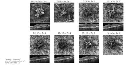
1 Images retinal microvasculature without dye injection Displays structure and function from a single imaging system Standard of Care-2011 DFE, Fundus Photos, VF 10-2, SD-OCT, FAF, or mferg 2016-AAO Baseline DFE At 5 years Annual screening:vf 10-2, SD-OCT Additional screening: merg, FAF Marmor MF, Kellner U, Lai TY, et al. Revised recommendations on screening for chloroquinine and hydroxychloroquinine retinopathy. Ophthalmology Feb;118(2): Retinal blood supply OCTA in disease Diabetic Retinopathy AMD Venous Occlusion Mac Tel II Other causes of CNV Choroidal Tumors Glaucoma FDA Approved Zeiss AngioPlex TM Optovue AngioVue TM OCTA uses motion contrast to detect flow from OCT data Rapidly acquires multiple cross-sectional images from a single location on the retina Flow is the difference in signal between two sequential B-scans 1


 Motion Artifacts in OCTA Images caused by patient motion,")


2 See retinal vasculature without referring patients out of the practice Visualize signs of disease earlier and make more intelligent referrals Manage more pathology to keep patients in the practice longer Elevate the practice with state-of-the-art imaging technology Longer Data Acquisition Times needed for multiple B-scan repeats (to boost OCTA flow signal) Motion Artifacts in OCTA Images caused by patient motion, fixation shifts and microsaccades Projection Artifacts caused by shadow projection of vessels above Observing dry AMD for conversion to wet Monitoring diabetic patients Visualizing vascularization in PEDs Identifying CNV in central serous Venous Occlusions Examining glaucoma patients for vascular changes 2


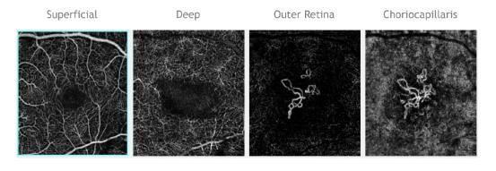
3 Spaide RF, Klancnik JM, Cooney MJ, Retinal Vascular Layers Imaged by Fluorescein Angiography and Optical Coherence Tomography Angiography, JAMA Ophthalmol, 2015;133: Delineation and quantification of nonperfusion Ischemic vs non ischemic? BRVO: Risk of post segment neo and vitreous/preretinal hemorrhage? CRVO: Risk of ant segment neo and NVG? Visualization of macular ischemia Improved visualization of collaterals Depth localization of abnormalities Visualization of deep capillary telangiectasias Earlier detection of posterior segment neo 3
 and deep (c) retinal plexus. Ischemic area due to capillary drop out is better delineated using OCTA.")
 Ectatic capillaries Atrophy of the neurosensory retina Depletion of the")


4 Comparison of fluorescein angiography image (a) in branch retinal vein occlusion with that of optical coherence tomography angiography (OCTA) images obtained at the superficial (b) and deep (c) retinal plexus. Ischemic area due to capillary drop out is better delineated using OCTA. However, leaking vessels due to neovascularization are not identified using OCTA. Neurodegenerative disease play a central role (loss of Muller cells) Ectatic capillaries Atrophy of the neurosensory retina Depletion of the macular pigment Dilated macular venules and arterioles with right angle bends Hyperplastic plaques of the RPE Neovascularization Multimodal Imaging of Macular Telangiectasia Type 2: Focus on Vascular Changes Using Optical Coherence Tomography Angiography Lisa Toto,1 Luca Di Antonio,1 Rodolfo Mastropasqua,2 Peter A. Mattei,1 Paolo Carpineto,1 Enrico Borrelli,1 Marco Rispoli,3 Br uno Lumbroso,3 and Leonardo Mastropasqua1 1Department of Medicine and Science of Aging, Ophthalmology Clinic, University G. d Annunzio Chieti-Pescara, Chieti, Italy 2Ophthalmology Clinic, University of Verona, Verona, Italy 3Italian Macular Center, Rome, Italy Multimodal Imaging of Macular Telangiectasia Type 2: Focus on Vascular Changes Using Optical Coherence Tomography Angiography Lisa Toto,1 Luca Di Antonio,1 Rodolfo Mastropasqua,2 Peter A. Mattei,1 Paolo Carpineto,1 Enrico Borrelli,1 Marco Rispoli,3 Bruno Lumbroso,3 and Leonardo Mastropasqua1 1Department of Medicine and Science of Aging, Ophthalmology Clinic, University G. d Annunzio Chieti-Pescara, Chieti, Italy 2Ophthalmology Clinic, University of Verona, Verona, Italy 3Italian Macular Center, Rome, Italy 4

: 1646.")
 Detection and localization of non perfusion Detailed evaluation of FAZ Visualize vascular abnormalities within the deep plexus")
5 Type 2 DM within 1 year 5-10% will have retinopathy Among patients with type 1 diabetes, 25%, 60% and 80% will have retinopathy after 5, 10 and 15 years of disease. Among patients with type 2 diabetes, 40% who use insulin and 24% who do not use insulin will have retinopathy at five years; the proportions will increase to 84% and 53%, respectively, at 19 years. Jason Noble MD.CMAJ Oct 19; 182(15): Sub clinical diabetic retinopathy detection Highlight and localize vascular abnormalities (IRMA, vascular loops, etc.) Detection and localization of non perfusion Detailed evaluation of FAZ Visualize vascular abnormalities within the deep plexus Differentiate IRMA from early NVE Localization of microaneurysms Early detection of PDR Precise definition of PDR Follow response to treatment IRMA 5
6 Choroidal ischemia detection/quantification Dry vs Wet and detection of CNV Quantify responsiveness to treatment Assess need for retreatment Detect quiescent (inactive) CNV membranes 6


7 Retinal OCT Thank You!! 7
Incorporating OCT Angiography Into Patient Care
 Incorporating OCT Angiography Into Patient Care Beth A. Steele, OD, FAAO OCT A: Introduction Isolates microvascular circulation from OCT image data Axial resolution = 5 microns (i.e. fine capillaries visible)
Will OCT-Angiography replace FA?
 ASL Roma A PRESIDIO TERRITORIALE NUOVO REGINA MARGHERITA AMBULATORIO PATOLOGIE RETINICHE Resp. Dott.ssa SUSANNA CATALANO CENTRO ITALIANO MACULA Will OCT-Angiography replace FA? Marco Rispoli, Luca di Antonio,
Visualize. Analyze. Personalize. OCT + OCTA
 Visualize. Analyze. Personalize. OCT + OCTA A New Approach to Protecting Vision AngioVue OCT Angiography brings valuable new information to clinical practice. Non-invasive visualization of retinal vasculature.
ZEISS AngioPlex OCT Angiography Overview ZEISS OCT Angiography
 ZEISS AngioPlex OCT Angiography Overview ZEISS OCT Angiography California, ZEISS AngioPlex Ultra-clear visualization of microvascular blood flow using non-invasive OCT angiography 2 AngioPlex OCT Angiography
OCT Angiography The Next Frontier
 Choroid Retina avascular 5/13/2017 OCT Angiography The Next Frontier Pierce Kenworthy OD, FAAO June 9, 2017 OCT Angiography (OCTA) 2016 Non-invasive, motion contrast imaging Represents erythrocyte movement
ZEISS AngioPlex OCT Angiography Making the revolutionary, routine.
 ZEISS AngioPlex OCT Angiography Making the revolutionary, routine. The moment that revolutionary insight becomes routine. // OCT ANGIOGRAPHY MADE BY ZEISS CIRRUS with AngioPlex creates a new era in both
OCT Interpretation in Retinal Disease
 OCT Interpretation in Retinal Disease Jay M. Haynie, OD, FAAO Financial Disclosure I have received honoraria or am on the advisory board for the following companies: Carl Zeiss Meditec Advanced Ocular
Clinical Study Optical Coherence Tomography Angiography in Retinal Vascular Diseases and Choroidal Neovascularization
 Hindawi Publishing Corporation Journal of Ophthalmology Volume 2015, Article ID 343515, 8 pages http://dx.doi.org/10.1155/2015/343515 Clinical Study Optical Coherence Tomography Angiography in Retinal
FA vs. OCTA? The status of OCTA, today. Fukuoka, JSOS 2016 Gerd Klose. Korobelnik J Fr Ophthalmol (2015)
") FA vs. OCTA? The status of OCTA, today Korobelnik J Fr Ophthalmol (2015) Fukuoka, JSOS 2016 Gerd Klose 1 2 FA / ICGA a well-founded Gold standard! Benefits Useful for many pathologies High contrast, detailed
FA vs. OCTA? The status of OCTA, today Korobelnik J Fr Ophthalmol (2015) Fukuoka, JSOS 2016 Gerd Klose 1 2 FA / ICGA a well-founded Gold standard! Benefits Useful for many pathologies High contrast, detailed
Is OCT-A Needed As An Investigative Tool During The Management Of Diabetic Macular Edema
 Is OCT-A Needed As An Investigative Tool During The Management Of Diabetic Macular Edema Ayman M Khattab MD, FRCS Professor of Ophthalmology Cairo University Diabetic Macular Edema (DME) Diabetic macular
OCT Angiography in Primary Eye Care
 OCT Angiography in Primary Eye Care An Image Interpretation Primer Julie Rodman, OD, MS, FAAO and Nadia Waheed, MD, MPH Table of Contents Diabetic Retinopathy 3-6 Choroidal Neovascularization 7-9 Central
OPTICAL COHERENCE TOMOGRAPHY ANGIOGRAPHY OF THE RETINA AND OPTIC NERVE. Lindsay B. Howse, OD
 OPTICAL COHERENCE TOMOGRAPHY ANGIOGRAPHY OF THE RETINA AND OPTIC NERVE Lindsay B. Howse, OD drlindsayhowse@gmail.com None. FINANCIAL DISCLOSURES OUTLINE Introduction/How OCTA works OCTA Analysis Advantages
Introducing ANGIOVUE ESSENTIAL. Built on the Avanti Widefield OCT Platform. OCT Angiography for Primary Eye Care
 Introducing ANGIOVUE ESSENTIAL Built on the Avanti Widefield OCT Platform OCT Angiography for Primary Eye Care Transform Your View of the Retina OCT Angiography (OCTA) is a quick non-invasive test that
OCT Angiography. Financial Disclosures: Pre-Test: Which one is Correct?
 OCT Angiography Brandon Lujan, MD Medical Director, Casey Reading Center Assistant Professor of Ophthalmology Financial Disclosures: Genentech (Consultant, Grant support, Educational training) UC Berkeley
ZEISS AngioPlex OCT Angiography. Clinical Case Reports
 Clinical Case Reports Proliferative Diabetic Retinopathy (PDR) Case Report 969 PROLIFERATIVE DIABETIC RETINOPATHY 1 1-year-old diabetic female presents for follow-up of proliferative diabetic retinopathy
CAPILLARY NETWORK ANOMALIES IN BRANCH RETINAL VEIN OCCLUSION ON OPTICAL COHERENCE TOMOGRAPHY ANGIOGRAPHY
 CAPILLARY NETWORK ANOMALIES IN BRANCH RETINAL VEIN OCCLUSION ON OPTICAL COHERENCE TOMOGRAPHY ANGIOGRAPHY MARCO RISPOLI, MD, MARIA CRISTINA SAVASTANO, MD, PHD, BRUNO LUMBROSO, MD Purpose: To analyze the
OCT Angiography. SriniVas Sadda, MD
 OCT Angiography SriniVas Sadda, MD Professor of Ophthalmology Director, Medical Retina Unit Ophthalmic Imaging Unit University of Southern California Los Angeles, California, USA Disclosure Consulting
PART 1: GENERAL RETINAL ANATOMY
 PART 1: GENERAL RETINAL ANATOMY General Anatomy At Ora Serrata At Optic Nerve Head Fundoscopic View Of Normal Retina What Is So Special About Diabetic Retinopathy? The WHO definition of blindness is
Deeper visualizations for intervening with confidence.
 CIRRUS OCT with AngioPlex from ZEISS Making the revolutionary routine New vascular quantification Deeper visualizations for intervening with confidence. CIRRUS OCT with AngioPlex from ZEISS can be a much
Swept-Source OCT Angiography: SS OCT Angio TM
 Swept-Source OCT Angiography: SS OCT Angio TM Not available in all countries, please check with your distributor. 2015.09 Swept-Source OCT Angiography: SS OCT Angio TM Introduction Optical coherence tomography
OCT Fundal Angiography Initial Experience The new era in Medical Retina Imaging Based on Cirrus 5000 AngioPlex 2016 Model Sheena George & Nicholas
 OCT Fundal Angiography Initial Experience The new era in Medical Retina Imaging Based on Cirrus 5000 AngioPlex 2016 Model Sheena George & Nicholas Lee Consultants Ophthalmologist at The Hillingdon Hospital
Optical Coherence Tomography in Diabetic Retinopathy. Mrs Samantha Mann Consultant Ophthalmologist Clinical Lead of SEL-DESP
 Optical Coherence Tomography in Diabetic Retinopathy Mrs Samantha Mann Consultant Ophthalmologist Clinical Lead of SEL-DESP Content OCT imaging Retinal layers OCT features in Diabetes Some NON DR features
Angio-OCT. Degenerazione Maculare Legata all Eta. Giuseppe Querques
 Angio-OCT Degenerazione Maculare Legata all Eta Giuseppe Querques Department of Ophthalmology, IRCCS Ospedale San Raffaele, University Vita Salute San Raffaele, Milan, Italy Financial Disclosure ADVISORY
ANSWERING THE WHY? Clinicians discuss the latest imaging technologies for retina practice BY PETER K. KAISER, MD
 Insert to March 2018 Sponsored by MULTI-MODALITY IMAGING: LATEST EVOLUTIONS IN OCTA AND UWF As the array of safe and efficacious medical and surgical options for retinal diseases expands, so does the need
Visualize. Analyze. Personalize. OCT + OCTA. with
 Visualize. Analyze. Personalize. OCT + OCTA with Avanti Widefield OCT with AngioVue OCTA Imaging Comprehensive Structural and Functional Imaging in a Single Imaging Platform Comprehensive OCT Imaging The
OCT-Angiography Clinical Cases. OCT-Angiography Clinical Cases
 OCT-Angiography Clinical Cases OCT-Angiography Clinical Cases NIDEK RS-3000 Advance AngioScan Daniela Bacherini Andrea Sodi Stanislao Rizzo CONTENTS Page Authors 3 Introduction 4 Case 1 Case 2 Case 3 Case
OCT Angiography: The Next Step in Retinal Imaging Jonathan Zelenak D.O.
 OCT Angiography: The Next Step in Retinal Imaging Jonathan Zelenak D.O. Hillsdale Hospital Michigan State University Overview Evolution of OCT How does OCT angiography work? Clinical examples Potential
Clinical Case Presentation. Branch Retinal Vein Occlusion. Sarita M. Registered Nurse Whangarei Base Hospital
 Clinical Case Presentation on Branch Retinal Vein Occlusion Sarita M. Registered Nurse Whangarei Base Hospital Introduction Case Study Pathogenesis Clinical Features Investigations Treatment Follow-up
EyePACS Grading System (Part 2): Detecting Presence and Severity of Background (Non-Proliferative) Diabetic Retinopathy Lesion
: Detecting Presence and Severity of Background (Non-Proliferative) Diabetic Retinopathy Lesion") EyePACS Grading System (Part 2): Detecting Presence and Severity of Background (Non-Proliferative) Diabetic Retinopathy Lesion George Bresnick MD MPA Jorge Cuadros OD PhD Anatomy of the eye: 3 Normal Retina
EyePACS Grading System (Part 2): Detecting Presence and Severity of Background (Non-Proliferative) Diabetic Retinopathy Lesion George Bresnick MD MPA Jorge Cuadros OD PhD Anatomy of the eye: 3 Normal Retina
The College of Optometrists - Learning outcomes for the Professional Certificate in Medical Retina
 Learning outcomes for the Professional Certificate in Medical Retina, incorporating diabetic retinopathy screening and age related macular degeneration The professional certificate is a prerequisite to
APRIL 8th 2016 Therapy
 APRIL 8th 2016 Therapy 09.00-10.00 SESSION 1: Age-related Macular Degeneration Moderators: A. Brucker, E. Souied, M. Stirpe, F. Boscia 09.00-09.15 TBD A. Brucker 09.15-09.30 Anti-PDGF and CNV fibrosis
The Common Clinical Competency Framework for Non-medical Ophthalmic Healthcare Professionals in Secondary Care
 The Common Clinical Competency Framework for Non-medical Ophthalmic Healthcare Professionals in Secondary Care Medical Retina November 2016 Association of Health Professions in Ophthalmology General basic
Why Is Imaging Critical in My Uveitis Practice?
 Why Is Imaging Critical in My Uveitis Practice? Dilraj S. Grewal, MD Developed in collaboration Imaging Is the Backbone of Uveitis Workup and Monitoring Treatment Response FP FAF B- scan Multimodal Imaging
OCT Angiography: An Upcoming Tool for Diagnosis and Treatment of Retinal Vascular Diseases
 E-ISSN 2454-2784 Recent Advances OCT Angiography: An Upcoming Tool for Diagnosis and Treatment of Retinal Vascular Diseases Purnima Sood 1, Nalini Saxena 2, Dinesh Talwar 3 1 Vitreo-Retina Consultant,
Diabetic Retinopathy
 Diabetic Retinopathy Diabetes can be classified into type 1 diabetes mellitus and type 2 diabetes mellitus, formerly known as insulin-dependent diabetes mellitus, and non-insulin diabetes mellitus, respectively.
The retinal function imager and clinical applications
 Su and Garg Eye and Vision (2018) 5:20 https://doi.org/10.1186/s40662-018-0114-1 REVIEW Open Access The retinal function imager and clinical applications Daniel Su and Sunir Garg * Abstract Background:
APRIL 8th 2016 Therapy
 APRIL 8th 2016 Therapy 09.00-10.00 SESSION 1: Age-related Macular Degeneration Moderators: A. Brucker, E. Souied, M. Stirpe, R. Brancato 09.00-09.15 TBD A. Brucker 09.15-09.30 TBD E. Souied 09.30-09.45
Disclosures. Definitions. Goals. Imaging and glaucoma 3/22/2016
 Pinakin Davey OD, PhD, FAAO Professor and Director of Research Disclosures Principal investigator for ivue OCT trial Principal investigator Topcon FDA trials for Maestro and OCT 2000 Consultant for Topcon
The Human Eye. Cornea Iris. Pupil. Lens. Retina
 The Retina Thin layer of light-sensitive tissue at the back of the eye (the film of the camera). Light rays are focused on the retina then transmitted to the brain. The macula is the very small area in
SOUTH-EAST EUROPEAN JOURNAL of OPHTHALMOLOGY 2015; 1 (1) 34 40
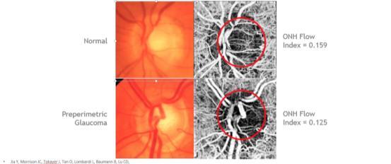
 34 40") Review article SOUTH-EAST EUROPEAN JOURNAL of OPHTHALMOLOGY 2015; 1 (1) 34 40 Retinal nerve fiber layer versus peripapillary capillary density assessment A powerful tool for detecting optic nerve head
Review article SOUTH-EAST EUROPEAN JOURNAL of OPHTHALMOLOGY 2015; 1 (1) 34 40 Retinal nerve fiber layer versus peripapillary capillary density assessment A powerful tool for detecting optic nerve head
November Volume 35 - Issue 11
 November 2015 - Volume 35 - Issue 11 pp: 2161-2431,e67-e72 Editorial Optical Coherence Tomography Angiography Spaide, Richard F.; Fujimoto, James G.; Waheed, Nadia K. Original Study IMAGE ARTIFACTS IN
Go With the Flow: An OCT Angiography Primer Lorne Yudcovitch, OD, MS, FAAO
 Go With the Flow: An OCT Angiography Primer Lorne Yudcovitch, OD, MS, FAAO yudcovil@pacificu.edu OCT Angiography (OCTA) History 2000 - First Doppler flowimetry OCT on human retina 2005 Speckle analysis
TOPCON EURETINA Clinical Advances and Applications With Swept Source OCT and Angiography. JANUARY/FEBRUARY 2019 VOL. 17, NO.
 SUPPLEMENT TO SPONSORED BY JANUARY/FEBRUARY 2019 VOL. 17, NO. 1 TOPCON EURETINA 2018 Clinical Advances and Applications With Swept Source OCT and Angiography. This supplement summarizes highlights from
Case Report: Indocyanine Green Dye Leakage from Retinal Artery in Branch Retinal Vein Occlusion
 Case Report: Indocyanine Green Dye Leakage from Retinal Artery in Branch Retinal Vein Occlusion Hiroki Fujita, Kyoko Ohno-Matsui, Soh Futagami and Takashi Tokoro Department of Visual Science, Tokyo Medical
Optical Coherence Tomography Angiography in Central Retinal Vein Occlusion: Correlation Between the Foveal Avascular Zone and Visual Acuity METHODS
 Special Issue Optical Coherence Tomography Angiography in Central Retinal Vein Occlusion: Correlation Between the Foveal Avascular Zone and Visual Acuity Manuel Casselholm de Salles, Anders Kvanta, Urban
Clinical Study Optical Coherence Tomography and Optical Coherence Tomography Angiography in Monitoring Coats Disease
 Hindawi Ophthalmology Volume 2017, Article ID 7849243, 8 pages https://doi.org/10.1155/2017/7849243 Clinical Study Optical Coherence Tomography and Optical Coherence Tomography Angiography in Monitoring
Clinically Significant Macular Edema (CSME)
") Clinically Significant Macular Edema (CSME) 1 Clinically Significant Macular Edema (CSME) Sadrina T. Shaw OMT I Student July 26, 2014 Advisor: Dr. Uwaydat Clinically Significant Macular Edema (CSME) 2
Clinically Significant Macular Edema (CSME) 1 Clinically Significant Macular Edema (CSME) Sadrina T. Shaw OMT I Student July 26, 2014 Advisor: Dr. Uwaydat Clinically Significant Macular Edema (CSME) 2
The Evaluation of Diabetic Macular Ischemia Using Optical Coherence Tomography Angiography
 Retina The Evaluation of Diabetic Macular Ischemia Using Optical Coherence Tomography Angiography Patrick D. Bradley, 1 Dawn A. Sim, 1 Pearse A. Keane, 1 João Cardoso, 1,2 Rupesh Agrawal, 1 Adnan Tufail,
Retinal Capillary Network and Foveal Avascular Zone in Eyes with Vein Occlusion and Fellow Eyes Analyzed With Optical Coherence Tomography Angiography
 Retinal Capillary Network and Foveal Avascular Zone in Eyes with Vein Occlusion and Fellow Eyes Analyzed With Optical Coherence Tomography Angiography The MIT Faculty has made this article openly available.
OCT Interpretation. Financial Disclosure. Jay M. Haynie, OD, FAAO. OCT Image Layers 7/21/2014
 OCT Interpretation Jay M. Haynie, OD, FAAO Financial Disclosure I have received honoraria or am on the advisory board for the following companies: Olympia Tacoma Renton Kennewick - Washington Carl Zeiss
Dr/ Marwa Abdellah EOS /16/2018. Dr/ Marwa Abdellah EOS When do you ask Fluorescein angiography for optic disc diseases???
 When do you ask Fluorescein angiography for optic disc diseases??? 1 NORMAL OPTIC DISC The normal optic disc on fluorescein angiography is fluorescent due to filling of vessels arising from the posterior
Research Article Diabetic Macular Ischemia Diagnosis: Comparison between Optical Coherence Tomography Angiography and Fluorescein Angiography
 Ophthalmology Volume 2016, Article ID 3989310, 6 pages http://dx.doi.org/10.1155/2016/3989310 Research Article Diabetic Macular Ischemia Diagnosis: Comparison between Optical Coherence Tomography Angiography
Building The Retina Company
 Building The Retina Company Optos devices produce ultra-widefield (UWF ), high resolution images (optomap ) of approximately 82% (200 ) of the retina. A single optomap can document the retina from the
Authors. Introduction. Introduction. Materials and Methods. Objective 10/27/2015
 Idiopathic Polypoidal Choroidal Vasculopathy (IPCV) in Thai Population Presenting with Choroidal Neovascularization (CNV) A multicenter study Authors Yonrawee Piyacomn 1, Chavakij Bhoomibunchoo 1, Yosanan
Diabetic Retinopatathy
 Diabetic Retinopatathy Jay M. Haynie, OD, FAAO Financial Disclosure I have received honoraria or am on the advisory board for the following companies: Carl Zeiss Meditec Arctic DX Macula Risk Advanced
LEE EYE CENTRE. YOUR VISION, OUR PASSION LEC EyeNews
 LEE EYE CENTRE YOUR VISION, OUR PASSION LEC EyeNews FOR INTERNAL CIRCULATION ONLY www.lec.com.my ISSUE 51/003 SEPT OCT 2017 The American Society of Cataract and Refractive Surgery is one of the leading
Optical Coherence Tomograpic Features in Idiopathic Retinitis, Vasculitis, Aneurysms and Neuroretinitis (IRVAN)
") Columbia International Publishing Journal of Ophthalmic Research (2014) Research Article Optical Coherence Tomograpic Features in Idiopathic Retinitis, Vasculitis, Aneurysms and Neuroretinitis (IRVAN)
Columbia International Publishing Journal of Ophthalmic Research (2014) Research Article Optical Coherence Tomograpic Features in Idiopathic Retinitis, Vasculitis, Aneurysms and Neuroretinitis (IRVAN)
Posterior Segment Update
 Posterior Segment Update Featured Speaker: Dr. Kyle Cheatham, FAAO, DIP ABO DISCLOSURE STATEMENT We have no direct financial or proprietary interest in any companies, products or services mentioned in
The Quick Guide to OCT Mastery 50 Real Cases with Expert Analysis
 OPTICAL COHERENCE TOMOGRAPHY The Quick Guide to OCT Mastery 50 Real Cases with Expert Analysis VOL 1 Sanjay Sharma, MD, FRCS, MSc (Epid), MBA Ophthalmologist, Epidemiologist Queen s University, Canada
Leo Semes, OD, FAAO UAB Optometry
 Leo Semes, OD, FAAO UAB Optometry Safe; inert Has long track record - over 45 years Mixes with plasma and highlights blood vessel compromise Using specific exciting (490 nm)and absorption (510 nm) filters
Clinical Study Choroidal Thickness in Eyes with Unilateral Ocular Ischemic Syndrome
 Hindawi Publishing Corporation Journal of Ophthalmology Volume 215, Article ID 62372, 5 pages http://dx.doi.org/1.1155/215/62372 Clinical Study Choroidal Thickness in Eyes with Unilateral Ocular Ischemic
Optical coherence tomographic angiography (OCTA) is a
 is a") Retina Distinctive Analysis of Macular Superficial Capillaries and Large Vessels Using Optical Coherence Tomographic Angiography in Healthy and Diabetic Eyes Jianqin Lei, 1 Enhui Yi, 2 Yan Suo, 3 Cheng
Retina Distinctive Analysis of Macular Superficial Capillaries and Large Vessels Using Optical Coherence Tomographic Angiography in Healthy and Diabetic Eyes Jianqin Lei, 1 Enhui Yi, 2 Yan Suo, 3 Cheng
Mark Dunbar: Disclosure
 Important Things to Understand About OCT Mark T. Dunbar, O.D., F.A.A.O. Bascom Palmer Eye Institute University of Miami, School of Medicine Mark Dunbar: Disclosure Optometry Advisory Board for: Allergan
Optical coherence tomography angiography of foveal hypoplasia
 Optical coherence tomography angiography of foveal hypoplasia Kaivon Pakzad-Vaezi, MD 1, Pearse Keane, MD 1,2, João Nobre Cardoso, MD 1, Catherine Egan, MD 1, Adnan Tufail, MD 1. Moorfields Eye Hospital
3/6/2014. Hoda MH Mostafa MD Associate Professor of Ophthalmology Cairo University. The author has no proprietary interest. Today s Objectives
 Hoda MH Mostafa MD Associate Professor of Ophthalmology Cairo University The author has no proprietary interest Today s Objectives Identify the CLINICAL SCENARIOS IN MACULAR EDEMA where OCT plays a MAJOR
Neovascular Glaucoma Associated with Cilioretinal Artery Occlusion Combined with Perfused Central Retinal Vein Occlusion
 Neovascular Glaucoma Associated with Cilioretinal Artery Occlusion Combined with Perfused Central Retinal Vein Occlusion Man-Seong Seo,* Jae-Moon Woo* and Jeong-Jin Seo *Department of Ophthalmology, Chonnam
Do You See What I See!!! Shane R. Kannarr, OD
 Do You See What I See!!! Shane R. Kannarr, OD skannarr@kannarreyecare.com Define Specialty Testing Additional Test to: Prove/Disprove Diagnosis To monitor progression of a condition To document a condition
The Common Clinical Competency Framework for Non-medical Ophthalmic Healthcare Professionals in Secondary Care
 The Common Clinical Competency Framework for Non-medical Ophthalmic Healthcare Professionals in Secondary Care Cataract November 2016 Association of Health Professions in Ophthalmology General basic competences
Stabilization of visual acuity with photodynamic therapy in eyes with chorioretinal anastomoses
 Graefe s Arch Clin Exp Ophthalmol (2004) 242:368 376 CLINICAL INVESTIGATION DOI 10.1007/s00417-003-0844-0 Rufino M. Silva José R. Faria de Abreu António Travassos José G. Cunha-Vaz Stabilization of visual
Jay M. Haynie, O.D.; F.A.A.O. Olympia Tacoma Renton Kennewick Washington
 Jay M. Haynie, O.D.; F.A.A.O. Olympia Tacoma Renton Kennewick Washington I Jay M. Haynie, OD, FAAO have received honoraria from the following companies: Reichert Technologies Notal Vision Carl Zeiss Meditec
NIH Public Access Author Manuscript Ophthalmology. Author manuscript; available in PMC 2015 January 01.
 NIH Public Access Author Manuscript Published in final edited form as: Ophthalmology. 2014 January ; 121(1): 180 187. doi:10.1016/j.ophtha.2013.09.002. Phase-Contrast Optical Coherence Tomography: A New
Optical Coherence Tomography: Pearls for the Anterior Segment Surgeon Basic Science Michael Stewart, M.D.
 Optical Coherence Tomography: Pearls for the Anterior Segment Surgeon Basic Science Michael Stewart, M.D. Disclosure OCT Optical Coherence Tomography No relevant financial relationships I will refer to
D JO. Optical Coherence Tomography Angiography: Principles and Application in Retinal Diseases
 E-ISSN 2454-2784 Abstract Optical Coherence Tomography Angiography: Principles and Application in Retinal Diseases Gabriella Moraes 1, Livia Faes 1, Bishwanath Pal 2 1 Research Centre at Moorfields Eye
Advances in OCT Murray Fingeret, OD
 Disclosures Advances in OCT Murray Fingeret, OD Consultant Alcon, Allergan, Bausch & Lomb, Carl Zeiss Meditec, Diopsys, Heidelberg Engineering, Reichert, Topcon Currently Approved OCT Devices OCT Devices
Experience Spectacular Retinal Imaging with the new NIDEK F-10 Digital Ophthalmoscope
 Experience Spectacular Retinal Imaging with the new NIDEK F-10 Digital Ophthalmoscope The F-10 was developed to give Ophthalmologists a high definition (HD) diagnostic imaging system. Designed to provide
Retinal Complications of Obstructive Sleep Apnea A Growing Concern!
 Retinal Complications of Obstructive Sleep Apnea A Growing Concern! Jay M. Haynie, OD, FAAO Financial Disclosure I have received honoraria or am on the advisory board for the following companies: Carl
EFFICACY OF ANTI-VASCULAR ENDOTHELIAL GROWTH FACTOR AGENTS IN RETINAL DISORDER FOR BETTER VISUAL ACUITY
 EFFICACY OF ANTI-VASCULAR ENDOTHELIAL GROWTH FACTOR AGENTS IN RETINAL DISORDER FOR BETTER VISUAL ACUITY Diwakar chaudhary *1, 2, Hu shuqiong, Long Yuan and Xiong kun 1 Yangtze University, 1 Nanhuan Road
Evaluation of efficacy of eplerenone in the management of chronic central serous choroidoretinopathy
 Original article: Evaluation of efficacy of eplerenone in the management of chronic central serous choroidoretinopathy Dr. Sushant Madaan* Department of Ophthalmology, NIMS Medical College and Hopsital,Jaipur,
Case Report Optical Coherence Tomography Angiography of Macular Telangiectasia Type 2 with Associated Subretinal Neovascular Membrane
 Hindawi Case Reports in Ophthalmological Medicine Volume 2017, Article ID 8186134, 4 pages https://doi.org/10.1155/2017/8186134 Case Report Optical Coherence Tomography Angiography of Macular Telangiectasia
What Is O.C.T. and Why Should I Give A Rip? OCT & Me How Optical Coherence Tomography Changed the Life of a Small Town Optometrist 5/19/2014
 OCT & Me How Optical Coherence Tomography Changed the Life of a Small Town Optometrist Email: myoder@wcoil.com Mark A. Yoder, O.D. 107 N. Main Street PO Box 123 Bluffton, OH 45817 @yoderod 115.02 Histoplasma
Diabetic Retinopathy A Presentation for the Public
 Diabetic Retinopathy A Presentation for the Public Ray M. Balyeat, MD The Eye Institute Tulsa, Oklahoma The Healthy Eye Light rays enter the eye through the cornea, pupil and lens. These light rays are
Role of Fluorescein angiography in evaluation of posterior segment disorders
 Original Article Role of Fluorescein angiography in evaluation of posterior segment disorders Arvind R, Surendar S 2, Ch. Jagan Mohan Rao 3 Associate Professor, 2 Postgraduate student, 3 Senior resident,
Fundus Autofluorescence
 Brittany Bateman, BS Fundus autofluorescence imaging is used to record fluorescence that may occur naturally in ocular structures or as a byproduct of a disease process. This technique allows the topographic
Venous Occlusive Diseases
 Venous Occlusive Diseases Bruce R. Saran, MD Adjunct Assistant Clinical Professor of Medicine Scheie Eye Institute University of Pennsylvania School of Medicine Philadelphia, PA -a division of: RVO Demographics
OCT Angiography: The Newest Frontier for the Revolutionary Technology
 Supplement April 2015 OCT Angiography: The Newest Frontier for the Revolutionary Technology OCT Angiography is a new non-invasive, motion contrast micro-vascular imaging modality. Based on two patented
Vascular Disease Ocular Manifestations of Systemic Hypertension
 Vascular Disease Ocular Manifestations of Systemic Hypertension Maynard L. Pohl, OD, FAAO Pacific Cataract & Laser Institute 10500 NE 8 th Street, Suite 1650 Bellevue, WA 98004 USA 425-462-7664 Cerebrovascular
CENTRAL SEROUS CHORIORETINOPATHY (CSC) IS
 IS") Association Between the Efficacy of Half-Dose Photodynamic Therapy With Indocyanine Green Angiography and Optical Coherence Tomography Findings in the Treatment of Central Serous Chorioretinopathy MASSIMO
Association Between the Efficacy of Half-Dose Photodynamic Therapy With Indocyanine Green Angiography and Optical Coherence Tomography Findings in the Treatment of Central Serous Chorioretinopathy MASSIMO
A COMPLETE GUIDE TO THE CLARUS 500 ULTRA-WIDEFIELD RETINAL CAMERA
 A COMPLETE GUIDE TO THE CLARUS 500 ULTRA-WIDEFIELD RETINAL CAMERA BY DANIEL EPSHTIEN, OD Originally published on newgradoptometry.com Ultra-widefield retinal imaging has had a contentious path to acceptance
The role of OCT-A in retinal disease management
 Graefe's Archive for Clinical and Experimental Ophthalmology (2018) 256:2019 2026 https://doi.org/10.1007/s00417-018-4109-3 REVIEW ARTICLE The role of OCT-A in retinal disease management Francisco J. Rodríguez
Measurement of Subfoveal Choroidal Thickness Using Spectral Domain Optical Coherence Tomography
 c l i n i c a l s c i e n c e Measurement of Subfoveal Choroidal Thickness Using Spectral Domain Optical Coherence Tomography Emily A. McCourt, MD; Brian C. Cadena, PhD; Cullen J. Barnett, CRA; Antonio
Optical coherence tomography (OCT) angiography
 angiography") CLINICAL TRIAL ENDPOINTS FOR OPTICAL COHERENCE TOMOGRAPHY ANGIOGRAPHY IN NEOVASCULAR AGE- RELATED MACULAR DEGENERATION EMILY D. COLE, BS,* DANIELA FERRARA, MD, PHD,* EDUARDO A. NOVAIS, MD,* RICARDO N.
CLINICAL TRIAL ENDPOINTS FOR OPTICAL COHERENCE TOMOGRAPHY ANGIOGRAPHY IN NEOVASCULAR AGE- RELATED MACULAR DEGENERATION EMILY D. COLE, BS,* DANIELA FERRARA, MD, PHD,* EDUARDO A. NOVAIS, MD,* RICARDO N.
Widefield Retinal Imaging with Auto Fluorescence Technology in the Optometric Practice
 Widefield Retinal Imaging with Auto Fluorescence Technology in the Optometric Practice This course will define ultra-widefield retinal imaging and autofluorescence for the attendee. Will show how it is
4/27/2010 INTRODUCTION TO RETINAL VASCULAR DISEASE VENOUS/VENULAR CENTRAL RETINAL VEIN OBSTRUCTION / CRVO ADDITIONAL FEATURES /COMPLICATIONS
 INTRODUCTION TO RETINAL VASCULAR DISEASE VENOUS/VENULAR Leo Semes, OD Professor, UAB Optometry 2 CENTRAL RETINAL VEIN OBSTRUCTION CENTRAL RETINAL VEIN OBSTRUCTION / OCCLUSION (CRVO) obstruction of the
PLEX Elite 9000 from ZEISS Swept-Source OCT
 PLEX Elite 9000 from ZEISS Swept-Source OCT Uncovering the undiscovered. ZEISS PLEX Elite 9000 // INNOVATION MADE BY ZEISS 2 Ultra-wide angiography En face montage Image courtesy of Prof. G. Querques,
Disease-Specific Fluorescein Angiography
 Ruth E. Picchiottino, CRA Disease-Specific Fluorescein Angiography 15 Disease-Specific Fluorescein Angiography Recommendations for tailoring retinal fluorescein angiography to diabetic retinopathy, macular
Diagnosis and treatment of diabetic retinopathy. Blake Cooper MD Ophthalmologist Vitreoretinal Surgeon Retina Associates Kansas City
 Diagnosis and treatment of diabetic retinopathy Blake Cooper MD Ophthalmologist Vitreoretinal Surgeon Retina Associates Kansas City Disclosures Consulted for Novo Nordisk 2017,2018. Will be discussing
World Sight Day Case Studies. Mark Frost Screening Manager South East London DESP
 World Sight Day 2015 Case Studies Mark Frost Screening Manager South East London DESP Introduction All of the following cases have been identified in our screening programme over the last 3 years. The
International Journal of Health Sciences and Research ISSN:
 International Journal of Health Sciences and Research www.ijhsr.org ISSN: 2249-9571 Original Research Article A Multivariate Analysis of Intravitreal Injection of Anti-VEGF Bevacizumab in the Treatment
The Common Clinical Competency Framework for Non-medical Ophthalmic Healthcare Professionals in Secondary Care
 The Common Clinical Competency Framework for Non-medical Ophthalmic Healthcare Professionals in Secondary Care Acute & Emergency Care November 2016 Association of Health Professions in Ophthalmology General
The diagnostic value of optical coherence tomography angiography in diabetic retinopathy: a systematic review
 https://doi.org/10.1007/s10792-018-1034-8 (0456789().,-volV) (0456789().,-volV) REVIEW The diagnostic value of optical coherence tomography angiography in diabetic retinopathy: a systematic review David