Microperimetric Sensitivity in Patients on Hydroxychloroquine (Plaquenil) Therapy
|
|
|
- Letitia Williamson
- 5 years ago
- Views:
Transcription
1 Microperimetric Sensitivity in Patients on Hydroxychloroquine (Plaquenil) Therapy Renu V. Jivrajka 1*, Mohamed A. Genead 2,3*, J. Jason McAnany 1, Clement C. Chow 1, William F. Mieler 1 1. Department of Ophthalmology and Visual Sciences, University of Illinois Chicago, Chicago IL 2. Pangere Center for Hereditary Retinal Diseases, The Chicago Lighthouse for People Who Are Blind or Visually Impaired, Chicago, IL 3. Allergan Inc., Irvine, CA *These authors contributed equally Corresponding Author: Renu Jivrajka, MD Department of Ophthalmology and Visual Sciences University of Illinois Chicago 1855 W Taylor ST, MC 648 Chicago, IL (312) Fax: (312) renujiv@gmail.com Funds from the Foundation Fighting Blindness, Columbia, Maryland; Pangere Corporation, Gary, Indiana; Grant Healthcare Foundation, Lake Forest, Illinois; NIH core grant EY01792; NIH research grant R00EY (JM). Illinois Society for Prevention of Blindness The sponsor or funding organization had no role in the design or conduct of this research. No conflicting relationship exists for any author. This material was previously presented at the Associate for Research in Vision and Ophthalmology, Fort Lauderdale, May Running head: Microperimetry sensitivity in Plaquenil patients
2 Abstract Purpose: The purpose of this study was to measure macular sensitivity using microperimetry in patients on Plaquenil therapy without evidence of retinopathy as assessed by recommended screening standards. Methods: Sixteen patients from a clinical practice treated with 200 or 400 mg/day of Plaquenil for more than 5 years, without visual complaints (visual acuity 20/25 or better), and without a history of diabetes or macular disease were included. Participants underwent a complete ophthalmic examination with spectral-domain optical coherence tomography (SD-OCT), 10-2 Humphrey visual field (HVF), fundus autofluoresecene (FAF), multifocal electroretinography (mferg) and microperimetry that covered the central 12 o of the visual field. Ten age-similar, visually normal subjects served as controls. Results: The average age of the study cohort was of 54.5 years, with an average daily Plaquenil dose of 4.00 mg/kg/day (range, mg/kg/day) and an average cumulative dose of 1485 grams (range, 256 to 3650 grams). All patients had normal ocular exams, and no evidence of retinopathy based on 10-2 HVF, FAF, mferg, and SD-OCT. The mean retinal sensitivity by microperimetry, was 15.0 db (OD) and 14.6 db (OS). The overall mean microperimetry sensitivity of the patients (14.7±1.9 db) was significantly lower (p < 0.001) than that of the controls
3 (16.5±2.1 db). Conclusions: Patients on Plaquenil without clinical evidence of retinal toxicity can have reduced retinal sensitivity, as assessed by microperimetry. The mean sensitivity difference between the patients and controls suggests that microperimetry can provide important information regarding visual function in patients on Plaquenil therapy. Key Words: plaquenil toxicity, microperimetry, retinopathy, screening
4 INTRODUCTION Thousands of patients currently use hydroxychloroquine (Plaquenil; Sanofi- Aventis, Bridgewater, New Jersey) for the management of various connective tissue disorders such as rheumatoid arthritis (RA) and systemic lupus erythematosus (SLE). 1 This medication is known to have a high affinity for binding to melanin granules and can therefore accumulate in the iris, choroid, ciliary body, and retinal pigment epithelium (RPE) on prolonged usage. 1-3 Although not entirely understood, retinal toxicity often begins with loss of rods and cones and degenerative changes of the RPE. Given the concern for retinal toxicity from Plaquenil use, early detection is an important responsibility faced by ophthalmologists. The American Academy of Ophthalmology has recently published revised screening guidelines to identify tests that provide the most useful information in detecting retinal toxicity from the use of Plaquenil. 1 The current standard of care based on this report involves a baseline ocular examination as well as 10-2 Humphrey visual fields (HVF) and one of three possible adjunct tests including spectral-domain optical coherence tomography (SD-OCT), fundus autofluoresecene (FAF), or multifocal electroretinography (mferg). Color vision testing and the Amsler grid are no longer recommended screening procedures. As the risk of toxicity approaches 1% after more than 7 years of Plaquenil usage, annual screening is recommended for patients
5 exceeding 5 years of exposure. 4 Early retinopathy is associated with an acquired paracentral visual field threshold elevation without any observable fundus changes. 5,6 Current screening methods for Plaquenil retinopathy are limited in that some detect toxicity only after significant structural damage has occurred, as seen on funduscopic examination, SD-OCT, and FAF. The need for comprehensive ophthalmic screening and early detection of retinopathy plays a critical role in the surveillance of patients on chronic Plaquenil therapy. Patients often exceed the recommended daily dosage based on lean body weight, even when prescribed the usual daily dose of 400 mg/day, leading to devastating visual consequences. 7 From a functional standpoint, 10-2 HVF is a subjective test that is widely available, although test reliability is limited by fixation instability and patient cooperation. Recently, mferg has been shown to detect subtle changes in earlier stages of toxicity. 8 However, mferg is limited by clinical availability, patient cooperation, specialized training for administration and interpretation, and cost. A non-invasive, sensitive, and reproducible test such as microperimetry could be another option for assessing retinal sensitivity in Plaquenil users. Although there exists no single definitive test, it is important to investigate detailed and comprehensive screening methods with the goal of detecting retinopathy, defined as structural or functional changes, before irreversible vision loss occurs.
6 The introduction of microperimetry testing with a scanning laser ophthalmoscope (SLO) using the OPKO microperimeter (OPKO Instrumentations, Miami, Florida, USA) provides clinicians with an additional tool to evaluate macular function in conjunction with SD-OCT. The microperimeter incorporates automated ocular tracking capabilities, which compensate for eye movements under real time conditions and permits good test reliability and reproducibility. 9 The operating system directly relates perimetric sensitivity values to SLO infrared images and associated SD-OCT thickness values by superimposing the acquired data, thus allowing direct correlations of structural and functional abnormalities. The purpose of this study is to examine retinal sensitivity and thickness in patients who are on Plaquenil for more than 5 years with no evidence of retinopathy based on 10-2 HVF, SD-OCT, FAF andmferg testing. PATIENTS AND METHODS Patient Selection: All patients were seen at a clinical practice and had been taking Plaquenil (100 to 400 mg/day) for a minimum of 5 years (range, 5-25 years) prior to presentation. These patients had no visual complaints and no clinical evidence of retinopathy. The study and data collection were carried out with prospective approval from an Institutional Review Board at the University of Illinois Chicago, informed consent
7 for the research was obtained from the subjects, and the study is in accordance with HIPAA regulations. Medical history including reason for Plaquenil usage was recorded from the patient s medical record. Ocular exam details including bestcorrected visual acuity (BCVA), intraocular pressure measurements using Goldmann applanation tonometry, color vision testing using Ishihara pseudoisochromatic color plates, slit-lamp biomicroscopy, and dilated fundus examination were recorded. Patients also underwent testing using the Humphrey 10-2 visual field, Heidelberg Spectralis SD-OCT, and Heidelberg Spectralis FAF, and mferg (VERIS system). Patients with visual complaints or BCVA worse than 20/25, history of diabetes or hypertension, weight fluctuation greater than 20% within 1 year, or any macular diseases including Plaquenil retinopathy were excluded from the study. Patients who met the inclusion criteria and had normal results from the above tests underwent microperimetry and SD-OCT testing with the OPKO instrument. Heidelberg SD-OCT and FAF Testing: Both SD-OCT and FAF were performed by the same skilled technician using a commercially available instrument (Spectralis; Heidelberg Engineering, Heidelberg, Germany). High resolution OCT images were obtained using a pattern size of 20 degrees (5.9 mm). A total of 19 B-scans were obtained from each eye. Real time FAF images were recorded over a 30 field of view. An excitatory argon
8 laser beam at 488 nm and a barrier filter at 500 nm were coupled in a collimating system for detecting FAF emission at wavelengths above 500 nm. To more optimally visualize the distribution of FAF intensities over the fundus, the pixel value distribution was normalized after calculation of the mean image. All patients underwent initial screening using the Spectralis SD-OCT unit prior to referral on a subsequent visit for MP testing and repeat SD-OCT testing using the OPKO Spectral OCT/SLO system. Microperimetry Testing: Microperimetry was performed using the OPKO Spectral OCT/SLO system by the same examiner (M.A.G.). All patients underwent testing in both eyes, with the exception of one patient who declined testing in the second eye. For those patients with reduced retinal sensitivities on initial testing, reproducibility was confirmed by a second test on each eye. Following dilation with 2.5% phenylephrine and 1% tropicamide, the subjects were seated in a darkened room and instructed to fixate on a red square with the tested eye while the non-tested eye was patched. Patients were then asked to press a button each time they saw the stimulus, which consisted of a spot of light that was equivalent in size to the Goldmann III (area 4 mm 2, diameter 26 minute of arc or 0.4 degrees). The stimulus was presented for 200 milliseconds using a 4-2 test strategy to estimate threshold. The Polar 3 testing grid was used in all patients, which is a standardized
9 grid of 28 points forming three concentric circles (2.3 degrees, 6.6 degrees, and 11 degrees). The inner circle is made of 4 points, while the middle and outer circles are composed of 12 points each. Sensitivity values were measured in 10 agesimilar, visually normal subjects with the same instrumentation for comparison with our patient cohort. SD-OCT Retinal Thickness Mapping: SD-OCT was performed using the spectral OCT/SLO system (OPKO instrumentations, Miami, FL) to obtain both OCT and SLO images with an axial resolution of < 10 microns, and a transverse resolution of 20 microns (in tissue). The system uses an SLO fundus image for alignment, orientation, and registration of the OCT image topographic maps. Both the Line scan (B-scan) and the 3- Dimensional Retinal Topography scan protocols were used for image acquisition. The Line scan mode allows the capture of cross-sectional B-scan OCT images of the vitreoretinal, retinal, and chorioretinal structures. A red scanning line on the SLO image indicated the exact location of the cross sectional OCT image. We used the Max Frame Count of 32 frames. The Max Frame Count is the maximum sequentially captured frames of OCT and SLO images, which are captured and displayed as individual frames. The 3D Retinal Topography mode covers an area of 9.0 x 9.0 mm with a 2.0 mm depth. After retinal thickness and microperimetry maps were generated, the images were aligned using native
10 software so that the retinal thickness at each MP test location is displayed. The OCT data acquired by the OPKO system were used for analysis in the present study. Multifocal ERG Testing: MfERG was performed on both eyes in 14 of 16 subjects using the International Society for Clinical Electrophysiology of Vision (ISCEV) guide lines. 10 Two patients were lost to follow up prior to completing mferg testing. Following dilation using 2.5 % phenylephrine and 1% tropicamide, recordings were performed unilaterally using a unipolar Burian-Allen contact lens electrode. MfERGs were recorded and analyzed using the VERIS Clinic system (Electro-Diagnostic Imaging, Inc. Redwood, CA, USA). Testing was completed in a darkened room by the same trained technician for all subjects. The stimulus consisted of a black-and-white pattern of 61 hexagons that was presented on a monitor (200 cd/m 2 for white, 99.3% contrast). The duration of data acquisition involved eight short intervals lasting a total of 4 to 5 minutes. Data Analysis: A three-way analysis of variance (ANOVA), with Bonferroni-corrected follow-up comparisons, was performed to examine differences in retinal sensitivity, as
11 assessed by microperimetry, between the patient and control groups (with a p value <0.05 considered significant). Subject group (patient vs. control), stimulus eccentricity (inner, middle, outer ring), and stimulus location (1 o clock through 12 o clock) were included as main factors. RESULTS Demographic Characteristics: The mean age of the patients was 54.5 ± 10.3 years (range, years) with 15 female subjects and 1 male subject. Twelve (75%) of the subjects were African American, 3 (19%) were Hispanic, and 1 (6%) was Asian. Of the 16 patients with a history of Plaquenil usage, 11 had a diagnosis of SLE, 4 with RA, and one with dermatomyositis (Table 1). The mean age of the visually normal control subjects was 53.4 ± 11.9 years (range, years). The mean age of the patients did not differ significantly from that of the controls (p = 0.81). The duration of Plaquenil exposure ranged from 5 to 25 years (median, 11 years). The average maximum daily dose for the study population was 4.00 mg/kg/day (range, mg/kg/day) with an average cumulative dose of 1485 grams (range, grams). The BCVA ranged from to 0.15 logmar (Snellen acuity equivalent of, 20/20 +3 to 20/25-3 ). The funduscopic exam was normal in all
12 subjects, as were the results from color vision testing. The 10-2 Humphrey visual field results were graded by 3 authors independently (M.A.G, C.C.C, and R.V.J).There was no evidence of retinopathy based on the pattern deviation maps, but, overall, sensitivity was reduced slightly for the patients, based on the mean deviation value. One patient had scattered outer rim defects on her visual field that were considered to be within the normal range. Both FAF and SD-OCT exams, graded by 3 retina specialists independently (M.A.G., C.C.C., and W.F.M.), showed normal autofluorescence and retinal structures and thickness, respectively. Two patients were noted to have few extrafoveal drusen outside the Polar 3 test grid, on FAF and SD-OCT that correlated with their funduscopic exams. Of the fourteen patients who underwent mferg testing a qualitative analysis of the results indicated that all had a normal waveform shape. (Table 2) Microperimetry Data: The overall mean sensitivity values for the right eye (15.0 ± 1.1 db) and left eye (14.6 ± 1.2 db) of the 15 patients who had measurements in both eyes were not significantly different (t = 0.87, p = 0.40). Consequently, data from the left eye were converted to right eye format and the data from the two eyes of each patient were averaged. All further analysis was based on the mean sensitivity value of the two eyes. The microperimetry sensitivity data are shown in Figure 1 for the controls (open circles) and the patients (filled circles). The bars represent the 5 th
13 and 95 th percentiles for the controls (dark bars) and the patients (light bars) and the horizontal lines indicate the mean for each group. A three-way ANOVA indicated a significant difference (p < 0.001) only for the main effect of subject group (overall mean sensitivities of 16.5 ± 2.1 db and 14.7 ± 1.9 db for the controls and patients, respectively). The overall microperimetry value was below the 5 th percentile of the controls for 5 of the 16 patients (first set of bars in Figure 1). For the inner ring (second set of bars), the microperimetry value for each patient fell within the 5 th and 95 th percentiles of the controls. For the middle ring (third set of bars), 6 patients were below the 5 th percentile of the controls, and 3 patients were below the 5 th percentile for the outer ring (fourth set of bars). Bonferroni-corrected follow-up comparisons indicated that the mean sensitivity values within the inner (Figure 1: second pair of bars), middle (Figure 1: third pair of bars), and outer (Figure 1: fourth pair of bars) rings were significantly less for the patients, as compared to the controls (p = 0.001, p = 0.004, p = 0.003, for the inner, middle, and outer rings, respectively). The sensitivity values for the patients did not significantly differ at the three eccentricities (mean sensitivities of 15.8 ± 2.3 db, 15.7 ± 1.9 db, and 14.9 ± 2.3 db for the inner, middle, and outer rings, respectively) or among the retinal locations within a ring. SD-OCT data/thickness map:
14 The overall mean thickness for right eye (281.0 ± 14.2 μm) and left eye (279.6 ± 24.3 μm) of the 15 patients who had measurements in both eyes was not significantly different (t = 0.34, p = 0.74). Consequently, data from the left eye were converted to right eye format and the data from the two eyes of each patient were averaged. The overall mean thickness of the controls (287.2 ± 14.0 μm) was not significantly different from that of the patients (280.3 ± 18.3 μm) (p = 0.32). Bonferroni-corrected follow-up comparisons indicated that the mean thickness values within the inner, middle, and outer rings were not significantly different for the patients, as compared to the controls (p = 0.17, p = 0.07, p = 0.99, for the inner, middle, and outer rings, respectively). Figures 2 and 3 summarize the OCT, microperimetry, HVF pattern deviation, and FAF results for two representative patients. DISCUSSION The current standard of care based on the recommended screening guidelines for patients on Plaquenil therapy requires the use of one of three objective screening tests including SD-OCT, FAF or mferg in addition to 10-2 HVF. Careful monitoring of these patients is needed, as long-term use of Plaquenil can result in bull s eye maculopathy and loss of retinal function. Although patients with normal
15 findings under the recommended screening guidelines are considered to have normal visual function, the data from the current study suggest that these patients can, as a group, have small but statistically significant reductions in microperimetry sensitivity. All patients in the present study had 10-2 HVF pattern deviations that had no clinically significant defects, minimal risk factors for toxicity including renal disease, liver disease, advanced age, and pre-existing retinal or macular disease, yet we found a statistically significant overall reduction in mean retinal sensitivity on microperimetry (16.5 ± 2.1 db and 14.7 ± 1.9 db for the controls and patients, respectively). In addition, this cohort of patients demonstrated normal outcomes on objective tests (SD-OCT, FAF and mferg) routinely used when screening for Plaquenil toxicity. Plaquenil toxicity is typically evaluated by the 10-2 HVF pattern deviation, which can identify subtle cases. 11 However, the mean deviation value, as an average of the reduction from normal, may also provide useful information regarding overall retinal sensitivity in these patients. Comparisons of 10-2 HVF and microperimetry have received minimal attention in the literature and a direct comparison between these testing methodologies is beyond the scope of this study. However, a previous study of glaucoma patients using microperimetry and 10-2 HVF found that the results of these techniques correlate well and that in some cases, microperimetry was able to detect abnormal areas in retinal sensitivity that 10-2 HVF did not. 12 Based on this result, it was proposed that in areas of macular
16 thinning, microperimetry might be more sensitive than standard automated perimetry in detecting paracentral visual field loss in glaucoma patients. An additional advantage of the microperimetry system used in the present study is its ability to obtain SD-OCT images and superimpose retinal sensitivity values on an SLO image with SD-OCT thicknesses, thus offering the ability to correlate functional responses with anatomical structure. An early sign of toxicity on SD- OCT is parafoveal thinning, which may involve damage to inner retinal layers, but with the evolution of toxicity, there is a progressive loss of parafoveal photoreceptors long before any RPE damage is visible clinically. 13 With advanced retinopathy, anatomic changes including perifoveal loss of the inner segment-outer segment (IS-OS) junction of the photoreceptors, outer retinal thinning, and posterior displacement of the inner retina towards the RPE have been reported, which are indicative of a severe level of toxicity. 14,15 Similar patterns of Plaquenil toxicity have been described using ultra-high resolution OCT. 16 These SD-OCT features, however, are seen largely in patients with paracentral scotomas or advanced maculopathy on HVF. None of our patients had these abnormalities. In addition, the overall mean retinal thickness of the patients in the present study did not differ significantly from that of the controls (287.2 ± 14.0 μm and ± 18.3 μm for the controls and patients, respectively).
17 Several reports suggest that mferg may detect decreased retinal function, in particular, in the susceptible perifoveal retina As a measure of retinal function, mferg generates a topograghical map of electrophysiologic activity which may help pinpoint selective areas of toxicity preferential to damage by Plaquenil consumption. The most specific waveform pattern seen in patients with Plaquenil toxicity is that of paracentral amplitude loss, however patients have been known to show reduction in the central area alone, the peripheral area alone and a generalized reduction. 18,19 On the other hand, qualitative analysis of waveforms in the present series of patients, demonstrated no identifiable pattern of waveform disruption attributable to Plaquenil toxicity. Although one limitation to the study is the lack of quantitative mferg data comparing ring ratio data of hydroxychloroquine patients to normal control subjects, the ring ratios were compared to published normative data in which there was no statistical difference. 20 While it is difficult to prevent retinal damage due to Plaquenil use, the goal of screening is to detect toxicity prior to any functional vision loss. The substantial variability of HVF sensitivity among Plaquenil patients who have a wide range of drug dosage and duration suggests the need for multiple screening modalities. A recent comparative study by Marmor summarized the results of individual screening tests in detection of early retinopathy and demonstrated that different individuals are more or less sensitive to different tests. 21 This study recommended
18 using multiple testing modalities in certain patients for more definitive results when there is a concern for toxicity. In conclusion, the devastating visual consequences of Plaquenil related retinal toxicity emphasize the importance of detailed screening. Despite our relatively small sample size, the results of this study suggest that, on average, patients on prolonged Plaquenil therapy (>5 years) with normal results on recommended screening procedures can have sensitivity losses throughout the central 12 degrees of the visual field. Large scale studies are needed to identify the factors underlying the deficits in patients with sensitivity losses, determine how common the losses are, and investigate the utility of microperimetry as a tool for screening for Plaquenil toxicity. Future longitudinal studies are needed, with repeat microperimetry testing over time, to determine if the reduced microperimetry sensitivities represent early detection of Plaquenil toxicity and whether the course of treatment should be altered in patients with microperimetry sensitivity reductions.
19 ACKNOWLEDGMENT Conflict of interest: The authors declare no conflict of interest.
20 REFERENCES 1. Marmor MF, Kellner U, Lai TY, Lyons JS, Mieler WF; American Academy of Ophthalmology. Revised recommendations on screening for chloroquine and hydroxychloroquine retinopathy. Ophthalmology 2011;118: Marmor MF, Carr RE, Easterbrook M, Farjo AA, Mieler WF; American Academy of Ophthalmology. Recommendations on screening for chloroquine and hydroxychlorquine retinopathy: a report by the American Academy of Ophthalmology. Ophthalmology 2002;109: Nebbioso M, Grenga R, Karavitis P. Early detection of macular changes with multifocal ERG in patients on antimalarial drug therapy. J Ocul Pharmacol Ther 2009;25: Mavrikakis I, Sfikakis PP, Mavrikakis E, Rougas K, Nikolaou A, Kostopoulos C, et al. The incidence of irreversible retinal toxicity in patients treated with hydroxychloroquine: a reappraisal. Ophthalmology 2003;110: Browning DJ. Hydroxychloroquine and chloroquine retinopathy: screening for drug toxicity. Am J Ophthalmol 2002;133: Easterbrook M. Detection and prevention of maculopathy associated with antimalarial agents. Int Ophthalmol Clin 1999;39: Payne JF, Hubbard GB 3rd, Aaberg TM Sr, Yan J. Clinical characteristics of hydroxychloroquine retinopathy. Br J Ophthalmol 2011;95:
21 8. Lyons JS, Severns ML. Detection of early hydroxychloroquine retinal toxicity enhanced by ring ratio analysis of multifocal electroretinography. Am J Ophthalmol 2007;143: Menke MN, Sato E, Van De Velde FJ, Feke GT. Combined use of SLO microperimetry and OCT for retinal functional and structural testing. Graefes Arch Clin Exp Ophthalmol 2006;244: Marmor MF, Hood DC, Keating D, Kondo M, Seeliger MW, Miyake Y; International Society for Clinical Electrophysiology of Vision. Guidelines for basic multifocal electroretinography (mferg). Doc Ophthalmol. 2003;106: Anderson C, Blaha GR, Marx JL. Humphrey visual field findings in hydroxychloroquine toxicity. Eye (Lond) 2011;25: Lima VC, Prata TS, De Moraes CG, Kim J, Seiple W, Rosen RB et al. A comparison between microperimetry and standard achromatic perimetry of the central visual field in eyes with glaucomatous paracentral visual-field defects. Br J Ophthalmol 2010;94: Pasadhika S, Fishman GA. Effects of chronic exposure to hydroxychloroquine or chloroquine on inner retinal structures. Eye (Lond) 2010;24: Chen E, Brown DM, Benz MS, Fish RH, Wong TP, Kim RY et al. Spectral domain optical coherence tomography as an effective screening test for hydroxychloroquine retinopathy (the flying saucer sign). Clin Ophthalmol 2010; 4:
22 15. Stepien KE, Han DP, Schell J, Godara P, Rha J, Carroll J. Spectral-domain optical coherence tomography and adaptive optics may detect hydroxychloroquine retinal toxicity before symptomatic vision loss. Trans Am Ophthalmol Soc 2009;107: Rodriguez-Padilla JA, Hedges TR 3rd, Monson B, Srinivasan V, Wojtkowski M, Reichel E et al. High-speed ultra-high-resolution optical coherence tomography findings in hydroxychloroquine retinopathy. Arch Ophthalmol 2007;125: So S, Hedges T, Schuman J, Amaro Quireza ML. Evaluation of Hydroxychloroquine Retinopathy with Multifocal Electroretinography. Ophthalmic Surg Lasers Imaging 2003;34: Maturi R, Yu M, Weleber R. Multifocal Electroretinographic Evaluation of Longterm Hydroxychloroquine Users. Arch Ophthalmol 2004;122: Lai T, Chan W, Li H, Lai RY, Lam DS. Multifocal Electroretinographic Changes in Patients Receiving Hydroxychloroquine Therapy. Am J Ophthalmol 2005;140: Lyons J, Severns M. Using multifocal ERG ring ratios to detect and follow Plaquenil retinal toxicity: a review. Doc Ophthalmol 2009;118: Marmor MF. Comparison of screening procedures in hydroxychloroquine toxicity. Arch Opthalmol 2012;130:
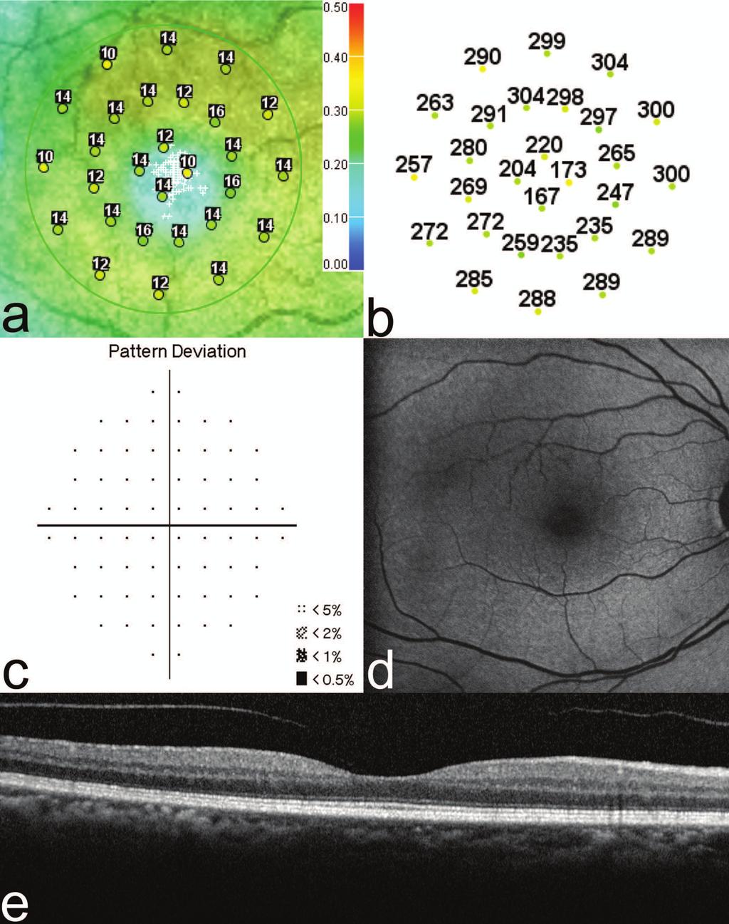
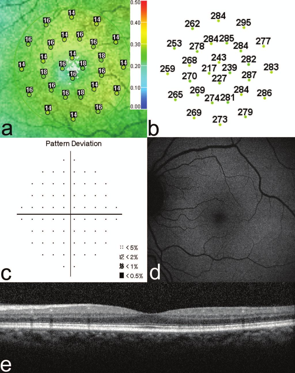
23 TITLES AND LEGENDS TO FIGURES FIGURE 1 Mean sensitivity for the control subjects (open circles) and patients (filled circles) for the verall mean and each of the three rings. The bars represent the 5 th and 95 th percentiles and the horizontal bars represent the mean for each group. FIGURE 2 Test results of a 60 year-old Hispanic female on Plaquenil for 6 years for treatment of Dermatomyositis (Maximum daily dose of 4.63 mg/kg/day and cumulative dose of 876 grams, BCVA 20/20-1 ). 2a. Microperimetry Polar 3 test grid superimposed on the scanning laser ophthalmoscope infrared image showing reduced mean retinal sensitivity 13.4 db). Small white crosses indicate fixation. 2b. Corresponding spectral domain ptical coherence tomography thickness at each of the 28 Polar 3 tested points shows o retinal thinning. 2c. Automated 10-2 Humphrey visual field shows a normal pattern eviation. 2d. Fundus autofluorescence shows no retinal pigment epithelium bnormalities. 2e. Spectral domain optical coherence tomography B-scan image shows ormal retinal thickness and an intact inner segment-outer segment junction of the photoreceptors. FIGURE 3 Test results of a 59 year-old African American female with a 17 year history of Plaquenil use for treatment of Rheumatoid Arthritis (Maximum daily dose of 6.77 mg/kg/day and cumulative dose of 2482 grams, BCVA 20/20-1 ). 3a. Microperimetry Polar 3 test grid
24 superimposed on the scanning laser ophthalmoscope infrared image showing reduced mean retinal sensitivity (14.1 db). 3b. Corresponding spectral domain optical coherence tomography thickness at each of the 28 Polar 3 tested points shows no retinal thinning. 3c. Her automated 10-2 Humphrey visual field shows a normal pattern deviation. 3d. Fundus autofluorescence shows no retinal pigment epithelium abnormalities. 3e. Spectral domain optical coherence tomography B-scan image shows normal retinal thickness and an intact inner segment-outer segment junction of the photoreceptors.
25
26
27
28 TABLE 1: SUMMARY OF CLINICAL DATA ject o Age (years) Race Sex Systemic diagnosis Years of exposure maximum daily dose (mg/kg/day) Cumulative Dose (grams) 1 76 AA F RA H F SLE/Sjogrens Asian F RA AA F RA AA F SLE H M SLE H F Dermatomyositis AA F SLE AA F SLE AA F SLE AA F SLE AA F SLE AA F RA AA F SLE AA F SLE AA F SLE : systemic lupus erythematosus, RA: rheumatoid arthritis; AA: African American H: Hispanic
29 TABLE 2: SUMMARY OF TESTING RESULTS Ishihara ject o Eye logmar Fundus Color Plates HVF 10-2 FAF mferg Retinal thickness (SD-OCT) MP (db) punctate 1 OS 0.02 drusen 17/17 full normal wnl normal OD 0 wnl 17/17 full normal wnl normal OS 0 wnl 17/17 full normal Not completed normal 16 3 OD 0 wnl 17/17 full normal Not completed normal OS 0 wnl 17/17 full normal Not completed normal OD 0.02 wnl 17/17 full normal Not completed normal OS punctate 0 drusen 17/17 full normal Not completed normal OD 0.06 wnl 17/17 full normal wnl normal OS 0 wnl 17/17 full normal wnl normal OD punctate 0.02 drusen 17/17 full normal wnl normal 15.5 temporal hypofluorescence 6 OS punctate 0 drusen 17/17 full corresponding to drusen wnl normal OD 0.02 wnl 17/17 full normal wnl normal OS 0.02 wnl 17/17 full normal wnl normal OD 0 wnl 17/17 full normal wnl normal OS 0 wnl 17/17 full normal wnl normal OD 0.02 wnl 17/17 full normal wnl normal OS extramacular 0.15 drusen 16/17 full normal wnl normal OD punctate drusen 17/17 full normal wnl normal OS punctate drusen 17/17 full normal wnl normal 14.7
30 11 OD 0 wnl 17/17 full inferior hypofluorescence corresponding to drusen wnl normal, extrafoveal drusen OS punctate 0 drusen 17/17 full inferior hypofluorescence corresponding to drusen wnl normal, extrafoveal drusen OD 0 wnl 17/17 full normal wnl normal OS 0 wnl 17/17 full normal wnl normal OD 0 wnl 17/17 full normal wnl normal OS 0.02 wnl 17/17 full normal wnl normal OD 0 wnl 17/17 full normal wnl normal OS 0 wnl 17/17 full normal wnl normal OD 0.1 wnl 17/17 full normal wnl normal OS 0 wnl 17/17 full normal wnl normal OD 0 wnl 11-Nov full normal wnl normal OS 0 wnl 11-Nov full normal wnl normal 16 : best corrected visual acuity; HVF: Humphrey visual fields; FAF: fundus autofluorescence; G: multi-focal electroretinogram; SD-OCT: spectral domain optical coherence tomography; MP: microperimetry; db: decibels
Retinal toxicity related to hydroxychloroquine in patients with systemic lupus erythematosus and rheumatoid arthritis
 Doc Ophthalmol (2017) 135:187 194 DOI 10.1007/s10633-017-9607-9 ORIGINAL RESEARCH ARTICLE Retinal toxicity related to hydroxychloroquine in patients with systemic lupus erythematosus and rheumatoid arthritis
Hydroxychloroquine: A Brief Review on Screening, Toxicity, and Progression
 Incorporating current trials and technology into clinical practice Howard F. Fine Co-Editor In 2011, the American Academy of Ophthalmology published updated guidelines on screening for retinal toxicity
Retinal toxicity associated with chronic exposure to hydroxychloroquine and its ocular screening. Review
 , pp.322-326 Retinal toxicity associated with chronic exposure to hydroxychloroquine and its ocular screening. Review Geamănu (Pancă) A*, Popa-Cherecheanu A*, Marinescu B**, Geamănu CD***, Voinea LM* *Ophthalmology
Spectral domain optical coherence tomography detects
 Spectral domain optical coherence tomography detects early stages of chloroquine retinopathy similar to multifocal electroretinography, fundus autofluorescence and near-infrared autofluorescence Simone
Fundus Autofluorescence. Jonathan A. Micieli, MD Valérie Biousse, MD
 Fundus Autofluorescence Jonathan A. Micieli, MD Valérie Biousse, MD The retinal pigment epithelium (RPE) has many important functions including phagocytosis of the photoreceptor outer segments Cone Rod
Clement C. Chow 1. Mohamed A. Genead 1. Anastasios Anastasakis 1. Felix Y. Chau¹. Gerald A. Fishman¹. Jennifer I. Lim¹
 1 Structural and Functional Correlation in Sickle Cell Retinopathy Using Spectral- Domain Optical Coherence Tomography and Scanning Laser Ophthalmoscope Microperimetry Clement C. Chow 1 Mohamed A. Genead
CLINICAL SCIENCES. Value of Red Targets and Pattern Deviation Plots in Visual Field Screening for Hydroxychloroquine Retinopathy
 CLINICAL SCIENCES Value of Targets and Pattern Deviation Plots in Visual Field Screening for Hydroxychloroquine Retinopathy Michael F. Marmor, MD; Fred Y. Chien, MD; Mark W. Johnson, MD Objective: To compare
SPECTRAL-DOMAIN OPTICAL COHERENCE TOMOGRAPHY AND ADAPTIVE OPTICS MAY DETECT HYDROXYCHLOROQUINE RETINAL TOXICITY BEFORE SYMPTOMATIC VISION LOSS
 SPECTRAL-DOMAIN OPTICAL COHERENCE TOMOGRAPHY AND ADAPTIVE OPTICS MAY DETECT HYDROXYCHLOROQUINE RETINAL TOXICITY BEFORE SYMPTOMATIC VISION LOSS BY Kimberly E. Stepien MD*, Dennis P. Han MD, Jonathan Schell
Clinical Trial Endpoints for Macular Diseases
 Clinical Trial Endpoints for Macular Diseases Developed in collaboration Learning Objective Upon completion, participants should be able to: Summarize types of biomarkers of progression and treatment response
Clinical Study Spectral-Domain Optical Coherence Tomography of Preclinical Chloroquine Maculopathy in Egyptian Rheumatoid Arthritis Patients
 Ophthalmology Volume 2015, Article ID 292357, 7 pages http://dx.doi.org/10.1155/2015/292357 Clinical Study Spectral-Domain Optical Coherence omography of Preclinical Chloroquine Maculopathy in Egyptian
Supplementary Appendix
 This appendix has been provided by the authors to give readers additional information about their work. Supplement to: Edwards TL, Jolly JK, MacLaren RE, et al.. N Engl J Med 206;374:996-8. DOI: 0.056/NEJMc50950
8/6/17. Disclosures Aerie Pharmaceuticals Alcon BioTissue Diopsys Optovue Shire
 Nathan Lighthizer, O.D., F.A.A.O. Associate Professor Assistant Dean for Clinical Care Director of Continuing Education Chief of Specialty Care Clinics Oklahoma College of Optometry Tahlequah, OK lighthiz@nsuok.edu
The Evolution of Fundus Perimetry
 The Evolution of Fundus Perimetry Company Profile CenterVue designs and manufactures highly automated medical devices for the diagnosis and management of ocular pathologies, including those that represent
Expanded spectral domain OCT findings in the early detection of hydroxychloroquine retinopathy and changes following drug cessation
 DOI 10.1186/s40942-016-0042-y International Journal of Retina and Vitreous ORIGINAL ARTICLE Open Access Expanded spectral domain OCT findings in the early detection of hydroxychloroquine retinopathy and
RETINAL TOXICITY FOLLOWING CHLOROQUINE THERAPY
 ARCH SOC ESP OFTALMOL 2007; 82: 103-108 SHORT COMMUNICATION RETINAL TOXICITY FOLLOWING CHLOROQUINE THERAPY TOXICIDAD RETINIANA SECUNDARIA A TRATAMIENTO CON CLOROQUINA FERRERAS A 1, PINILLA I 1, ABECIA
Yasser R. Serag, MD Tamer Wasfi, MD El- Saied El-Dessoukey, MD Magdi S. Moussa, MD Anselm Kampik, MD
 Microperimetric Evaluation of Brilliant Blue G- assisted Internal Limiting Membrane Peeling By Yasser R. Serag, MD Tamer Wasfi, MD El- Saied El-Dessoukey, MD Magdi S. Moussa, MD Anselm Kampik, MD The internal
Assessment of macular function by multifocal electroretinogram before and after macular hole surgery
 420 Department of Ophthalmology, Gunma University School of Medicine, Japan Y-J Si S Kishi K Aoyagi Correspondence to: Ying-Jie Si, MD, Department of Ophthalmology, Gunma University School of Medicine,
Electrodiagnostics Alphabet Soup
 Nathan Lighthizer, O.D., F.A.A.O Assistant Professor, NSUOCO Chief of Specialty Care Clinics Chief of Electrodiagnostics Clinic What is electrodiagnostics testing? Visual Pathway Basic Understanding VEP
Subject: Electroretinography
 01-92000-28 Original Effective Date: 05/15/15 Reviewed: 03/22/18 Revised: 01/01/19 Subject: Electroretinography THIS MEDICAL COVERAGE GUIDELINE IS NOT AN AUTHORIZATION, CERTIFICATION, EXPLANATION OF BENEFITS,
Cirrus TM HD-OCT. Details defi ne your decisions
 Cirrus TM HD-OCT Details defi ne your decisions 2 With high-defi nition OCT Carl Zeiss Meditec takes you beyond standard spectral domain Built on 10 years experience at the vanguard of innovation, Carl
Seong Joon Ahn *, Jooyoung Joung, Sang Hyup Lee and Byung Ro Lee *
 Ahn et al. BMC Ophthalmology (2018) 18:310 https://doi.org/10.1186/s12886-018-0985-x CASE REPORT Open Access Intravitreal dexamethasone implant therapy for the treatment of cystoid macular Oedema due to
Cirrus TM HD-OCT. Details define your decisions
 Cirrus TM HD-OCT Details define your decisions 2 With high-definition OCT Carl Zeiss Meditec takes you beyond standard spectral domain Built on 10 years experience at the vanguard of innovation, Carl Zeiss
OCT and muti-focal ERG findings in spontaneous closure of bilateral traumatic macular holes
 Doc Ophthalmol (2008) 116:159 164 DOI 10.1007/s10633-008-9113-1 CASE REPORT OCT and muti-focal ERG findings in spontaneous closure of bilateral traumatic macular holes Hongling Chen Æ Mingzhi Zhang Æ Shizhou
Retinal functional changes measured by frequency-doubling technology in patients treated with hydroxychloroquine
 Graefes Arch Clin Exp Ophthalmol (2011) 249:715 721 DOI 10.1007/s00417-010-1612-6 MISCELLANEOUS Retinal functional changes measured by frequency-doubling technology in patients treated with hydroxychloroquine
Andrew J. Barkmeier, MD; Benjamin P. Nicholson, MA; Levent Akduman, MD
 c l i n i c a l s c i e n c e Effectiveness of Laser Photocoagulation in Clinically Significant Macular Edema With Focal Versus Diffuse Parafoveal Thickening on Optical Coherence Tomography Andrew J. Barkmeier,
In office electrodiagnostics: what can it do for you
 9/6/6 In office electrodiagnostics: what can it do for you Nathan Lighthizer, O.D., F.A.A.O Assistant Professor, NSUOCO Chief of Specialty Care Clinics Chief of Electrodiagnostics Clinic Course Outline/Objective
Often asymptomatic but can cause a reduction in BCVA and distortion of vision.
 Christopher Wolfe, OD, FAAO, Dipl. ABO Epiretinal Membrane (ERM) and Vitreomacular Traction (VMT) Epiretinal membrane (macular pucker, cellophane maculopathy, premacular fibrosis) consists of a layer of
Macular Ganglion Cell Complex Measurement Using Spectral Domain Optical Coherence Tomography in Glaucoma
 Med. J. Cairo Univ., Vol. 83, No. 2, September: 67-72, 2015 www.medicaljournalofcairouniversity.net Macular Ganglion Cell Complex Measurement Using Spectral Domain Optical Coherence Tomography in Glaucoma
RETINA 2018 OBJECTIVES OCT VERY USEFUL INFORMATION SAFE AND FRIENDLY 1/11/2018 KELLY MITCHELL
 RETINA 2018 KELLY MITCHELL OBJECTIVES HIGHLIGHT NEW DIAGNOSTIC & TREATMENT OPTIONS REVIEW DIAGNOSTIC KEYS OF SELECT RETINAL DISEASES DISCUSS USE OF IMAGING AND REFERRAL RECOURSES FOR PATIENT BENEFIT OCT
Efficacy of Topical Dorzolamide for Treatment of Cystic Macular Lesions in a. Patient with Enhanced S-Cone Syndrome
 1 Efficacy of Topical Dorzolamide for Treatment of Cystic Macular Lesions in a Patient with Enhanced S-Cone Syndrome Mohamed A. Genead. Gerald A. Fishman. J. Jason McAnany Institute affiliation Department
Hydroxychloroquine and Chloroquine Retinopathy: Recommendations on Screening
 linical Guidelines Hydroxychloroquine and hloroquine Retinopathy: Recommendations on Screening February 2018 - Review date: February 2021 Executive Summary Recent data have highlighted that hydroxychloroquine
Clinical Ocular Grand Rounds
 Clinical Ocular Grand Rounds SRC 2013 Melbourne, Australia Blair Lonsberry, MS,, MEd., FAAO Diplomate, American Board of Optometry Clinic Director and Professor of Optometry Pacific University College
High Definition Spectral Domain Optical Coherence Tomography Findings in Three Patients with Solar Retinopathy and Review of the Literature
 The Open Ophthalmology Journal, 2012, 6, 29-35 29 Open Access High Definition Spectral Domain Optical Coherence Tomography Findings in Three Patients with Solar Retinopathy and Review of the Literature
Optical Coherence Tomograpic Features in Idiopathic Retinitis, Vasculitis, Aneurysms and Neuroretinitis (IRVAN)
") Columbia International Publishing Journal of Ophthalmic Research (2014) Research Article Optical Coherence Tomograpic Features in Idiopathic Retinitis, Vasculitis, Aneurysms and Neuroretinitis (IRVAN)
Columbia International Publishing Journal of Ophthalmic Research (2014) Research Article Optical Coherence Tomograpic Features in Idiopathic Retinitis, Vasculitis, Aneurysms and Neuroretinitis (IRVAN)
International Journal of Basic and Applied Physiology
 Multifocal Electroretinography in Assessment Of Diseases Of Posterior Pole Of Retina JagdeepKaur S. Dani*, Mitesh M. Sinha**, Archana H. Patel**, Anju B. Mehta ***, Geeta B. Nair**** *Associate Professor,
The MP-1 Microperimeter Clinical Applications in Retinal Pathologies
 The MP-1 Microperimeter Clinical Applications in Retinal Pathologies Nelson R. Sabates, MD Director, Retina/Vitreous Service Vice-Chairman Department of Ophthalmology University of Missouri Kansas City
Fundus Autofluorescence
 Brittany Bateman, BS Fundus autofluorescence imaging is used to record fluorescence that may occur naturally in ocular structures or as a byproduct of a disease process. This technique allows the topographic
Topical Dorzolamide for Treatment of Cystoid Macular Edema in Patients with Choroideremia
 Topical Dorzolamide for Treatment of Cystoid Macular Edema in Patients with Choroideremia Mohamed A. Genead 1,2, J. Jason McAnany 1 and Gerald A. Fishman 1,2 Institute affiliations 1 Department of Ophthalmology
High Resolution Imaging in Patients with Retinal Dystrophies
 High Resolution Imaging in Patients with Retinal Dystrophies Ophthalmic Photographers Society Annual Midyear Meeting April 2, 213 Jacque Duncan, M.D. UCSF Department of Ophthalmology How can retinal imaging
Method for comparing visual field defects to local RNFL and RGC damage seen on frequency domain OCT in patients with glaucoma.
 Method for comparing visual field defects to local RNFL and RGC damage seen on frequency domain OCT in patients with glaucoma. Donald C. Hood 1,2,* and Ali S. Raza 1 1 Department of Psychology, Columbia
Diagnosis in AMD. Managing your AMD Patients
 Managing your AMD Patients Robert W. Dunphy, O.D., F.A.A.O. Diagnosis in AMD Have suspicion Identify relative risk Conduct surveillance Biometry Utilize technology to facilitate detection of change / stability
Retinal Nerve Fiber Layer Measurements in Myopia Using Optical Coherence Tomography
 Original Article Philippine Journal of OPHTHALMOLOGY Retinal Nerve Fiber Layer Measurements in Myopia Using Optical Coherence Tomography Dennis L. del Rosario, MD and Mario M. Yatco, MD University of Santo
CLINICAL SCIENCES. Characterizing the Phenotype and Genotype of a Family With Occult Macular Dystrophy
 CLINICAL SCIENCES Characterizing the Phenotype and Genotype of a Family With Occult Macular Dystrophy Connie J. Chen, MD; Hendrik P. N. Scholl, MD, MA; David G. Birch, PhD; Takeshi Iwata, PhD; Neil R.
Ultrahigh Speed Imaging of the Rat Retina Using Ultrahigh Resolution Spectral/Fourier Domain OCT
 Ultrahigh Speed Imaging of the Rat Retina Using Ultrahigh Resolution Spectral/Fourier Domain OCT The MIT Faculty has made this article openly available. Please share how this access benefits you. Your
OCT in the Diagnosis and Follow-up of Glaucoma
 OCT in the Diagnosis and Follow-up of Glaucoma Karim A Raafat MD. Professor Of Ophthalmology Cairo University Hmmmm! Do I have Glaucoma or not?! 1 Visual Function 100% - N Gl Structure : - 5000 axon /
Clinical Study Choroidal Thickness in Eyes with Unilateral Ocular Ischemic Syndrome
 Hindawi Publishing Corporation Journal of Ophthalmology Volume 215, Article ID 62372, 5 pages http://dx.doi.org/1.1155/215/62372 Clinical Study Choroidal Thickness in Eyes with Unilateral Ocular Ischemic
What You Should Know About Acute Macular Neuroretinopathy
 What You Should Know About Acute Macular Neuroretinopathy David J. Browning MD, PhD Chong Lee BS Acute macular neuroretinopathy is a condition characterized by the sudden, painless onset of paracentral
The Effect of Pupil Dilation on Scanning Laser Polarimetry With Variable Corneal Compensation
 C L I N I C A L S C I E N C E The Effect of Pupil Dilation on Scanning Laser Polarimetry With Variable Corneal Compensation Amjad Horani, MD; Shahar Frenkel, MD, PhD; Eytan Z. Blumenthal, MD BACKGROUND
Sensitivity and Specificity of Multifocal Electroretinography in Detecting Chloroquine and Hydroxychloroquine retinal toxicity
 Sensitivity and Specificity of Multifocal Electroretinography in Detecting Chloroquine and Hydroxychloroquine retinal toxicity Sina Ahmadi Pirshahid This thesis is submitted to the Faculty of Graduate
CLINICALCASE PROVOST J, SEKFALI R, AMOROSO F, ZAMBROWSKI O, MIERE A
 CLINICALCASE PROVOST J, SEKFALI R, AMOROSO F, ZAMBROWSKI O, MIERE A Department of ophthalmology, Souied E. (MD,PhD) Centre Hospitalier Intercommunal de Créteil Université Paris Est HISTORY 13 years old
R&M Solutions
 Mohamed Hosny El-Bradey, MD., Assistant Professor of Ophthalmology, Tanta University. Wael El Haig, MD., Professor of Ophthalmology. Zagazeeg University. 1 Myopic CNV is considered the most common vision
The beneficial effects of focal photocoagulation for clinically
 Retinal Function in Diabetic Macular Edema after Focal Laser Photocoagulation Vivienne C. Greenstein, 1 Haifan Chen, 1 Donald C. Hood, 2 Karen Holopigian, 1 William Seiple, 1 and Ronald E. Carr 1 PURPOSE.
The New Frontier of Microperimetry
 Macular Integrity Assessment The New Frontier of Microperimetry Microperimetry is attracting our attention more and more as a method that is superior to standard automated perimetry for visual function
The New Frontier of Microperimetry
 Macular Integrity Assessment The New Frontier of Microperimetry Index 4 Company Profile Microperimetry is attracting our attention more and more as a method that is superior to standard automated perimetry
Overview. Macular OCT Artifact Study
 Imaging Artifacts Sarah Moyer, CRA, OCT-C Director, Ophthalmic Imaging Kittner Eye Center University of North Carolina Chapel Hill, NC Disclose financial interest now Overview Sarah s Thoughts on Artifacts
Translating data and measurements from stratus to cirrus OCT in glaucoma patients and healthy subjects
 Romanian Journal of Ophthalmology, Volume 60, Issue 3, July-September 2016. pp:158-164 GENERAL ARTICLE Translating data and measurements from stratus to cirrus OCT in glaucoma patients and healthy subjects
Efficacy of Anti-VEGF Agents in the Treatment of Age-Related Macular Degeneration
 Efficacy of Anti-VEGF Agents in the Treatment of Age-Related Macular Degeneration Marilita M. Moschos Abstract- Purpose: To evaluate by OCT and mf-erg the macular function in eyes with CNV due to ARMD
Moving forward with a different perspective
 Moving forward with a different perspective The Leader In Vision Diagnostics Offers A New Perspective Marco has served the eyecare community by offering exceptional lane products and automated high tech
Optic Disk Pit with Sudden Central Visual Field Scotoma
 Optic Disk Pit with Sudden Central Visual Field Scotoma The Harvard community has made this article openly available. Please share how this access benefits you. Your story matters. Citation Published Version
PRIMUS 200 from ZEISS The essential OCT
 PRIMUS 200 from ZEISS The essential OCT Seeing beyond the surface. ZEISS PRIMUS 200 // INNOVATION MADE BY ZEISS Clear Visualization. Advanced Technology. Reliability. Essential elements of your first OCT.
Case Report Optic Disk Pit with Sudden Central Visual Field Scotoma
 Case Reports in Ophthalmological Medicine Volume 2016, Article ID 1423481, 4 pages http://dx.doi.org/10.1155/2016/1423481 Case Report Optic Disk Pit with Sudden Central Visual Field Scotoma Nikol Panou
Quantitative analysis of central visual field defects in macular edema using three-dimensional computer-automated threshold Amsler grid testing
 Graefes Arch Clin Exp Ophthalmol (2009) 247:165 170 DOI 10.1007/s00417-008-0971-8 RETINAL DISORDERS Quantitative analysis of central visual field defects in macular edema using three-dimensional computer-automated
Rates and Predictors of Hydroxychloroquine Retinal Toxicity in Patients With Rheumatoid Arthritis and Systemic Lupus Erythematosus
 Arthritis Care & Research Vol. 62, No. 6, June 2010, pp 775 784 DOI 10.1002/acr.20133 2010, American College of Rheumatology SPECIAL ARTICLE: DRUG SAFETY IN THE RHEUMATIC DISEASES Rates and Predictors
The effect of oral acetazolamide on cystoid macular edema in hydroxychloroquine retinopathy: a case report
 Hong et al. BMC Ophthalmology (2017) 17:124 DOI 10.1186/s12886-017-0517-0 CASE REPORT Open Access The effect of oral acetazolamide on cystoid macular edema in hydroxychloroquine retinopathy: a case report
PRIMUS 200 from ZEISS The essential OCT
 EN 00_00I The contents of the brochure may differ from the current status of approval of the product in your country. Please contact your regional representative for more information. Subject to change
Glaucoma: Diagnostic Modalities
 Glaucoma: Diagnostic Modalities - Dr. Barun Kumar Nayak, Dr. Sarika Ramugade Glaucoma is a leading cause of blindness in the world, especially in older people. Early detection and treatment by ophthalmologist
Macular function assessed with mferg before and after panretinal photocoagulation in patients with proliferative diabetic retinopathy.
 Macular function assessed with mferg before and after panretinal photocoagulation in patients with proliferative diabetic retinopathy. Lövestam Adrian, Monica; Andréasson, Sten; Ponjavic, Vesna Published
Macular pseudoholes (MPHs) are well-demarcated, DEVELOPMENT OF MACULAR PSEUDOHOLES. A 36-Month Period of Follow-up
 are well-demarcated, DEVELOPMENT OF MACULAR PSEUDOHOLES. A 36-Month Period of Follow-up") DEVELOPMENT OF MACULAR PSEUDOHOLES A 36-Month Period of Follow-up MONICA VARANO, MD,* CECILIA SCASSA, MD,* NICOLETTA CAPALDO, MD,* MARTA SCIAMANNA, MD,* VINCENZO PARISI, MD* Purpose: To assess the changes
DEVELOPMENT OF MACULAR PSEUDOHOLES A 36-Month Period of Follow-up MONICA VARANO, MD,* CECILIA SCASSA, MD,* NICOLETTA CAPALDO, MD,* MARTA SCIAMANNA, MD,* VINCENZO PARISI, MD* Purpose: To assess the changes
Retina Grand Rounds. Stephen Huddleston MD Charles Retina Institute University of Tennessee Hamilton Eye Institute
 Retina Grand Rounds Stephen Huddleston MD Charles Retina Institute University of Tennessee Hamilton Eye Institute Fundus Autoflourescence 2013 2016 Plaquenil Toxicity Excellent treatment for a variety
ACUTE ZONAL OCCULT OUTER RETINOPATHY
 Outer Retinal Structure in Patients With Acute Zonal Occult Outer Retinopathy MARIANNA MKRTCHYAN, BRANDON J. LUJAN, DAVID MERINO, CHARLES E. THIRKILL, AUSTIN ROORDA, AND JACQUE L. DUNCAN PURPOSE: To correlate
Ganglion cell complex scan in the early prediction of glaucoma
 Original article in the early prediction of glaucoma Ganekal S Nayana Super Specialty Eye Hospital and Research Center, Davangere, Karnataka, India Abstract Objective: To compare the macular ganglion cell
PROSPECTIVE THREE-DIMENSIONAL ANALYSIS OF STRUCTURE AND FUNCTION IN VITREOMACULAR ADHESION CURED BY PHARMACOLOGIC VITREOLYSIS
 PROSPECTIVE THREE-DIMENSIONAL ANALYSIS OF STRUCTURE AND FUNCTION IN VITREOMACULAR ADHESION CURED BY PHARMACOLOGIC VITREOLYSIS Kevin R. Tozer, BS,* Wolfgang Fink, PhD, ** Alfredo A. Sadun, MD, PhD, FARVO,
Abstracts. Edited by Dr. Tahir Mahmood. The association between thyroid problems and glaucoma
 Abstracts Edited by Dr. Tahir Mahmood The association between thyroid problems and glaucoma Cross JM, Girkin CA, Owsley C, McGwin Jr G Br J Ophthalmol 2008; 92:1503-5. Primary open angle (OAC) glaucoma
Structural examina.on: Imaging
 ManaMa: Glaucoma Structural examina.on: Imaging Luís Abegão Pinto, MD, PhD Department of Ophthalmology CHLC Lisbon Faculty of Medicine, Lisbon University 1 11-10- 2013 Structural changes Qualitative changes
Introducing ANGIOVUE ESSENTIAL. Built on the Avanti Widefield OCT Platform. OCT Angiography for Primary Eye Care
 Introducing ANGIOVUE ESSENTIAL Built on the Avanti Widefield OCT Platform OCT Angiography for Primary Eye Care Transform Your View of the Retina OCT Angiography (OCTA) is a quick non-invasive test that
Clinically Significant Macular Edema (CSME)
") Clinically Significant Macular Edema (CSME) 1 Clinically Significant Macular Edema (CSME) Sadrina T. Shaw OMT I Student July 26, 2014 Advisor: Dr. Uwaydat Clinically Significant Macular Edema (CSME) 2
Clinically Significant Macular Edema (CSME) 1 Clinically Significant Macular Edema (CSME) Sadrina T. Shaw OMT I Student July 26, 2014 Advisor: Dr. Uwaydat Clinically Significant Macular Edema (CSME) 2
Five Things You re Missing with Your Fundus Camera
 ebook Five Things You re Missing with Your Fundus Camera By Donald J. Siegel, OD, Sun City West Eye Care Sponsored by: Before I began incorporating EIDON true-color imaging into my practice, my retinal
OPHTHALMIC MOLECULAR GENETICS. SECTION EDITOR: JANEY L. WIGGS, MD, PhD
 OPHTHALMIC MOLECULAR GENETICS SECTION EDITOR: JANEY L. WIGGS, MD, PhD Phenotypic Characterization of 3 Families With Autosomal Dominant Retinitis Pigmentosa Due to Mutations in KLHL7 Yuquan Wen, PhD; Kirsten
53 year old woman attends your practice for routine exam. She has no past medical history or family history of note.
 Case 1 Normal Tension Glaucoma 53 year old woman attends your practice for routine exam. She has no past medical history or family history of note. Table 1. Right Eye Left Eye Visual acuity 6/6 6/6 Ishihara
Two Pits in a Pod: Using EDI-OCT to evaluate the lamina cribrosa in a patient with openangle glaucoma and multiple optic pits
 Two Pits in a Pod: Using EDI-OCT to evaluate the lamina cribrosa in a patient with openangle glaucoma and multiple optic pits Abstract: EDI-OCT imaging is used to evaluate glaucoma by examining the thinning
Mark Dunbar: Disclosure
 Important Things to Understand About OCT Mark T. Dunbar, O.D., F.A.A.O. Bascom Palmer Eye Institute University of Miami, School of Medicine Mark Dunbar: Disclosure Optometry Advisory Board for: Allergan
Course # Getting to Know Your OCT
 Course # 140 Getting to Know Your OCT Course Title: Lecturer: Getting to Know Your OCT Brad Sutton, OD, FAAO IU School of Optometry Financial Disclosures No financial disclosures Optical Coherence Tomography-OCT
Retina Conference. Janelle Fassbender, MD, PhD University of Louisville Department of Ophthalmology and Visual Sciences 09/04/2014
 Retina Conference Janelle Fassbender, MD, PhD University of Louisville Department of Ophthalmology and Visual Sciences 09/04/2014 Subjective CC/HPI: 64 year old Caucasian female referred by outside ophthalmologist
Comparison of Near-Infrared and Short-Wavelength Autofluorescence in Retinitis Pigmentosa
 Retina Comparison of Near-Infrared and Short-Wavelength Autofluorescence in Retinitis Pigmentosa Tobias Duncker, Mirela R. Tabacaru, Winston Lee, Stephen H. Tsang, Janet R. Sparrow, and Vivienne C. Greenstein
Optical Coherence Tomography (OCT) in Uveitis Piergiorgio Neri, BMedSc, MD, PhD Head Ocular Immunology Unit
 in Uveitis Piergiorgio Neri, BMedSc, MD, PhD Head Ocular Immunology Unit") The Eye Clinic Polytechnic University of Marche Head: Prof Alfonso Giovannini November, 1991 Optical Coherence Tomography (OCT) in Uveitis Piergiorgio Neri, BMedSc, MD, PhD Head Ocular Immunology Unit
The Eye Clinic Polytechnic University of Marche Head: Prof Alfonso Giovannini November, 1991 Optical Coherence Tomography (OCT) in Uveitis Piergiorgio Neri, BMedSc, MD, PhD Head Ocular Immunology Unit
New Concepts in Glaucoma Ben Gaddie, OD Moderator Murray Fingeret, OD Louis Pasquale, MD
 New Concepts in Glaucoma Ben Gaddie, OD Moderator Murray Fingeret, OD Louis Pasquale, MD New Concepts in Glaucoma Optical Coherence Tomography: Is it necessary and needed to diagnose and monitor glaucoma?
Reproducibility of Nerve Fiber Layer Thickness Measurements by Use of Optical Coherence Tomography
 Reproducibility of Nerve Fiber Layer Thickness Measurements by Use of Optical Coherence Tomography Eytan Z. Blumenthal, MD, 1 Julia M. Williams, BS, 1 Robert N. Weinreb, MD, 1 Christopher A. Girkin, MD,
THE STRUCTURE-FUNCTION JUNCTION
 THE STRUCTURE-FUNCTION JUNCTION Craig Thomas, O.D. 3900 West Wheatland Road Dallas, Texas 75237 972-780-7199 thpckc@yahoo.com Paul M. Karpecki, O.D., FAAO 120 N Eagle Creek Drive # 431 Lexington, KY 40509
Clinical Features of Bilateral Acute Idiopathic Maculopathy
 Clinical Features of Bilateral Acute Idiopathic Maculopathy Toru Nakazawa,, Katsuhiro Yamaguchi, Masahiko Shimura, Madoka Yoshida, Yuki Yoshioka and Makoto Tamai Department of Ophthalmology, Katta General
THE OPHTHALMOLOGIST S NEEDS FOR THE ANALYSIS OF THE RETINA
 biophotonics end-users needs THE OPHTHALMOLOGIST S NEEDS FOR THE ANALYSIS OF THE RETINA Dr Matonti Frédéric CHU Nord / INT AMU Marseille ANATOMY OF THE RETINA ANATOMY OF THE RETINA ANATOMY OF THE RETINA
Author(s) Sekiya, Takuro; Yoshimura, Nagahisa. Citation Japanese journal of ophthalmology (
 Sekiya, Takuro; Yoshimura, Nagahisa. Citation Japanese journal of ophthalmology (") Title Concentric division of 10 visual f pigmentosa. Author(s) Ogino, Ken; Otani, Atsushi; Oishi, Sekiya, Takuro; Yoshimura, Nagahisa Citation Japanese journal of ophthalmology ( Issue Date 2013-05 URL
Title Concentric division of 10 visual f pigmentosa. Author(s) Ogino, Ken; Otani, Atsushi; Oishi, Sekiya, Takuro; Yoshimura, Nagahisa Citation Japanese journal of ophthalmology ( Issue Date 2013-05 URL
Dry AMD: the regulatory view
 EMA Ophthalmology Workshop 2011 Dry AMD: the regulatory view Marco Coassin, MD PhD University of Rome Campus Bio-Medico - Italy In this presentation Personal views Previous scientific advices No currently
Optical Coherence Tomography (OCT)
") Understanding and Interpreting OCT Mark Dunbar: Disclosure The Swiss Army Pocket Knife of Eye Care Mark T. Dunbar, O.D., F.A.A.O. Bascom Palmer Eye Institute University of Miami, School of Medicine Consultant
Understanding and Interpreting OCT Mark Dunbar: Disclosure The Swiss Army Pocket Knife of Eye Care Mark T. Dunbar, O.D., F.A.A.O. Bascom Palmer Eye Institute University of Miami, School of Medicine Consultant
OCCULT MACULAR DYStrophy
 CLINICAL SCIENCES Fundus Autofluorescence in Autosomal Dominant Occult Macular Dystrophy Kaoru Fujinami, MD; Kazushige Tsunoda, MD, PhD; Gen Hanazono, MD; Kei Shinoda, MD, PhD; Hisao Ohde, MD, PhD; Yozo
Advances in OCT Murray Fingeret, OD
 Disclosures Advances in OCT Murray Fingeret, OD Consultant Alcon, Allergan, Bausch & Lomb, Carl Zeiss Meditec, Diopsys, Heidelberg Engineering, Reichert, Topcon Currently Approved OCT Devices OCT Devices
The Blood-Retinal Barrier in Chloroquine Retinopathy
 Investigative Ophthalmology & Visual Science, Vol. 30, No. 8, August 1989 Copyright Association for Research in Vision and Ophthalmology The Blood-Retinal Barrier in Chloroquine Retinopathy ichael. Raines,
Collaboration in the care of glaucoma patients and glaucoma suspects. Barry Emara MD FRCS(C) Nico Ristorante November 29, 2012
 Nico Ristorante November 29, 2012") Collaboration in the care of glaucoma patients and glaucoma suspects Barry Emara MD FRCS(C) Nico Ristorante November 29, 2012 Goals of Collaboration Patient-centred and evidence based approach Timely access
Collaboration in the care of glaucoma patients and glaucoma suspects Barry Emara MD FRCS(C) Nico Ristorante November 29, 2012 Goals of Collaboration Patient-centred and evidence based approach Timely access
The ideal tool for early detection and monitoring of AMD.
 The ideal tool for early detection and monitoring of AMD. presenting maia 1 MAIA, the new frontier of Fundus Perimetry (microperimetry) assesses the function of the macula representing an effective clinical
Diagnosis and treatment of diabetic retinopathy. Blake Cooper MD Ophthalmologist Vitreoretinal Surgeon Retina Associates Kansas City
 Diagnosis and treatment of diabetic retinopathy Blake Cooper MD Ophthalmologist Vitreoretinal Surgeon Retina Associates Kansas City Disclosures Consulted for Novo Nordisk 2017,2018. Will be discussing
Use of Scanning Laser Ophthalmoscope Microperimetry in Clinically Significant Macular Edema in Type 2 Diabetes Mellitus
 Use of Scanning Laser Ophthalmoscope Microperimetry in Clinically Significant Macular Edema in Type 2 Diabetes Mellitus Fumihiko Mori, Satoshi Ishiko, Norihiko Kitaya, Taiichi Hikichi, Eiichi Sato, Akira