Marshall Scale for Head Trauma Mark C. Oswood, MD PhD Department of Radiology Hennepin County Medical Center, Minneapolis, MN
|
|
|
- Marjorie Young
- 5 years ago
- Views:
Transcription
1 Marshall Scale for Head Trauma Mark C. Oswood, MD PhD Department of Radiology Hennepin County Medical Center, Minneapolis, MN
2 History of Marshall scale Proposed by Marshall, et al in 1991 to classify head injury Used to identify patients at higher risk for mortality Evaluates 3 imaging findings Basilar cisterns Midline shift High/mixed density mass lesions Correlation shown between category and mortality Category 1: 10% mortality Category 4: >50% mortality
3 Imaging findings seen in Head Trauma Subarachnoid hemorrhage Subdural hemorrhage Epidural hemorrhage Herniation Edema Intra-parenchymal Hemorrhage Fracture

4 Subarachnoid hemorrhage Trauma is most frequent cause of SAH Complications include vasospasm and hydrocephalus May require CTA to differentiate from aneurysmal SAH
5 Subdural hemorrhage Prognosis depends on size and chronicity Increased density in acute SDH May require surgical drainage if large and/or acute
6 Epidural hemorrhage Usually associated with fracture Require urgent surgical drainage if enlarging (arterial bleed) Small EDH often managed conservatively
7 Herniation Subfalcine herniation (midline shift) Measure shift of septum pellucidum from midline Can result in hydrocephalus or infarction

8 Edema Loss of normal sulci Loss of gray-white differentiation May be associated with hypoxic-ischemic injury
9 Intra-parenchyma l Hemorrhage High/mixed density mass Most common in frontal and temporal lobes Small lesions frequently seen with traumatic axonal injury
 Volume of ellipsoid is calculated by formula: Volume = (A * B* C) / 2 Newman G, Stroke.")
10 Measurement of high/mixed density masses Measure maximum transverse, anterior-posterior, and cranio-caudal dimensions (A, B, C) Volume of ellipsoid is calculated by formula: Volume = (A * B* C) / 2 Newman G, Stroke. 2007;38:862 A B C
 / 2 = 96 cc 4 cm 6 cm 8")
11 Measurement of hemorrhage volume A = 4cm, B = 8 cm, C = 6 cm Volume = (4 * 8 * 6) / 2 = 96 cc 4 cm 6 cm 8 cm
12 Fracture
13 Categories of Marshall scale 1: Normal for age 2: High/mixed density mass less than 25cc, midline shift less than 5mm, basilar cisterns preserved 3: Basilar cisterns effaced 4: Midline shift greater than 5mm Evacuated mass lesion: High/mixed density mass >25cc which was surgically evacuated Non-evacuated mass lesion: High/mixed density mass >25cc not surgically treated
14 Examples of each category Findings to evaluate for Marshall score: Volume of high/mixed density mass Basilar cisterns Midline shift Other findings: Fracture Pneumocephalus Subarachnoid hemorrhage


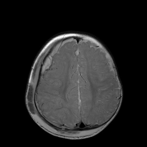
15 Category 1 Basilar cisterns patent No midline shift

16 Category 2 Sphenoid fracture and pneumocephalus Epidural hemorrhage

17 Category 2 Basilar cisterns patent No midline shift

18 Category 3 Basilar cisterns compressed No midline shift


19 Category 4 Basilar cisterns compressed Midline shift > 5mm

20 Evacuated mass Subdural hemorrhage greater than 25cc volume Midline shift resolved after surgical drainage


21 Evacuated mass Subdural hemorrhage greater than 25cc volume Basilar cistern compression resolved after surgical drainage

22 Unevacuated mass Subdural hemorrhage > 25cc Midline shift with compression of basilar cisterns

23 Unevacuated mass Patient was not surgical candidate due to neurologic examination Duret hemorrhage in brainstem
24 HCMC Marshall scale template Marshall Traumatic Brain Injury Scale: MARSHALL DIAGNOSTIC CATEGORIES OF ABNORMALITIES VISUALIZED ON CT SCANNING FOR TRAUMATIC BRAIN INJURY: Diffuse Injury 1: No visible intracranial pathology seen on CT scan. Diffuse Injury 2: Cisterns are present with shift 0-5 mm and/or lesion densities present. No high or mixed density lesion>25ml. May include bone fragments and foreign bodies. Diffuse Injury 3 (swelling): Cisterns compressed or absent with shift 0-5mm. No high or mixed density lesion>25ml. Diffuse Injury 4 (shift): Shift>5mm. No high or mixed density lesion>25ml. Evacuated mass lesion: Any surgically evacuated lesion. Non evacuated mass lesion: High or mixed-density lesion>25ml. Not surgically evacuated.
25 High/mixed density mass greater than 25 cc? No Basilar cisterns patent? No Midline shift greater than 5 mm? Yes No Yes Category 2 Category 3 Yes Surgically drained? No Non-Evacuated Mass Lesion Category 4 Yes Evacuated Mass Lesion

26 Differentiating Category 2 & 3 Category 2: Basilar cisterns patent Category 3: Basilar cisterns compressed
27 Prognosis from Marshall scale Prognosis depends on: Age Motor function Pupil reactivity CT scan findings including subarachnoid hemorrhage Marshall scale score can be used as one component of prognosis Marshall score alone is poor predictor of functional outcome
28 Limitations of Marshall scale Does not consider location of hemorrhage: subarachnoid, subdural, epidural, parenchymal Does not evaluate for traumatic axonal injury Does not differentiate degree of subfalcine or uncal herniation Intended to measure mortality, not likelihood of functional recovery
29 Alternatives to Marshall scale Rotterdam Stockholm Helsinki Use additional information from subarachnoid hemorrhage, intraventricular hemorrhage, degree of midline shift, location of high density mass May be more accurate in predicting prognosis More complicated to use with more interobserver variability
30 References 1: Thelin EP, Nelson DW, Vehviläinen J, Nyström H, Kivisaari R, Siironen J,Svensson M, Skrifvars MB, Bellander BM, Raj R. Evaluation of novel computerizedtomography scoring systems in human traumatic brain injury: An observational,multicenter study. PLoS Med Aug 3;14(8). 2: Charry JD, Tejada JH, Pinzon MA, Tejada WA, Ochoa JD, Falla M, Tovar JH,Cuellar-Bahamón AM, Solano JP. Predicted Unfavorable Neurologic Outcome IsOverestimated by the Marshall Computed Tomography Score, CorticosteroidRandomization After Significant Head Injury (CRASH), and International Missionfor Prognosis and Analysis of Clinical Trials in Traumatic Brain Injury (IMPACT)Models in Patients with Severe Traumatic Brain Injury Managed with EarlyDecompressive Craniectomy. World Neurosurg May;101: : Steyerberg EW, Mushkudiani N, Perel P, Butcher I, Lu J, McHugh GS, Murray GD,Marmarou A, Roberts I, Habbema JD, Maas AI. Predicting outcome after traumaticbrain injury: development and international validation of prognostic scores basedon admission characteristics. PLoS Med Aug 5;5(8):e165; discussion e165. 4: Marshall LF, Marshall SB, Klauber MR, Van Berkum Clark M, Eisenberg H, JaneJA, Luerssen TG, Marmarou A, Foulkes MA. The diagnosis of head injury requires aclassification based on computed axial tomography. J Neurotrauma Mar;9Suppl 1:S
Head CT Scan Interpretation: A Five-Step Approach to Seeing Inside the Head Lawrence B. Stack, MD
 Head CT Scan Interpretation: A Five-Step Approach to Seeing Inside the Head Lawrence B. Stack, MD Five Step Approach 1. Adequate study 2. Bone windows 3. Ventricles 4. Quadrigeminal cistern 5. Parenchyma
NEURO IMAGING 2. Dr. Said Huwaijah Chairman of radiology Dep, Damascus Univercity
 NEURO IMAGING 2 Dr. Said Huwaijah Chairman of radiology Dep, Damascus Univercity I. EPIDURAL HEMATOMA (EDH) LOCATION Seventy to seventy-five percent occur in temporoparietal region. CAUSE Most likely caused
HEAD AND NECK IMAGING. James Chen (MS IV)
") HEAD AND NECK IMAGING James Chen (MS IV) Anatomy Course Johns Hopkins School of Medicine Sept. 27, 2011 OBJECTIVES Introduce cross sectional imaging of head and neck Computed tomography (CT) Review head
HEAD AND NECK IMAGING James Chen (MS IV) Anatomy Course Johns Hopkins School of Medicine Sept. 27, 2011 OBJECTIVES Introduce cross sectional imaging of head and neck Computed tomography (CT) Review head
Midline Shift is Unrelated to Subjective Pupillary Reactivity Assessment on Admission in Moderate and Severe Traumatic Brain Injury
 Neurocrit Care (2018) 29:203 213 https://doi.org/10.1007/s12028-018-0526-8 ORIGINAL ARTICLE Midline Shift is Unrelated to Subjective Pupillary Reactivity Assessment on Admission in Moderate and Severe
NEURORADIOLOGY DIL part 3
 NEURORADIOLOGY DIL part 3 Bleeds and hemorrhages K. Agyem MD, G. Hall MD, D. Palathinkal MD, Alexandre Menard March/April 2015 OVERVIEW Introduction to Neuroimaging - DIL part 1 Basic Brain Anatomy - DIL
Using Abbreviated Injury Scale (AIS) codes to classify Computed Tomography (CT) features in the Marshall System
 codes to classify Computed Tomography (CT) features in the Marshall System") RESEARCH ARTICLE Open Access Using Abbreviated Injury Scale (AIS) codes to classify Computed Tomography (CT) features in the System Mehdi M Lesko 1*, Maralyn Woodford 1, Laura White 1, Sarah J O Brien
RESEARCH ARTICLE Open Access Using Abbreviated Injury Scale (AIS) codes to classify Computed Tomography (CT) features in the System Mehdi M Lesko 1*, Maralyn Woodford 1, Laura White 1, Sarah J O Brien
THREE HUNDRED AND ten TBI patients with a
 Acute Medicine & Surgery 2014; 1: 31 36 doi: 10.1002/ams2.5 Original Article Outcome prediction model for severe traumatic brain injury Jiro Iba, 1 Osamu Tasaki, 2 Tomohito Hirao, 2 Tomoyoshi Mohri, 3
Cerebro-vascular stroke
 Cerebro-vascular stroke CT Terminology Hypodense lesion = lesion of lower density than the normal brain tissue Hyperdense lesion = lesion of higher density than normal brain tissue Isodense lesion = lesion
Benign brain lesions
 Benign brain lesions Diagnostic and Interventional Radiology Hung-Wen Kao Department of Radiology, Tri-Service General Hospital, National Defense Medical Center Computed tomography Hounsfield unit (HU)
Commonly available CT characteristics and prediction of outcome in traumatic brain injury patients
 Romanian Neurosurgery Volume XXXI Number 1 2017 January - March Article Commonly available CT characteristics and prediction of outcome in traumatic brain injury patients Anil Kumar, Umamaheswara Reddy
Acute cerebral MCA ischemia with secondary severe head injury and acute intracerebral and subdural haematoma. Case report
 214 Balasa et al - Acute cerebral MCA ischemia Acute cerebral MCA ischemia with secondary severe head injury and acute intracerebral and subdural haematoma. Case report D. Balasa 1, A. Tunas 1, I. Rusu
Sequential changes in Rotterdam CT scores related to outcomes for patients with traumatic brain injury who undergo decompressive craniectomy
 clinical article J Neurosurg 124:1640 1645, 2016 Sequential changes in Rotterdam CT scores related to outcomes for patients with traumatic brain injury who undergo decompressive craniectomy Kenji Fujimoto,
Evaluation of Craniocerebral Trauma Using Computed Tomography
 IOSR Journal of Dental and Medical Sciences (IOSR-JDMS) e-issn: 2279-0853, p-issn: 2279-0861.Volume 13, Issue 9 Ver. IV (Sep. 2014), PP 57-62 Evaluation of Craniocerebral Trauma Using Computed Tomography
Moron General Hospital Ciego de Avila Cuba. Department of Neurological Surgery
 Moron General Hospital Ciego de Avila Cuba Department of Neurological Surgery Early decompressive craniectomy in severe head injury with intracranial hypertension Angel J. Lacerda MD PhD, Daisy Abreu MD,
How to interpret an unenhanced CT brain scan. Part 2: Clinical cases
 How to interpret an unenhanced CT brain scan. Part 2: Clinical cases Thomas Osborne a, Christine Tang a, Kivraj Sabarwal b and Vineet Prakash c a Radiology Registrar; b Radiology Foundation Year 1 Doctor;
Traumatic Brain Injuries
 Traumatic Brain Injuries Scott P. Sherry, MS, PA-C, FCCM Assistant Professor Department of Surgery Division of Trauma, Critical Care and Acute Care Surgery DISCLOSURES Nothing to disclose Discussion of
Predicting Outcome after Traumatic Brain Injury: Development and International Validation of Prognostic Scores Based on Admission Characteristics
 Predicting Outcome after Traumatic Brain Injury: Development and International Validation of Prognostic Scores Based on Admission Characteristics Ewout W. Steyerberg 1*, Nino Mushkudiani 1, Pablo Perel
Classical CNS Disease Patterns
 Classical CNS Disease Patterns Inflammatory Traumatic In response to the trauma of having his head bashed in GM would have experienced some of these features. NOT TWO LITTLE PEENY WEENY I CM LACERATIONS.
Early prediction of outcome after severe traumatic brain injury: a simple and practical model
 Rizoli et al. BMC Emergency Medicine (2016) 16:32 DOI 10.1186/s12873-016-0098-x RESEARCH ARTICLE Open Access Early prediction of outcome after severe traumatic brain injury: a simple and practical model
WHITE PAPER: A GUIDE TO UNDERSTANDING SUBARACHNOID HEMORRHAGE
 WHITE PAPER: A GUIDE TO UNDERSTANDING SUBARACHNOID HEMORRHAGE Subarachnoid Hemorrhage is a serious, life-threatening type of hemorrhagic stroke caused by bleeding into the space surrounding the brain,
8/29/2011. Brain Injury Incidence: 200/100,000. Prehospital Brain Injury Mortality Incidence: 20/100,000
 Traumatic Brain Injury Almario G. Jabson MD Section Of Neurosurgery Asian Hospital And Medical Center Brain Injury Incidence: 200/100,000 Prehospital Brain Injury Mortality Incidence: 20/100,000 Hospital
The central nervous system
 Sectc.qxd 29/06/99 09:42 Page 81 Section C The central nervous system CNS haemorrhage Subarachnoid haemorrhage Cerebral infarction Brain atrophy Ring enhancing lesions MRI of the pituitary Multiple sclerosis
Simplifying the use of prognostic information in traumatic brain injury. Part 2: Graphical presentation of probabilities
 CLINICAL ARTICLE J Neurosurg 128:1621 1634, 2018 Simplifying the use of prognostic information in traumatic brain injury. Part 2: Graphical presentation of probabilities Gordon D. Murray, MA, PhD, 1 Paul
Validation of CRASH Model in Prediction of 14-day Mortality and 6-month Unfavorable Outcome of Head Trauma Patients
 Emergency 2016; 4 (4): 196-201 ORIGINAL RESEARCH Validation of CRASH Model in Prediction of 14-day Mortality and 6-month Unfavorable Outcome of Head Trauma Patients Behrooz Hashemi 1, Mahnaz Amanat 1,
SUPPLEMENTARY FIG. S2. (A) Risk of bias and applicability concerns graph by marker. Review authors judgments about each domain presented as
 Risk of bias and applicability concerns graph by marker. Review authors judgments about each domain presented as") Supplementary Data SUPPLEMENTARY FIG. S1. Graphical depiction of (A) influence and (B) outlier detection analyses of S100 calcium binding protein B (S100B) 0.10 0.11lg/L cutoff value studies. (C) Summary
Supplementary Data SUPPLEMENTARY FIG. S1. Graphical depiction of (A) influence and (B) outlier detection analyses of S100 calcium binding protein B (S100B) 0.10 0.11lg/L cutoff value studies. (C) Summary
2. Subarachnoid Hemorrhage
 Causes: 2. Subarachnoid Hemorrhage A. Saccular (berry) aneurysm - Is the most frequent cause of clinically significant subarachnoid hemorrhage is rupture of a saccular (berry) aneurysm. B. Vascular malformation
secondary effects and sequelae of head trauma.
 Neuroimaging of vascular/secondary secondary effects and sequelae of head trauma. Andrès Server Alonso Department of Neuroradiology Division of Radiology Ullevål University Hospital Oslo, Norway. Guidelines
PREDICTION OF PROGNOSIS IN PATIENTS OF DIFFUSE BRAIN INJURY USING PROGNOSTIC PREDICTIVE MODEL DEVELOPED BY NIMHANS
 PREDICTION OF PROGNOSIS IN PATIENTS OF DIFFUSE BRAIN INJURY USING PROGNOSTIC PREDICTIVE MODEL DEVELOPED BY NIMHANS Devendra Singh Dhaker, Yogendra Singh Bhakuni, Ashish Kumar Dwivedi, A. K. Chaurasia,
Update sulle lesioni emorragiche posttraumatiche
 Update sulle lesioni emorragiche posttraumatiche Corrado Iaccarino Neurochirurgia-Neurotraumatologia AOU Parma Neurochirurgia d'urgenza IRCCS ASMN Reggio Emilia LAW UPDATING This document provides recommendations
Meninges and Ventricles
 Meninges and Ventricles Irene Yu, class of 2019 LEARNING OBJECTIVES Describe the meningeal layers, the dural infolds, and the spaces they create. Name the contents of the subarachnoid space. Describe the
INCREASED INTRACRANIAL PRESSURE
 INCREASED INTRACRANIAL PRESSURE Sheba Medical Center, Acute Medicine Department Irene Frantzis P-Year student SGUL 2013 Normal Values Normal intracranial volume: 1700 ml Volume of brain: 1200-1400 ml CSF:
Classification of traumatic brain injury PREDICTION OF OUTCOME IN TRAUMATIC BRAIN INJURY CLINICAL STUDIES
 CLINICAL STUDIES PREDICTION OF OUTCOME IN TRAUMATIC BRAIN INJURY WITH COMPUTED TOMOGRAPHIC CHARACTERISTICS: A COMPARISON BETWEEN THE COMPUTED TOMOGRAPHIC CLASSIFICATION AND COMBINATIONS OF COMPUTED TOMOGRAPHIC
Introduction. Materials and Methods. Young Hwan Choi, Tea Kyoo Lim, and Sang Gu Lee. 108 Copyright 2017 Korean Neurotraumatology Society
 CLINICAL ARTICLE Korean J Neurotrauma 2017;13(2):108-112 pissn 2234-8999 / eissn 2288-2243 https://doi.org/10.13004/kjnt.2017.13.2.108 Clinical Features and Outcomes of Bilateral Decompression Surgery
Clinical Outcome of Borderline Subdural Hematoma with 5-9 mm Thickness and/or Midline Shift 2-5 mm
 Original Article Print ISSN: 2321-6379 Online ISSN: 2321-595X DOI: 10.17354/ijss/2017/300 Clinical Outcome of Borderline Subdural Hematoma with 5-9 mm Thickness and/or Midline Shift 2-5 mm Raja S Vignesh
Imaging of Acute Cerebral Trauma
 July, 2005 Imaging of Acute Cerebral Trauma Louis Rivera, Harvard Medical School, Year III 46 y/o Female s/p Trauma - Unrestrained? MVC requiring Med Flight - Facial bruising/swelling - DEEP COMA - SEIZURES
Midline shift in relation to thickness of traumatic acute subdural hematoma predicts mortality
 Bartels et al. BMC Neurology (2015) 15:220 DOI 10.1186/s12883-015-0479-x RESEARCH ARTICLE Midline shift in relation to thickness of traumatic acute subdural hematoma predicts mortality Open Access Ronald
Correspondence should be addressed to Sorayouth Chumnanvej;
 Neurology Research International Volume 2016, Article ID 2737028, 7 pages http://dx.doi.org/10.1155/2016/2737028 Research Article Assessment and Predicting Factors of Repeated Brain Computed Tomography
Radiological parameters for surgical indication in traumatic brain injury
 Radiological parameters for surgical indication in traumatic brain injury Poster No.: C-1821 Congress: ECR 2011 Type: Educational Exhibit Authors: A. Elías Mas, C. Corbella Sala, S. Pasetto, F. Caiazzo,
Intracranial air on computerized tomography ANNE G. OSBORN, M.D., JONATHAN H. DAINES, M.D., S. DOUGLAS WING, M.D., AND ROBERT E. ANDERSON, M.D.
 J Neurosurg 48:355-359, 1978 Intracranial air on computerized tomography ANNE G. OSBORN, M.D., JONATHAN H. DAINES, M.D., S. DOUGLAS WING, M.D., AND ROBERT E. ANDERSON, M.D. Department of Radiology, University
ISCHEMIC STROKE IMAGING
 ISCHEMIC STROKE IMAGING ผศ.พญ พญ.จ ร ร ตน ธรรมโรจน ภาคว ชาร งส ว ทยา คณะแพทยศาสตร มหาว ทยาล ยขอนแก น A case of acute hemiplegia Which side is the abnormality, right or left? Early Right MCA infarction
THE ROLE OF IMAGING IN DIAGNOSIS OF SUBDURAL HEMATOMA: REVIEW ARTICLE
 THE ROLE OF IMAGING IN DIAGNOSIS OF SUBDURAL HEMATOMA: REVIEW ARTICLE * Dr. Sumendra Raj Pandey, Prof. Dr. Liu Pei WU, Dr. Sohan Kumar Sah, Dr. Lalu Yadav, Md. Sadam Husen Haque and Rajan KR. Chaurasiya
Perioperative Management Of Extra-Ventricular Drains (EVD)
") Perioperative Management Of Extra-Ventricular Drains (EVD) Dr. Vijay Tarnal MBBS, FRCA Clinical Assistant Professor Division of Neuroanesthesiology Division of Head & Neck Anesthesiology Michigan Medicine
Perioperative Management Of Extra-Ventricular Drains (EVD) Dr. Vijay Tarnal MBBS, FRCA Clinical Assistant Professor Division of Neuroanesthesiology Division of Head & Neck Anesthesiology Michigan Medicine
Case Conference: Neuroradiology. Case 1: Tumor Case 1: 22yo F w/ HA and prior Seizures
 Case Conference: Neuroradiology Case 1: 22yo F w/ HA and prior Seizures David E. Rex, MD, PhD Stanford University Hospital Department of Radiology Case 1: Tumor Most likely gangiloglioma, oligodendroglioma,
HHS Public Access Author manuscript Neurocrit Care. Author manuscript; available in PMC 2017 February 01.
 Derivation of a Predictive Score for Hemorrhagic Progression of Cerebral Contusions in Moderate and Severe Traumatic Brain Injury Randall Z. Allison 1, Kazuma Nakagawa 2,3, Michael Hayashi 4,5, Daniel
Case 1. Case 5/30/2013. Traumatic Brain Injury : Review, Update, and Controversies
 Case 1 Traumatic Brain Injury : Review, Update, and Controversies Shirley I. Stiver MD, PhD 32 year old male s/p high speed MVA Difficult extrication Intubated at scene Case BP 75 systolic / palp GCS 3
Role of Invasive ICP Monitoring in Patients with Traumatic Brain Injury: An Experience of 98 Cases
 31 Original Article Indian Journal of Neurotrauma (IJNT) 2006, Vol. 3, No. 1, pp. 31-36 Role of Invasive ICP Monitoring in Patients with Traumatic Brain Injury: An Experience of 98 Cases Deepak Kumar Gupta
UPSTATE Comprehensive Stroke Center. Neurosurgical Interventions Satish Krishnamurthy MD, MCh
 UPSTATE Comprehensive Stroke Center Neurosurgical Interventions Satish Krishnamurthy MD, MCh Regional cerebral blood flow is important Some essential facts Neurons are obligatory glucose users Under anerobic
Surgical Management & Clinical Outcome of Severe Brain Trauma due to Acute Subdural Hematoma.
 International Journal of Sciences: Basic and Applied Research (IJSBAR) ISSN 2307-4531 (Print & Online) http://gssrr.org/index.php?journal=journalofbasicandapplied ----------------------------------------------------------------------------------------------------------------
Imaging for the Diagnosis and Management of Traumatic Brain Injury
 Neurotherapeutics: The Journal of the American Society for Experimental NeuroTherapeutics Imaging for the Diagnosis and Management of Traumatic Brain Injury Jane J. Kim and Alisa D. Gean Department of
PRACTICE GUIDELINE. DEFINITIONS: Mild head injury: Glasgow Coma Scale* (GCS) score Moderate head injury: GCS 9-12 Severe head injury: GCS 3-8
 score Moderate head injury: GCS 9-12 Severe head injury: GCS 3-8") PRACTICE GUIDELINE Effective Date: 9-1-2012 Manual Reference: Deaconess Trauma Services TITLE: TRAUMATIC BRAIN INJURY GUIDELINE OBJECTIVE: To provide practice management guidelines for traumatic brain
PRACTICE GUIDELINE Effective Date: 9-1-2012 Manual Reference: Deaconess Trauma Services TITLE: TRAUMATIC BRAIN INJURY GUIDELINE OBJECTIVE: To provide practice management guidelines for traumatic brain
Study the Prognostic Value of Computed Tomographic Characteristics in Cases of Traumatic Brain Injury
 Study the Prognostic Value of Computed Tomographic Characteristics in Cases of Traumatic Brain Injury Tamer Abdullah Helmy 1, Mohammed Nasr-Eldeen Elsayd 1, Mamdoh Ahmed Zidan 2, Mohamed Farid 3 (1) Critical
For Emergency Doctors. Dr Suzanne Smallbane November 2011
 For Emergency Doctors Dr Suzanne Smallbane November 2011 A: Orbit B: Sphenoid Sinus C: Temporal Lobe D: EAC E: Mastoid air cells F: Cerebellar hemisphere A: Frontal lobe B: Frontal bone C: Dorsum sellae
Outcome of Traumatic Subarachnoid Hemorrhage Nazar Husain, Muhammad Akmal Husain, Tariq Ahmad
 Original Article Outcome of Traumatic Subarachnoid Hemorrhage Nazar Husain, Muhammad Akmal Husain, Tariq Ahmad ABSTRACT Objectives: Traumatic Subarachnoid Hemorrhage (Tr SAH) is a part of traumatic brain
Outcomes after severe traumatic brain injury (TBI)
") CLINICAL ARTICLE J Neurosurg 129:234 240, 2018 Clinical characteristics and temporal profile of recovery in patients with favorable outcomes at 6 months after severe traumatic brain injury Aditya Vedantam,
CLINICAL ARTICLE J Neurosurg 129:234 240, 2018 Clinical characteristics and temporal profile of recovery in patients with favorable outcomes at 6 months after severe traumatic brain injury Aditya Vedantam,
Head trauma - interpreting CT scans
 Head trauma - interpreting CT scans Poster No.: C-2075 Congress: ECR 2014 Type: Educational Exhibit Authors: T. Rihtar; Zagreb/HR Keywords: Trauma, Hemorrhage, Edema, Diagnostic procedure, CT, Neuroradiology
Impact of traumatic subarachnoid hemorrhage on outcome in nonpenetrating head injury
 J Neurosurg 83:445 452, 1995 Impact of traumatic subarachnoid hemorrhage on outcome in nonpenetrating head injury Part I: A proposed computerized tomography grading scale KARL A. GREENE, M.D., PH.D., FREDERICK
Do Prognostic Models Matter in Neurocritical Care?
 Do Prognostic Models Matter in Neurocritical Care? Alexis F. Turgeon MD MSc FRCPC Associate Professor and Director of Research Department of Anesthesiology and Critical Care Medicine Division of Critical
INTRACRANIAL PRESSURE -!!
 INTRACRANIAL PRESSURE - Significance raised ICP main cause of death in severe head injury main cause of morbidity in moderate and mild head injury main target and prognostic indicator in the ITU setting
NEURO IMAGING OF ACUTE STROKE
 1 1 NEURO IMAGING OF ACUTE STROKE ALICIA RICHARDSON, MSN, RN, ACCNS-AG, ANVP-BC WENDY SMITH, MA, RN, MBA, SCRN, FAHA LYNN HUNDLEY, APRN, CNRN, CCNS, ANVP-BC 2 2 1 DISCLOSURES Alicia Richardson: Stryker
How to Read a Head CT. Andrew D. Perron, MD, FACEP. Head CT. Head CT. Head CT. Head CT. EM Residency Program Director
 Blood Can Be Very Bad How to Read a Head CT EM Residency Program Director (or How I learned to stop worrying and love computed tomography ) Department of Emergency Medicine Maine Medical Center Portland,
Extradural hematoma (EDH) accounts for 2% of all head injuries (1). In
 accounts for 2% of all head injuries (1). In") CASE REPORT Conservative management of extradural hematoma: A report of sixty-two cases A. Rahim H. Zwayed 1, Brandon Lucke-Wold 2 Zwayed ARH, Lucke-wold B. Conservative management of extradural hematoma:
CASE REPORT Conservative management of extradural hematoma: A report of sixty-two cases A. Rahim H. Zwayed 1, Brandon Lucke-Wold 2 Zwayed ARH, Lucke-wold B. Conservative management of extradural hematoma:
Brain Injuries. Presented By Dr. Said Said Elshama
 Brain Injuries Presented By Dr. Said Said Elshama Types of head injuries 1- Scalp injuries 2- Skull injuries 3- Intra Cranial injuries ( Brain ) Anatomical structure of meninges Intra- Cranial Injuries
North Oaks Trauma Symposium Friday, November 3, 2017
 Traumatic Intracranial Hemorrhage Aaron C. Sigler, DO, MS Neurosurgery Tulane Neurosciences None Disclosures Overview Anatomy Epidural hematoma Subdural hematoma Cerebral contusions Outline Traumatic ICH
Acute Ischaemic Stroke
 Acute Ischaemic Stroke CT or MR SCA READIG FORM SCA ID: DATE OF READIG: SCA QUALIT: Good Moderate Poor Comment: READER ID: TPE OF SCA: CT: Without contrast: With contrast: MR: Diffusion: Perfusion ote
Non-Traumatic Neuro Emergencies
 Department of Radiology University of California San Diego Non-Traumatic Neuro Emergencies John R. Hesselink, M.D. Nontraumatic Neuroemergencies 1. Acute focal neurological deficit 2. Worst headache of
Intraoperative contralateral extradural hematoma during evacuation of traumatic acute extradural hematoma: A case report with review of literature
 Intraoperative contralateral extradural hematoma during evacuation of traumatic acute extradural hematoma: A case report with review of literature Anand Sharma 1, Arti Sharma 2, Yashbir Dewan 1 1 Artemis
To date, head injury remains the leading cause of. Outcome in patients with blunt head trauma and a Glasgow Coma Scale score of 3 at presentation
 J Neurosurg 111:683 687, 2009 Outcome in patients with blunt head trauma and a Glasgow Coma Scale score of 3 at presentation Clinical article Ro u k o z B. Ch a m o u n, M.D., Cl a u d i a S. Ro b e r
What Are We Going to Do? Fourth Year Meds Clinical Neuroanatomy. Hydrocephalus and Effects of Interruption of CSF Flow. Tube Blockage Doctrine
 Fourth Year Meds Clinical Neuroanatomy Ventricles, CSF, Brain Swelling etc. David A. Ramsay, Neuropathologist, LHSC What Are We Going to Do? Hydrocephalus and some effects of the interruption of CSF flow
Surgical Management of Stroke Brandon Evans, MD Department of Neurosurgery
 Surgical Management of Stroke Brandon Evans, MD Department of Neurosurgery 2 Stroke Stroke kills almost 130,000 Americans each year. - Third cause of all deaths in Arkansas. - Death Rate is highest in
Neuroimaging of TBI: Current Clinical Guidelines and Future Direction Brain Injury Alliance of Colorado 2017
 Neuroimaging of TBI: Current Clinical Guidelines and Future Direction Brain Injury Alliance of Colorado 2017 Peter E. Ricci, M.D. Staff Neuroradiologist Radiology Imaging Associates OBJECTIVES 1. Understand
PROPOSAL FOR MULTI-INSTITUTIONAL IMPLEMENTATION OF THE BRAIN INJURY GUIDELINES
 PROPOSAL FOR MULTI-INSTITUTIONAL IMPLEMENTATION OF THE BRAIN INJURY GUIDELINES INTRODUCTION: Traumatic Brain Injury (TBI) is an important clinical entity in acute care surgery without well-defined guidelines
Diagnostic modalities of the Central Nervous System
 NEURORADIOLOGY Kinga Karlinger, MD, PhD Associate Professor Semmelweis University, Budapest Diagnostic modalities of the Central Nervous System X-ray: screening is not used any more, x-ray images instead
J Neurosurg Pediatrics 13:
 J Neurosurg Pediatrics 13:307 314, 2014 AANS, 2014 Computed tomography characteristics in pediatric versus adult traumatic brain injury Clinical article Korak Sarkar, M.D., 1 Krista Keachie, M.D., 2 UyenThao
Bilateral superior ophthalmic vein enlargement associated with diffuse cerebral swelling
 J Neurosurg 86:893 897, 1997 Bilateral superior ophthalmic vein enlargement associated with diffuse cerebral swelling Report of 11 cases ROHIT K. KHANNA, M.D., CHRISTOPHER J. PHAM, D.O., GHAUS M. MALIK,
Glasgow Coma Scale score at intensive care unit discharge predicts the 1-year outcome of patients with severe traumatic brain injury
 Eur J Trauma Emerg Surg (2013) 39:285 292 DOI 10.1007/s00068-013-0269-3 ORIGINAL ARTICLE Glasgow Coma Scale score at intensive care unit discharge predicts the 1-year outcome of patients with severe traumatic
Ventriculostomy and Risk of Upward Herniation in Patients with Obstructive Hydrocephalus from Posterior Fossa Mass Lesions
 https://doi.org/10.1007/s12028-017-0487-3 ORIGINAL ARTICLE Ventriculostomy and Risk of Upward Herniation in Patients with Obstructive Hydrocephalus from Posterior Fossa Mass Lesions Sherri A. Braksick
Usefulness of Intracranial CT Angiography with Spiral CT in Brain Death - A Preliminary Report -
 Usefulness of Intracranial CT Angiography with Spiral CT in Brain Death - A Preliminary Report - Jong-Ho Park, M.D., Hong-Ki Song, M.D., Dae-Young Yoon, M.D. Department of Neurology and Radiology*, Hallym
CNS pathology Third year medical students. Dr Heyam Awad 2018 Lecture 5: disturbed fluid balance and increased intracranial pressure
 CNS pathology Third year medical students Dr Heyam Awad 2018 Lecture 5: disturbed fluid balance and increased intracranial pressure ILOs Understand causes and symptoms of increased intracranial pressure.
Cerebral autoregulation is a complex intrinsic control. Time course for autoregulation recovery following severe traumatic brain injury
 J Neurosurg 111:695 700, 2009 Time course for autoregulation recovery following severe traumatic brain injury Clinical article Gi l l E. Sv i r i, M.D., M.Sc., 1 Ru n e Aa s l i d, Ph.D., 2 Co l l e e
Pediatric Head Trauma August 2016
 PEDIATRIC HEAD TRAUMA AUGUST 2016 Pediatric Head Trauma August 2016 EDUCATION COMMITTEE PEER EDUCATION Quick Review of Pathophysiology of TBI Nuggets of knowledge to keep in mind with TBI Intracranial
Stroke School for Internists Part 1
 Stroke School for Internists Part 1 November 4, 2017 Dr. Albert Jin Dr. Gurpreet Jaswal Disclosures I receive a stipend for my role as Medical Director of the Stroke Network of SEO I have no commercial
Virtual Mentor American Medical Association Journal of Ethics August 2008, Volume 10, Number 8:
 Virtual Mentor American Medical Association Journal of Ethics August 2008, Volume 10, Number 8: 516-520. CLINICAL PEARL The Hazards of Stopping a Brain in Motion: Evaluation and Classification of Traumatic
Prognostic Value of Secondary Insults in Traumatic Brain Injury: Results from the IMPACT Study ABSTRACT
 JOURNAL OF NEUROTRAUMA Volume 24, Number 2, 2007 Mary Ann Liebert, Inc. Pp. 287 293 DOI: 10.1089/neu.2006.0031 Prognostic Value of Secondary Insults in Traumatic Brain Injury: Results from the IMPACT Study
Characteristic features of CNS pathology. By: Shifaa AlQa qa
 Characteristic features of CNS pathology By: Shifaa AlQa qa Normal brain: - The neocortex (gray matter): six layers: outer plexiform, outer granular, outer pyramidal, inner granular, inner pyramidal, polymorphous
The "filling defect" sign helps localise the site of intracranial aneurysm rupture on an unenhanced CT
 The "filling defect" sign helps localise the site of intracranial aneurysm rupture on an unenhanced CT Poster No.: C-3380 Congress: ECR 2010 Type: Topic: Authors: Keywords: DOI: Educational Exhibit Neuro
Index. aneurysm, 92 carotid occlusion, 94 ICA stenosis, 95 intracranial, 92 MCA, 94
 A ADC. See Apparent diffusion coefficient (ADC) Aneurysm cerebral artery aneurysm, 93 CT scan, 93 gadolinium, 93 Angiography, 13 Anoxic brain injury, 25 Apparent diffusion coefficient (ADC), 7 Arachnoid
Spectrum of various patterns of injuries in cranio-cerebral trauma: CT evaluation
 Original article: Spectrum of various patterns of injuries in cranio-cerebral trauma: CT evaluation 1Dr. Kaleem Ahmad, 2 Dr. RK Rauniyar, 3 Dr. Sajid Ansari, 4 Dr. Mukesh Kumar Gupta 1Associate Professor,
Neurotrauma. Béla Faludi Dept.. of Neurology University of PécsP
 Neurotrauma Béla Faludi Dept.. of Neurology University of PécsP Emergency!!! Why here? Opened cranial injury visible: neurosurgery Closed injuries sometimes diagnosed by neurologist Masking situation:
CEREBRAL INFARCTION AS NEUROSURGICAL POST OPERATIVE COMPLICATION : SERIAL CASES
 CEREBRAL INFARCTION AS NEUROSURGICAL POST OPERATIVE COMPLICATION : SERIAL CASES Oleh Ferry Wijanarko, dr, Sp BS DR Agus Turchan, dr, Sp BS BEDAH SARAF SOLO Secretariat : Neurosurgery Subdivision Dr Moewardi
Head Injury: Classification Most Severe to Least Severe
 Head Injury: Classification Most Severe to Least Severe Douglas I. Katz, MD Professor, Dept. Neurology, Boston University School of Medicine, Boston MA Medical Director Brain Injury Program, HealthSouth
MR imaging as predictor of delayed posttraumatic cerebral hemorrhage
 J Neurosurg 69:203-209, 1988 MR imaging as predictor of delayed posttraumatic cerebral hemorrhage TOKUTARO TANAKA, M.D., TSUNEO SAKAI, M.D., KENICHI UEMURA, M.D., ATSUSHI TERAMURA, M.D., ICHIRO FUJISHIMA,
Disclosure. Seizure Prophylaxis in Traumatic Head Injury
 Seizure Prophylaxis in Traumatic Head Injury Anthony Angelow, PhD(c), ACNPC, AGACNP-BC, CEN Associate Lecturer, Fitzgerald Health Education Associates Clinical practice Division of Trauma Surgery and Division
ARTERIOVENOUS MALFORMATION OR CONTUSION : A DIAGNOSTIC DILEMMA. Yong Pei Yee, Ibrahim Lutfi Shuaib, Jafri Malin Abdullah*
 Malaysian Journal of Medical Sciences, Vol. 8, No. 2, July 2001 (47-51) CASE REPORT ARTERIOVENOUS MALFORMATION OR CONTUSION : A DIAGNOSTIC DILEMMA Yong Pei Yee, Ibrahim Lutfi Shuaib, Jafri Malin Abdullah*
Acute Clinical Deterioration of Posterior Fossa Epidural Hematoma: Clinical Features, Risk Factors and Outcome
 Original Article 271 Acute Clinical Deterioration of Posterior Fossa Epidural Hematoma: Clinical Features, Risk Factors and Outcome Tsung-Ming Su, MD; Tsung-Han Lee, MD; Tao-Chen Lee, MD; Ching-Hsiao Cheng,
Efficacy of neuroendoscopic evacuation of traumatic intracerebral or intracerebellar hematoma
 Original Contribution Kitasato Med J 2017; 47: 141-147 Efficacy of neuroendoscopic evacuation of traumatic intracerebral or intracerebellar hematoma Hiroyuki Koizumi, 1,2 Daisuke Yamamoto, 1 Yasushi Asari,
Neuroradiological Findings in Non- Accidental Trauma Educational Pictorial Review
 Neuroradiological Findings in Non- Accidental Trauma Educational Pictorial Review M B Moss, MD; L Lanier, MD; R Slater; C L Sistrom, MD; R G Quisling, MD; I M Schmalfuss, MD; and D Rajderkar, MD Contact:
Importance of Hematoma Removal Ratio in Ruptured Middle Cerebral Artery Aneurysm Surgery with Intrasylvian Hematoma
 Journal of Cerebrovascular and Endovascular Neurosurgery pissn 2234-8565, eissn 2287-3139, http://dx.doi.org/10.7461/jcen.2017.19.1.5 Original Article Importance of Hematoma Removal Ratio in Ruptured Middle
Chronic Brain-Dead Patients Who Exhibit Lazarus Sign
 CASE REPORT Korean J Neurotrauma 2017;13(2):153-157 pissn 2234-8999 / eissn 2288-2243 https://doi.org/10.13004/kjnt.2017.13.2.153 Chronic Brain-Dead Patients Who Exhibit Lazarus Sign Department of Neurosurgery,
Introduction to Neurosurgical Subspecialties:
 Introduction to Neurosurgical Subspecialties: Trauma and Critical Care Neurosurgery Brian L. Hoh, MD 1, Gregory J. Zipfel, MD 2 and Stacey Q. Wolfe, MD 3 1 University of Florida, 2 Washington University,