Imaging in a confused patient: Infections and Inflammation
|
|
|
- Clare McKenzie
- 5 years ago
- Views:
Transcription


1 American Society of Neuroimaging Imaging in a confused patient: Infections and Inflammation January 21, 2017 Los Angeles, California Joshua P. Klein, MD, PhD, FANA, FAAN, FASN Chief, Division of Hospital Neurology Associate Professor of Neurology and Radiology Brigham and Women s Hospital & Harvard Medical School
2 Disclosure I have no financial interests or disclosures to report that are relevant to the content of this presentation.
3 Inflammation as a cause of confusion Infectious meningitis, encephalitis, vasculitis, abscess Non-infectious meningitis, encephalitis, vasculitis, tumor, demyelination, trauma, toxic, metabolic
4 Confusion History: Symptoms, pace, review of systems, coincident non-neurologic disease, medication changes, drug exposures Physical exam: Level of consciousness, localizing signs Secondary phenomena: hydrocephalus, mass effect, herniation
5 Confusion plus syndromes 1. Viral encephalitis: + fever, seizures 2. Limbic encephalitis: + seizures, behavior change 3. Wernicke: + ataxia, oculomotor paresis 4. ADEM: + fever, preceding illness 5. CJD: + progressive, seizures, myoclonus 6. NPH: + ataxia, incontinence 7. PML: + immunosuppression 8. PRES: + hypertension, exposure to chemo 9. Non-convulsive status epilepticus: + seizures 10. Serotonin syndrome, NMS: + med exposures 11. Acute hepatic encephalopathy: + elevated ammonia 12. Pontine (and extrapontine) myelinolysis: + hypo-na

6 Confusion What is the relationship of confusion to consciousness and arousal? Reticular Activating System Danaila L and Pascu ML, 2013
7 Confusion Conscious: state of awareness of self and environment, and responsiveness to external stimulation and inner need. Levels of consciousness or arousal: awake, drowsy, stuporous, obtunded, comatose Unconscious: state of unawareness of self and environment, coupled with diminished responsiveness to environmental stimuli.
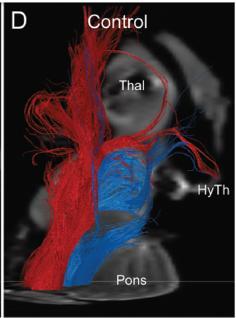
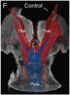
8 Neuroanatomy, alteration of arousal JNEN 2013;72:505
9 NEJM 2007;356:166

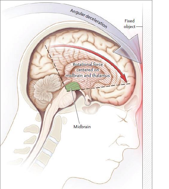
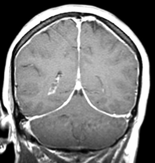
10 A 53 year-old woman who fell down stairs

11 A 53 year-old woman who fell down stairs JNEN 2013;72:505 JNEN 2013;72(6):505

12 Coma, trauma to ARAS


13 Coma, injury to ARAS thalamic projections Basilar thrombosis CNS lymphoma

14 Coma, ischemia to ARAS cortical projections
15 Meningeal inflammation

16 Meningitis, compartments (pachymeninges) (leptomeninges) Skull Dura Outer arachnoid ~ CSF ~ inner arachnoid Pia Brain epidural subdural subarachnoid intraparenchymal intraventricular
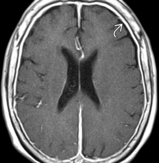
17 Pachymeningitis (dura)
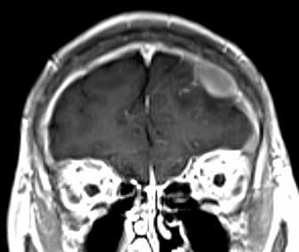
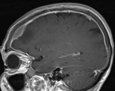
18 Pachymeningitis (dural myeloid sarcoma) T1-post DWI ADC
19 Leptomeningitis (pia, carcinomatous)

20 Meningitis (carcinomatous) Klein JP, Handb Clin Neurol 2016;136:923

21 Leptomeningitis in sarcoidosis Klein JP, in Youmans and Winn Neurological Surgery, 7 th Ed, 2016

22 Leptomeningitis in tuberculosis Axial graphic shows the early capsule Klein formation of JP, an abscess in Youmans with central liquified and necrosis Winn and inflammatory Neurological debris. Collagen and Surgery, reticulin form the 2016 well-defined abscess wall. Note the surrounding edema.

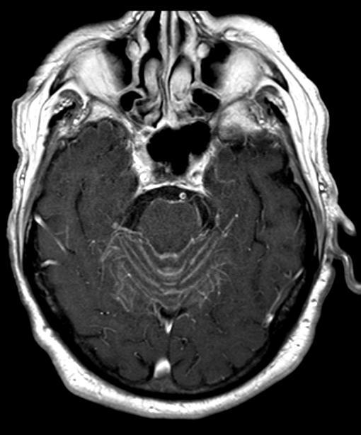
23 Leptomeningitis and vasculitis Lymphocytic meningitis due to HSV-2 Ischemic infarctions and hemorrhages Leptomeningeal enhancement J Neurovirology 2014;20:419
 in")
24 Meningitis (dura & pia) in infection
25 Cerebral inflammation

26 Encephalitis, limbic (VGKC with thymoma)
27 Encephalitis, EEE virus

28 HIV encephalitis AIDS Pt Care STDS 2012;26:383


29 HSV-1 encephalitis Adams and Victor s Principles of Neurology, 10 th Ed, 2014


30 HSV-3 (VZV) encephalitis, vasculitis NEJM 2000;342:635


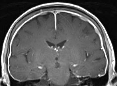
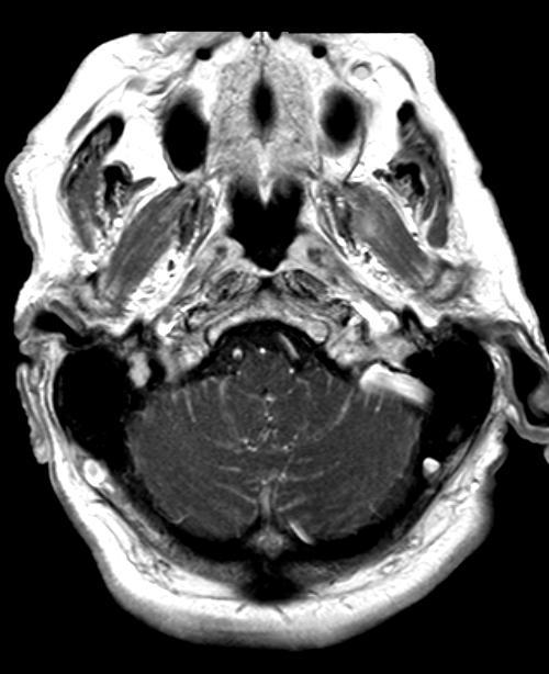
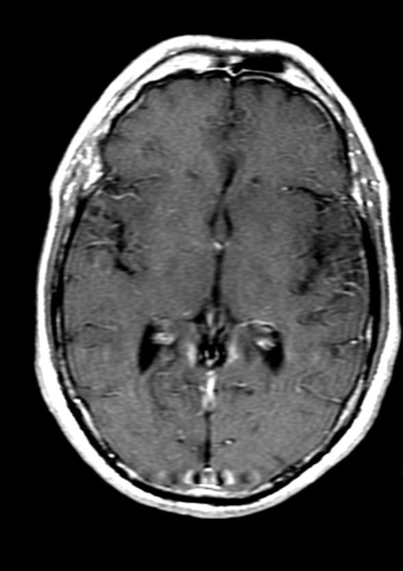
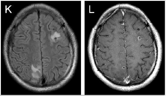
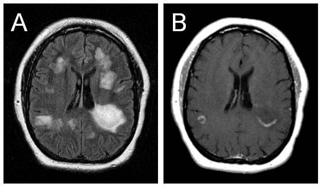
31 HHV-5 (CMV) ventriculo-encephalitis opportunistic, associated with ventriculo-encephalitis, also, meningitis, transverse myelitis, and radiculomyelitis STATdx


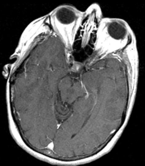
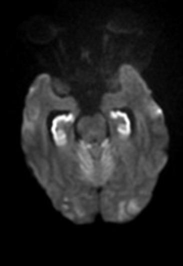
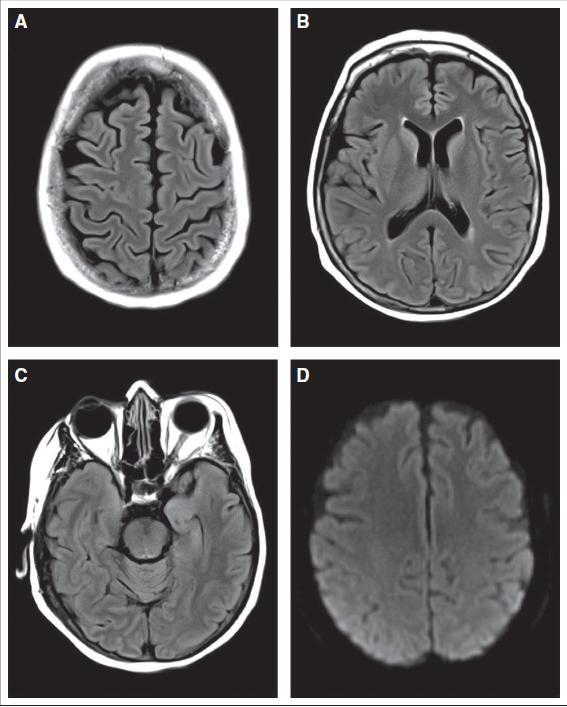
32 HHV-6 encephalitis opportunistic, medial temporal lobe tropism STATdx
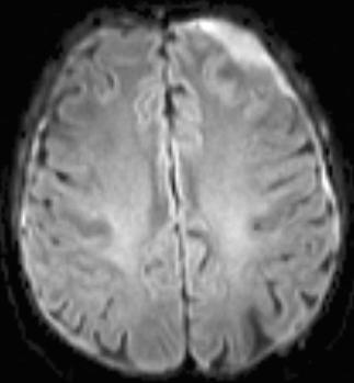
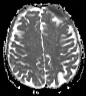
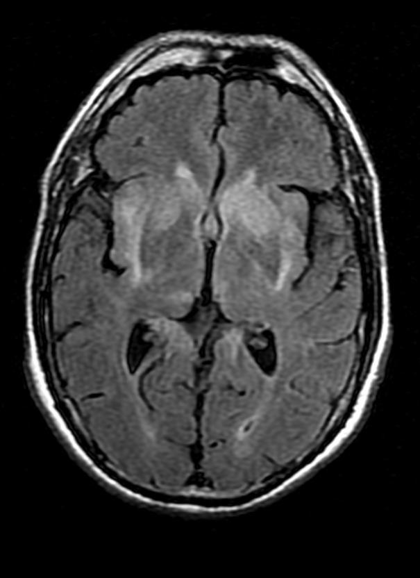
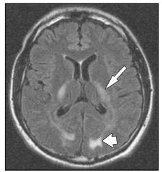
33 JC virus encephalitis Am J Hematol 2016;91:1057

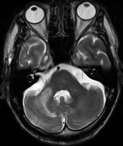
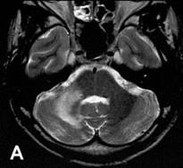
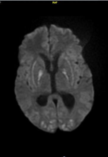
34 Creutzfeldt-Jakob disease, sporadic JNNP 2007;78:664

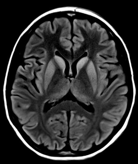
35 Creutzfeldt-Jakob disease, sporadic
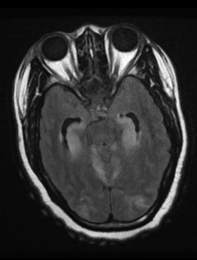
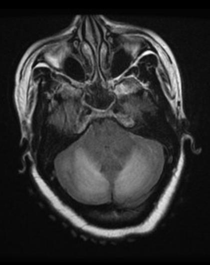
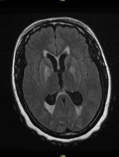
36 Hypoxemic encephalopathy

37 Hypoxemic-ischemic encephalopathy

38 Delayed post-hypoxic leukoencephalopathy 4 weeks post-inhalation 3 weeks later 10 months later

39 Heroin inhalation toxicity AJR 2003;180:847


40 Cerebritis Abscess Axial graphic shows early cerebritis, the initial phase of abscess formation, in the frontal lobe. There is a focal unencapsulated mass of petechial hemorrhage, inflammatory cells, and edema. STATdx
41 Cerebritis Abscess Early cerebritis: Patchy enhancement masses of PMNs, hyperemia Late cerebritis: Intense, irregular rim enhancement coalescence of masses, necrosis Early capsule: Well-defined, thin-wall, enhancing rim collagenous capsule, liquefied core Late capsule: Cavity collapses, capsule thickens thick wall of collagen and fibroblasts Ddx: glioblastoma, metastases, demyelination, hematoma, subacute infarction.


42 Cerebral abscess T1-post DWI ADC


43 Tuberculoma Axial graphic shows the early capsule formation of an abscess with central liquified necrosis and J inflammatory Clin Neurosci debris. Collagen and 2013;20:1599 reticulin form the well-defined abscess wall. Note the surrounding edema.
44 Neurocysticercosis

45 Tumefactive demyelination Klein JP, in Youmans and Winn Neurological Surgery, 2016
46 ADEM

47 PRES J Clin Oncology 2016;34(2):1
48 Summary Confusion may result from inflammation of the brain and meninges. Infectious and non-infectious inflammation can cause meningitis, encephalitis, vasculitis.
49 Summary The history and physical exam are key. Identify the clinical syndrome, then correlate to imaging. Assess all compartments. Anticipate secondary effects of primary disease process.
An Approach. to Brain. Infection. 37F found down. Disclosures. Approach to CNS Infection. Objectives. Parenchymal. None.
 An Approach Disclosures to Brain None. Infection Jason Shewchuk, MD Clinical Associate Professor Head of Neuroradiology UBC European Course in Neuroradiology 2018 Objectives Following this session the
Disclosure. + Outline. Case-based approach to neurological emergencies that might present to the ED
 Kathleen R. Fink, MD University of Washington 5 th Nordic Emergency Radiology Course May 21, 2015 Disclosure My spouse receives research salary support from: Bracco BayerHealthcare Guerbet Outline Case-based
Brain Pain Infections of the CNS
 FRIDAY, OCTOBER 28, 2016 Brain Pain Infections of the CNS Suyash Mohan MD, PDCC Assistant Professor of Radiology & Neurosurgery Division of Neuroradiology, Department of Radiology Perelman School of Medicine
Imaging findings in CNS infections and differential diagnosis. M. Lequin
 Imaging findings in CNS infections and differential diagnosis M. Lequin OUTLINE Introduction and terminology Diagnosis & Differential diagnosis Pediatric brain infections viral infections Meningitis Encephalitis
Kathleen R. Fink, MD Virginia Mason Medical Center. 6 th Nordic Emergency Radiology Course 2017
 Kathleen R. Fink, MD Virginia Mason Medical Center 6 th Nordic Emergency Radiology Course 2017 Disclosure My spouse receives research salary support from: Guerbet Outline Indications for imaging CNS infections
Interactive Cases: Demyelinating Diseases and Mimics. Disclosures. Case 1 25 yo F with nystagmus; look for tumor 4/14/2017
 Interactive Cases: Demyelinating Diseases and Mimics Disclosures None Brad Wright, MD 27 March 2017 Case 1 25 yo F with nystagmus; look for tumor What do you suspect? A. Demyelinating disease B. Malignancy
Failure to wake. Robin S. Howard
 Failure to wake Robin S. Howard National Hospital for Neurology and Neurosurgery, Queen Square St. Thomas Hospital, Guys & St. Thomas NHS (Foundation) Trust Royal College of Physicians - November 2017
COMA. DIAH MUSTIKA HW,SpS,KIC INTENSIVE CARE UNIT of EMERGENCY DEPARTMENT
 COMA DIAH MUSTIKA HW,SpS,KIC INTENSIVE CARE UNIT of EMERGENCY DEPARTMENT NAVAL HOSPITAL dr RAMELAN, SURABAYA DEFINITIONS Coma State of unresponsiveness to external or internal stimuli in which a patient
Index. aneurysm, 92 carotid occlusion, 94 ICA stenosis, 95 intracranial, 92 MCA, 94
 A ADC. See Apparent diffusion coefficient (ADC) Aneurysm cerebral artery aneurysm, 93 CT scan, 93 gadolinium, 93 Angiography, 13 Anoxic brain injury, 25 Apparent diffusion coefficient (ADC), 7 Arachnoid
Neuroradiology of AIDS
 Neuroradiology of AIDS Frank Minja,, HMS IV Gillian Lieberman MD September 2002 AIDS 90% of HIV patients have CNS involvement 1 10% of AIDS patients present first with neurological symptoms 2 73-80% of
Non-Traumatic Neuro Emergencies
 Department of Radiology University of California San Diego Non-Traumatic Neuro Emergencies John R. Hesselink, M.D. Nontraumatic Neuroemergencies 1. Acute focal neurological deficit 2. Worst headache of
MRI OF THE THALAMUS. Mohammed J. Zafar, MD, FAAN Kalamazoo, MI
 1 MRI OF THE THALAMUS Mohammed J. Zafar, MD, FAAN Kalamazoo, MI Objectives: The thalamic nuclei can be involved in a wide variety of conditions. A systematic imaging approach would be useful for narrowing
RINGS N THINGS: Imaging Patterns in Differential Diagnosis. Anne G. Osborn, M.D.
 RINGS N THINGS: Imaging Patterns in Differential Diagnosis Anne G. Osborn, M.D. ExpDDxs: Intra-axial (Parenchymal) Lesions Ring-enhancing lesions, solitary 1 Ring-enhancing lesion crossing corpus callosum
Organic Mental Disorders. Organic Mental Disorders. Axes. Damrongsak Bulyalert Department of Internal Medicine
 Organic Mental Disorders Damrongsak Bulyalert Department of Internal Medicine www.metadon.net 1 Organic Mental Disorders In DSM (Diagnostic and Statistical Manual of Mental Disorders), OMD includes Delirium,
Demyelinating Diseases of the Brain
 Department of Radiology University of California San Diego Demyelinating Diseases of the Brain John R. Hesselink, M.D. T1-Weighted Images Normal White Matter Contents Axons with envelope of myelin Neuroglia
IMAGING OF INTRACRANIAL INFECTIONS
 IMAGING OF INTRACRANIAL INFECTIONS Dr Carolina Kachramanoglou LYSHOLM DEPARTMENT OF NEURORADIOLOGY NATIONAL HOSPITAL FOR NEUROLOGY AND NEUROSURGERY Plan Introduce MR sequences that are useful in the diagnosis
EVALUATION OF COMATOSE PATIENT. Prof. G. Zuliani
 EVALUATION OF COMATOSE PATIENT Prof. G. Zuliani Consciousness Two components of conscious behavior: Vigilance (arousal): appearance of wakefulness Awareness (content): the sum of cognitive and affective
Cerebrovascular diseases-2
 Cerebrovascular diseases-2 Primary angiitis of CNS - Other causes of infarction i. Hypercoagulable states ii. Drug-abuse such as amphetamine, heroin and cocain Note - The venous side of the circulation
Classical CNS Disease Patterns
 Classical CNS Disease Patterns Inflammatory Traumatic In response to the trauma of having his head bashed in GM would have experienced some of these features. NOT TWO LITTLE PEENY WEENY I CM LACERATIONS.
CT and MR findings of systemic lupus erythematosus involving the brain: Differential diagnosis based on lesion distribution
 CT and MR findings of systemic lupus erythematosus involving the brain: Differential diagnosis based on lesion distribution Poster No.: C-2723 Congress: ECR 2010 Type: Educational Exhibit Topic: Neuro
Index. Note: Page numbers of article titles are in boldface type.
 Note: Page numbers of article titles are in boldface type. A Abdominal trauma in pregnant patients coma due to, 986 987 Absence seizures impaired consciousness effects on, 803 807 Acute adrenal failure,
Unit VIII Problem 6 Pathology: Meningitis
 Unit VIII Problem 6 Pathology: Meningitis - Important terms: Meningitis: it is inflammation of meninges (coverings of the central nervous system) caused by infection. They are classified to: Pachymeningitis:
NEURORADIOLOGY DIL part 3
 NEURORADIOLOGY DIL part 3 Bleeds and hemorrhages K. Agyem MD, G. Hall MD, D. Palathinkal MD, Alexandre Menard March/April 2015 OVERVIEW Introduction to Neuroimaging - DIL part 1 Basic Brain Anatomy - DIL
Head Injury: Classification Most Severe to Least Severe
 Head Injury: Classification Most Severe to Least Severe Douglas I. Katz, MD Professor, Dept. Neurology, Boston University School of Medicine, Boston MA Medical Director Brain Injury Program, HealthSouth
Role of MRI in acute disseminated encephalomyelitis
 Original Research Article Role of MRI in acute disseminated encephalomyelitis Shashvat Modiya 1*, Jayesh Shah 2, C. Raychaudhuri 3 1 1 st year resident, 2 Associate Professor, 3 HOD and Professor Department
The Neurology of HIV Infection. Carolyn Barley Britton, MD, MS Associate Professor of Clinical Neurology Columbia University
 The Neurology of HIV Infection Carolyn Barley Britton, MD, MS Associate Professor of Clinical Neurology Columbia University HIV/AIDS Epidemiology World-wide pandemic, 40 million affected U.S.- Disproportionate
Case 9 10/29/2018. CJD (Creutzfeldt -Jakob Disease) CJD (Creutzfeldt -Jakob Disease) CJD (Creutzfeldt -Jakob Disease)
 CJD (Creutzfeldt -Jakob Disease) CJD (Creutzfeldt -Jakob Disease)") CJD (Creutzfeldt -Jakob Disease) Rare fatal neurodegen dz caused by infectious protein Prion (lacks nucleic acid)- causes spongiform changes of the brain and neuronal death. 4 types: scjd- 85% of cases
CJD (Creutzfeldt -Jakob Disease) Rare fatal neurodegen dz caused by infectious protein Prion (lacks nucleic acid)- causes spongiform changes of the brain and neuronal death. 4 types: scjd- 85% of cases
Disclosure. Learner Objectives. Congenital Infections. Question. Main Categories 4/26/2016
 Communicating Communicability: Imaging of CNS Infections Aaron P. Kamer, MD Assistant Professor of Clinical Radiology Neuroradiology Section April 26, 2016 Disclosure Within the past 12 months: I have
Cerebro-vascular stroke
 Cerebro-vascular stroke CT Terminology Hypodense lesion = lesion of lower density than the normal brain tissue Hyperdense lesion = lesion of higher density than normal brain tissue Isodense lesion = lesion
CNS Infections in the Pediatric Age Group
 CNS Infections in the Pediatric Age Group Introduction CNS infections are frequently life-threatening In the Philippines, bacterial meningitis is one of the top leading causes of mortality in children
Vascular Disorders. Nervous System Disorders (Part B-1) Module 8 -Chapter 14. Cerebrovascular disease S/S 1/9/2013
 Module 8 -Chapter 14. Cerebrovascular disease S/S 1/9/2013") Nervous System Disorders (Part B-1) Module 8 -Chapter 14 Overview ACUTE NEUROLOGIC DISORDERS Vascular Disorders Infections/Inflammation/Toxins Metabolic, Endocrinologic, Nutritional, Toxic Neoplastic Traumatic
Nervous System Disorders (Part B-1) Module 8 -Chapter 14 Overview ACUTE NEUROLOGIC DISORDERS Vascular Disorders Infections/Inflammation/Toxins Metabolic, Endocrinologic, Nutritional, Toxic Neoplastic Traumatic
Meninges and Ventricles
 Meninges and Ventricles Irene Yu, class of 2019 LEARNING OBJECTIVES Describe the meningeal layers, the dural infolds, and the spaces they create. Name the contents of the subarachnoid space. Describe the
MRI imaging in meningeal diseases
 Original article MRI imaging in meningeal diseases 1Dr. Narendrakumar M Shah, 2 Dr Vaishali D M 1Associate professor, Department of Radiodiagnosis, SDM Medical college, Dharwad 2Consultant radiologist,
Imaging the Spinal Cord & Intradural Disease
 Department of Radiology University of California San Diego Imaging the Spinal Cord & Intradural Disease John R. Hesselink, M.D. Spinal Cord Diseases Tumors Syringohydromyelia Trauma Ischemia / Infarction
Elsevier's Encyclopedia of euroscience
 REPRINTED FROM Elsevier's Encyclopedia of euroscience e Edited by George Adelman Barry H. Smith Editorial Manager Jennifer De Pasquale 1999 Elsevier Science B.V. All rights reserved. Visit the Encyclopedia's
8th Annual NKY TBI Conference 3/28/2014
 Closed Head Injury: Headache to Herniation A N T H O N Y T. K R A M E R U N I V E R S I T Y O F C I N C I N N A T I B L U E A S H E M S T E C H N O L O G Y P R O G R A M Objectives Describe the pathological
Central Nervous System Infection
 Central Nervous System Infection Ashley H. Aiken KEYWORDS CNS infections Meningitis Abscess Encephalitis Subdural empyema Infections of the brain and its linings pose a growing, worldwide health problem.
Cerebral malaria: MR imaging spectrum
 Cerebral malaria: MR imaging spectrum Poster No.: C-2705 Congress: ECR 2010 Type: Educational Exhibit Topic: Neuro Authors: P. S. Naphade, M. D. Agrawal, S. S. Sankhe, K. M. Siva, B. K. Jain; Mumbai/IN
V. CENTRAL NERVOUS SYSTEM TRAUMA
 V. CENTRAL NERVOUS SYSTEM TRAUMA I. Concussion - Is a clinical syndrome of altered consiousness secondary to head injury - Brought by a change in the momentum of the head when a moving head suddenly arrested
A Guide to the Radiologic Evaluation of Extra-Axial Hemorrhage
 July 2013 A Guide to the Radiologic Evaluation of Extra-Axial Hemorrhage John Dickson, Harvard Medical School Year III Agenda 1. Define extra-axial hemorrhage and introduce its subtypes 2. Review coup
Moath Darweesh. Zaid Emad. Anas Abu -Humaidan
 3 Moath Darweesh Zaid Emad Anas Abu -Humaidan Introduction: First two lectures we talked about acute and chronic meningitis, which is considered an emergency situation. If you remember, CSF examination
Normal and abnormal meningeal enhancement: MRI features
 Normal and abnormal meningeal enhancement: MRI features Poster No.: C-3381 Congress: ECR 2010 Type: Scientific Exhibit Topic: Neuro Authors: I. Hasni Bouraoui, W. Gamaoun, N. Mama, H. Moulahi, A. Daadoucha,
Marchiafava Bignami Disease (MBD) and Diffusion Tensor Image (DTI) Tractography. Priscilla Chukwueke, MD, MPH
 and Diffusion Tensor Image (DTI) Tractography. Priscilla Chukwueke, MD, MPH") Marchiafava Bignami Disease (MBD) and Diffusion Tensor Image (DTI) Tractography Priscilla Chukwueke, MD, MPH INTRODUCTION Definition: A rare CNS disease characterized by demyelination of the Corpus Callosum.
Marchiafava Bignami Disease (MBD) and Diffusion Tensor Image (DTI) Tractography Priscilla Chukwueke, MD, MPH INTRODUCTION Definition: A rare CNS disease characterized by demyelination of the Corpus Callosum.
LOSS OF CONSCIOUSNESS & ASSESSMENT. Sheba Medical Center Acute Medicine Department MATTHEW WRIGHT
 LOSS OF CONSCIOUSNESS & ASSESSMENT Sheba Medical Center Acute Medicine Department MATTHEW WRIGHT OUTLINE Causes Head Injury Clinical Features Complications Rapid Assessment Glasgow Coma Scale Classification
Opportunistic infections in the era of cart, still a problem in resource-limited settings
 Opportunistic infections in the era of cart, still a problem in resource-limited settings Cristiana Oprea Victor Babes Clinical Hospital for Infectious and Tropical Diseases, Bucharest, Romania Assessment
CEREBROVASCULAR DISEASES. By: Shifaa AlQa qa
 CEREBROVASCULAR DISEASES By: Shifaa AlQa qa Cerebrovascular diseases Brain disorders caused by pathologic processes involving blood vessels 3 pathogenic mechanisms (1) thrombotic occlusion, (2) embolic
MRI and differential diagnosis in patients suspected of having MS
 Andrea Falini Italy MRI and differential diagnosis in patients suspected of having MS IMPROVING THE PATIENT S LIFE THROUGH MEDICAL EDUCATION www.excemed.org Outline of presentation - Diagnostic criteria
Neurology Clerkship Learning Objectives
 Neurology Clerkship Learning Objectives Clinical skills Perform a neurological screening examination of the cranial nerves, motor system, reflexes, and sensory system under the observation and guidance
Imaging of Acute Cerebral Trauma
 July, 2005 Imaging of Acute Cerebral Trauma Louis Rivera, Harvard Medical School, Year III 46 y/o Female s/p Trauma - Unrestrained? MVC requiring Med Flight - Facial bruising/swelling - DEEP COMA - SEIZURES
CNS pathology Third year medical students. Dr Heyam Awad 2018 Lecture 5: disturbed fluid balance and increased intracranial pressure
 CNS pathology Third year medical students Dr Heyam Awad 2018 Lecture 5: disturbed fluid balance and increased intracranial pressure ILOs Understand causes and symptoms of increased intracranial pressure.
CNS pathology Third year medical students. Dr Heyam Awad 2018 Lecture 7: Non traumatic brain haemorrhage
 CNS pathology Third year medical students Dr Heyam Awad 2018 Lecture 7: Non traumatic brain haemorrhage ILOS To list the causes of intracranial haemorrhage. To understand the pathogenesis of each cause.
ISCHEMIC STROKE IMAGING
 ISCHEMIC STROKE IMAGING ผศ.พญ พญ.จ ร ร ตน ธรรมโรจน ภาคว ชาร งส ว ทยา คณะแพทยศาสตร มหาว ทยาล ยขอนแก น A case of acute hemiplegia Which side is the abnormality, right or left? Early Right MCA infarction
Appendix 2 (as supplied by the authors): ICD codes to identify high-risk children
: ICD codes to identify high-risk children") Appendix 2 (as supplied by the authors): ICD codes to identify high-risk children ICD-9 codes to identify high risk children in physician claims database Category of condition Condition ICD-9 code Bacterial
Appendix 2 (as supplied by the authors): ICD codes to identify high-risk children ICD-9 codes to identify high risk children in physician claims database Category of condition Condition ICD-9 code Bacterial
Dr Paul Holmes Guy s and St Thomas NHS Foundation Trust, London
 Dr Paul Holmes Guy s and St Thomas NHS Foundation Trust, London HIV and Lumbar punctures in 2018 Paul Holmes Consultant Neurologist Guy s and St Thomas Hospitals I have no competing interests Summary of
NEURO IMAGING 2. Dr. Said Huwaijah Chairman of radiology Dep, Damascus Univercity
 NEURO IMAGING 2 Dr. Said Huwaijah Chairman of radiology Dep, Damascus Univercity I. EPIDURAL HEMATOMA (EDH) LOCATION Seventy to seventy-five percent occur in temporoparietal region. CAUSE Most likely caused
Role of imaging (images) in my practice. Dr P Senthur Nambi Consultant Infectious Diseases
 in my practice. Dr P Senthur Nambi Consultant Infectious Diseases") Role of imaging (images) in my practice Dr P Senthur Nambi Consultant Infectious Diseases Medical images: My thoughts Images are just images Subject to the intellect of the interpreter View it in conjuction
Role of imaging (images) in my practice Dr P Senthur Nambi Consultant Infectious Diseases Medical images: My thoughts Images are just images Subject to the intellect of the interpreter View it in conjuction
Who Gets Epilepsy? Etiologies and Risk Factors for Seizures. David Spencer, MD Professor of Neurology Director, OHSU Epilepsy Center Portland, OR
 Who Gets Epilepsy? Etiologies and Risk Factors for Seizures David Spencer, MD Professor of Neurology Director, OHSU Epilepsy Center Portland, OR Epidemiology Risk Factors Febrile seizures CNS infection
EEG IN FOCAL ENCEPHALOPATHIES: CEREBROVASCULAR DISEASE, NEOPLASMS, AND INFECTIONS
 246 Figure 8.7: FIRDA. The patient has a history of nonspecific cognitive decline and multiple small WM changes on imaging. oligodendrocytic tumors of the cerebral hemispheres (11,12). Electroencephalogram
IV. Cerebrovascular diseases
 IV. Cerebrovascular diseases - Cerebrovascular disease denotes brain disorders caused by pathologic processes involving the blood vessels. - The three main pathogenic mechanisms are: 1. Thrombotic occlusion
Laura Tormoehlen, M.D. Neurology and EM-Toxicology Indiana University
 Laura Tormoehlen, M.D. Neurology and EM-Toxicology Indiana University Disclosures! No conflicts of interest to disclose Neuroimaging 101! Plain films! Computed tomography " Angiography " Perfusion! Magnetic
HYPERTENSIVE ENCEPHALOPATHY
 HYPERTENSIVE ENCEPHALOPATHY Reversible posterior leukoencephalopathy syndrome Cause Renal disease Pheochromocytoma Disseminated vasculitis Eclampsia Acute toxemia Medications & illicit drugs (cocaine)
2. Subarachnoid Hemorrhage
 Causes: 2. Subarachnoid Hemorrhage A. Saccular (berry) aneurysm - Is the most frequent cause of clinically significant subarachnoid hemorrhage is rupture of a saccular (berry) aneurysm. B. Vascular malformation
VIRAL ENCEPHALITIS EASY TO MISS
 TAMORISH KOLE MBBS MRCS(EDIN) FRSM(UK) SENIOR CONSULTANT & HEAD, EMERGENCY MEDICINE, MAX HEALTHCARE, NEW DELHI, INDIA ADJUNCT ASSISTANT PROFESSOR, EMERGENCY MEDICINE, GEORGE WASHINGTON UNIVERSITY, WASHINGTON
Delirium & Dementia. Nicholas J. Silvestri, MD
 Delirium & Dementia Nicholas J. Silvestri, MD Outline Delirium vs. Dementia Neural pathways relating to consciousness Encephalopathy Stupor Coma Dementia Delirium vs. Dementia Delirium Abrupt onset Lasts
Chapter 57: Nursing Management: Acute Intracranial Problems
 Chapter 57: Nursing Management: Acute Intracranial Problems NORMAL INTRACRANIAL PRESSURE Intracranial pressure (ICP) is the hydrostatic force measured in the brain CSF compartment. Normal ICP is the total
Pathologic Analysis of CNS Surgical Specimens
 2015 Kenneth M. Earle Memorial Neuropathology Review Pathologic Analysis of CNS Surgical Specimens Peter C. Burger, MD Interdisciplinary Quality Control Familiarity with entities Use of diagnostic algorithm
CNS INFECTIONS II Reid Heffner, M.D. Department of Pathology and Anatomical Sciences December 13, 2018
 CNS INFECTIONS II Reid Heffner, M.D. Department of Pathology and Anatomical Sciences December 13, 2018 Robbins Basic Pathology. 10 th ed. Chap. 23, pp. 862-870 I HAVE NO CONFLICTS OF INTEREST OR DISCLOSURES
Traumatic Brain Injury TBI Presented by Bill Masten
 1 2 Cerebrum two hemispheres and four lobes. Cerebellum (little brain) coordinates the back and forth ballet of motion. It judges the timing of every movement precisely. Brainstem coordinates the bodies
Neuroimaging in Pregnancy
 Neuroimaging in Pregnancy January 18, 2014 Sarasota, FL Joshua P. Klein, M.D., Ph.D. Departments of Neurology and Radiology Brigham and Women s Hospital and Harvard Medical School American Society of Neuroimaging
A challenging neurological complication in a young HIV-infected woman
 A challenging neurological complication in a young HIV-infected woman Ianache Irina-Cristiana Vi tor Ba es Clini al Hospital for Infectious and Tropical Diseases Bucharest - HIV/AIDS department Assessment
Encephalitis. HSV Encephalitis. Encephalitis. Viral CNS Infection. WNV Encephalitis GRAY MATTER. Zoran Rumboldt
 Encephalitis Viral CNS Infection Hematogenous dissemination ( along peripheral nerves ) Zoran Rumboldt University of Rijeka Medical University of South Carolina Telemedicine Clinic MarinMed Clinic Many
A pictorial review of neurological complications of systemic lupus erythematosus and antiphospholipid syndrome
 A pictorial review of neurological complications of systemic lupus erythematosus and antiphospholipid syndrome Poster No.: C-2780 Congress: ECR 2010 Type: Educational Exhibit Topic: Neuro Authors: E. Tavernaraki,
Masses of the Corpus Callosum
 Masses of the Corpus Callosum Kesav Raghavan, HMS Year III Dr. Agenda Corpus Callosum Development and Anatomy Our Patient: Clinical Presentation Differential Diagnosis of Masses in the Corpus Callosum
Key Features. Rapidly Progressive Dementia 2/13/2010
 Key Features Winston Chiong MD, PhD Gary M. Abrams MD Andrew Bollen DVM, MD Rapidly progressive dementia onset with fatigue and rapid decline over 3-4 months Focal neurological deficits left visual field
A Neurologist s Approach to Altered Mental Status
 A Neurologist s Approach to Altered Mental Status S. Andrew Josephson, MD Department of Neurology University of California San Francisco October 23, 2008 The speaker has no disclosures Case 1 A 71 year-old
Role of MRI in unidentified focal neurological deficit. Ibrahim ARZIMAN, LCDR, MD Gulhane Military Medical Academy Department of Emergency Medicine
 Role of MRI in unidentified focal neurological deficit Ibrahim ARZIMAN, LCDR, MD Gulhane Military Medical Academy Department of Emergency Medicine Focal neurological deficit impairments of nerve, spinal
COPYRIGHT 2012 THE TRANSVERSE MYELITIS ASSOCIATION. ALL RIGHTS RESERVED
 The Transverse Myelitis Association...advocating for those with acute disseminated encephalomyelitis, neuromyelitis optica, optic neuritis and transverse myelitis ACUTE DISSEMINATED ENCEPHALOMYELITIS (ADEM)
May He Rest in Peace
 May He Rest in Peace Neurologic Complications of AIDS Medical Knowledge Fiesta 2012 Paul K. King MD pkingmd@yahoo.com Objectives definition of HIV/AIDS what are the neurologic complications of AIDS how
Stroke School for Internists Part 1
 Stroke School for Internists Part 1 November 4, 2017 Dr. Albert Jin Dr. Gurpreet Jaswal Disclosures I receive a stipend for my role as Medical Director of the Stroke Network of SEO I have no commercial
Benign brain lesions
 Benign brain lesions Diagnostic and Interventional Radiology Hung-Wen Kao Department of Radiology, Tri-Service General Hospital, National Defense Medical Center Computed tomography Hounsfield unit (HU)
Fundamental Clinical Brain MR Imaging Applications and Protocols
 Continuing Education Seminar for Radiologic Technologists Fundamental Clinical Brain MR Imaging Applications and Protocols Darren P. O Neill, MD Indiana University Neuroradiology Objectives Review fundamental
FIRST COAST SERVICE OPTIONS FLORIDA MEDICARE PART B LOCAL COVERAGE DETERMINATION
 FIRST COAST SERVICE OPTIONS FLORIDA MEDICARE PART B LOCAL COVERAGE DETERMINATION CPT/HCPCS Codes 70450 Computed tomography, head or brain; without contrast material 70460 with contrast material(s) 70470
Once a vessel is torn, blood accumulating under arterial pressure can dissect the tightly applied dura away from the inner skull surface producing a
 Once a vessel is torn, blood accumulating under arterial pressure can dissect the tightly applied dura away from the inner skull surface producing a hematoma that compresses the brain surface. - Clinically,
Traumatic brain injuries are caused by external mechanical forces such as: - Falls - Transport-related accidents - Assault
 PP2231 Brain injury Cerebrum consists of frontal, parietal, occipital and temporal lobes Diencephalon consists of thalamus, hypothalamus Cerbellum Brain stem consists of midbrain, pons, medulla Central
Who Gets Epilepsy? Etiologies and Risk Factors for Seizures. David Spencer, MD Professor of Neurology Director, OHSU Epilepsy Center Portland, OR
 Who Gets Epilepsy? Etiologies and Risk Factors for Seizures David Spencer, MD Professor of Neurology Director, OHSU Epilepsy Center Portland, OR Epidemiology Risk Factors Febrile seizures CNS infection
INCREASED INTRACRANIAL PRESSURE
 INCREASED INTRACRANIAL PRESSURE Sheba Medical Center, Acute Medicine Department Irene Frantzis P-Year student SGUL 2013 Normal Values Normal intracranial volume: 1700 ml Volume of brain: 1200-1400 ml CSF:
A RARE CASE OF: MARCHIAFAVA-BIGNAMI DISEASE Manisha Panchal 1, Maulik Jethva 2, Anjana Trivedi 3, Pinkal Patel 4, Manish Yadav 5
 A RARE CASE OF: MARCHIAFAVA-BIGNAMI DISEASE Manisha Panchal 1, Maulik Jethva 2, Anjana Trivedi 3, Pinkal Patel 4, Manish Yadav 5 HOW TO CITE THIS ARTICLE: Manisha Panchal, Maulik Jethva, Anjana Trivedi,
Scope. EEG patterns in Encephalopathy. Diffuse encephalopathy. EEG in adult patients with. EEG in diffuse encephalopathy
 Scope EEG patterns in Encephalopathy Dr.Pasiri Sithinamsuwan Division of Neurology Department of Medicine Phramongkutklao Hospital Diffuse encephalopathy EEG in specific encephalopathies Encephalitides
Management of Immune Reconstitution Inflammatory Syndrome (IRIS)
") Management of Immune Reconstitution Inflammatory Syndrome (IRIS) Adult Clinical Guideline from the New York State Department of Health AIDS Institute www.hivguidelines.org Purpose of the IRIS Guideline
Management of Immune Reconstitution Inflammatory Syndrome (IRIS) Adult Clinical Guideline from the New York State Department of Health AIDS Institute www.hivguidelines.org Purpose of the IRIS Guideline
Chapter Fifteen. Neurological Disorders
 Chapter Fifteen Neurological Disorders Causes of Neurological Disorders Head Injuries Tumors Seizures Drugs (primary effects, side effects, and withdrawal) Circulation Issues Circulation Issues STROKES!
Initial symptom or syndrome: (1) FOCAL WEAKNESS OR NUMBNESS
 FOCAL WEAKNESS OR NUMBNESS") View the referenced DVD patient cases, especially if few hospital or clinic patients are encountered for any one symptom or syndrome. The DVD patient cases are referenced by initial symptom or syndrome
View the referenced DVD patient cases, especially if few hospital or clinic patients are encountered for any one symptom or syndrome. The DVD patient cases are referenced by initial symptom or syndrome
Course Syllabus. Medical Neuroscience NSC Office hours: M, W noon-12:45 PM. Feel free to drop by my office anytime. Door is open if I am there.
 Course Syllabus Medical Neuroscience NSC 4351 Van Miller MD PhD JO 4.214 phone 972-883-4229 (no voice mail) van.miller@utdallas.edu (best way to contact me) Office hours: M, W noon-12:45 PM. Feel free
Babak Tamizi Far MD. Assistant professor of internal medicine Al-zahra hospital, Isfahan university of medical sciences
 Babak Tamizi Far MD. Assistant professor of internal medicine Al-zahra hospital, Isfahan university of medical sciences ٢ Level of consciousness is depressed Stuporous patients respond only to repeated
Paraparesis. Differential Diagnosis. Ran brauner, Tel Aviv university
 Paraparesis Differential Diagnosis Ran brauner, Tel Aviv university Definition Loss of motor power to both legs Paraparesis (paraplegia) refers to partial (- paresis) or complete (-plegia) loss of voluntary
MR neuroimaging of HIV infected patients : A pictorial review
 MR neuroimaging of HIV infected patients : A pictorial review Poster No.: R-0198 Congress: 2014 CSM Type: Scientific Exhibit Authors: P. F. Kwan, R. Thomas, A. Dixon; SOUTH YARRA/AU Keywords: Neuroradiology
DIAH MUSTIKA HW SpS,KIC Intensive Care Unit of Emergency Department Naval Hospital dr RAMELAN, Surabaya
 DIAH MUSTIKA HW SpS,KIC Intensive Care Unit of Emergency Department Naval Hospital dr RAMELAN, Surabaya Encephalopathy is a common complication of systemic illness or direct brain injury. Acute confusional
Dementia and Delirium: A Neurologist s Approach to Altered Mental Status. Case 1 4/7/11. Which of the following evaluations is your next step?
 Dementia and Delirium: A Neurologist s Approach to Altered Mental Status S. Andrew Josephson, MD Director, Neurohospitalist Program Medical Director, Inpatient Neurology University of California San Francisco
Characteristic features of CNS pathology. By: Shifaa AlQa qa
 Characteristic features of CNS pathology By: Shifaa AlQa qa Normal brain: - The neocortex (gray matter): six layers: outer plexiform, outer granular, outer pyramidal, inner granular, inner pyramidal, polymorphous
FOCAL NEUROLOGICAL DEFICIT in HIV PATIENTS -a case based approach. Dr Jency Maria Koshy, CMC, Ludhiana
 FOCAL NEUROLOGICAL DEFICIT in HIV PATIENTS -a case based approach Dr Jency Maria Koshy, CMC, Ludhiana Case 1 Middle aged gentleman Diagnosed to have HIV 5 months prior to admission CD4 at the time of detection-132