SPINAL AVM: CLASSIFICATION AND MANAGEMENT STRATEGIES. Presented by : Anuj K Tripathi
|
|
|
- Jade Nicholson
- 6 years ago
- Views:
Transcription
1 SPINAL AVM: CLASSIFICATION AND MANAGEMENT STRATEGIES Presented by : Anuj K Tripathi
2 SPINAL AVM Spinal vascular malformations represent a rare and insufficiently studied pathological entity Great difficulties are caused by lack of a clear structural hemodynamic classification of spinal AVM.
3 Spinal vascular malformations: an historical perspective Perry Black, Neurosurgical FOCUS Dec 2006, Vol. 21, No. 6: 1-7. Early Observations:1860s to 1912 Based on autopsy material, Virchow provided the earliest classification of spinal vascular lesions, which he described as neoplasms. Two large groups Angioma cavernosum, an absence of parenchyma between the blood vessels Angioma racemosum (hamartoma), vessels were separated by parenchyma. In 1910, Fedor Krause was the first to recognize a spinal lesion observed at laminectomy as a vascular abnormality.
4 Spinal vascular malformations: an historical perspective Perry Black, Neurosurgical FOCUS Dec 2006, Vol. 21, No. 6: 1-7. The Middle Ages : 1912 to 1960 The evolution of understanding and classification of spinal vascular lesions Elsberg s classification of spinal vascular lesions Aneurysm Angioma, Dilation of veins In their monograph published in 1928, Cushing and Bailey devoted their attention briefly to spinal vascular lesions.
5 Cushing & Bailey (1928) I. Hemangioblastomas vascular neoplasms of spinal cord (blood vessels and network of reticulum) II. Vascular malformations a. plexus of dilated veins b. aneurysmal varix c. venous angioma d. telangiectasiae.
6 The Modern Era: 1960 to the Present The remarkable studies that occurred in neuroimaging, pathology and in surgical technique resulted in a better understanding of the angioarchitecture and pathology of the lesions which enhanced clarity in classification of these entities. collaborative effort among neuroradiologists and neurosurgeons in England, France, and the US.
7 Type I. Dural (intradural or extradural) AVF (also referred to as Type I spinal AVM or as angioma racemosum venosum, nidus, or true AVM), usually in the dural sleeve of a spinal root, associated with a single-coiled vessel on dorsal pial surface of the spinal cord Type II. Glomus AVMs Type III. Juvenile AVMs (nidus usually intramedullary) Type IV. Direct spinal AVF
8 Spetzler, Detwiler, Riina, Porter (2002) Three Broad Categories 1. Neoplastic vascular lesions a. hemangioblastoma b. cavernous malformation 2. Spinal aneurysms (occur rarely) 3. Arteriovenous lesions a. AVFs extradural intradural (dorsal or ventral) b. AVMs extradural intradural intradural intramedullary intramedullary extramedullary conus medullaris Spetzler RF, et al: Modified classification of spinal cord vascular lesions. J Neurosurg 96 (2 Suppl): , 2002
9 YURI P. ZOZULYA, EUGENE I. SLIN KO, AND IYAD I. AL-QASHQISH, (2006) I. intramedullary II. intradural or perimedullary III. dural IV. epidural V. intravertebral VI. Combined Spinal arteriovenous malformations: new classification and surgical treatment Yuri P. Zozulya, et al Neurosurgical FOCUS May 2006, Vol. 20, No. 5: 1-17.
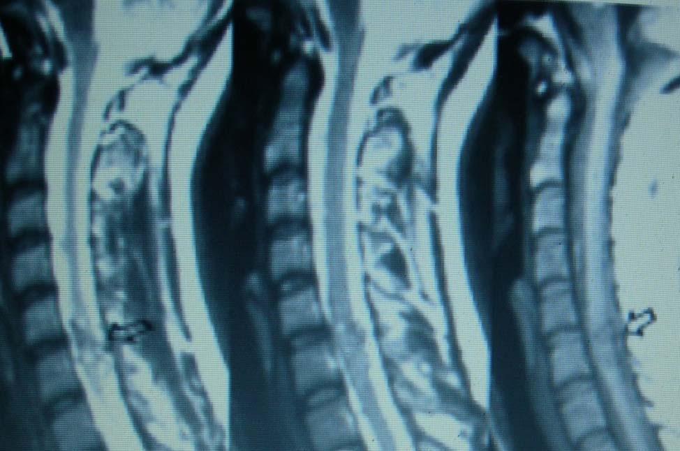
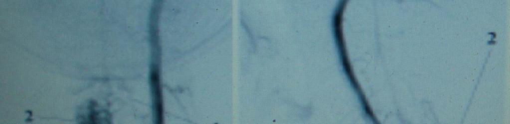
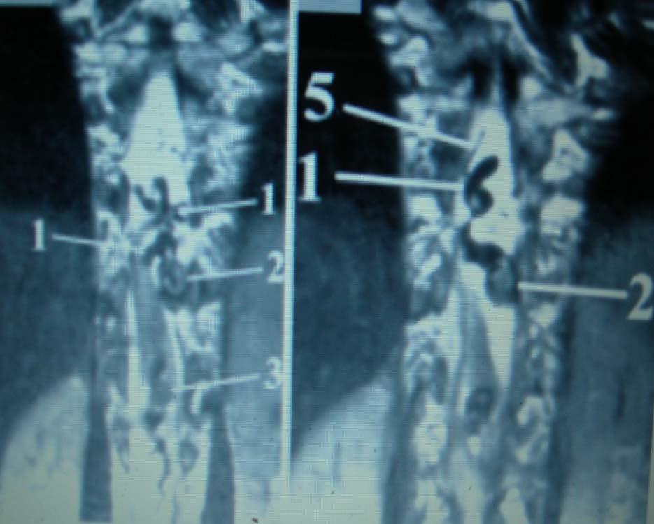
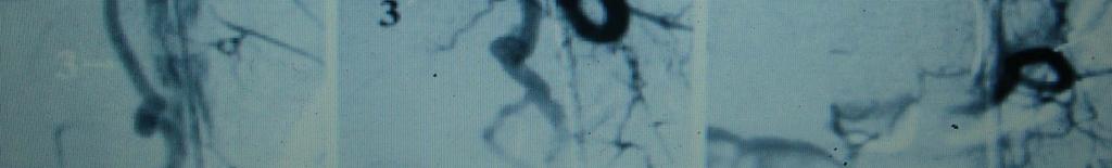
10 Intramedullary AVMs. AVM feeding vessels passed from the ventral or dorsal spinal arteries, and sometimes from the radiculopial arteries. The AVM was drained by the perimedullary veins. The spinal cord seemed expanded in the region of the malformation; sometimes expanded perimedullary draining veins are discovered around the cord on MR images. Changes of the spinal cord density are observed around the vascular nidus.
11 The vessels are densely packed in glomus AVMs and scattered in the spinal cord in diffuse AVMs The selective spinal angiography studies revealed a vascular conglomerate consisting of vessels that either adjoined each other tightly (glomus type) or were scattered in the spinal cord matter (diffuse type). According to the MR imaging and surgical findings, intramedullary AVMs are limited by spinal cord or the conglomeration of vessels spread on the surface of the spinal cord. clustering zones of low-intensity MR signals in the spinal cord
12
13


14
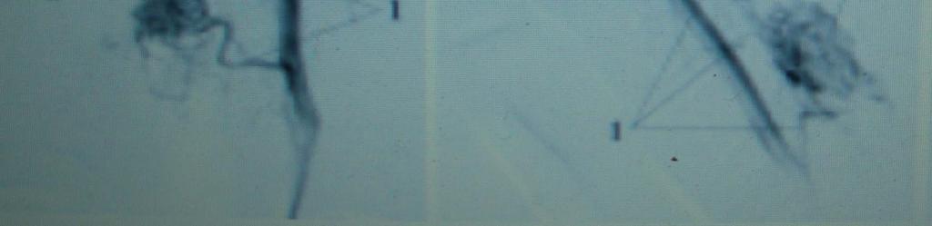
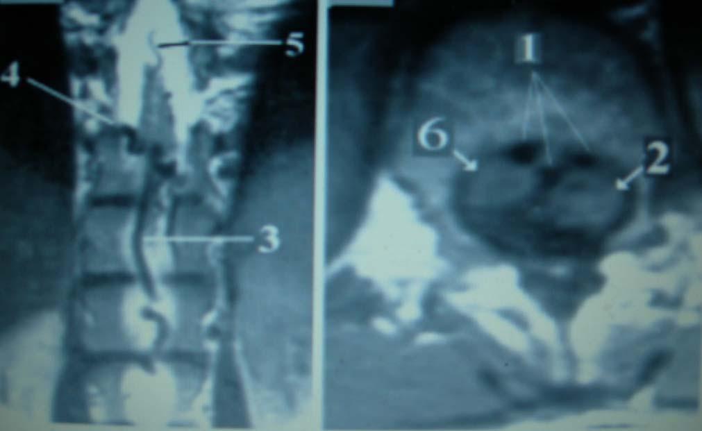
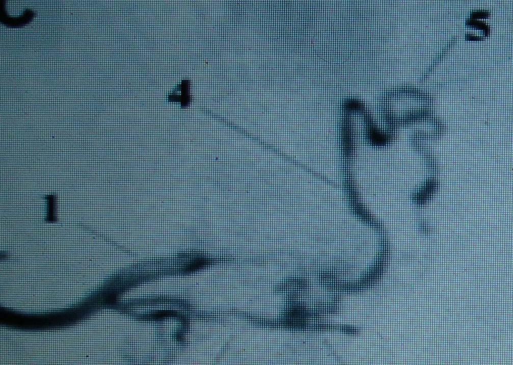
15 Intradural or Perimedullary AVMs. AVM feeding vessels passed from ventral or dorsal radiculomedullary arteries These vessels drained into the ventral or dorsal perimedullary veins These vessels can be localized on the ventral as well as on the dorsal or lateral spinal cord surface. These lesions are visualized on MR imaging as conglomerations of vessels in the form of low-intensity zones around the spinal cord on MR images.
16 The spinal cord is not expanded; in most cases its compression and displacement by the malformation are observed. Spinal cord edema is rare with this type of malformation. The selective spinal angiography studies demonstrate feeding vessels from the ventral or dorsal radiculomedullary arteries. Comparing the MR imaging and selective spinal angiography data, found thrombosis in AVM vessels.
17
18
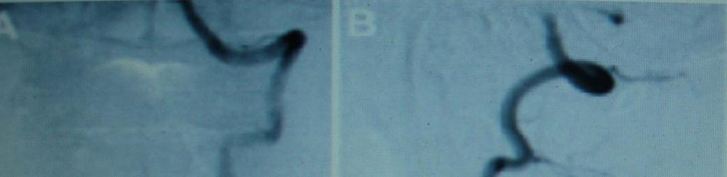
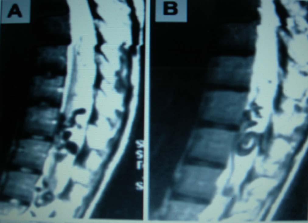
19 Dural AVMs With Retrograde Drainage Into Perimedullary Veins. In dural AVMs with retrograde drainage into perimedullary veins, expansion of these veins are found on MR imaging studies. In patients with an insignificant amount of blood shunting, the perimedullary vein expansion look like serpiginous flow voids on the dorsal surface of the spinal cord, most often in the middle and lower thoracic spine. vast spinal cord edema and thickening are typical
20 Angiographic studies reveal expanded radiculomeningeal arteries, which through a vascular conglomerate in the region of the intervertebral neural foramen shunted into the expanded perimedullary veins. Most often, a malformation has only one tributary a nidus characterized by slow blood flow; several tributaries are rarely found. The blood flow in spinal cord arteries is also slower

21
22
23
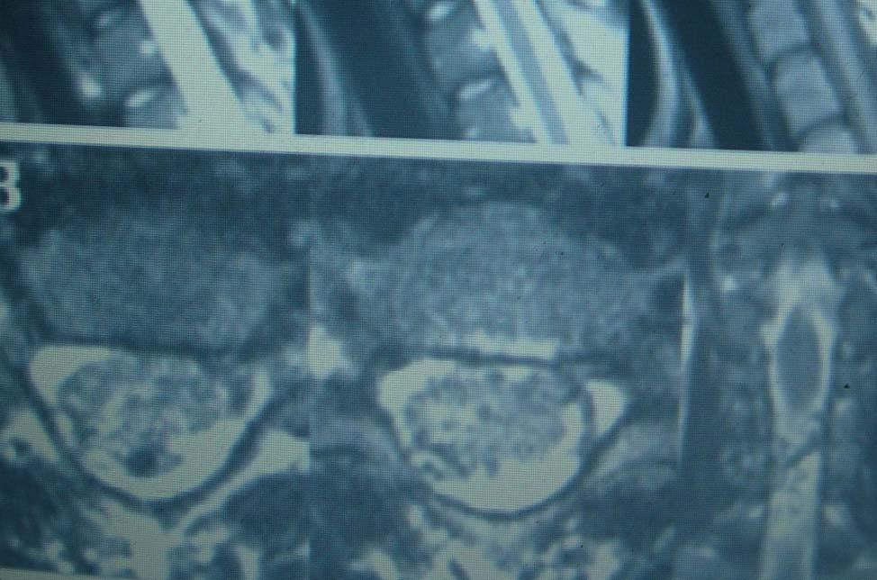
24 Epidural AVMs These lesions are characterized by lowintensity-signal zones located epidurally on MR imaging, and has caused a compression of the duramater. Selective spinal angiography demonstrate the feeding vessels spreading out directly from the spinal branch or from the postcentral, prelaminar branches. The vascular conglomerate of the AVMs are not large; it consist of small vessels.
25 Intravertebral AVMs intravertebral AVMs are discovered in the form of large vessels with intense blood flow, which are situated more often inside the vertebrae or with paravertebral spreading from these structures. Expanded epidural or paravertebral veins draining these AVMs are visible on MR images.
26 Selective spinal angiography identify AVM feeding vessels from the ventrolateral branches of segmental arteries (postcentral and prelaminar branches). The AVMs are drained through the epidural or the paravertebral veins into the ascending lumbar veins, the inferior venacava, and the azygous and hemiazygous veins

27
28 Combined Malformations. Combined lesions are situated in several adjacent anatomical structures. If combined glomus AVMs are located mainly intradurally, they received tributaries chiefly from the radiculomedullary arteries and has perimedullary venous drainage. In case of primary extradural localization, the combined glomera of the AVMs receive tributaries from the spinal branches and are drained mainly by the epidural and paravertebral veins. However, these AVMs often have equally intradural and extradural locations
29 ANGIOGRAPHIC FINDINGS
30 AVM Arterial supply Venous drainage type flow Intramedullary ant spinal artery post spinal artery radiculopial artery combined Perimedullary veins Glomus diffuse low mod high Intradural (perimedullary) ant spinal artery ant radiculomedullary. post radiculomedullary combined ant perimedullary post perimedullary glomus low mod high Dural radiculomeningeal artery retrograde into antperimedullary veins retrograde into post perimedullary veins antegrade into epidural veins Micro glomus low
31 epidural VAs spinal branch of segmental arteries post central branches. prelaminar branches combined epidural veins paravertebral veins glomus low mod high combined mainly from spinal branches mainly from radiculomedullary arteries intravertebral ventrolateral branches of segmental arteries postcentral branches prelaminar branches combined 1 one side 2 both sides perimedullaryveins extradural veins paravertebral veins combined epidural veins paravertebral veins combined mainly intradural glomus AVM. mainly epidural glomus glomus AVM AVM limited by vertebra glomus AVM w/ para vertebral spreading low mod high low mod high
32 Clinical features Equal incidence Mean age-3rd decade Uniform distribution Higher incidence of association with other vascular malformation Congenital lesion Acute initial presentation Haemorrhage Stepwise progression of deficit Progressive loss of neurological deficit Pregnancy,exercise,minor trauma-rapid progression Bruit over spinal cord
33 Pathophysiology Venous congestion and hypertension Reduced arterial perfusion Ischemia Compressive myelopathy
34 author Rosenblum et al 1987 Yasargil et al 1984 cases Biondi et al 1990 Age at onset Mean 27 yr 76%<41yr Mean 20 yr Initial symptom Acute onset in 50% SAH in 59% SAH in 58% At diagnosis SAH in 52 % 74% disabled SAH in 76%, 63% disabled SAH in 68%, 75% disabled Progressive evolution Associiated with deterioration - Step wise progression- 40% Posture-17% Pregnency-6% Valsalva-13% Activity-15% 31%- relapse,with worsening
35 Surgical approaches
36 Open surgical interventions are advisable necessary to occlude only the feeding vessels, one has failed to embolize them endovascularly, embolization of the tributaries threatens to occlude the arteries feeding the spinal cord. embolization of the main tributaries will not result in complete occlusion of the blood flow; selective spinal angiography does not identify all of the tributaries diameter is too small
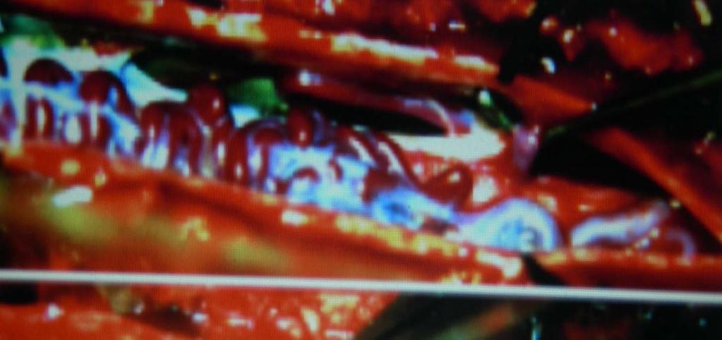
37 Occlusion of the feeding and draining vessels and malformation resection Intramedullary glomus AVM Perimedullary AVM Epidural AVM Combined AVM
38 Indications for occluding only the feeding vessels Intramedullary diffuse AVM Intravertebral AVM Combined AVM Dural AVM Conus medullaris AVM
39 Combining surgical intervention with endovascular embolization High flow AVM and numerous large feeding vessels running into it After the endovascular embolization a mass effect due to AVM blood flow remains.
40 Intramedullary AVM Depended on type of feeding vessels and location of the nidus. ventral approaches Feeder from the anterior spinal artery ventral regions ventral exophytic spreading posterior approaches Feeder from the dorsal spinal arteries dorsal regions dorsal exophytic spreading,
41 Intramedullary glomus AVMs, Two variants of nidus resection 1) isolate the vessels near the nidus and coagulate, then dissect the nidus and resect 2) the vessels are cut off in the nidus itself during its separation, and resection of the nidus.
42 Intramedullary diffuse AVMs, Cut off the feeding vessels near the nidus, then perform a myelotomy, partially isolated the vessels in the nidus, coagulate and intersected them, but left them in situ..
43 Conusmedullaris AVM In intramedullary AVM of the conusmedullaris, because of the possible pelvic disturbances, only performe occlusion of feeding vessels, leaving the malformation in situ.
44 Perimedullary AVMs Occlusion of the feeding vessels right at the nidus as the first step, then cut off the draining perimedullary veins and perform total resection of the AVM During this procedure, try to preserve the pial vascular plexus of the spinal cord.
45 Dural AVMs two variants of surgical technique 1) Occluding the malformation in the dural leaf of the spinal nerve root or cutting off the feeding vessels immediately outside the root. 2) occluding the radicular vein, which provides retrograde blood shunting from the AVM into the perimedullary veins.
46 Epidural AVMs Coagulate and section the direct tributaries: postcentral, prelaminar spinal branches. Coagulate and section the spinal branch of the segmental artery laterally at the point of its entry in to the intervertebral foramen, coagulated the intervertebral veins, and the epidural veins.
47 intravertebral malformations combine endovascular obliteration of feeding vessels and direct surgical intervention. Vertebral body affected but no body expansion and dura matter compression - vertebroplasty Vertebral body expansion and dura mater compression - occlude the vessels and resect the affected VB
48 Combined AVMs Primary extradural localization - endovascular technology Mainly intradural location and spinal cord compression -combination of endovascular and microsurgical methods
49 Author Rosenbl um et al 1987 Yasargil et al 1984 Conolly et al 1998 Cases YURI P et al 2006 Follow up 3yr 3yr 3.8yr 4mth- 8yr Improved % Unchang ed% Worse%
50 Embolization. Indication Occlude only the feeding vessels Combined AVM and intervertebral AVM AVMs with a pronounced blood flow Two variations of the conventional technique are applied: 1) super selective obliteration of the feeding vessels 2) obliteration of the main tributary, usually the segmental artery.
51 If these arteries ended in the AVM it is possible to apply selective embolization. If these arteries extend branches to the AVM and continued farther, feeding the spinal cord, avoid embolization. a nonselective technique of obliteration of the segmental main tributary, if this artery do not feed the spinal cord,staged obliteration of tributaries with a few days between each stage
52 Embolic agents Particulate materials Poly vinyl alcohol( micro) Gelfoam Sponge microparticulate Balloon occlusion Liquid agents N-butyl cyanoacrylate ethylene vinyl alcohol copolymer)
53 embolic agent N-butyl cyanoacrylate Onyx (ethylene vinyl alcohol copolymer) type liquid liquid dilution Lipiodil DMSO and Tantalum proximal reflux venous penetration gluing of the catheter +++ _ delayed venous thrombosis +++ _ J Neurosurg (Spine 2) 93: , 2000
54 Biondi et al Embolization with particles Mean follow up- 6 years Total cases- 35 Recurrent avm- 35(100%) outcome Improved 63% Unchanged 26% Worse 11% Rufus A. Corkill, et al Embolization with liquid embolic agent mean follow-up 24.3 months Total cases- 17 outcome total obliteration 6 patients (37.5%), subtotal obliteration 5 patients (31.25%), partial obliteration 5 patients (31.25%). Improvement 14 patients (82%). Radiology177: ,1990 Journal of Neurosurgery: Spine Nov 2007
55 The cause of the initial neurological deterioration following embolization Edema in the spinal cord following occlusion of the nidus and thrombosis Occlusion of the ASA due to reflux Direct toxic effect of DMSO Thrombosis of the spinal veins J Neurosurg (Spine 2) 93: , 2000
56 Risks of open surgical or endovascular treatment Skin infection or cellulitis Bleeding Injury to nervous tissue, causing paralysis, bladder or bowel dysfunction, or sexual dysfunction Chronic pain syndromes Thrombosis of epidural veins and neurologic loss Spinal cord infarction James S Harrop et al Department of Neurosurgery, JFK Medical Center, Edison, New Jersey emedicine.com
57 Complications that result from open surgical ligation or resection Infection of meninges (meningitis) Cerebrospinal fluid leak Wound dehiscence Complications that result from the endovascular technique Femoral hematoma Pseudoaneurysms and thrombosis Arterial dissection
58 Stereotactic radiosurgery Single high dose Hypofractionated irradiation 20 to 30% rate of occlusion. Internal fiducial markers and imageguided radiation allow stereotactic irradiation for spinal disease with real-time verification and an accuracy of ±1 mm for every 0.03 seconds Stereotactic Irradiation for Intramedullary AVMs Ten Years Experience Hokkaido University Collection of Scholarly and Academic Papers JAPAN
59 Thank you
Historical perspective
 SPINAL AVM Introduction Vascular malformations of spinal cord are a rare clinical entity, representing 5% of all primary spinal cord lesions, with arteriovenous malformations(avm) & cavernous malformations
Spinal arteriovenous malformations: new classification and surgical treatment
 Neurosurg Focus 20 (5):E7, 2006 Spinal arteriovenous malformations: new classification and surgical treatment YURI P. ZOZULYA, M.D., PH.D., EUGENE I. SLIN KO, M.D., PH.D., AND IYAD I. AL-QASHQISH, M.D.
SPINAL EPIDURAL ARTERIOVENOUS MALFORMATIONS: REPORT OF A CASE WITH DISCUSSION OF CLASSIFICATION AND TREATMENT
 SPINAL EPIDURAL ARTERIOVENOUS MALFORMATIONS: REPORT OF A CASE WITH DISCUSSION OF CLASSIFICATION AND TREATMENT Caitlin M. Clark, B.A. and W. Craig Clark, M.D., Ph.D., FAANS, FACS, FICS INTRODUCTION True
SURGICAL TREATMENT OF SPINAL ARTERIOVENOUS MALFORMATIONS: VASCULAR ANATOMY AND SURGICAL OUTCOME
 Spinal Arteriovenous Malformations SURGICAL TREATMENT OF SPINAL ARTERIOVENOUS MALFORMATIONS: VASCULAR ANATOMY AND SURGICAL OUTCOME Po-An Tai, Yong-Kwang Tu, and Hon-Man Liu 1 Background and purpose: Spinal
Spinal Arteriovenous Shunts: Angioarchitecture and Historical Changes in Classification
 REVIEW ARTICLE doi: 10.2176/nmc.ra.2016-0316 Neurol Med Chir (Tokyo) 57, 356 365, 2017 Spinal Arteriovenous Shunts: Angioarchitecture and Historical Changes in Classification Keisuke Takai 1 Online May
Spinal Vascular Lesions
 Spinal Vascular Lesions Spinal Vascular Lesions Spinal cord infarction Hemangioblastoma Cavernous malformation Vascular malformations (Type 1-4) Spinal artery aneurysm Troy Hutchins, MD Assistant Professor
Results of the surgical treatment of perimedullary arteriovenous fistulas with special reference to embolization
 Neurosurg Focus 5 (4): Article 9, 1998 Results of the surgical treatment of perimedullary arteriovenous fistulas with special reference to embolization Kazutoshi Hida, M.D., Yoshinobu Iwasaki, M.D., Katsuya
SDAVFs are rare acquired vascular lesions predominantly
 CLINICAL REPORT W.J. van Rooij R.J. Nijenhuis J.P. Peluso M. Sluzewski G.N. Beute B. van der Pol Spinal Dural Fistulas without Swelling and Edema of the Cord as Incidental Findings SUMMARY: SDAVFs cause
MR Imaging of Spinal Cord Arteriovenous Malformations at 0.5 T: Study of 34 Cases
 833 MR Imaging of Spinal Cord Arteriovenous Malformations at 0.5 T: Study of 34 Cases D. Dormont 1 F. Gelbert 2 E. Assouline 2 D. Reizine 2 A. Helias 2 M. C. Riche 2 J. Chiras 1 J. Sories 1 J. J. Merland
Management of spinal dural arteriovenouse fistula
 Romanian Neurosurgery Volume XXXI Number 4 2017 October-December Article Management of spinal dural arteriovenouse fistula A. Chiriac, Georgiana Ion, N. Dobrin, I. Poeată ROMANIA DOI: 10.1515/romneu-2017-0074
Dural Arteriovenous Malformations and Fistulae (DAVM S DAVF S)
") Jorge Guedes Campos NEUROIMAGING DEPARTMENT HOSPITAL SANTA MARIA UNIVERSITY OF LISBON PORTUGAL DEFINITION region of arteriovenous shunting confined to a leaflet of packymeninges often adjacent to a major
Jorge Guedes Campos NEUROIMAGING DEPARTMENT HOSPITAL SANTA MARIA UNIVERSITY OF LISBON PORTUGAL DEFINITION region of arteriovenous shunting confined to a leaflet of packymeninges often adjacent to a major
Clinical Features and Outcomes of Spinal Cord Arteriovenous Malformations Comparison Between Nidus and Fistulous Types
 Clinical Features and Outcomes of Spinal Cord Arteriovenous Malformations Comparison Between Nidus and Fistulous Types Young-Jun Lee, MD, PhD; Karel G. Terbrugge, MD, FRCP(C); Guillaume Saliou, MD, PhD;
Vascular Malformations
 Vascular Malformations LTC Robert Shih Chief of Neuroradiology Walter Reed Medical Center Special thanks to LTC Alice Smith (retired) Disclosures: None. This presentation reflects the personal views of
Brain Arteriovenous Malformations Endovascular Therapy and Associated Therapeutic Protocols Jorge Guedes Cabral de Campos
 Endovascular Therapy and Associated Therapeutic Protocols Jorge Guedes Cabral de Campos Neuroradiology Department Hospital de Santa Maria University of Lisbon CEREBRAL AVM CLINICAL / EPIDEMIOLOGY Brain
Cerebrovascular Malformations in the Elderly Indications for Treatment
 Cerebrovascular Malformations in the Elderly Indications for Treatment Johanna T. Fifi, MD, FAHA, FSVIN Director of Endovascular Ischemic Stroke Assistant Professor of Neurology, Neurosurgery, and Radiology
Modern treatment of brain arteriovenous malformation
 ORIGINAL RESEARCH W.J. van Rooij M. Sluzewski G.N. Beute Brain AVM Embolization with Onyx BACKGROUND AND PURPOSE: To report the initial experience by using a new liquid embolic agent (Onyx) for embolization
Treatment of Slow-Flow (Type I) Perimedullary Spinal Arteriovenous Fistulas with Special Reference to Embolization
 Perimedullary Spinal Arteriovenous Fistulas with Special Reference to Embolization") AJNR Am J Neuroradiol 26:2582 2586, November/December 2005 Case Report Treatment of Slow-Flow (Type I) Perimedullary Spinal Arteriovenous Fistulas with Special Reference to Embolization Ismail Oran, Mustafa
AJNR Am J Neuroradiol 26:2582 2586, November/December 2005 Case Report Treatment of Slow-Flow (Type I) Perimedullary Spinal Arteriovenous Fistulas with Special Reference to Embolization Ismail Oran, Mustafa
Role of Three-Dimensional Rotational Angiography in the Treatment of Spinal Dural Arteriovenous Fistulas
 Open Access Case Report DOI: 10.7759/cureus.1932 Role of Three-Dimensional Rotational Angiography in the Treatment of Spinal Dural Arteriovenous Fistulas Yigit Ozpeynirci 1, Bernd Schmitz 2, Melanie Schick
Surgical Privileges Form: "Neurosurgery" Clinical Privileges Request. Requested (To be completed by the applicant) Not Recommended (For committee use)
 Not Recommended (For committee use)") Surgical Form: Clinical Request "Neurosurgery" Applicant s Name:. License No. (If Any):... Date:... Scope of Practice:. Facility:.. Place of Work:. the applicant) CATEGORY I: Core : 1. Interpretation of
Surgical Form: Clinical Request "Neurosurgery" Applicant s Name:. License No. (If Any):... Date:... Scope of Practice:. Facility:.. Place of Work:. the applicant) CATEGORY I: Core : 1. Interpretation of
Recently, diagnostic imaging methods for spinal
 J Neurosurg Spine 18:398 408, 2013 AANS, 2013 Three-dimensional angioarchitecture of spinal dural arteriovenous fistulas, with special reference to the intradural retrograde venous drainage system Clinical
A.J. Hauer Intracranial dural arteriovenous fistulae
 A.J. Hauer 27-06-2018 Intracranial dural arteriovenous fistulae Dural arteriovenous fistulae (davfs) epidemiology Pathological anastomoses (within the dural leaflets) between meningeal arteries and dural
Peripheral Spinal Cord Hypointensity on T2-weighted MR Images: A Reliable Imaging Sign of Venous Hypertensive Myelopathy
 AJNR Am J Neuroradiol 21:781 786, April 2000 Peripheral Spinal Cord Hypointensity on T2-weighted MR Images: A Reliable Imaging Sign of Venous Hypertensive Myelopathy Robert W. Hurst and Robert I. Grossman
Pial arteriovenous fistula of the spine in a child with hemiplegia
 CASE REPORT Pial arteriovenous fistula of the spine in a child with hemiplegia Kazuki Hatayama 1, Shinichiro Goto 1, Ayumi Nishida 2 & Masaru Inoue 1 1 Department of Pediatrics, Okayama Red-Cross Hospital,
Endovascular Treatment of Cerebral Arteriovenous Malformations. Bs. Nguyễn Ngọc Pi Doanh- Bs Đặng Ngọc Dũng Khoa Ngoại Thần Kinh
 Endovascular Treatment of Cerebral Arteriovenous Malformations Bs. Nguyễn Ngọc Pi Doanh- Bs Đặng Ngọc Dũng Khoa Ngoại Thần Kinh Stroke Vascular Malformations of the Brain Epidemiology: - Incidence: 0.1%,
Materials and Methods. A. Biondi, 1 2 J.J. Merland, 1 J.E. Hodes, 1 A. Aymard, 1 and D. Reizine 1
 Aneurysms of Spinal Arteries Associated with Intramedullary Arteriovenous Malformations. II. Results of A V M Endovascular Treatment and Hemodynamic Considerations I A. Biondi, 1 2 J.J. Merland, 1 J.E.
Researcher 2018;10(3) Management of Vascular Myelopathy.
 Management of Vascular Myelopathy.") Management of Vascular Myelopathy Mustafa El-sayed Mohammed El-sayed 1, Magdy Asaad El-Hawar 1, Wafik Ebrahim Aly 2 and Ahmed Mohammed Abd El-Fatah Deabes 3 1 Neurosurgery Department, Faculty of Medicine,
Management of Vascular Myelopathy Mustafa El-sayed Mohammed El-sayed 1, Magdy Asaad El-Hawar 1, Wafik Ebrahim Aly 2 and Ahmed Mohammed Abd El-Fatah Deabes 3 1 Neurosurgery Department, Faculty of Medicine,
Leo Happel, PhD Professor, Neurology, Neurosurgery, Physiology, and Neuroscience LSU Health Science Center
 Leo Happel, PhD Professor, Neurology, Neurosurgery, Physiology, and Neuroscience LSU Health Science Center Leo Happel disclosed no financial relationships Vascular Lesions of the Spinal Cord The vast majority
Occlusive hyperemia: a theory for the hemodynamic complications following resection of intracerebral arteriovenous malformations
 J Neurosurg 78: 167-175, 1993 Occlusive hyperemia: a theory for the hemodynamic complications following resection of intracerebral arteriovenous malformations NAYEF R. F. AL-RODHAN, M.D., PH.D., THORALF
PA SYLLABUS. Syllabus for students of the FACULTY OF MEDICINE No.2
 Approved At the meeting of the Faculty Council Medicine No. of Approved At the meeting of the chair of Neurosurgery No. of Dean of the Faculty Medicine No.2 PhD, associate professor M. Betiu Head of the
Brain AVM with Accompanying Venous Aneurysm with Intracerebral and Intraventricular Hemorrhage
 Cronicon OPEN ACCESS EC PAEDIATRICS Case Report Brain AVM with Accompanying Venous Aneurysm with Intracerebral and Intraventricular Hemorrhage Dimitrios Panagopoulos* Neurosurgical Department, University
Chapter 4 Section 20.1
 Surgery Chapter 4 Section 20.1 Issue Date: August 29, 1985 Authority: 32 CFR 199.4(c)(2) and (c)(3) Copyright: CPT only 2006 American Medical Association (or such other date of publication of CPT). All
Chapter 4 Section 20.1
 Surgery Chapter 4 Section 20.1 Issue Date: August 29, 1985 Authority: 32 CFR 199.4(c)(2) and (c)(3) 1.0 CPT 1 PROCEDURE CODES 61000-61626, 61680-62264, 62268-62284, 62290-63048, 63055-64484, 64505-64595,
Chapter 4 Section 20.1
 Surgery Chapter 4 Section 20.1 Issue Date: August 29, 1985 Authority: 32 CFR 199.4(c)(2) and (c)(3) Copyright: CPT only 2006 American Medical Association (or such other date of publication of CPT). All
Recanalization of spinal arteriovenous malformations following embolization
 J Neurosurg 7:74-72, 989 Recanalization of spinal arteriovenous malformations following embolization WALTER A. HALL, M.D., EDWARD H. OLDFIELD, M.D., AND JOHN L. DOPPMAN, M.D. Clinical Neurosurgery Section,
Partially trombosed glomus type spinal arteriovenous malformation case presentation
 442 Gorgan et al Partially trombosed glomus type spinal arteriovenous malformation Partially trombosed glomus type spinal arteriovenous malformation case presentation M.R. Gorgan, Narcisa Bucur, Catioara
Interventional Radiology in Trauma. Vikash Prasad, MD, FRCPC Vascular and Interventional Radiology The Moncton Hospital
 Interventional Radiology in Trauma Vikash Prasad, MD, FRCPC Vascular and Interventional Radiology The Moncton Hospital Disclosures None relevant to this presentation Shareholder Johnson and Johnson Goal
Complex dural arteriovenous fistulas. Results of combined endovascular and neurosurgical treatment in 16 patients
 J Neurosurg 71:352-358,1989 Complex dural arteriovenous fistulas Results of combined endovascular and neurosurgical treatment in 16 patients STANLEY L. BARNWELL, M.D., PH.D., VAN V. HALBACH, M.D., RANDALL
Spontaneous occlusion of a cerebral arteriovenous malformation after subtotal endovascular embolisation
 206 Chiriac et al Spontaneous occlusion of a cerebral arteriovenous malformation Spontaneous occlusion of a cerebral arteriovenous malformation after subtotal endovascular embolisation A. Chiriac, N. Dobrin*,
Transvenous Embolization of Cavernous Sinus Dural Arteriovenous Fistulas with Shunts Involving the Laterocavernous Sinus
 Journal of Neuroendovascular Therapy 2017; 11: 1 7 Online November 9, 2016 DOI: 10.5797/jnet.oa.2016-0062 Transvenous Embolization of Cavernous Sinus Dural Arteriovenous Fistulas with Shunts Involving
Essentials of Clinical MR, 2 nd edition. 51. Primary Neoplasms
 51. Primary Neoplasms As with spinal central canal neoplasms in other regions, those of the lumbar spine may be classified as extradural, intradural extramedullary, and medullary. If an extradural lesion
CLEAR III TRIAL : UPDATE ON SURGICAL MATTERS THAT MATTER
 CLEAR III TRIAL : UPDATE ON SURGICAL MATTERS THAT MATTER CLEAR Surgical Center Team July 2011 Trial Enrollment Status Updates Insert latest enrollment update chart from most recent CLEAR newsletter Imaging
What Is an Arteriovenous malformation (AVM)?
?") American Society of Neuroradiology What Is an Arteriovenous malformation (AVM)? From the Cerebrovascular Imaging and Intervention Committee of the American Heart Association Cardiovascular Council Randall
American Society of Neuroradiology What Is an Arteriovenous malformation (AVM)? From the Cerebrovascular Imaging and Intervention Committee of the American Heart Association Cardiovascular Council Randall
Spinal dural AV fistula: One stop shop imaging with MR?
 Spinal dural AV fistula: One stop shop imaging with MR? Poster No.: C-3378 Congress: ECR 2010 Type: Educational Exhibit Topic: Neuro Authors: J. C. Röper-Kelmayr 1, C. Ginthör 1, D. Flöry 1, R. Chapot
Dr. Shakir Husain MD, DM, FINR Consultant & Chief of Services Department of NeuroEndoVascular Therapy & Stroke. Program Director
 EGAS MUNIZ FELLOWSHIP INTERVENTIONAL NEUROLOGY & STROKE Neurointervention is fast becoming an important subspecialty of neurosciences. There are many unexplored dimensions of these techniques, which may
N-butyl 2-cyanoacrylate Embolization of Spinal Dural Arteriovenous Fistulae
 AJNR Am J Neuroradiol : 7, January N-butyl -cyanoacrylate Embolization of Spinal Dural Arteriovenous Fistulae Joon K. Song, Y. Pierre Gobin, Gary R. Duckwiler, Yuichi Murayama, John G. Frazee, Neil A.
Title. CitationNeurologia medico-chirurgica, 49(3): Issue Date Doc URL
: Issue Date Doc URL") Title Cervical Epidural Arteriovenous Fistula With Radicul Author(s)Kawabori, Masahito; Hida, Kazutoshi; Yano, Shunsuke; CitationNeurologia medico-chirurgica, 49(3): 108-113 Issue Date 2009-03 Doc URL
Title Cervical Epidural Arteriovenous Fistula With Radicul Author(s)Kawabori, Masahito; Hida, Kazutoshi; Yano, Shunsuke; CitationNeurologia medico-chirurgica, 49(3): 108-113 Issue Date 2009-03 Doc URL
Endovascular Treatment for Spontaneous Vertebral Arteriovenous Fistula in Neurofibromatosis Type 1: A Case Report
 Endovascular Treatment for Spontaneous Vertebral Arteriovenous Fistula in Neurofibromatosis Type 1: A Case Report Case Report Yon Kwon Ihn, MD 1, Won Sang Jung, MD 1, Bum-Soo Kim, MD 1 We report the case
Neurosurgical Techniques
 Neurosurgical Techniques Neurosurgical Techniques Laminectomy for the Removal of Spinal Cord Tumors J. GRAFTON LOVE, M.D. Section of Neurologic Surgery, Mayo Clinic and Mayo Foundation, Rochester, Minnesota
Introduction to Neuroimaging spine. John J. McCormick MD
 Introduction to Neuroimaging spine John J. McCormick MD Neuroanatomy Netter drawings Radiographic Anatomy Cervical Spine Cervical Spine Oblique View Cervical Spine Dens View Thoracic Spine Lumbar Spine
Spine. Neuroradiology. Spine. Spine Pathology. Distribution of fractures. Radiological algorithm. Role of radiology 18/11/2015
 Spine Neuroradiology Spine Prof.Dr.Nail Bulakbaşı X Ray: AP/L/Oblique Vertebra & disc spaces CT & CTA Vertebra, discs, vessels MRI & MRA Vertebra, disc, vessels, meninges Spinal cord & nerves Myelography
B Umesh, P Pramodbhai, C Laxmidas, M Taiyabali, G Jayesh, R A
 ISPUB.COM The Internet Journal of Radiology Volume 14 Number 1 B Umesh, P Pramodbhai, C Laxmidas, M Taiyabali, G Jayesh, R A Citation B Umesh, P Pramodbhai, C Laxmidas, M Taiyabali, G Jayesh, R A.. The
Overview. Spinal Anatomy Spaces & Meninges Spinal Cord. Anatomy of the dura. Anatomy of the arachnoid. Anatomy of the spinal meninges
 European Course in Neuroradiology Module 1 - Anatomy and Embryology Dubrovnik, October 2018 Spinal Anatomy Spaces & Meninges Spinal Cord Johan Van Goethem Overview spinal meninges & spaces spinal cord
Kathleen R. Fink, MD Virginia Mason Medical Center. 6 th Nordic Emergency Radiology Course 2017
 Kathleen R. Fink, MD Virginia Mason Medical Center 6 th Nordic Emergency Radiology Course 2017 Disclosure My spouse receives research salary support from: Guerbet Outline Acute neck and back pain Acute
Spinal intradural arteriovenous fistulas acquired in late adulthood: absent spinal venous drainage in pathogenesis and pathophysiology
 J Neurosurg Spine 3:488 494, 2005 Spinal intradural arteriovenous fistulas acquired in late adulthood: absent spinal venous drainage in pathogenesis and pathophysiology Report of two cases GABRIEL C. TENDER
Type II Endoleak Embolization Choice of Materials: EVOH, Glue, Thrombin & Coils. Michael S. Rosenberg, MD Assistant Professor of Radiology
 Type II Endoleak Embolization Choice of Materials: EVOH, Glue, Thrombin & Coils Michael S. Rosenberg, MD Assistant Professor of Radiology Michael Rosenberg, M. D. No relevant financial relationship reported
Case Log Mapping Update: April 2018 Review Committee for Neurological Surgery
 Case Log Mapping Update: April 2018 Review Committee for Neurological Surgery The Review Committee has made the following changes to the CPT code mappings: The following previously untracked CPT codes
Spinal DAVFs, perimedullary AVFs, and spinal. Clinical features and treatment outcomes of the spinal arteriovenous fistulas and malformations
 J Neurosurg Spine 19:207 216, 2013 AANS, 2013 Clinical features and treatment outcomes of the spinal arteriovenous fistulas and malformations Clinical article Won-Sang Cho, M.D., 1 Ki-Jeong Kim, M.D.,
Intracranial dural arteriovenous fistulas (DAVFs) with retrograde
 with retrograde") ORIGINAL RESEARCH W.J. van Rooij M. Sluzewski G.N. Beute Dural Arteriovenous Fistulas with Cortical Venous Drainage: Incidence, Clinical Presentation, and Treatment BACKGROUND AND PURPOSE: Our purpose
ORIGINAL RESEARCH W.J. van Rooij M. Sluzewski G.N. Beute Dural Arteriovenous Fistulas with Cortical Venous Drainage: Incidence, Clinical Presentation, and Treatment BACKGROUND AND PURPOSE: Our purpose
Venous Congestive Myelopathy due to Chronic Inferior Vena Cava Thrombosis Treated with Endovascular Stenting: Case Report and Review of the Literature
 Venous Congestive Myelopathy due to Chronic Inferior Vena Cava Thrombosis Treated with Endovascular Stenting: Case Report and Review of the Literature Diego Z. Carvalho, MD 1, Joshua D. Hughes, MD 2, Greta
Features compression after open and endovascular operation in vascular malformation
 Features compression after open and endovascular operation in vascular malformation Sapelkin Sergey Institute of Surgery named A.V. Vishnevsky, Moscow, Russia 21.10.2017 CIRC Meeting, Grassau AV-malformations:
EMBOLIZATION OF ARTERIOVENOUS FISTULA AFTER RADIOSURGERY FOR MULTIPLE CEREBRAL ARTERIOVENOUS MALFORMATIONS
 Arteriovenous fistula after radiosurgery for multiple CAVM EMBOLIZATION OF ARTERIOVENOUS FISTULA AFTER RADIOSURGERY FOR MULTIPLE CEREBRAL ARTERIOVENOUS MALFORMATIONS Chao-Bao Luo, Wan-Yuo Guo, Michael
Treatment of brain AVMs includes different modalities
 ORIGINAL RESEARCH W.J. van Rooij S. Jacobs M. Sluzewski B. van der Pol G.N. Beute M.E. Sprengers Curative Embolization of Brain Arteriovenous Malformations with Onyx: Patient Selection, Embolization Technique,
Vascular malformations: Venous malformations anomalous veins drain normal brain tissue for 65% of all cases 2.5%. was 0, 3% per year
 Vascular malformations: 1. Venous malformations: congenital venous anomalies pathologically characterised by anomalous veins (thickened and hyalinised walls) separated by normal brain. These anatomically
(http://thejns.org/doi/abs/ / focus11344)
") Neurosurg Focus 32 (5):E3, 2012 Surgical treatment of Type I spinal dural arteriovenous fistulas Alexander E. Ropper, M.D., Bradley A. Gross, M.D., and Rose Du, M.D., Ph.D. Department of Neurosurgery,
Neurosurg Focus 32 (5):E3, 2012 Surgical treatment of Type I spinal dural arteriovenous fistulas Alexander E. Ropper, M.D., Bradley A. Gross, M.D., and Rose Du, M.D., Ph.D. Department of Neurosurgery,
Overview of Cerebrovascular Malformations
 Overview of Cerebrovascular Malformations Pursuit of Neurovascular Excellence 8 th annual Barbara Albani, MD Chief, Neurointerventional Surgery Christiana Care Health Systems Newark, DE Financial Disclosures
Untangling Cerebral Dural Arteriovenous Fistulas
 Untangling Cerebral Dural Arteriovenous Fistulas Bradley A. Gross, MD Assistant Professor, Dept of Neurosurgery, University of Pittsburgh September 2017 davfs Definition Clinical Presentation Natural History
CENTRAL NERVOUS SYSTEM TRAUMA and Subarachnoid Hemorrhage. By: Shifaa AlQa qa
 CENTRAL NERVOUS SYSTEM TRAUMA and Subarachnoid Hemorrhage By: Shifaa AlQa qa Subarachnoid Hemorrhage Causes: Rupture of a saccular (berry) aneurysm Vascular malformation Trauma Hematologic disturbances
VASCULAR MALFORMATIONS. Owen Samuels, MD Adam Webb, MD Emory University
 VASCULAR MALFORMATIONS Owen Samuels, MD Adam Webb, MD Emory University Introduction Brain and spinal cord vascular malformations can be separated into five main categories: 1) Arteriovenous malformation,
A Case of Carotid-Cavernous Fistula
 A Case of Carotid-Cavernous Fistula By : Mohamed Elkhawaga 2 nd Year Resident of Ophthalmology Alexandria University A 19 year old male patient came to our outpatient clinic, complaining of : -Severe conjunctival
Sp i n a l davfs are acquired vascular lesions characterized
 Neurosurg Focus 26 (1):E4, 2009 Intracranial subarachnoid hemorrhage resulting from cervical spine dural arteriovenous fistulas: literature review and case presentation Da n i e l R. Fa s s e t t, M.D.,
Imaging abdominal vascular emergencies. V.Stoynova
 Imaging abdominal vascular emergencies V.Stoynova Abdominal vessels V. Stoynova 2 Acute liver bleeding trauma anticoagulant therapy liver disease : HCC, adenoma, meta, FNH, Hemangioma Diagnosis :CT angiography
Transverse-Sigmoid Sinus Dural Arteriovenous Malformations
 Transverse-Sigmoid Sinus Dural Arteriovenous Malformations Kenan I. Amautovic, M.D., and Ali F. Krisht, M.D. '-...--- Learning Objectives: After reading this article, the participant should: 1. Have an
Spontaneous Obliteration of Pial Arteriovenous Malformations: A Review of 27 Cases
 AJNR Am J Neuroradiol :, March 00 Spontaneous Obliteration of Pial Arteriovenous Malformations: A Review of ases Maneesh. Patel, Timothy J. Hodgson, Andras A. Kemeny, and David M. Forster BAKGROUND AND
Differences between CS-DAVF and TCCF to reveal and redefine CS-DAVF
 Pan et al. Chinese Neurosurgical Journal (2018) 4:26 https://doi.org/10.1186/s41016-018-0121-z CHINESE MEDICAL ASSOCIATION COMMENTARY Differences between CS-DAVF and TCCF to reveal and redefine CS-DAVF
Neurophysiologic Monitoring and Pharmacologic Provocative Testing for Embolization of Spinal Cord Arteriovenous Malformations
 AJNR Am J Neuroradiol 25:1131 1138, August 2004 Neurophysiologic Monitoring and Pharmacologic Provocative Testing for Embolization of Spinal Cord Arteriovenous Malformations Yasunari Niimi, Francesco Sala,
The superior ophthalmic vein approach for the treatment of carotid-cavernous fistulas: our first experience
 230 Chiriac et al - Superior ophthalmic vein approach The superior ophthalmic vein approach for the treatment of carotid-cavernous fistulas: our first experience A. Chiriac, N. Dobrin 1, Georgiana Ion
Department of Neurological Surgery
 Department of Neurological Surgery CAT 1 A Basic Privileges: Patient management, including H & Ps and diagnostic and therapeutic treatments, procedures and interventions, Requiring a level of training
Vascular Malformations of the Brain. William A. Cox, M.D. Forensic Pathologist/Neuropathologist. September 8, 2014
 Vascular Malformations of the Brain William A. Cox, M.D. Forensic Pathologist/Neuropathologist September 8, 2014 Vascular malformations of the brain are classified into four principal groups: arteriovenous
CASE OF THE WEEK PROFESSOR YASSER METWALLY
 CLINICAL PICTURE CLINICAL PICTURE: CASE OF THE WEEK PROFESSOR YASSER METWALLY A 29 years old male patients presented with proptosis, ecchymoses of the left eye with both subjective and objective bruit
Neuroradiology Subspecialty Exam Study Guide
 Neuroradiology Subspecialty Exam Study Guide The exam will consist of three equal parts; Brain, Spine and Head & Neck. Pediatric cases are included within each exam section. Each section will consist of
Visceral aneurysm. Diagnosis and Interventions M.NEDEVSKA
 Visceral aneurysm Diagnosis and Interventions M.NEDEVSKA History 1953 De Bakeyand Cooley Visceral aneurysm VAAs rare, reported incidence of 0.01 to 0.2% on routine autopsies. Clinically important Potentially
General Data. Gender: Female Birthday and age: 1932/11/03, 73 y/o Occupation: house keeper Date of Admission: 2005/03/30
 General Data Gender: Female Birthday and age: 1932/11/03, 73 y/o Occupation: house keeper Date of Admission: 2005/03/30 Chief Complain Dizziness and light headache for recent 1 year. Present illness Hypertension
LUMBAR SPINAL STENOSIS
 LUMBAR SPINAL STENOSIS Always occurs in the mobile segment. Factors play role in Stenosis Pre existing congenital or developmental narrowing of the lumbar spinal canal Translation of one anatomic segment
WHITE PAPER: A GUIDE TO UNDERSTANDING SUBARACHNOID HEMORRHAGE
 WHITE PAPER: A GUIDE TO UNDERSTANDING SUBARACHNOID HEMORRHAGE Subarachnoid Hemorrhage is a serious, life-threatening type of hemorrhagic stroke caused by bleeding into the space surrounding the brain,
Venous Infarction of the Spinal Cord Resulting from Dural Arteriovenous Fistula: MR Imaging
 739 Venous Infarction of the Spinal Cord Resulting from Dural Arteriovenous Fistula: MR Imaging Findings E-M. Larsson 1 P. Desai 1 C. W. Hardin 2 J. Story 3 J. R. Jinkins 1 Three patients with spinal dural
Vascular Malformations of the Brain: A Review of Imaging Features and Risks
 Vascular Malformations of the Brain: A Review of Imaging Features and Risks Comprehensive Neuroradiology: Best Practices October 27-30, 2016 Sudhakar R. Satti, MD Associate Director Neurointerventional
Pankaj Sharma, Alok Ranjan, Rahul Lath
 Asian Spine Journal Asian Spine Case Journal Report Asian Spine J 2014;8(3):365-370 Filum terminale http://dx.doi.org/10.4184/asj.2014.8.3.365 arteriovenous fistula 365 Arteriovenous Fistula of the Filum
Dilemma in Imaging Diagnosis, Endovascular Management and Complications
 Vascular anomaly at the craniocervical junction presenting with sub arachnoid hemorrhage: Dilemma in Imaging Diagnosis, Endovascular Management and Complications Ajeet 1* 1. Department of Radiology, St
Three Cases of Dural Arteriovenous Fistula of the Anterior Condylar Vein within the Hypoglossal Canal
 AJNR Am J Neuroradiol 20:2016 2020, November/December 1999 Case Report Three Cases of Dural Arteriovenous Fistula of the Anterior Condylar Vein within the Hypoglossal Canal Robert Ernst, Robert Bulas,
The treatment of brain arteriovenous malformations. Neurologic Complications of Arteriovenous Malformation Embolization Using Liquid Embolic Agents
 ORIGINAL RESEARCH M.V. Jayaraman M.L. Marcellus S. Hamilton H.M. Do D. Campbell S.D. Chang G.K. Steinberg M.P. Marks Neurologic Complications of Arteriovenous Malformation Embolization Using Liquid Embolic
Division of Neurological Surgery, Barrow Neurological Institute, St. Joseph s Hospital and Medical Center, Phoenix, Arizona
 clinical article J Neurosurg 122:876 882, 2015 The role of microscope-integrated near-infrared indocyanine green videoangiography in the surgical treatment of intracranial dural arteriovenous fistulas
22110 vertebral segment; cervical vertebral segment; thoracic vertebral segment; lumbar
 The following codes are authorized by Palladian Health for applicable product lines. Visit palladianhealth.com to request authorization and to access guidelines. Palladian Musculoskeletal Program Codes
An arteriovenous fistula (AVF) is an anomalous
 is an anomalous") Arteriovenous Fistulas: Etiology and Treatment Important considerations from initial evaluation to treatment planning. y Rafiuddin Patel M Ch (Hons), MRCS, FRCR, and Anthony A. Nicholson, Sc, MSc, M Ch,
Arteriovenous Fistulas: Etiology and Treatment Important considerations from initial evaluation to treatment planning. y Rafiuddin Patel M Ch (Hons), MRCS, FRCR, and Anthony A. Nicholson, Sc, MSc, M Ch,
Tentorial Dural Arteriovenous Fistulas: A Single-Center Cohort of 12 Patients
 Journal of Cerebrovascular and Endovascular Neurosurgery pissn 2234-8565, eissn 2287-3139, http://dx.doi.org/10.7461/jcen.2017.19.4.284 Original Article Tentorial Dural Arteriovenous Fistulas: A Single-Center
Cervical Spine Surgery: Approach related outcome
 Cervical Spine Surgery: Approach related outcome Hez Progect Israel 2016 Ran Harel, MD Spine Surgery Unit, Department of Neurosurgery, Sheba Medical Center, Ramat-Gan, Israel Sackler Medical School, Tel-Aviv
How to manage the left subclavian and left vertebral artery during TEVAR
 How to manage the left subclavian and left vertebral artery during TEVAR Jürg Schmidli Chief of Vascular Surgery Inselspital Hamburg 2017 Dept Cardiovascular Surgery, Bern, Switzerland Disclosure No Disclosures
Blood Supply of the CNS
 Blood Supply of the CNS Lecture Objectives Describe the four arteries supplying the CNS. Follow up each artery to its destination. Describe the circle of Willis and its branches. Discuss the principle
Transarterial Occlusion of Solitary Intracerebral Arteriovenous Fistulas
 747 Transarterial Occlusion of Solitary Intracerebral Arteriovenous Fistulas Van V. Halbach 1 2 Randall T. Higashida 1 2 Grant B. Hieshima 1 2 Carl W. Hardin 1 Christopher F. Dowd 1 Stanley L. Barnwell
Recanalization of the Left Common Iliac Vein for MayeThurner Syndrome Associated with Arteriovenous Fistula
 EJVES Short Reports (2015) 29, 3e7 SHORT REPORT Recanalization of the Left Common Iliac Vein for MayeThurner Syndrome Associated with Arteriovenous Fistula H. Yuan a, J. Sun b,h.t.qi c, X. Jin a, X.J.