Blindness In An Elderly Woman
|
|
|
- Osborn Williams
- 6 years ago
- Views:
Transcription
1 Blindness In An Elderly Woman A 74 y/o woman with a chief complaint of: a cloud in front of my right eye and I can t t see through it Symptoms began 24 hours prior to examination. Visual loss was painless and confirmed to be monocular by patient s s cross cover testing
2 Blindness There was no associated acute headache, constitutional upset, or other neuro- logical symptoms. The patient wore refractive lens and had experienced no other visual symptoms, specifically no flashes (photopsias) floaters nor visual distortions
3 Blindness PMH: excellent health Hypertension rxed with diuretics Surgeries: Hysterectomy FH: Mother living and well age 92. Father killed age 39.
4 Blindness Review of systems: HEENT: Frequent mild, temporal headaches. Scalp tenderness while combing hair. Pain in front of ears while chewing.
5 Blindness ROS (continued) General: vague sense of malaise for several months; 12 pound weight loss. Lungs: neg Heart: neg Abdominal: Evaluation for postprandial al abdominal pain 3 months ths prior nonrevealing Extremities: migratory muscle and joint pains without stiffness, swelling, s tenderness, or weakness. Neuropsych: one episode of painless isolated horizontal double vision lasting g 40 minutes 2 months prior to eval
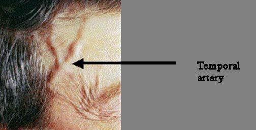
6 Blindness Physical Examination HEENT: Soft right carotid bruit Scalp tenderness to tap temporal fossa; Weak temporal pulses. General: Unremarkable Neurol : positive only for visual findings:
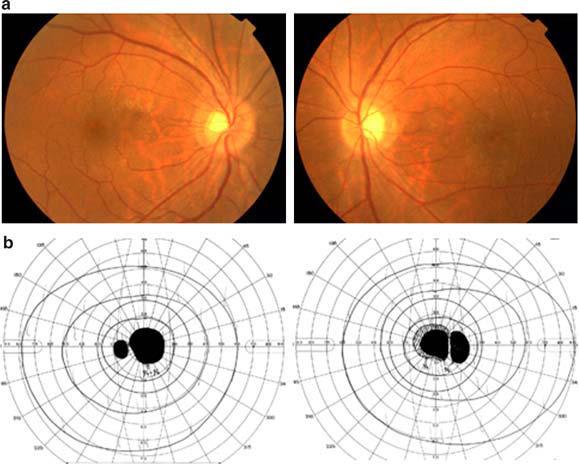
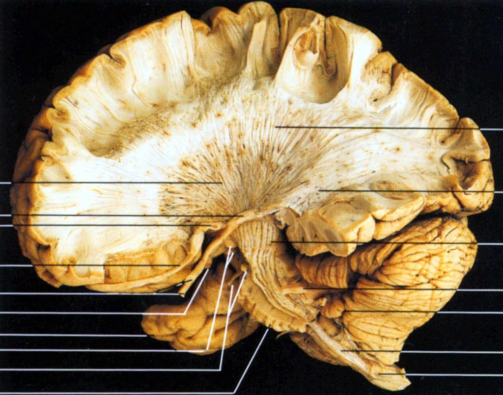
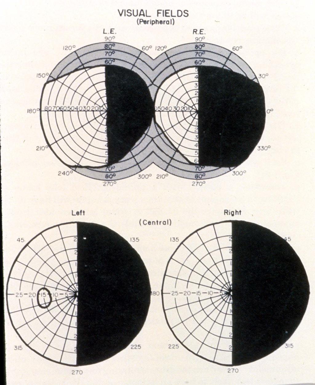
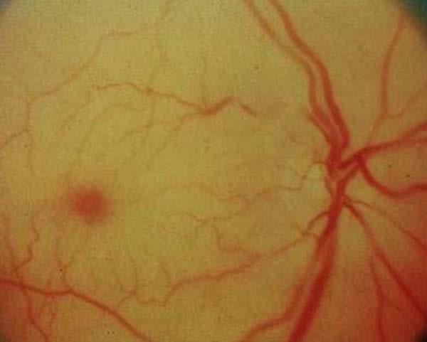
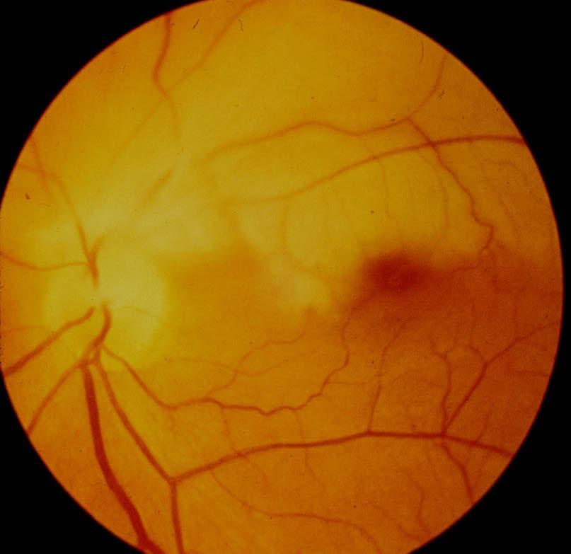
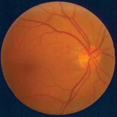
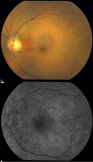
7 Blindness Neuro ophthalmologic examination: Normal appearance to inspection VA: 20/150 OD 20/ 25 OS Color: 3/6 HRR plates OD 6/6 OS VF: Full field OS ; Central scotoma with inferior breakout OD Pupils: OD sluggish with an afferent defect; briskly reactive OS Fundus: Disk head swelling with fine peripapillary hemorrhage OD Normal appearing disc OS.
8
9 Blindness Laboratory Studies CBC: wbc 7200 with nl dif. Hb 11.4 Platelets: 490,000
10 Blindness Laboratory Studies CMP: unremarkable Serum amylase- nl C-reactive protein (CRP) Erythrocyte sedimentation rate (ESR) 77mm/hour-
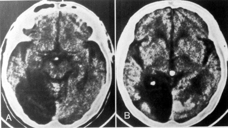
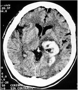
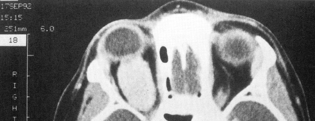
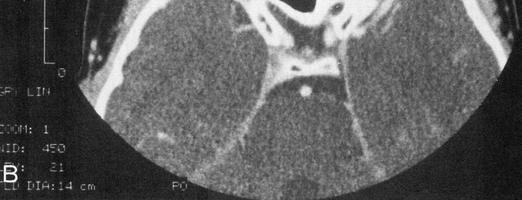
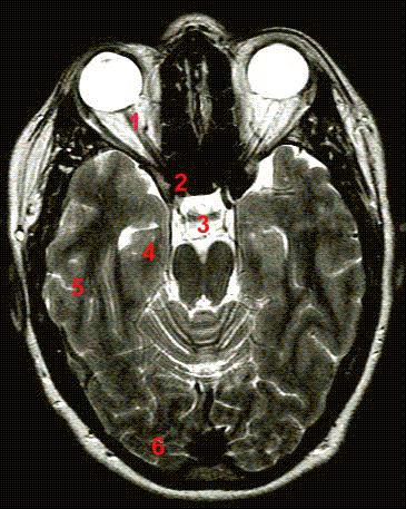
11 Blindness Additional Studies Chest Xray: : normal EKG : nonspecific ST wave change MRI : After evaluation treatment was initiated and a diagnostic procedure was performed
12
13
14
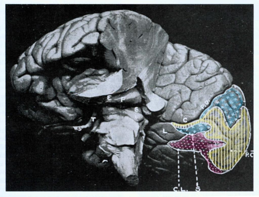
15 Blindness Cortical blindness always produces balanced visual loss
16
17
18
19
20 Grand Rounds Hemispheric infarcts will not produce unilateral visual loss because the preserved hemisphere contains signals generated from half of the macula on each eye. Same applies to optic tracts/ radiations
21 Grand Rounds Chiasmal lesions may at times cause monocular visual loss But there has to be a visual field defect in the other eye
22
23
24
25

26
27
28 Grand Rounds Prechiasmal intracranial monocular visual loss can be due to: Meningioma But tempo is wrong (meningioma( is common but slow growing)
29
30
31
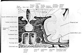
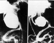
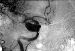
32 Grand Rounds Prechiasmal intracranial monocular visual loss may be due to: Ophthalmic artery aneurysm is a consideration But A lesion in this location will not result in disc head swelling MRI virtually always reveals lesion
33 Grand Rounds Therefore The location of visual failure lies in the anterior visual pathway: retina or optic nerve The absence of subjective complaints of photopsias and floaters plus the fundic findings of disc swelling exclude the retina.
34 Grand Rounds The optic nerve is the site of injury. Dif Dx: Compressive Infiltrative Inflammatory Ischemic
35
36 Could this be optic neuritis?
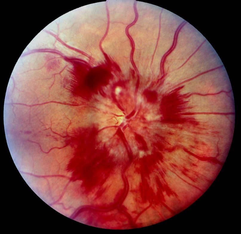
37 Grand Rounds Picture inflamed ON
38
39
40
41 Grand Rounds Is this inflammatory? ie.. optic neuritis? Pros: Sudden visual loss Swollen nerve Suggestive VF loss Cons: Age Absence of pain on eye movement Presence of constitutional sx. Absence of signal change on MRI Presence of hemorrhages Preservation of color vision
42 Grand Rounds Is the lesion ischemic? Ischemia takes two forms Central retinal artery occlusion Posterior ciliary vessel occlusion
43
44
45 Grand Rounds Optic Disc infarction- 2 types Arteritic versus nonarteritic Identical in appearance!
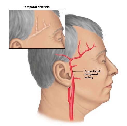
46 Esus
47 Grand Rounds Non arteritic anterior ischemic optic neuropathy ( NAION): Younger population Hypertensive, vasculopathic Opposite optic disc is tight
48

49
50 Grand Rounds Anterior Ischemic Optic Neuropathy (AION) Normal optic cup opposite eye Older age group, > 70y/o Constitutional symptoms, temporal headaches, and particularly jaw claudication!
51
52 Grand Rounds Case summary: Older individual Ischemic optic neuropathy Normal optic cup Constitutional symptoms Jaw claudication Transient diplopia Blood work indicates inflammation
53 Blindness DX: GIANT CELL ARTERITIS aka TEMPORAL ARTERITIS
54 Giant Cell Arteritis A chronic granulomatous vasculitis of large and medium sized vessels, etiology unknown, occurring in the elderly. Most common cause of vasculitis in elderly. Occurrence is a medical emergency with potential systemic and ophthalmic complications Prevention of blindness depends on prompt diagnosis and initiation of steroid therapy
55 Giant Cell Arteritis Pathology: affects the cranial branches of arteries originating from the aortic arch Usually associated with marked elevated acute-phase reactants Closely related to polymyalgia rheumatica 10% - 15% of cases involve extracranial vessels and present as transient ischemic attacks particularly amaurosis fugax With AION risk to other eye is high
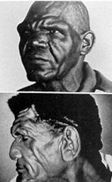
56 Giant Cell Arteritis Typical Features New onset headache Scalp tenderness Jaw Claudication Polymyalgia Rheumatica Fever, anorexia, weight loss PE: Tender, nodular temporal arteries scalp tenderness, temporalis muscle atrophy ophthalmic signs.
57
58 Giant Cell Arteritis Epidemiology 1/5000 people > 50y/o Peak between y/o Women twice as frequently Cyclic pattern of increased incidence every seven years. Frequency increased at higher latitude Black = Hispanic =White
59 Giant Cell Arteritis Laboratory Examination. Erythrocytic sedimentation rate (ESR) = or > 50 mm/hr. NB: 10% are nl Platelets frequently > than 450,000. C Reactive protein ( CRP) most sensitive not affected by erythrocyte number or shape, immunoglobulins renal function or cholesterol.
60 Giant cell arteritis Definitive diagnosis: Temporal artery biopsy
61 Pathology Sebastian R. Alston, M.D. September 19, 2008

62 Giant Cells Elastic lamina Media Temporal Arteritis Fragmented internal elastic lamina Chronic granulomatous inflammation with giant cells No necrosis
63 Temporal Arteritis Giant Cells (arrow) are characteristic but are not always present Intimal Hyperplasia Necrosis not usually present

64 Giant-Cell Arteritis of the Temporal Artery Weyand C and Goronzy J. N Engl J Med 2003;349:
65 Temporal Arteritis Giant cells may be marker of more aggressive course (e.g. blindness) If active inflammation is not present, temporal arteritis is indistinguishable from arteriosclerosis Skip lesions occur Step through vessel Immunohistochemistry Treatment may cause differences in morphology
66 References Armstrong AT, Tyler WB, Wood GC, Harrington TM, Clinical importance of giant cells in temporal arteritis.. J Clin Pathol (2008) 61: Cox M, Gilks B, Healed or quiescent temporal arteritis versus senescent changes in temporal artery biopsy specimens. Pathology (2001) 33: (Abstract) Font RL, Prabhakaran VC, Histological parameters in recognizing steroid-treated treated temporal arteritis: : an analysis of 35 cases. Br J Ophthalmol (2007) 91: Gooi P, Brownstein S, Rawlings N, Temporal arteritis: : a dilemma in clinical and pathological diagnosis. Can J Ophthalmol (2008) 43: Poller DN, Van Wyk Q, Jeffrey MJ, The importance of skip lesions in temporal arteritis.. J Clin Path (2000) 53: Weyand CM and Goronzy JJ, Medium- and large-vessel vasculitis.. N Engl J Med (2003) 349:
67
68 Giant cell arteritis American College of Rheumatology Age onset > 50y/o New headache Temporal artery abnormality ESR > 50 Abnormal artery biopsy
69 Giant Cell Arteritis Pathogenesis Unknown adventitial antigen attracts T cells through vasovasorum. Interferon gamma macrophage differentiation and migration.
70 Giant Cell Arteritis Pathogenesis Adventitia: Cytokines Media: Metalloproteins Intima: : Nitric Oxide Synthase 2 + repair mechanisms = intimal luminal hyperplasia/ degradation of internal elastic lamina.
71 Giant Cell Arteritis Granulomatous infiltrate: 50 % Nonspecific lymph infiltrate: 50% Biopsy > 1.5 cm due to skip lesions Absent giant cells doesn t negate dx Start steroids immediately (2 week window)
Neuro-Ocular Grand Rounds
 Neuro-Ocular Grand Rounds Anthony B. Litwak,OD, FAAO VA Medical Center Baltimore, Maryland Dr. Litwak is on the speaker and advisory boards for Alcon and Zeiss Meditek COMMON OPTIC NEUROPATHIES THAT CAN
9/11/11. Temporal Arteritis. Background. Background. Richard E. Castillo, OD, DO NORTHEASTERN STATE UNIVERSITY Director, Ophthalmic Surgery Service
 Temporal Arteritis Richard E. Castillo, OD, DO NORTHEASTERN STATE UNIVERSITY Director, Ophthalmic Surgery Service 1 Background Giant Cell Arteritis Temporal Arteritis Cranial Arteritis Granulomatous Arteritis
Neuro-Ocular Grand Rounds Anthony B. Litwak,OD, FAAO VA Medical Center Baltimore, Maryland
 Neuro-Ocular Grand Rounds Anthony B. Litwak,OD, FAAO VA Medical Center Baltimore, Maryland Dr. Litwak is on the speaker and advisory boards for Alcon and Zeiss Meditek COMMON OPTIC NEUROPATHIES THAT CAN
Professor Helen Danesh-Meyer. Eye Institute Auckland
 Professor Helen Danesh-Meyer Eye Institute Auckland Bitten by Ophthalmology Emergencies Helen Danesh-Meyer, MBChB, MD, FRANZCO Sir William and Lady Stevenson Professor of Ophthalmology Head of Glaucoma
Non-arteritic anterior ischemic optic neuropathy (NAION) with segmental optic disc edema. Jonathan A. Micieli, MD Valérie Biousse, MD
 with segmental optic disc edema. Jonathan A. Micieli, MD Valérie Biousse, MD") Non-arteritic anterior ischemic optic neuropathy (NAION) with segmental optic disc edema Jonathan A. Micieli, MD Valérie Biousse, MD A 75 year old white woman lost vision in the inferior part of her visual
Non-arteritic anterior ischemic optic neuropathy (NAION) with segmental optic disc edema Jonathan A. Micieli, MD Valérie Biousse, MD A 75 year old white woman lost vision in the inferior part of her visual
I have nothing to disclose but I
 OPTIC NEUROPATHIES Robert L. Tomsak MD PhD Professor of Ophthalmology and Neurology Wayne State t University it Sh School of Mdii Medicine I have nothing to disclose but I wish I did. dd Road map for this
Sequential non-arteritic anterior ischemic optic neuropathy (NAION) Jonathan A. Micieli, MD Valérie Biousse, MD
 Jonathan A. Micieli, MD Valérie Biousse, MD") Sequential non-arteritic anterior ischemic optic neuropathy (NAION) Jonathan A. Micieli, MD Valérie Biousse, MD A 68 year old white woman had a new onset of floaters in her right eye and was found to have
Sequential non-arteritic anterior ischemic optic neuropathy (NAION) Jonathan A. Micieli, MD Valérie Biousse, MD A 68 year old white woman had a new onset of floaters in her right eye and was found to have
Sudden Vision Loss. Brendan Girschek, MD, FRCSC, FACS Vitreoretinal Surgery Cedar Valley Medical Specialists
 Sudden Vision Loss Brendan Girschek, MD, FRCSC, FACS Vitreoretinal Surgery Cedar Valley Medical Specialists My Credentials -Residency in Ophthalmology at the LSU Eye Center in New Orleans, LA -Fellowship
Rafik Girgis. Consultant Ophthalmic Surgeon ( Cataract & Primary Care)
") Rafik Girgis Consultant Ophthalmic Surgeon ( Cataract & Primary Care) Blepharitis Is a very common condition which usually bilateral & symmetrical. The main types are: Anterior, posterior or mixed Complications:
Rafik Girgis Consultant Ophthalmic Surgeon ( Cataract & Primary Care) Blepharitis Is a very common condition which usually bilateral & symmetrical. The main types are: Anterior, posterior or mixed Complications:
Anterior Ischemic Optic Neuropathy (AION)
") Anterior Ischemic Optic Neuropathy (AION) Your doctor thinks you have suffered an episode of anterior ischemic optic neuropathy (AION). This is the most common cause of sudden decreased vision in patients
Anterior Ischemic Optic Neuropathy (AION) Your doctor thinks you have suffered an episode of anterior ischemic optic neuropathy (AION). This is the most common cause of sudden decreased vision in patients
Patient with Daily Headache NTERNATIONAL CLASSIFICATION HEADACHE DISORDERS. R. Allan Purdy, MD, FRCPC,FACP. Professor of Medicine (Neurology)
") Patient with Daily Headache NTERNATIONAL CLASSIFICATION of R. Allan Purdy, MD, FRCPC,FACP HEADACHE DISORDERS Professor of Medicine (Neurology) 2nd edition (ICHD-II) Learning Issues Headaches in the elderly
Patient with Daily Headache NTERNATIONAL CLASSIFICATION of R. Allan Purdy, MD, FRCPC,FACP HEADACHE DISORDERS Professor of Medicine (Neurology) 2nd edition (ICHD-II) Learning Issues Headaches in the elderly
Alan G. Kabat, OD, FAAO (901)
") THE SWOLLEN OPTIC DISC: EMERGENCY OR ANOMALY? Alan G. Kabat, OD, FAAO (901) 252-3691 Memphis, Tennessee alan.kabat@alankabat.com Course description: The swollen disc presents a diagnostic dilemma. While
THE SWOLLEN OPTIC DISC: EMERGENCY OR ANOMALY? Alan G. Kabat, OD, FAAO (901) 252-3691 Memphis, Tennessee alan.kabat@alankabat.com Course description: The swollen disc presents a diagnostic dilemma. While
Preventing blindness: Ultrasound in Giant cell arteritis
 Preventing blindness: Ultrasound in Giant cell arteritis Elizabeth Jernberg, MD Associate Clinical Professor of Medicine Division of Rheumatology University of Washington Virginia Mason Medical Center
Vasculitis. Edward Dwyer, M.D. Division of Rheumatology. Vasculitis
 Edward Dwyer, M.D. Division of Rheumatology VASCULITIS is a primary inflammatory disease process of the vasculature Determinants of the Clinical Manifestations of : Target organ involved Size of vessel
Vasculitis local: systemic
 Vasculitis Inflammation of the vessel wall. Signs and symptoms: 1- local: according to the involved tissue 2- systemic:(fever, myalgia, arthralgias, and malaise) Pathogenesis 1- immune-mediated 2- infectious
Giant Cell Arteritis. Leonid Skorin, Jr., DO, OD, MS, FAAO, FAOCO 1 & Rebecca Lange, OD 2 INTRODUCTION SYMPTOMS & SIGNS EPIDEMIOLOGY REVIEW ARTICLE
 Osteopathic Family Physician (2018) 17-21 17 REVIEW ARTICLE Leonid Skorin, Jr., DO, OD, MS, FAAO, FAOCO 1 & Rebecca Lange, OD 2 1 Mayo Clinic Health System, Albert Lea, MN 2 Whidbey Vision Care, Oak Harbor,
Vasculitis local: systemic
 Vasculitis Inflammation of the vessel wall. Signs and symptoms: 1- local: according to the involved tissue 2- systemic:(fever, myalgia, arthralgias, and malaise) Pathogenesis 1- immune-mediated inflammation
Giant cell arteritis
 Postgraduate Medical Journal (May 1974) 50, 265-269. R. G. TURNER M.B., B.S. J. HENRY M.B., B.S. Giant cell arteritis A. I. FRIEDMANN F.R.C.S. D. GERAINT JAMES M.D., F.R.C.P. The Medical Ophthalmology
Case Presentation VASCULITIS. Case Presentation. Case Presentation. Vasculitis
 Case Presentation VASCULITIS The patient is a 24 year old woman who presented to the emergency room with left-sided weakness. She was confused and complained of a severe headache. She was noted to have
Delayed Choroidal Perfusion in Giant Cell Arteritis
 Tournai of Clinical Neuro-ophthalmology 11(4): 221-227,1991. 1991 Raven Press, Ltd., New York Delayed Choroidal Perfusion in Giant Cell Arteritis H. G. Mack, B. Me SC., M.B.B.S., J. O'Day, F.R.A.C.P.,
VASCULITIS. Case Presentation. Case Presentation
 VASCULITIS Case Presentation The patient is a 24 year old woman who presented to the emergency room with left-sided weakness. She was confused and complained of a severe headache. She was noted to have
Optic Nerve Disorders: Structure and Function and Causes
 Optic Nerve Disorders: Structure and Function and Causes Using Visual Fields, OCT and B-scan Ultrasound to Diagnose and Follow Optic Nerve Visual Losses Ohio Ophthalmological Society and Ophthalmic Tech
Jacqueline Theis, O.D., F.A.A.O.
 Neuro-Ophthalmological Emergencies Presenting in Primary Care Optometry Describes the symptoms, signs, and management of neuro-ophthalmological emergencies. Signs/Symptoms to be Concerned about (especially
OPTIC NEUROPATHIES Optic Neuritis vs AION. Jacqueline M.S. Winterkorn, Ph.D., M.D.
 OPTIC NEUROPATHIES Optic Neuritis vs AION Jacqueline M.S. Winterkorn, Ph.D., M.D. OPTIC NEUROPATHIES Inflammatory Optic Neuritis Ischemic Optic Neuropathy Compressive Optic Neuropathy Traumatic Optic
Objectives. Unexplained Vision Loss: Where Do I Go From Here. History. History. Drug Induced Vision Loss
 Objectives Unexplained Vision Loss: Where Do I Go From Here Denise Goodwin, OD, FAAO Coordinator, Neuro-ophthalmic Disease Clinic Pacific University College of Optometry goodwin@pacificu.edu Know the importance
Evaluation of ONH Pallor in Glaucoma Patients and Suspects. Leticia Rousso, O.D. Joseph Sowka, O.D
 Evaluation of ONH Pallor in Glaucoma Patients and Suspects Leticia Rousso, O.D Joseph Sowka, O.D I. Abstract This case report will evaluate a young glaucoma suspect with unilateral sectoral optic nerve
Neuropathy (NAION) and Avastin. Clinical Assembly of the AOCOO-HNS Foundation May 9, 2013
 and Avastin. Clinical Assembly of the AOCOO-HNS Foundation May 9, 2013") Non Arteritic Ischemic Optic Neuropathy (NAION) and Avastin Shalom Kelman, MD Clinical Assembly of the AOCOO-HNS Foundation May 9, 2013 Anterior Ischemic Optic Neuropathy Acute, painless, visual loss,
Non Arteritic Ischemic Optic Neuropathy (NAION) and Avastin Shalom Kelman, MD Clinical Assembly of the AOCOO-HNS Foundation May 9, 2013 Anterior Ischemic Optic Neuropathy Acute, painless, visual loss,
3/16/2018. Optic Nerve Examination. Hassan Eisa Swify FRCS Ed (Ophthalmology) Air Force Hospital
 Air Force Hospital") Optic Nerve Examination Hassan Eisa Swify FRCS Ed (Ophthalmology) Air Force Hospital 1 Examination Structure ( optic disc) Function Examination of the optic disc The only cranial nerve (brain tract) which
Optic Nerve Examination Hassan Eisa Swify FRCS Ed (Ophthalmology) Air Force Hospital 1 Examination Structure ( optic disc) Function Examination of the optic disc The only cranial nerve (brain tract) which
Neuro-ophthalmologyophthalmology. Marek Michalec, MD.
 Neuro-ophthalmologyophthalmology Marek Michalec, MD. Neuro-ophthalmology Study integrating ophthalmology and neurology Disorders affecting parts of CNS devoted to vision or eye: Afferent system (visual
Overview INTRODUCTION 3/15/2018. Headache Emergencies. Other way to differentiate between them? Is there an easy way to differentiate between them?
 Overview Headache Emergencies Primary versus Secondary headache disorder Red flags 4 cases of unusual headache emergencies Disclaimer: we will not talk about brain bleed as patients usually go the ED.
Grand Rounds. Eddie Apenbrinck M.D. University of Louisville School of Medicine Department of Ophthalmology & Visual Sciences 6/20/2014
 Grand Rounds Eddie Apenbrinck M.D. University of Louisville School of Medicine Department of Ophthalmology & Visual Sciences 6/20/2014 Subjective CC: sudden painless loss of vision OD HPI: 75 year old
Case Follow Up. Sepi Jooniani PGY-1
 Case Follow Up Sepi Jooniani PGY-1 Triage 54 year old M Pt presents to prelim states noticed today he had reddness to eyes, states worse in R eye. Pt denies any pain or itching. No further complaints.
Anterior Ischemic Optic Neuropathy
 UNIVERSITY OF ZAGREB SCHOOL OF MEDICINE Laurent Martini Anterior Ischemic Optic Neuropathy GRADUATE THESIS Zagreb, 2016. This graduate thesis was made at the department of Neuro-ophthalmology of KBC Retro,
12/2/16. Ways to differentiate:
 Nate Lighthizer, O.D., F.A.A.O. Assistant Dean for Clinical Care Services Director of CE Chief of Specialty Care Clinics Chief of Electrodiagnostics Clinic Oklahoma College of Optometry lighthiz@nsuok.edu
OCCLUSIVE VASCULAR DISORDERS OF THE RETINA
 OCCLUSIVE VASCULAR DISORDERS OF THE RETINA Learning outcomes By the end of this lecture the students would be able to Classify occlusive vascular disorders (OVD) of the retina. Correlate the clinical features
OCT : retinal layers. Extraocular muscles. History. Central vs Peripheral vision. History: Temporal course. Optical Coherence Tomography (OCT)
") Optical Coherence Tomography (OCT) OCT : retinal layers 7 Central vs Peripheral vision Extraocular muscles RPE E Peripheral Vision: Rods (95 million) 30% Ganglion cells Central Vision: Cones (5 million)
Optical Coherence Tomography (OCT) OCT : retinal layers 7 Central vs Peripheral vision Extraocular muscles RPE E Peripheral Vision: Rods (95 million) 30% Ganglion cells Central Vision: Cones (5 million)
Neuro Ocular Grand Rounds Anthony B. Litwak, OD, FAAO VA Medical Center Baltimore, MD
 Neuro Ocular Grand Rounds Anthony B. Litwak, OD, FAAO VA Medical Center Baltimore, MD 58 YOWM! C/O I think there is something wrong with my vision, but I m not sure what it is.! +PMH for HTN, atrial fibrillation,
Speaker Disclosure Statement. " Dr. Tim Maillet and Dr. Vladimir Kozousek have no conflicts of interest to disclose.
 Speaker Disclosure Statement Dr. Tim Maillet and Dr. Vladimir Kozousek have no conflicts of interest to disclose. Diabetes Morbidity Diabetes doubles the risk of stroke. Diabetes quadruples the risk of
CMS Limitations Guide - Radiology Services
 CMS Limitations Guide - Radiology Services Starting October 1, 2015, CMS will update their existing medical necessity limitations on tests and procedures to correspond to ICD-10 codes. This limitations
Learn Connect Succeed. JCAHPO Regional Meetings 2015
 Learn Connect Succeed JCAHPO Regional Meetings 2015 OPTIC NEUROPATHY AS EASY AS 1,2,3,4 OPTIC NERVE ANATOMY M. Tariq Bhatti, MD Departments of Ophthalmology and Neurology Duke Eye Center and Duke University
Concise guidance: diagnosis and management of giant cell arteritis
 ONISE GUIDANE linical Medicine 2010, Vol 10, No 4: 381 6 oncise guidance: diagnosis and management of giant cell arteritis Bhaskar Dasgupta on behalf of the Giant ell Arteritis Guideline Development Group*
Sahand Ensafi PA, CCPA, B.H.Sc.,Department of Emergency Medicine, University Health Network
 Sahand Ensafi PA, CCPA, B.H.Sc.,Department of Emergency Medicine, University Health Network No Disclosures Definitions Ophthalmologic Blindness Practical definition? WHO V/A less than 3/60 (snellen)
NANOS Patient Brochure
 NANOS Patient Brochure Transient Visual Loss Copyright 2016. North American Neuro-Ophthalmology Society. All rights reserved. These brochures are produced and made available as is without warranty and
Temporal arteritis. Occurrence of ocular complications 7 years after diagnosis. University of Edinburgh, and Royal Infirmary of Edinburgh
 Brit. J. Ophthal. (I 972) 56, 584 Temporal arteritis Occurrence of ocular complications 7 years after diagnosis JAMES F. CULLEN Department of Ophthalmology, University of Edinburgh, and Royal Infirmary
Neurological Dilemmas in Primary Care
 Neurological Dilemmas in Primary Care David Clark, DO dclark@oregonneurology.com When to test? How to test? Pitfalls in testing? When to treat? How to treat? How long to treat? Neurological Dilemmas Seizure
Treatment of central retinal artery occlusions
 Treatment of central retinal artery occlusions Jeffrey N. Weiss Retina Associates of South Florida, Margate, Florida, USA To the Editor: As a practicing retinal specialist for the last 28 years, I read
Pearls, Pitfalls and Advances in Neuro-Ophthalmology
 Pearls, Pitfalls and Advances in Neuro-Ophthalmology Nancy J. Newman, MD Emory University Atlanta, GA Consultant for Gensight Biologics, Santhera Data Safety Monitoring Board for Quark AION Study Medical-legal
ISCHEMIC OPTIC neuropathy (ION)
") OBSERVATION Ischemic Optic Neuropathy Associated With Internal Carotid Artery Dissection Valérie Biousse, MD; Monique Schaison, MD; Pierre-Jean Touboul, MD; Jacques D Anglejan-Chatillon, MD; Marie-Germaine
OBSERVATION Ischemic Optic Neuropathy Associated With Internal Carotid Artery Dissection Valérie Biousse, MD; Monique Schaison, MD; Pierre-Jean Touboul, MD; Jacques D Anglejan-Chatillon, MD; Marie-Germaine
EYE TRAUMA: INCIDENCE
 Introduction EYE TRAUMA: INCIDENCE 2.5 million eye injuries per year in U.S. 40,000 60,000 of eye injuries lead to visual loss Introduction Final visual outcome of many ocular emergencies depends on prompt,
Ischaemic optic neuropathy: the Singapore scene
 O r i g i n a l A r t i c l e Singapore Med J 2007; 48 (4) : 281 Ischaemic optic neuropathy: the Singapore scene Cullen J F, Por Y M Abstract The commonest cause of an optic neuropathy in Singapore is
Dr/ Marwa Abdellah EOS /16/2018. Dr/ Marwa Abdellah EOS When do you ask Fluorescein angiography for optic disc diseases???
 When do you ask Fluorescein angiography for optic disc diseases??? 1 NORMAL OPTIC DISC The normal optic disc on fluorescein angiography is fluorescent due to filling of vessels arising from the posterior
GIANT CELL ARTERITIS. Page 1 of 6 Reproduction of this material requires written permission of the Vasculitis Foundation. Copyright 2018.
 What is giant cell arteritis (GCA)? Giant cell arteritis (GCA) is a form of vasculitis a family of rare disorders characterized by inflammation of the blood vessels, which can restrict blood flow and damage
CHAPTER 13 CLINICAL CASES INTRODUCTION
 2 CHAPTER 3 CLINICAL CASES INTRODUCTION The previous chapters of this book have systematically presented various aspects of visual field testing and is now put into a clinical context. In this chapter,
Learn Connect Succeed. JCAHPO Regional Meetings 2017
 Learn Connect Succeed JCAHPO Regional Meetings 2017 NO FINANCIAL DISCLOSURES Technician s Role in Neuro-Ophthalmology Workup Beth Koch COT, ROUB Cleveland 9/16/2017 What Tests Are You Expected To Perform?
Identify the choice that best completes the statement or answers the question.
 Chapter 5. The Eye Multiple Choice Identify the choice that best completes the statement or answers the question. 1. The most common type of eye disorder is: A. Refractive errors B. Macular conditions
10 EYE EMERGENCIES. Who goes, who you better not send! Brant Slomovic, MD, FRCPC University Health Network
 10 EYE EMERGENCIES Who goes, who you better not send! Brant Slomovic, MD, FRCPC University Health Network DISCLOSURES I have none PVD CASE 1 WHAT IS A PVD? a process of aging (45-55) liquefaction of vitreous
Introduction. Overview
 Temporal arteritis James Goodwin MD ( Dr. Goodwin of the University of Illinois at Chicago has no relevant financial relationships to disclose. ) Originally released May 14, 1996; last updated September
Five steps: Overview
 Optic atrophy is not a diagnosis Andrew G. Lee, MD Professor of Ophthalmology, Neurology and Neurosurgery, Weill Cornell Medical College Chair, Department of Ophthalmology, Houston Methodist Hospital,
Vasculitides in Surgical Neuropathology Practice
 Vasculitides in Surgical Neuropathology Practice USCAP requires that all faculty in a position to influence or control the content of CME disclose any relevant financial relationship WITH COMMERCIAL INTERESTS
Hyperbaric Oxygen Therapy in two patients with Non-arteritic Anterior Optic Neuropathy who did not respond to Prednisone
 Case Report Hyperbaric Oxygen Therapy in two patients with Non-arteritic Anterior Optic Neuropathy who did not respond to Prednisone L BOJIĆ, M. IVANIŠEVIĆ, G.GOŠOVIĆ Eye Clinic, Clinical Hospital Split
Faculty Financial Disclosure. Learning Objectives: Office Ophthalmology. Basic Eye Exam: What s in your pocket/office? Office Ophthalmology
 Faculty Financial Disclosure Office Ophthalmology Lynn K. Gordon, MD, PhD, has no financial relationships to disclose. Lynn K. Gordon, MD, PhD Professor and Vernon O Underwood Family Chair Department of
Papilledema. Golnaz Javey, M.D. and Jeffrey J. Zuravleff, M.D.
 Papilledema Golnaz Javey, M.D. and Jeffrey J. Zuravleff, M.D. Papilledema specifically refers to optic nerve head swelling secondary to increased intracranial pressure (IICP). Optic nerve swelling from
Sudden loss of vision
 Sudden loss of vision Abstract Du Toit N, MBChB, DipOphth(SA), FRCS(Ed), FCOphth(SA), MMed, Senior Lecturer University of Cape Town; Groote Schuur Hospital, Cape Town Correspondence to: Nagib du Toit,
APPROACH TO PATIENTS WITH POLYARTHRALGIA
 APPROACH TO PATIENTS WITH POLYARTHRALGIA Scott Vogelgesang, MD Division of Immunology University of Iowa No conflicts of interest DEFINITIONS Arthralgia joint pain with no evidence of inflammation Arthritis
Learn Connect Succeed. JCAHPO Regional Meetings 2017
 Learn Connect Succeed JCAHPO Regional Meetings 2017 You have some Nerve Asking Me to Work Up that Patient! What I Need to know about the Neuro- Ophthalmology Patient Financial Disclosures No relevant financial
CAN WE REPLACE TEMPORAL ARTERY BIOPSY WITH CRANIAL ULTRASOUND FOR THE DIAGNOSIS OF GIANT CELL ARTERITIS?
 CAN WE REPLACE TEMPORAL ARTERY BIOPSY WITH CRANIAL ULTRASOUND FOR THE DIAGNOSIS OF GIANT CELL ARTERITIS? Adam P. Croft (ST3 Rheumatology) Susan Mollan, Paresh Jobunputra Speaker has no disclosures TAB
OCULAR MANIFESTATIONS OF SYSTEMIC DISEASES THUCANH MULTERER, MD
 OCULAR MANIFESTATIONS OF SYSTEMIC DISEASES THUCANH MULTERER, MD UNDERGRADUATE: Philadelphia College of Pharmacy and Science 1996 MEDICAL SCHOOL: MCP Hahnemann School of Medicine, Philadelphia PA 2000 RESIDENCY:
Questions to ponder today
 Questions to ponder today What clinical signs/symptoms help me identify a retinal artery occlusion (RAO)? Past RAO? What is my responsibility to patient when I diagnose a RAO? Lab test? Imaging? If my
Carotid Cavernous Fistula
 Chief Complaint: Double vision. Carotid Cavernous Fistula Alex W. Cohen, MD, PhD; Richard Allen, MD, PhD May 14, 2010 History of Present Illness: A 46 year old female patient presented to the Oculoplastics
COMMUNICATIONS. ARTERITIS*t CILIARY ARTERY INVOLVEMENT IN GIANT CELL. generalized vascular disease was emphasized by Cooke, Cloake, Govan, and
 Brit. J. Ophthal. (1967) 51, 505 COMMUNICATIONS CILIARY ARTERY INVOLVEMENT IN GIANT CELL ARTERITIS*t BY P. A. MAcFAUL Department of Pathology, Institute of Ophthalmology, University oflondon THIS condition
Analysis of Fundus Photography and Fluorescein Angiography in Nonarteritic Anterior Ischemic Optic Neuropathy and Optic Neuritis
 pissn: 1011-8942 eissn: 2092-9382 Korean J Ophthalmol 2016;30(4):289-294 http://dx.doi.org/10.3341/kjo.2016.30.4.289 Original Article Analysis of Fundus Photography and Fluorescein Angiography in Nonarteritic
5/2/2016 EYE EMERGENCIES. Nathaniel Pelsor, O.D., FAAO Talley Medical-Surgical Eye Care Associates. Anatomy. Tools
 EYE EMERGENCIES Nathaniel Pelsor, O.D., FAAO Talley Medical-Surgical Eye Care Associates Anatomy Tools 1 Contact dermatitis Blepharitis HSV Preseptal Cellulitis Anterior Chamber Subconjunctival hemorrhage
Vasculitis Prof. Dr. med. Katharina Glatz Pathologie
 Vasculitis 08-21-2018 Prof. Dr. med. Katharina Glatz Pathologie Agenda Anatomy and histology Vasculitis: Chapel Hill Classification Examples Giant cell arteritis Single organ vasculitis Artery or Vein?
Ischemic optic neuropathies are the most common acute. Ischemic Optic Neuropathies REVIEW ARTICLE
 REVIEW ARTICLE Ischemic Optic Neuropathies Katie Luneau, MD,* Nancy J. Newman, MD,* and Valérie Biousse, MD* Abstract: Anterior ischemic optic neuropathy (AION) is the most common cause of acute optic
NON-ATHEROSCLEROTIC PATHOLOGY OF THE CAROTID ARTERIES
 NON-ATHEROSCLEROTIC PATHOLOGY OF THE CAROTID ARTERIES Leslie M. Scoutt, MD, FACR Professor of Diagnostic Radiology & Surgery Vice Chair, Dept of Radiology & Biomedical Imaging Chief, Ultrasound Section
The Joints are Painful & Swollen: Do I give Steroids? Dr Tom Kennedy
 The Joints are Painful & Swollen: Do I give Steroids? Dr Tom Kennedy Learning Objectives When to use an acute rheumatology service Appropriate use of steroids by condition Injection or Oral or Intramuscular
Headache Assessment In Primary Eye Care
 Headache Assessment In Primary Eye Care Spencer Johnson, O.D., F.A.A.O. Northeastern State University Oklahoma College of Optometry johns137@nsuok.edu Course Objectives Review headache classification Understand
Fundus Autofluorescence. Jonathan A. Micieli, MD Valérie Biousse, MD
 Fundus Autofluorescence Jonathan A. Micieli, MD Valérie Biousse, MD The retinal pigment epithelium (RPE) has many important functions including phagocytosis of the photoreceptor outer segments Cone Rod
Takayasu s Arteritis: A Case Report With Global Arterial Involvement
 1 Case Report Takayasu s Arteritis: A Case Report With Global Arterial Involvement Waqas Ahmed, Zeeshan Ahmad* From Shifa International Hospital H-8/4, Islamabad, Pakistan Correspondence: Dr Waqas Ahmed,
Dr Jo-Anne Pon. Consultant Ophthalmologist and Oculoplastic Surgeon Southern Eye Specialists Christchurch
 Dr Jo-Anne Pon Consultant Ophthalmologist and Oculoplastic Surgeon Southern Eye Specialists Christchurch 12:15-12:30 Visual Migraines to be Worried About Visual Migraines To Be Worried About Jo-Anne Pon
The focus of this week s lab will be pathology of the cardiovascular system.
 LAB 3: THE MUSCLE AND CARDIOVASCULAR SYSTEM The focus of this week s lab will be pathology of the cardiovascular system. The cases we will cover are: A. Atherosclerosis Refer to virtual slide p_8, should
Dr Kusala S. Gunasekara MBBS(Col),MD(Med),MRCP(UK) Acting Consultant Rheumatologist DGH-Matale
,MD(Med),MRCP(UK) Acting Consultant Rheumatologist DGH-Matale") Dr Kusala S. Gunasekara MBBS(Col),MD(Med),MRCP(UK) Acting Consultant Rheumatologist DGH-Matale Patient 67 yr old female First presentation to the rheumatology unit in May 2013 Referred by GP as the patient
Dr Kusala S. Gunasekara MBBS(Col),MD(Med),MRCP(UK) Acting Consultant Rheumatologist DGH-Matale Patient 67 yr old female First presentation to the rheumatology unit in May 2013 Referred by GP as the patient
10/27/2013. Optic Red Herrings
 Optic Red Herrings 1 Optic neuropathy Compressive Inflammatory Toxic Glaucomatous Ischemic Post traumatic GLAUCOMATOUS OPTIC NEUROPATHY Glaucoma: Traditionally defined as a progressive optic neuropathy
Unexplained visual loss in seven easy steps
 Unexplained visual loss in seven easy steps Andrew G. Lee, MD Chair Ophthalmology, Houston Methodist Hospital, Professor, Weill Cornell MC; Adjunct Professor, Baylor COM, U Iowa, UTMB Galveston, UT MD
Evaluation of optic disc blood flow of intraconal orbital tumors using laser speckle flowgraphy.
 Research Article http://www.alliedacademies.org/ophthalmic-and-eye-research/ Evaluation of optic disc blood flow of intraconal orbital tumors using laser speckle flowgraphy. Hideki Chuman*, Takako Hidaka,
Neuro-Ophthalmic Masqueraders
 Neuro-Ophthalmic Masqueraders Leonid Skorin, Jr., OD, DO, MS, FAAO, FAOCO Mayo Clinic Health System in Albert Lea Denise Goodwin, OD, FAAO Pacific University College of Optometry Please silence all mobile
Bilateral Posterior Ischemic Optic Neuropathy in a Patient with Severe Diabetic Ketoacidosis
 Bilateral Posterior Ischemic Optic Neuropathy in a Patient with Severe Diabetic Ketoacidosis The Harvard community has made this article openly available. Please share how this access benefits you. Your
Case Report A Case of Recurrent Transient Monocular Visual Loss after Receiving Sildenafil
 Case Reports in Ophthalmological Medicine Volume 2011, Article ID 645089, 4 pages doi:10.1155/2011/645089 Case Report A Case of Recurrent Transient Monocular Visual Loss after Receiving Sildenafil Asaad
NIH Public Access Author Manuscript Arch Neurol. Author manuscript; available in PMC 2011 December 15.
 NIH Public Access Author Manuscript Published in final edited form as: Arch Neurol. 2011 April ; 68(4): 517 520. doi:10.1001/archneurol.2011.64. Varicella Zoster Virus Ischemic Optic Neuropathy and Subclinical
Polymyalgia, Temporal Arteritis and pineapples
 Polymyalgia, Temporal Arteritis and pineapples Rod Hughes Consultant Rheumatologist Ashford St Peter s Hospital Trust Chertsey Wed 11 th May 2011 Meeting aims Pineapples their significance in disease Defining
CME for Family Medicine Specialists. Evelyn Sutton, MD, FRCPC, FACP November 17, 2018
 CME for Family Medicine Specialists Evelyn Sutton, MD, FRCPC, FACP November 17, 2018 Disclosures Received $ from Advisory Board Consultant: Amgen, Abvie, Pfizer, Actelion, Lilly, Grants: Arthritis Society,
Differences between Non-arteritic Anterior Ischemic Optic Neuropathy and Open Angle Glaucoma with Altitudinal Visual Field Defect
 pissn: 1011-8942 eissn: 2092-9382 Korean J Ophthalmol 2015;29(6):418-423 http://dx.doi.org/10.3341/kjo.2015.29.6.418 Original Article Differences between Non-arteritic Anterior Ischemic Optic Neuropathy
Role Of Various Factors In The Treatment Of Optic Neuritis----A Study Abstract Aim: Materials & Methods Discussion: Conclusion: Key words
 IOSR Journal of Dental and Medical Sciences (IOSR-JDMS) e-issn: 2279-0853, p-issn: 2279-0861.Volume 15, Issue 9 Ver. X (September). 2016), PP 51-57 www.iosrjournals.org Role Of Various Factors In The Treatment
Grand Rounds Clinical Cases from Alex D. Gibberman, O.D. Harpers Point Eye Associates
 Grand Rounds Clinical Cases from 2016 Alex D. Gibberman, O.D. Harpers Point Eye Associates Relevant Financial Interests -none Case 1: 54 year old African American Female CC: Noticed a green line in
Shared embryology Eye and brain develop from neuro-ectoderm
 The Patient with Visual Loss: Localization of Neuropathologic Disease and Select Diseases of Neuropathologic Interest Steven A. Kane, M.D., Ph.D. The Edward S. Harkness Eye Institute Shared embryology
LAB 4: THE MUSCLE AND CARDIOVASCULAR SYSTEM THE MUSCLE AND CARDIOVASCULAR SYSTEM
 LAB 4: THE MUSCLE AND CARDIOVASCULAR SYSTEM THE MUSCLE AND CARDIOVASCULAR SYSTEM The focus of this week s lab will be pathology of the cardiovascular system. The cardiovascular system is composed of the
Recurrent transient visual loss in a middle aged woman
 Recurrent transient visual loss in a middle aged woman Chow SY, Draman N, Teh WM, Azhany Y Chow SY, Draman N, Teh WM, et al. Recurrent transient visual loss in a middle aged woman. Malays Fam Physician.
Slide 4. Slide 5. Slide 6
 Slide 1 Slide 4 Demographics El Paso Eye Care Border Healthcare-Based Grand Rounds Derek N. Cunningham, O.D. 80-90% Mexican-Americans Diabetes Hypertension Hyperlipidemia Obesity 70% uninsured High poverty
Sudden loss of vision History and examination
 THEME vision at risk Lucy Goold MBBS, MMed(OphthSc), is ophthalmology resident and Associate Clinical Lecturer, South Australian Institute of Ophthalmology, Royal Adelaide Hospital, South Australia. lgoold@med.usyd.edu.au
Polymyalgia rheumatica and giant cell arteritis
 Polymyalgia rheumatica and giant cell arteritis What is polymyalgia rheumatica? Polymyalgia rheumatica is a rheumatic disorder associated with moderate-to-severe musculoskeletal pain and stiffness in the