Sahand Ensafi PA, CCPA, B.H.Sc.,Department of Emergency Medicine, University Health Network
|
|
|
- Emily Wilkinson
- 5 years ago
- Views:
Transcription

1 Sahand Ensafi PA, CCPA, B.H.Sc.,Department of Emergency Medicine, University Health Network

2

3
4 No Disclosures
5 Definitions Ophthalmologic Blindness Practical definition? WHO V/A less than 3/60 (snellen) or equivalent No chart? Our definition? Systematic Approach Rapid Fire
6 General Approach Vision Loss Painless vs. Painful Media vs Retina vs Neural Pathway History (Painless) HPI Truly acute? Prodrome? Trauma? Visual field/eye affected PMHx: Vascular Risk Fx, RE?, Surgery? Meds: Anticholinergics, sildenafil (AION), OCP (ischemia), Tox
7 General Approach Physical Examination General Inspection V/A Corrected/PH, CF to 1-2ft -> HM -> LP Pupils Symmetry, reactivity/reflex, RAPD Pressure (+/-) EOM (3, 4, 6) VF (confrontation) SLE +/- flourescein Ocular U/S

8 Pupillary Light Reflex/RAPD

9 Case 1 36 yo male, hit in the eye with a baseball ball 2 hours ago. Now V/A left eye - CF at 4ft. No c/o of pain.

10 Hyphema Overview Blood in AC Traumatic or spontaneous?open globe/associated injuries Treatment Raise head of bed Dilate Manage Pressure Timolol/apraclonidine, IV Mannitol, Diamox Follow-up? If > 1/3 of AC may require admission for monitoring Complications Rebleed 3-5d in 30% Glaucoma Corneal blood staining


11 Lens Dislocation Overview Partial vs complete disruption of zonules Traumatic or spontaneous Blurred vision due to RE Monocular diplopia with subluxation Treatment Nil if v/a mantained OR for lens replacement if opacity/complications Complications Glaucoma


12 Lens Dislocation

13
14 Case 2 65 yo female, loss of vision in right eye while on her phone 1 hour ago, v/a only 3ft on presentation to ED.
 Thrombotic vs embolic 90%>50, Vascular")
 Embolic/Thrombotic 50-70, A.")
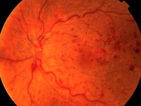
15 Sudden Severe Vision Loss Vitreous Hemorrhage Blood in vitreous cavity RD, PVD, Retinopathy, CRVO, Trauma?anticoags CRVO Obstruction of retinal venous outflow (At or prox to lamina cribrosa) Thrombotic vs embolic 90%>50, Vascular risk fx Young: OCP, Collagen vasc disease, coagulopathy Ischemic (33%) vs Non-ischemic (67%) CRAO Obstruction of arterial circulation of retina (At or prox to lamina cribrosa) Embolic/Thrombotic 50-70, A. Fib, GCA, Vascular risk fx Anoxia = irreversible damage within ~90min
16 Presentation Vitreous Hemorrhage CRVO CRAO Vision Floaters -> LP (min to hours) Sudden (sec to min), <20/200 (scotoma to CF/LP) *Non-Ischemic typically not as severe Sudden (sec), CF -> LP/No LP?Amaurosis prior Pupils Reactive, (-) RAPD Reactive, (+) RAPD Dilated, not/minimally reactive, (+RAPD)

17 Presentation

18 Presentation

19 Presentation

20 Presentation


21 Presentation

22 Presentation
23 Summary of Fundi Vitreous Hemorrhage CRVO Fundi Normal/Hazy -> LP Dilated, Tortous veins, Hemorrhages, Cotton Wool spots Blood and thunder CRAO Pale/milky white retina, minimal/absent arteries, cherry red spot

24 Work- Up and Management Vitreous Hemorrhage?Anti-coags Elevation of head Call ophtha CRVO Thrombosis w/u,?underlying cause Decrease IOP Call Ophtha CRAO CALL OPHTHA Stroke W/U Relieve spasm/dislodge embolus Digital Massage/Acetazolamide Breath into paper bag


25 Case 3 70 yo male complaining of dark shadow covering the inferior half of left visual field for 1 day.


26 Sudden Painless Field Loss BRVO/BRAO Superior Temporal Retinal Detachment Rhegmatogenous, exudative, tractional Myopia, Age > 45, Previous hx AION Arteritic (5-10%) Infarction of retrolaminar optic nerve from occlusion/decreased perfusion of posterior ciliary arteries Non-arteritic (90-95%) Disc at risk /Poor flow
27 Presentation Retinal Detachment Non-arteritic AION BRVO BRAO Vision Flashes/Floa ters/curtain Decreased V/A (40% < 20/200), Chromotopsia, altitudinal VF loss Asymptomatic -> Scotoma/blurri ng of VF affected Decreased VA -> blurring/vf loss Pupils Reactive, +/- RAPD Reactive, +RAPD Reactive, +/- RAPD Reactive, +/- RAPD Fundi grayishwhite, raised retina Pallid disc swelling, venous dilation, Same as CRVO limited to branch Same as CRAO limited to branch


28 Presentation

29 Presentation


30 Presentation
31 Work Up and Management Unilateral VF Defect Call Ophtha - follow-up within 12-24h depending BRAO/BRVO Similar to Central Retinal Detachment Follow up? 1. Hollands H, et al. Acute-Onset Floaters and Flashes: Is this Patient at Risk for Retinal Detachment? JAMA 2009;302(20): Laser/OR AION Rule out GCA NAION Not enough evidence for steroids, Anecdotal evidence for ASA
32 Summary Detailed history Vital signs of the eye Generalized vs VF Curtain defect does not = RD RAPD = BAD = Retina/Neural Pathway Acute VF loss = Ophtha Majority with acute severe visual loss will require urgent if not emergent followup

33
34 References Friedman, Neil J., Peter K. Kaiser, and Roberto Pineda. The Massachusetts Eye and Ear Infirmary Illustrated Manual of Ophthalmology. Philadelphia, PA: Saunders/Elsevier, PDF. Hockberger, Robert S., and Ron M. Walls. "Acute Visual Loss." Rosen's Emergency Medicine - Concepts and Clinical Practice 8th Ed. Ed. John A. Marx. 8th ed. Vol. 1. N.p.: Elsevier, n.d Print. Hollands H, et al. Acute-Onset Floaters and Flashes: Is this Patient at Risk for Retinal Detachment? JAMA 2009;302(20): Jogi, Renu. Basic Ophthalmology. New Delhi: Jaypee Brothers Medical, n.d. PDF. Long, Brit. "Acute Visual Loss in the Emergency Department: Pearls and Pitfalls - Emdocs." Emdocs. Emdocs, 26 Apr Web. 10 Oct Tintinalli, Judith E. "Eye Emergencies." Tintinallis Emergency Medicine A Comprehensive Study Guide. 7th ed. N.p.: McGraw Hill, Print. Tsai, James C. Oxford American Handbook of Ophthalmology. Oxford: Oxford UP, Print.
Case Follow Up. Sepi Jooniani PGY-1
 Case Follow Up Sepi Jooniani PGY-1 Triage 54 year old M Pt presents to prelim states noticed today he had reddness to eyes, states worse in R eye. Pt denies any pain or itching. No further complaints.
LECTURE # 7 EYECARE REVIEW: PART III
 LECTURE # 7 EYECARE REVIEW: PART III HOW TO TRIAGE EYE EMERGENCIES STEVE BUTZON, O.D. EYECARE REVIEW: HOW TO TRIAGE EYE EMERGENCIES FOR PRIMARY CARE PHYSICIANS Steve Butzon, O.D. Member Director IDOC President
Rapid Visual Loss. Dr Michael Johnson PhD FCOptom DipOrth DipGlauc DipTp(IP) Independent Prescribing Optometrist
 Independent Prescribing Optometrist") Rapid Visual Loss Dr Michael Johnson PhD FCOptom DipOrth DipGlauc DipTp(IP) Independent Prescribing Optometrist Outline Pathophysiology Differential diagnosis. Patient scenarios in community practice:
Rapid Visual Loss Dr Michael Johnson PhD FCOptom DipOrth DipGlauc DipTp(IP) Independent Prescribing Optometrist Outline Pathophysiology Differential diagnosis. Patient scenarios in community practice:
OCCLUSIVE VASCULAR DISORDERS OF THE RETINA
 OCCLUSIVE VASCULAR DISORDERS OF THE RETINA Learning outcomes By the end of this lecture the students would be able to Classify occlusive vascular disorders (OVD) of the retina. Correlate the clinical features
For details on measurement and recording of visual acuity, refer to Annex 1. VISION INTERPRETING RESULTS ABSTRACT
 management update on functional decline in older adults 2012 Unit No. 5 VISION Dr Au Eong Kah Guan, Ms Yulianti, Ms Fifiana ABSTRACT Among Singaporean adults of Chinese origin aged 40 to 79 years old,
Course # Flashes and Floaters and Curtains, Oh My!
 Course # 132 Flashes and Floaters and Curtains, Oh My! FLASHES and FLOATERS and CURTAINS, OH MY!!! FLASHES OF LIGHT Vitreous is the villain Retinal traction Retinal hole Retinal tear Migraine Classic migraine
Course # Flashes and Floaters and Curtains, Oh My!
 Course # 132 Flashes and Floaters and Curtains, Oh My! FLASHES and FLOATERS and CURTAINS, OH MY!!! FLASHES OF LIGHT Vitreous is the villain Retinal traction Retinal hole Retinal tear Migraine Classic migraine
Sudden Vision Loss. Brendan Girschek, MD, FRCSC, FACS Vitreoretinal Surgery Cedar Valley Medical Specialists
 Sudden Vision Loss Brendan Girschek, MD, FRCSC, FACS Vitreoretinal Surgery Cedar Valley Medical Specialists My Credentials -Residency in Ophthalmology at the LSU Eye Center in New Orleans, LA -Fellowship
Mild NPDR. Moderate NPDR. Severe NPDR
 Diabetic retinopathy Diabetic retinopathy is the most common cause of blindness in adults aged 35-65 years-old. Hyperglycaemia is thought to cause increased retinal blood flow and abnormal metabolism in
Grand Rounds. Eddie Apenbrinck M.D. University of Louisville School of Medicine Department of Ophthalmology & Visual Sciences 6/20/2014
 Grand Rounds Eddie Apenbrinck M.D. University of Louisville School of Medicine Department of Ophthalmology & Visual Sciences 6/20/2014 Subjective CC: sudden painless loss of vision OD HPI: 75 year old
Chris Brown, M.D. Eye Specialty Group, PLC Continuing Education Series
 Chris Brown, M.D. Eye Specialty Group, PLC 2018 Continuing Education Series Disclaimer I have no financial interests in this lecture or any information discussed therein Objectives Fluorescein Angiogram
10/6/2016. HYPERTENSIVE RETINOPATHY Amiee Ho, O.D.
 1 2 3 4 5 6 HYPERTENSIVE RETINOPATHY Amiee Ho, O.D. COURSE DESCRIPTION This course focuses on the clinical features, diagnosis and management of hypertensive retinopathy. Additionally, some background
5/2/2016 EYE EMERGENCIES. Nathaniel Pelsor, O.D., FAAO Talley Medical-Surgical Eye Care Associates. Anatomy. Tools
 EYE EMERGENCIES Nathaniel Pelsor, O.D., FAAO Talley Medical-Surgical Eye Care Associates Anatomy Tools 1 Contact dermatitis Blepharitis HSV Preseptal Cellulitis Anterior Chamber Subconjunctival hemorrhage
3/16/2018. Optic Nerve Examination. Hassan Eisa Swify FRCS Ed (Ophthalmology) Air Force Hospital
 Air Force Hospital") Optic Nerve Examination Hassan Eisa Swify FRCS Ed (Ophthalmology) Air Force Hospital 1 Examination Structure ( optic disc) Function Examination of the optic disc The only cranial nerve (brain tract) which
Optic Nerve Examination Hassan Eisa Swify FRCS Ed (Ophthalmology) Air Force Hospital 1 Examination Structure ( optic disc) Function Examination of the optic disc The only cranial nerve (brain tract) which
Clinical Case Presentation. Branch Retinal Vein Occlusion. Sarita M. Registered Nurse Whangarei Base Hospital
 Clinical Case Presentation on Branch Retinal Vein Occlusion Sarita M. Registered Nurse Whangarei Base Hospital Introduction Case Study Pathogenesis Clinical Features Investigations Treatment Follow-up
Dr/ Marwa Abdellah EOS /16/2018. Dr/ Marwa Abdellah EOS When do you ask Fluorescein angiography for optic disc diseases???
 When do you ask Fluorescein angiography for optic disc diseases??? 1 NORMAL OPTIC DISC The normal optic disc on fluorescein angiography is fluorescent due to filling of vessels arising from the posterior
EYE TRAUMA: INCIDENCE
 Introduction EYE TRAUMA: INCIDENCE 2.5 million eye injuries per year in U.S. 40,000 60,000 of eye injuries lead to visual loss Introduction Final visual outcome of many ocular emergencies depends on prompt,
Faculty Financial Disclosure. Learning Objectives: Office Ophthalmology. Basic Eye Exam: What s in your pocket/office? Office Ophthalmology
 Faculty Financial Disclosure Office Ophthalmology Lynn K. Gordon, MD, PhD, has no financial relationships to disclose. Lynn K. Gordon, MD, PhD Professor and Vernon O Underwood Family Chair Department of
Neuropathy (NAION) and Avastin. Clinical Assembly of the AOCOO-HNS Foundation May 9, 2013
 and Avastin. Clinical Assembly of the AOCOO-HNS Foundation May 9, 2013") Non Arteritic Ischemic Optic Neuropathy (NAION) and Avastin Shalom Kelman, MD Clinical Assembly of the AOCOO-HNS Foundation May 9, 2013 Anterior Ischemic Optic Neuropathy Acute, painless, visual loss,
Non Arteritic Ischemic Optic Neuropathy (NAION) and Avastin Shalom Kelman, MD Clinical Assembly of the AOCOO-HNS Foundation May 9, 2013 Anterior Ischemic Optic Neuropathy Acute, painless, visual loss,
Bleeding in the anterior chamber, obstructing vision Caused by surgery, injury, coagulopathy, sickle cell or idiopathic Needs urgent care to prevent
 Bleeding in the anterior chamber, obstructing vision Caused by surgery, injury, coagulopathy, sickle cell or idiopathic Needs urgent care to prevent long-term vision loss TX by elevating head of bed, reducing
NEPTUNE RED BANK BRICK
 NEPTUNE RED BANK BRICK Diabetes & The Eye Diabetics are more likely to develop Cataracts at a younger age. Diabetics are twice as likely to develop Glaucoma when compared to non-diabetics. The primary
Supplementary Online Content
 Supplementary Online Content Park KH, Kim YK, Woo SJ, et al. Iatrogenic occlusion of the ophthalmic artery after cosmetic facial filler injections: a national survey by the Korean Retina Society. JAMA
Ocular Lecture. Sue Bednar NP Ali Atwater PA-C
 Ocular Lecture Sue Bednar NP Ali Atwater PA-C Triaging Ocular Complaints Painful Eye/Red eye +/-blurry vision +/-visual loss +/-floaters +/-fevers If any of the above findings exist, pt is likely to have
OPHTHALMOLOGY REFERRAL GUIDE FOR GPS
 OPHTHALMOLOGY REFERRAL GUIDE FOR GPS A guidebook to support general practitioners in the management and referral of a range of common eye problems. Contents 3 Introduction 4 Ophthalmic Workup 6 Acute Visual
10 EYE EMERGENCIES. Who goes, who you better not send! Brant Slomovic, MD, FRCPC University Health Network
 10 EYE EMERGENCIES Who goes, who you better not send! Brant Slomovic, MD, FRCPC University Health Network DISCLOSURES I have none PVD CASE 1 WHAT IS A PVD? a process of aging (45-55) liquefaction of vitreous
FRANZCO, MD, MBBS. Royal Darwin Hospital
 Diabetes and Eye By Dr. Nishantha Wijesinghe FRANZCO, MD, MBBS Consultant Ophthalmologist Royal Darwin Hospital 98% of Diabetics do not need to suffer from severe visual loss Yet Diabetic eye disease is
Neuro-Ocular Grand Rounds
 Neuro-Ocular Grand Rounds Anthony B. Litwak,OD, FAAO VA Medical Center Baltimore, Maryland Dr. Litwak is on the speaker and advisory boards for Alcon and Zeiss Meditek COMMON OPTIC NEUROPATHIES THAT CAN
UC SF. g h. Eye Trauma. Martha Neighbor, MD Emergency Services San Francisco General Hospital University of California
 UC SF Eye Trauma sf g h Martha Neighbor, MD Emergency Services San Francisco General Hospital University of California Goals Recognize vision threatening eye emergencies Treat them when we can Know when
Neovascular Glaucoma Associated with Cilioretinal Artery Occlusion Combined with Perfused Central Retinal Vein Occlusion
 Neovascular Glaucoma Associated with Cilioretinal Artery Occlusion Combined with Perfused Central Retinal Vein Occlusion Man-Seong Seo,* Jae-Moon Woo* and Jeong-Jin Seo *Department of Ophthalmology, Chonnam
OCULAR MANIFESTATIONS OF SYSTEMIC DISEASES THUCANH MULTERER, MD
 OCULAR MANIFESTATIONS OF SYSTEMIC DISEASES THUCANH MULTERER, MD UNDERGRADUATE: Philadelphia College of Pharmacy and Science 1996 MEDICAL SCHOOL: MCP Hahnemann School of Medicine, Philadelphia PA 2000 RESIDENCY:
Recurrent intraocular hemorrhage secondary to cataract wound neovascularization (Swan Syndrome)
") Recurrent intraocular hemorrhage secondary to cataract wound neovascularization (Swan Syndrome) John J. Chen MD, PhD; Young H. Kwon MD, PhD August 6, 2012 Chief complaint: Recurrent vitreous hemorrhage,
Recurrent intraocular hemorrhage secondary to cataract wound neovascularization (Swan Syndrome) John J. Chen MD, PhD; Young H. Kwon MD, PhD August 6, 2012 Chief complaint: Recurrent vitreous hemorrhage,
Neuro-Ocular Grand Rounds Anthony B. Litwak,OD, FAAO VA Medical Center Baltimore, Maryland
 Neuro-Ocular Grand Rounds Anthony B. Litwak,OD, FAAO VA Medical Center Baltimore, Maryland Dr. Litwak is on the speaker and advisory boards for Alcon and Zeiss Meditek COMMON OPTIC NEUROPATHIES THAT CAN
A Case of Carotid-Cavernous Fistula
 A Case of Carotid-Cavernous Fistula By : Mohamed Elkhawaga 2 nd Year Resident of Ophthalmology Alexandria University A 19 year old male patient came to our outpatient clinic, complaining of : -Severe conjunctival
The Case: A 64 yo man with chronic back pain has elective multilevel lumbar spinal surgery
 The Case: A 64 yo man with chronic back pain has elective multilevel lumbar spinal surgery The Case: Upon awakening from anesthesia, he is blind in both eyes After Non-Ocular Surgeries Nancy J. Newman,
Case #1: 68 M with floaters OS
 Case #1: 68 M with floaters OS Point-of-Care Ocular Sonography for the Emergency Department Nate Teismann MD Dept of Emergency Medicine, UCSF Topics in EM 2012 Acute onset of dark spots in L eye 2 days
Speaker Disclosure Statement. " Dr. Tim Maillet and Dr. Vladimir Kozousek have no conflicts of interest to disclose.
 Speaker Disclosure Statement Dr. Tim Maillet and Dr. Vladimir Kozousek have no conflicts of interest to disclose. Diabetes Morbidity Diabetes doubles the risk of stroke. Diabetes quadruples the risk of
Sudden loss of vision
 Sudden loss of vision Abstract Du Toit N, MBChB, DipOphth(SA), FRCS(Ed), FCOphth(SA), MMed, Senior Lecturer University of Cape Town; Groote Schuur Hospital, Cape Town Correspondence to: Nagib du Toit,
The Human Eye. Cornea Iris. Pupil. Lens. Retina
 The Retina Thin layer of light-sensitive tissue at the back of the eye (the film of the camera). Light rays are focused on the retina then transmitted to the brain. The macula is the very small area in
Professor Helen Danesh-Meyer. Eye Institute Auckland
 Professor Helen Danesh-Meyer Eye Institute Auckland Bitten by Ophthalmology Emergencies Helen Danesh-Meyer, MBChB, MD, FRANZCO Sir William and Lady Stevenson Professor of Ophthalmology Head of Glaucoma
Learn Connect Succeed. JCAHPO Regional Meetings 2015
 Learn Connect Succeed JCAHPO Regional Meetings 2015 OPTIC NEUROPATHY AS EASY AS 1,2,3,4 OPTIC NERVE ANATOMY M. Tariq Bhatti, MD Departments of Ophthalmology and Neurology Duke Eye Center and Duke University
Neuro Ocular Grand Rounds Anthony B. Litwak, OD, FAAO VA Medical Center Baltimore, MD
 Neuro Ocular Grand Rounds Anthony B. Litwak, OD, FAAO VA Medical Center Baltimore, MD 58 YOWM! C/O I think there is something wrong with my vision, but I m not sure what it is.! +PMH for HTN, atrial fibrillation,
Ophthalmology. Juliette Stenz, MD
 Ophthalmology Juliette Stenz, MD Required Slide Disclosures NO SIGNIFICANT FINANCIAL, GENERAL, OR OBLIGATION INTERESTS TO REPORT Required Slide At the end of this session, students will be able to: 1.
Pearls, Pitfalls and Advances in Neuro-Ophthalmology
 Pearls, Pitfalls and Advances in Neuro-Ophthalmology Nancy J. Newman, MD Emory University Atlanta, GA Consultant for Gensight Biologics, Santhera Data Safety Monitoring Board for Quark AION Study Medical-legal
Grand Rounds Clinical Cases from Alex D. Gibberman, O.D. Harpers Point Eye Associates
 Grand Rounds Clinical Cases from 2016 Alex D. Gibberman, O.D. Harpers Point Eye Associates Relevant Financial Interests -none Case 1: 54 year old African American Female CC: Noticed a green line in
The Prevalence of diabetic optic neuropathy in type 2 diabetes mellitus
 The Prevalence of diabetic optic neuropathy in type 2 diabetes mellitus Received: 25/4/2016 Accepted: 8/12/2016 Introduction Diabetic papillopathy is an atypical form of non-arteritic anterior ischemic
Ophthalmic Trauma Update
 Ophthalmic Trauma Update Richard S. Davidson, M.D. Professor of Ophthalmology Vice Chair for Quality and Clinical Affairs UCHealth Eye Center University of Colorado School of Medicine August 5, 2017 Financial
UNDERSTAND MORE ABOUT UVEITIS UVEITIS
 UNDERSTAND MORE ABOUT UVEITIS UVEITIS Uveitis What is uveitis? Uveitis is inflammation of the uvea, the middle layer of your eye. The eye is shaped much like a tennis ball, with three different layers
World Sight Day Case Studies. Mark Frost Screening Manager South East London DESP
 World Sight Day 2015 Case Studies Mark Frost Screening Manager South East London DESP Introduction All of the following cases have been identified in our screening programme over the last 3 years. The
PART 1: GENERAL RETINAL ANATOMY
 PART 1: GENERAL RETINAL ANATOMY General Anatomy At Ora Serrata At Optic Nerve Head Fundoscopic View Of Normal Retina What Is So Special About Diabetic Retinopathy? The WHO definition of blindness is
Diagnosis and treatment of diabetic retinopathy. Blake Cooper MD Ophthalmologist Vitreoretinal Surgeon Retina Associates Kansas City
 Diagnosis and treatment of diabetic retinopathy Blake Cooper MD Ophthalmologist Vitreoretinal Surgeon Retina Associates Kansas City Disclosures Consulted for Novo Nordisk 2017,2018. Will be discussing
OCT : retinal layers. Extraocular muscles. History. Central vs Peripheral vision. History: Temporal course. Optical Coherence Tomography (OCT)
") Optical Coherence Tomography (OCT) OCT : retinal layers 7 Central vs Peripheral vision Extraocular muscles RPE E Peripheral Vision: Rods (95 million) 30% Ganglion cells Central Vision: Cones (5 million)
Optical Coherence Tomography (OCT) OCT : retinal layers 7 Central vs Peripheral vision Extraocular muscles RPE E Peripheral Vision: Rods (95 million) 30% Ganglion cells Central Vision: Cones (5 million)
A Patient s Guide to Diabetic Retinopathy
 Diabetic Retinopathy A Patient s Guide to Diabetic Retinopathy 840 Walnut Street, Philadelphia PA 19107 www.willseye.org Diabetic Retinopathy 1. Definition Diabetic retinopathy is a complication of diabetes
 measure of your overall performance. An isolated glucose test is helpful to let you know what your sugar level is at one moment, but it doesn t tell you whether or not your diabetes is under adequate control
FA Conference. Lara Rosenwasser Newman, M.D. 10/2/14 University of Louisville Department of Ophthalmology and Visual Sciences
 FA Conference Lara Rosenwasser Newman, M.D. 10/2/14 University of Louisville Department of Ophthalmology and Visual Sciences Patient Presentation CC: (sent by optometrist) Blurry/foggy vision HPI: 62 yo
Ophthamology Directorate. Eye Injection for Macular Disorders Information for Patients
 Ophthamology Directorate Eye Injection for Macular Disorders Information for Patients As discussed at your appointment today, please call the Medical Retinal Services Coordinator as soon as possible (within
OCULAR HEMORRHAGES. ROSCOE J. KENNEDY, M.D. Department of Ophthalmology
 OCULAR HEMORRHAGES ROSCOE J. KENNEDY, M.D. Department of Ophthalmology Ocular hemorrhages are important not only because they produce visual loss but also because they usually indicate a disorder elsewhere
For more information about how to cite these materials visit
 Author: Jonathan Trobe, MD, 2009 License: Unless otherwise noted, this material is made available under the terms of the Creative Commons Attribution 3.0 License: http://creativecommons.org/licenses/by/3.0/
Age-Related Macular Degeneration
 Age-Related Macular Degeneration Age-Related Macular Degeneration Age-related macular degeneration (AMD) is one of the most common causes of poor vision after age 60. AMD is a deterioration or breakdown
Eyes on Diabetics: How to Avoid Blindness in Diabetic Patient
 Eyes on Diabetics: How to Avoid Blindness in Diabetic Patient Rova Virgana FK Unpad Pusat Mata Nasional RS Mata Cicendo Bandung Eye Center (Hospital and Clinic) PIT IDI Jabar 2018 Keys Facts from WHO
Eyes, ears, teeth and everything in between
 Eyes, ears, teeth and everything in between E M E R G E N C Y D E P A R T M E N T J U N I O R T E A C H created 14/11/10 by S.R. Bruijns, version 1.0 Objectives Eyes Ears Teeth Maxilla- facial EYES Approaching
Outline. Outline. Vitreous Development & Anatomy OPT - 243
 2010 OPT - 243 Vitreous Disorders & Vitreoretinal Disorders of the Posterior Pole I Leo Semes, OD, FAAO 100% 0% 0% 0% 0% Which of these gives the best resolution for studying vitreoretinal disorders of
Questions to ponder today
 Questions to ponder today What clinical signs/symptoms help me identify a retinal artery occlusion (RAO)? Past RAO? What is my responsibility to patient when I diagnose a RAO? Lab test? Imaging? If my
Brampton Hurontario Street Brampton, ON L6Y 0P6
 Diabetic Retinopathy What is Diabetic Retinopathy Diabetic retinopathy is one of the leading causes of blindness world-wide. Diabetes damages blood vessels in many organs of the body including the eyes.
Themes for conferences No 42
 Themes for conferences No 42 Systemic Arterial Hypertension Venous Occlusive Disease Arterial Occlusive Disease Ocular Ischemic Syndrome Med. pract. Anton R. Xavier Dr. med. Claudia Zawinka Dr. med. Stephan
Neuro-Ophthalmic Masqueraders
 Neuro-Ophthalmic Masqueraders Leonid Skorin, Jr., OD, DO, MS, FAAO, FAOCO Mayo Clinic Health System in Albert Lea Denise Goodwin, OD, FAAO Pacific University College of Optometry Please silence all mobile
Diagnosing and Managing Ocular Emergencies and Urgencies
 Diagnosing and Managing Ocular Emergencies and Urgencies Blair Lonsberry, MS, OD, MEd., FAAO Professor of Optometry Pacific University College of Optometry blonsberry@pacificu.edu Disclosures I have received
Diabetic Retinopathy Screening in Hong Kong. Dr. Rita Gangwani M.S, FRCS (Ophth), FCOphth(HK), FHKAM Eye Institute, The University of Hong Kong
, FCOphth(HK), FHKAM Eye Institute, The University of Hong Kong") Diabetic Retinopathy Screening in Hong Kong Dr. Rita Gangwani M.S, FRCS (Ophth), FCOphth(HK), FHKAM Eye Institute, The University of Hong Kong Co-Investigators Prof. David Wong Prof. Sarah McGhee Dr. Wico
Diabetic Retinopathy Screening in Hong Kong Dr. Rita Gangwani M.S, FRCS (Ophth), FCOphth(HK), FHKAM Eye Institute, The University of Hong Kong Co-Investigators Prof. David Wong Prof. Sarah McGhee Dr. Wico
Interactive Grand Rounds
 Interactive Grand Rounds Blair Lonsberry, MS, OD, MEd., FAAO Professor of Optometry Pacific University College of Optometry blonsberry@pacificu.edu CASE 1 Case History 38 black male, complaining that the
A LITTLE ANATOMY. three layers of eye: 1. outer: corneosclera. 2. middle - uvea. anterior - iris,ciliary body. posterior - choroid
 GLAUCOMA A LITTLE ANATOMY three layers of eye: 1. outer: corneosclera 2. middle - uvea anterior - iris,ciliary body posterior - choroid connection at the pars plana between post and ant uvea 3. retina
Neuro-ophthalmologyophthalmology. Marek Michalec, MD.
 Neuro-ophthalmologyophthalmology Marek Michalec, MD. Neuro-ophthalmology Study integrating ophthalmology and neurology Disorders affecting parts of CNS devoted to vision or eye: Afferent system (visual
Identify the choice that best completes the statement or answers the question.
 Chapter 5. The Eye Multiple Choice Identify the choice that best completes the statement or answers the question. 1. The most common type of eye disorder is: A. Refractive errors B. Macular conditions
EYE INJURIES OBJECTIVES COMMON EYE EMERGENCIES 7/19/2017 IMPROVE ASSESSMENT OF EYE INJURIES
 EYE INJURIES BRITTA ANDERSON D.O. DMC PRIMARY CARE SPORTS MEDICINE ASSOCIATE TEAM PHYSICIAN DETROIT TIGERS OBJECTIVES IMPROVE ASSESSMENT OF EYE INJURIES UNDERSTAND WHAT IS CONSIDERED AN EMERGENCY DEVELOP
I have nothing to disclose but I
 OPTIC NEUROPATHIES Robert L. Tomsak MD PhD Professor of Ophthalmology and Neurology Wayne State t University it Sh School of Mdii Medicine I have nothing to disclose but I wish I did. dd Road map for this
Retinal Vein Occlusion (RVO) Treatment pathway- Northeast England. Retinal Vein Occlusion (RVO) with Macular oedema (MO)
 Treatment pathway- Northeast England. Retinal Vein Occlusion (RVO) with Macular oedema (MO)") Retinal Vein Occlusion (RVO) Treatment pathway- Northeast England (Royal Victoria Infirmary, Sunderland Eye Infirmary, James Cook University Hospital, Darlington Memorial Hospital, University Hospital
Retinal Vein Occlusion (RVO) Treatment pathway- Northeast England (Royal Victoria Infirmary, Sunderland Eye Infirmary, James Cook University Hospital, Darlington Memorial Hospital, University Hospital
Patient Symptoms- What They Might Mean. Sarah Dougherty Wood, OD, MS, FAAO Heart of America, February 2011 Paraoptometric Lecture
 Patient Symptoms- What They Might Mean Sarah Dougherty Wood, OD, MS, FAAO Heart of America, February 2011 Paraoptometric Lecture Basic ocular anatomy and physiology Movie projector analogy Blur at near/eye
Fast Facts. Fast Facts: Ophthalmology. Peter Simcock and Andre Burger. Second edition
 Fast Facts Fast Facts: Ophthalmology Peter Simcock and Andre Burger Second edition A fantastic companion for all clinicians involved in primary eye care. Dr Amit Jinabhai, Lecturer in Optometry, University
Outline. Preventing & Treating Diabetes Related Blindness. Eye Care Center Doctors. Justin Kanoff, MD. Eye Care Center of Northern Colorado
 Outline Preventing & Treating Diabetes Related Blindness Justin Kanoff, MD Eye Care Center of Northern Colorado 303 974 4302 Introduction to Eye Care Center of Northern Colorado How the eye works Eye problems
4/27/2010 INTRODUCTION TO RETINAL VASCULAR DISEASE VENOUS/VENULAR CENTRAL RETINAL VEIN OBSTRUCTION / CRVO ADDITIONAL FEATURES /COMPLICATIONS
 INTRODUCTION TO RETINAL VASCULAR DISEASE VENOUS/VENULAR Leo Semes, OD Professor, UAB Optometry 2 CENTRAL RETINAL VEIN OBSTRUCTION CENTRAL RETINAL VEIN OBSTRUCTION / OCCLUSION (CRVO) obstruction of the
CASE PRESENTATION. DR.Sravani 1 st yr PG Dept of Ophthalmology
 CASE PRESENTATION DR.Sravani 1 st yr PG Dept of Ophthalmology Name : X X X X X Age : 50yrs Sex : male Occupation : Farmer Residence : Mothkur CHIEF COMPLAINTS : - Diminision of vision in Right Eye since
Year 2 MBChB Clinical Skills Session Ophthalmoscopy. Reviewed & ratified by: Mr M Batterbury Consultant Ophthalmologist
 Year 2 MBChB Clinical Skills Session Ophthalmoscopy Reviewed & ratified by: o Mr M Batterbury Consultant Ophthalmologist Learning objectives o To understand the anatomy and physiology of the external and
An Update on Branch Retinal Vein Occlusion Treatment Studies. Amiee Ho, O.D. Pacific University College of Optometry
 An Update on Branch Retinal Vein Occlusion Treatment Studies Amiee Ho, O.D. Pacific University College of Optometry Course Description This course focuses on current treatment options available for macular
Non-arteritic anterior ischemic optic neuropathy (NAION) with segmental optic disc edema. Jonathan A. Micieli, MD Valérie Biousse, MD
 with segmental optic disc edema. Jonathan A. Micieli, MD Valérie Biousse, MD") Non-arteritic anterior ischemic optic neuropathy (NAION) with segmental optic disc edema Jonathan A. Micieli, MD Valérie Biousse, MD A 75 year old white woman lost vision in the inferior part of her visual
Non-arteritic anterior ischemic optic neuropathy (NAION) with segmental optic disc edema Jonathan A. Micieli, MD Valérie Biousse, MD A 75 year old white woman lost vision in the inferior part of her visual
Alan G. Kabat, OD, FAAO (901)
") THE SWOLLEN OPTIC DISC: EMERGENCY OR ANOMALY? Alan G. Kabat, OD, FAAO (901) 252-3691 Memphis, Tennessee alan.kabat@alankabat.com Course description: The swollen disc presents a diagnostic dilemma. While
THE SWOLLEN OPTIC DISC: EMERGENCY OR ANOMALY? Alan G. Kabat, OD, FAAO (901) 252-3691 Memphis, Tennessee alan.kabat@alankabat.com Course description: The swollen disc presents a diagnostic dilemma. While
INTRODUCTION AND SYMPTOMS
 CHAPTER 1 INTRODUCTION AND SYMPTOMS Introduction of Diabetic Retinopathy Diabetic retinopathy (DR) is a potentially blinding complication of diabetes. It is defined as presence of one or more definite
The Foundation WHAT IS THE RETINA?
 The Foundation American Society of Retina Specialists Committed to improving the quality of life of all people with retinal disease. Branch Retinal Vein Occlusion Retinal vein occlusions occur when there
Case : The glaucoma consult Case: The Glaucoma Consult Case: The Glaucoma Consult Case : The Glaucoma Consult Case : The weekend call you don t want
 1 2 3 4 5 6 Case : The glaucoma consult CC: second opinion on glaucoma HPI: OU/ 1 mos/optometrist thinks I have glaucoma/ no Rx yet / no POAG family hx 62 BM, VA OD = 20/40, VA OS = 20/30 NS +1 IOP: 20
Vascular Disease Ocular Manifestations of Systemic Hypertension
 Vascular Disease Ocular Manifestations of Systemic Hypertension Maynard L. Pohl, OD, FAAO Pacific Cataract & Laser Institute 10500 NE 8 th Street, Suite 1650 Bellevue, WA 98004 USA 425-462-7664 Cerebrovascular
Clinical Ocular Grand Rounds
 Clinical Ocular Grand Rounds Blair Lonsberry, MS, OD, MEd., FAAO Diplomate, American Board of Optometry Professor of Optometry Pacific University College of Optometry blonsberry@pacificu.edu CASE 1 Case
THE EYE: RETINA AND GLOBE
 Neuroanatomy Suzanne Stensaas February 24, 2011, 10:00-12:00 p.m. Reading: Waxman Ch. 15. Your histology and gross anatomy books should be useful. Reading: Histology of the Eye from any histology book
Ocular Urgencies and Emergencies
 Ocular Urgencies and Emergencies Pam Boyce, O.D., F.A.A.O. Boyce Family Eye Care, Ltd. 528 Devon Ave. Park Ridge, IL 60068 847-518-0303 Somebody s going to lose an eye Epidemiology 2.4 million ocular and
RVO RETINAL VEIN OCCLUSION
 RVO RETINAL VEIN OCCLUSION A guide to understanding RVO Take some time to learn about RVO - it may help you hold on to your vision Retinal vein occlusion is a common disorder of the retina and a leading
Diabetic Retinopathy
 Diabetic Retinopathy Introduction People with diabetes are more likely to have eye problems that can lead to blindness. Diabetic retinopathy is a disease of the eye s retina that is caused by diabetes.
Evaluation of ONH Pallor in Glaucoma Patients and Suspects. Leticia Rousso, O.D. Joseph Sowka, O.D
 Evaluation of ONH Pallor in Glaucoma Patients and Suspects Leticia Rousso, O.D Joseph Sowka, O.D I. Abstract This case report will evaluate a young glaucoma suspect with unilateral sectoral optic nerve
Blindness In An Elderly Woman
 Blindness In An Elderly Woman A 74 y/o woman with a chief complaint of: a cloud in front of my right eye and I can t t see through it Symptoms began 24 hours prior to examination. Visual loss was painless
Retinal Tear and Detachment
 Retinal Tear and Detachment Introduction The retina is the layer of tissue in the back of the eye that is responsible for vision. It is attached to the choroid tissue, which supplies the retina with blood.
ASSESSING THE EYES. Structures. Eyelids Extraocularmuscles Eyelashes Lacrimal glands: Lacrimal ducts Cornea Conjunctiva Sclera Pupils Iris.
 ASSESSING THE EYES Structures External Eyelids Extraocularmuscles Eyelashes Lacrimal glands: Lacrimal ducts Cornea Conjunctiva Sclera Pupils Iris 1 2 Structures Internal Optic disc Physiological cup Retinal
Preliminary report on effect of retinal panphotocoagulation on rubeosis iridis and
 British Journal of Ophthalmology, 1977, 61, 278-284 Preliminary report on effect of retinal panphotocoagulation on rubeosis iridis and neovascular glaucoma LEILA LAATIKAINEN From Moorfields Eye Hospital,
10/4/2013. Bruce K.Williams, MSN, RN,ACNP-BC Sisters of Charity Providence Hospitals. What is the worst thing that can go wrong with an eye?
 Red Eyes, Red Alert! Bruce K.Williams, MSN, RN,ACNP-BC Sisters of Charity Providence Hospitals Red Eyes, Red Alert! Red Eyes, Red Alert! What is the worst thing that can go wrong with an eye? 1 Red Eyes,