Learn Connect Succeed. JCAHPO Regional Meetings 2015
|
|
|
- Maud Gibson
- 5 years ago
- Views:
Transcription

1 Learn Connect Succeed JCAHPO Regional Meetings 2015


2 OPTIC NEUROPATHY AS EASY AS 1,2,3,4 OPTIC NERVE ANATOMY M. Tariq Bhatti, MD Departments of Ophthalmology and Neurology Duke Eye Center and Duke University Medical Center OPTIC NEUROPATHY As Easy As 1, 2, 3, 4 OPTIC NERVE ANATOMY I. Optic Nerve a. Anatomy b. Physiology c. Vascular supply d. Clinical examination II. Case Studies III. Summary 1.2 million axons/nerve (1% subserve pupil) White matter tract covered by: Pia mater Arachnoid mater Dura mater Myelinated by oligodendrocytes at lamina cribosa Astrocytes (supporting cells) OPTIC NERVE ANATOMY OPTIC NERVE ANATOMY Photoreceptor Ganglion cell Axon 4 segments: intraocular (2 mm) intraorbital (20 30 mm) intracanalicular (6 10 mm) intracranial (3 16 mm) 1

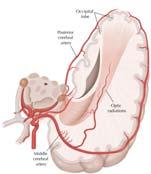
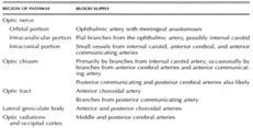
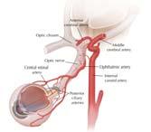
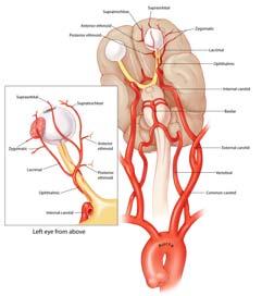

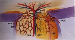
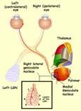
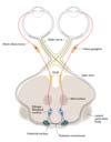
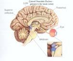
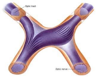
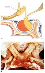
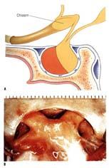
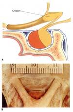
3 OPTIC NERVE ANATOMY OPTIC NERVE PROJECTIONS 4 segments: intraocular (2 mm) intraorbital (20 30 mm) intracanalicular (6 10 mm) intracranial (3 16 mm) VISION PUPIL SACCADES/FIXATION CIRCADIAN RHYTHM VISUAL PATHWAY BLOOD SUPPLY OPTIC CHIASM ANATOMY OPTIC NERVE HEAD BLOOD SUPPLY OPTIC CHIASM Pre fixed chiasm Post fixed chiasm 2
")


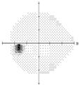
4 Onset and Duration Progression (tempo) Description of visual loss Associated symptoms Medications Surgery Review of systems Visual acuity range: 20/15 NLP Levitan s Swinging Flash Light Test (Relative Afferent Pupillary Defect) 3

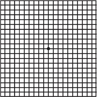
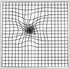
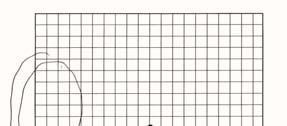
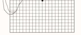
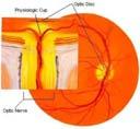
5 Visual Evoked Potentials (VEP) Electroretinogram (ERG) AMSLER GRID OPTIC NERVE VERSUS MACULA 4
 Compressive optic neuropathy Toxic/nutritional optic neuropathy Infectious (syphilitic) optic neuropathy Hereditary")
 Papilledema (often bilateral) 4 Responses: As Easy as 1, 2, 3, 4 1. Nothing 2. Cupping 3. Swelling 4.")
 Anomalous blood vessel branching pattern (congenital) Optociliary shunt vessel (optic nerve tumor) Associated Retinal Findings Macular star")
 Embolus (carotid or cardiac disease) Peripapillary telangiectatic")
6 Normal Optic Nerve Optic neuritis Traumatic optic neuropathy Compressive optic neuropathy Posterior ischemic optic neuropathy Radiation optic neuropathy Optic Nerve Pallor (on presentation) Compressive optic neuropathy Toxic/nutritional optic neuropathy Infectious (syphilitic) optic neuropathy Hereditary optic neuropathy Optic Nerve Edema Optic neuritis Anterior ischemic optic neuropathy Optic nerve glioma/optic nerve sheath meningioma Leber hereditary optic neuropathy (pseudoedema) Papilledema (often bilateral) 4 Responses: As Easy as 1, 2, 3, 4 1. Nothing 2. Cupping 3. Swelling 4. Palloring Associated Optic Nerve Findings Peripapillary hemorrhages: superficial (acquired) vs subretinal (congenital) Crowded fellow optic nerve (NA-AION) Anomalous blood vessel branching pattern (congenital) Optociliary shunt vessel (optic nerve tumor) Associated Retinal Findings Macular star (neuroretinitis) Choroidal folds (hypotony, orbital tumor) Retinal hemorrhages (CRVO) Retinitis, white dots, retinal scars (infection, inflammation) Associated Retinovascular Findings Cotton wool spots (GCA, hypertension, vasculitis) Embolus (carotid or cardiac disease) Peripapillary telangiectatic vessels (LHON) BOW TIE ATROPHY 5


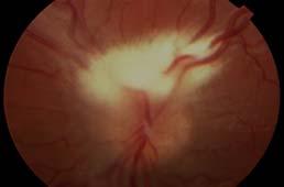
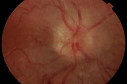
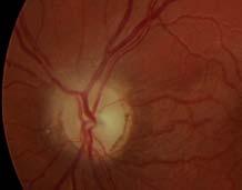
7 OPTIC NEUROPATHY Elevated ICP Neoplastic (compressive) Inflammatory Infectious Vascular Metabolic/Toxic/Medication Hereditary Ocular Congenital Traumatic Ophthalmoscopic Features: PSEUDOPAPILLEDEMA COURTESY OF MAYS A. EL DAIRI, MD elevated disc: margins obscured absence of central cup anomalous branching pattern of blood vessels no obscuration of blood vessels SVP s present no exudates no cotton wool spots rare hemorrhages 6


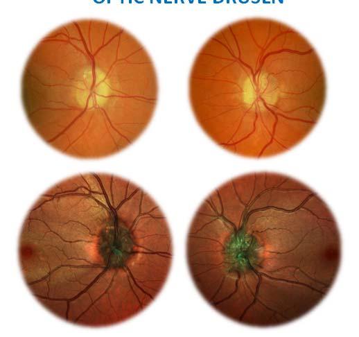
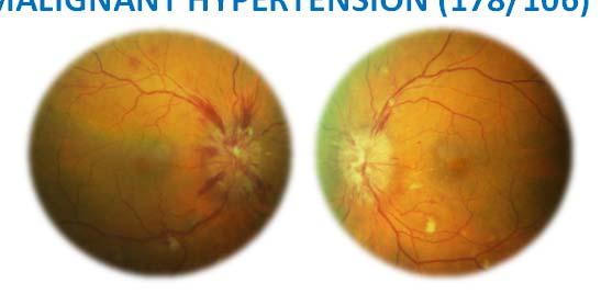
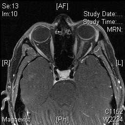
8 OPTIC NERVE DRUSEN MALIGNANT HYPERTENSION (178/106) OPTIC NERVE DRUSEN TILTED OPTIC NERVE MYELINATED NERVE FIBER LAYER VITREORETINAL TRACTION 7


 OPTIC")


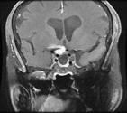
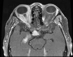
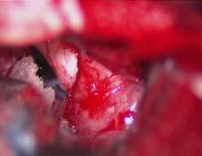
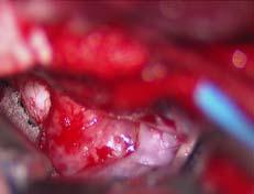
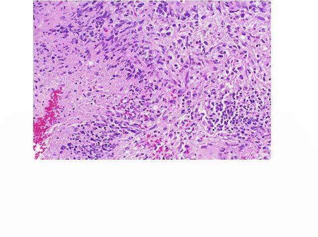
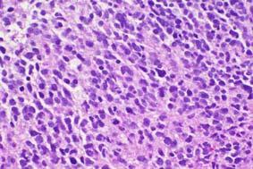
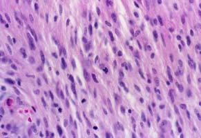
9 MORNING GLORY ANOMALY CASE STUDIES MENINGOENCEPHALOCELE OPTIC NERVE HYPOPLASIA Case 1: A 33 year old black female woke up with a blind spot in the vision in the right eye associated with pain with eye movements. Past medical history notable for aneurysm repair 4.5 years previously. On examination: Visual acuity: 20/25 both eyes Color vision: decreased right eye Pupils: right relative afferent pupillary defect Fundus exam: normal SEPTO OPTIC DYSPLASIA (DE MORSIER SYNDROME) OPTIC NERVE PIT Unsuccessful clipping of right supraclinoid internal carotid artery aneurysm, distal to the origin of the ophthalmic artery. Wrapped with muslin,coated with cyanoacrylate glue and subsequently embolized. COLOBOMA 8


10 Recalls visiting parents several weeks ago and playing with their new kittens. Mother diagnosed with cat scratch disease. Serology positive for Bartonella henselae. Eight days later: DIAGNOSIS: MUSLINOMA DIAGNOSIS: CAT SCRATCH DISEASE Case3: 61 year old Caucasian woman with 2 months of painless decreased vision OD. Vision 20/30. 20/70 VA < 20/50 PH 20/40 Pupils: RAPD OD 14 IOP < 14 no control Color Plates < 7.5/ 10 EOM: Full SLE: 1+NS OU Case 2: 31 year old woman suddenly noticed black spot in vision of left eye. No pain with eye movements. Brief viral illness 1 week previously. No past medical history. On examination: Visual acuity: right eye 20/20 left eye CF Color vision: right eye 11/11 left eye 0/11 Pupils: left RAPD 9

11 DIAGNOSIS: MALIGNANT OPTIC NERVE GLIOMA 10


 Case 5: 20 year old")


12 Case 4: 41 yo monocular female developed headaches and 1 week later noticed blurred vision right eye with a pull like sensation. On Examination: visual acuity: right eye 4/200 color: right eye 1/11 slit lamp exam: normal motility: normal CSF, CxR, ACE, ANA, C ANCA, P ANCA, RPR, ESR, Bartonella and Lyme titers: all negative or normal. Admitted to hospital and received IV Solu Medrol. 1 week later: increase pain and decrease vision DIAGNOSIS: HZV ACUTE RETINAL NECROSIS (ARN) Case 5: 20 year old Caucasian woman with one day history of decreased vision in the left eye associated with ocular pain with eye movements. Past ocular, medical, surgical and neurological histories unremarkable. Medications: birth control pills. Review of systems: denies cat scratch or tick bite. Visual acuity: right eye 20/20 left eye 20/400 Color vision: right eye 11/11 left eye 0/11 Left relative afferent pupillary defect (Marcus Gunn pupil) Eye Movements: full Neurological exam: normal 11


13 Case 1: FOLLOW UP VISUAL FIELD DIAGNOSIS: ACUTE DEMYELINATING OPTIC NEURITIS Case 6: 39 yo black female noticed blurred vision right eye after grease splattered in eye 3 days previously. Vision continued to worsen for the next 3 weeks. No ocular pain. On examination: visual acuity: LP right eye color: 0 right eye Right RAPD 20/20 left eye 11/11 left eye slit lamp, motility: normal neuro exam: normal Visual acuity: 20/20 right eye 20/25 left eye Color vision: 11/11 right eye 8/11 left eye Subtle left afferent pupillary defect SIX WEEKS LATER MRI, syphilis serology, ANA, C ANCA, Bartonella and Lyme: all normal or negative ESR: elevated DIAGNOSIS: SARCOIDOSIS 12


")


14 SUMMARY OPTIC NEUROPATHY Case 7: Healthy 11 year old boy with one history of decrease vision right eye and pressure over right eye followed within several weeks with decrease vision left eye. Medications: none Family History: maternal uncle with visual loss in 20s Differential diagnosis extensive Requires detailed history, physical examination and paraclinical studies Elevated ICP (papilledema) Neoplastic (compressive) Inflammatory Infectious Vascular Metabolic/Toxic/Nutritional/Medication Hereditary Ocular Congenital Traumatic OPTIC NEUROPATHY As Easy as 1, 2, 3, 4, 5,6,7,8,9,10. Work up: MRI enhancement right optic nerve. Blood tests: positive toxoplasmosis titers Lumbar puncture: normal 3 days IV solumedrol with continued loss of vision DIAGNOSIS: LEBER HEREDITARY OPTIC NEUROPATHY THANK YOU 13
Neuro-Ocular Grand Rounds Anthony B. Litwak,OD, FAAO VA Medical Center Baltimore, Maryland
 Neuro-Ocular Grand Rounds Anthony B. Litwak,OD, FAAO VA Medical Center Baltimore, Maryland Dr. Litwak is on the speaker and advisory boards for Alcon and Zeiss Meditek COMMON OPTIC NEUROPATHIES THAT CAN
Neuro-Ocular Grand Rounds
 Neuro-Ocular Grand Rounds Anthony B. Litwak,OD, FAAO VA Medical Center Baltimore, Maryland Dr. Litwak is on the speaker and advisory boards for Alcon and Zeiss Meditek COMMON OPTIC NEUROPATHIES THAT CAN
Alan G. Kabat, OD, FAAO (901)
") THE SWOLLEN OPTIC DISC: EMERGENCY OR ANOMALY? Alan G. Kabat, OD, FAAO (901) 252-3691 Memphis, Tennessee alan.kabat@alankabat.com Course description: The swollen disc presents a diagnostic dilemma. While
THE SWOLLEN OPTIC DISC: EMERGENCY OR ANOMALY? Alan G. Kabat, OD, FAAO (901) 252-3691 Memphis, Tennessee alan.kabat@alankabat.com Course description: The swollen disc presents a diagnostic dilemma. While
3/16/2018. Optic Nerve Examination. Hassan Eisa Swify FRCS Ed (Ophthalmology) Air Force Hospital
 Air Force Hospital") Optic Nerve Examination Hassan Eisa Swify FRCS Ed (Ophthalmology) Air Force Hospital 1 Examination Structure ( optic disc) Function Examination of the optic disc The only cranial nerve (brain tract) which
Optic Nerve Examination Hassan Eisa Swify FRCS Ed (Ophthalmology) Air Force Hospital 1 Examination Structure ( optic disc) Function Examination of the optic disc The only cranial nerve (brain tract) which
Papilledema. Golnaz Javey, M.D. and Jeffrey J. Zuravleff, M.D.
 Papilledema Golnaz Javey, M.D. and Jeffrey J. Zuravleff, M.D. Papilledema specifically refers to optic nerve head swelling secondary to increased intracranial pressure (IICP). Optic nerve swelling from
12/2/16. Ways to differentiate:
 Nate Lighthizer, O.D., F.A.A.O. Assistant Dean for Clinical Care Services Director of CE Chief of Specialty Care Clinics Chief of Electrodiagnostics Clinic Oklahoma College of Optometry lighthiz@nsuok.edu
Neuro-ophthalmologyophthalmology. Marek Michalec, MD.
 Neuro-ophthalmologyophthalmology Marek Michalec, MD. Neuro-ophthalmology Study integrating ophthalmology and neurology Disorders affecting parts of CNS devoted to vision or eye: Afferent system (visual
Five steps: Overview
 Optic atrophy is not a diagnosis Andrew G. Lee, MD Professor of Ophthalmology, Neurology and Neurosurgery, Weill Cornell Medical College Chair, Department of Ophthalmology, Houston Methodist Hospital,
Pearls, Pitfalls and Advances in Neuro-Ophthalmology
 Pearls, Pitfalls and Advances in Neuro-Ophthalmology Nancy J. Newman, MD Emory University Atlanta, GA Consultant for Gensight Biologics, Santhera Data Safety Monitoring Board for Quark AION Study Medical-legal
Neuro Ocular Grand Rounds Anthony B. Litwak, OD, FAAO VA Medical Center Baltimore, MD
 Neuro Ocular Grand Rounds Anthony B. Litwak, OD, FAAO VA Medical Center Baltimore, MD 58 YOWM! C/O I think there is something wrong with my vision, but I m not sure what it is.! +PMH for HTN, atrial fibrillation,
Dr/ Marwa Abdellah EOS /16/2018. Dr/ Marwa Abdellah EOS When do you ask Fluorescein angiography for optic disc diseases???
 When do you ask Fluorescein angiography for optic disc diseases??? 1 NORMAL OPTIC DISC The normal optic disc on fluorescein angiography is fluorescent due to filling of vessels arising from the posterior
OCCLUSIVE VASCULAR DISORDERS OF THE RETINA
 OCCLUSIVE VASCULAR DISORDERS OF THE RETINA Learning outcomes By the end of this lecture the students would be able to Classify occlusive vascular disorders (OVD) of the retina. Correlate the clinical features
Optic Nerve Disorders: Structure and Function and Causes
 Optic Nerve Disorders: Structure and Function and Causes Using Visual Fields, OCT and B-scan Ultrasound to Diagnose and Follow Optic Nerve Visual Losses Ohio Ophthalmological Society and Ophthalmic Tech
I have nothing to disclose but I
 OPTIC NEUROPATHIES Robert L. Tomsak MD PhD Professor of Ophthalmology and Neurology Wayne State t University it Sh School of Mdii Medicine I have nothing to disclose but I wish I did. dd Road map for this
Optic Disc: Anatomy, Variants, Unusual discs. Kathleen B. Digre, MD Professor Neurology, Ophthalmology
 Optic Disc: Anatomy, Variants, Unusual discs Kathleen B. Digre, MD Professor Neurology, Ophthalmology THE OPHTHALMOSCOPE DIRECT OPHTHALMOSCOPY Jan Purkinje 1823 Hermann von Helmholtz 1851 Hand held ophthalmoscope
OPTIC NERVE SWELLING IN CHILDHOOD
 OPTIC NERVE SWELLING IN CHILDHOOD Melissa W. Ko, MD, FAAN One of the main findings on a pediatric neurologic examination that can instill fear and lead to an urgent referral to neuro-ophthalmology is the
10/27/2013. Optic Red Herrings
 Optic Red Herrings 1 Optic neuropathy Compressive Inflammatory Toxic Glaucomatous Ischemic Post traumatic GLAUCOMATOUS OPTIC NEUROPATHY Glaucoma: Traditionally defined as a progressive optic neuropathy
THE SWOLLEN DISC. Valerie Biousse, MD Emory University School of Medicine Atlanta, GA
 THE SWOLLEN DISC Valerie Biousse, MD Emory University School of Medicine Atlanta, GA Updated from: Neuro-Ophthalmology Illustrated. Biousse V, Newman NJ. Thieme, New-York,NY. 2 nd Ed, 2016. Edema of the
Typical idiopathic intracranial hypertension Optic nerve appearance and brain MRI findings. Jonathan A. Micieli, MD Valérie Biousse, MD
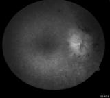
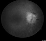
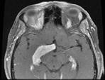
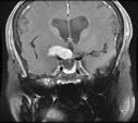
 Typical idiopathic intracranial hypertension Optic nerve appearance and brain MRI findings Jonathan A. Micieli, MD Valérie Biousse, MD A 24 year old African American woman is referred for bilateral optic
LECTURE # 7 EYECARE REVIEW: PART III
 LECTURE # 7 EYECARE REVIEW: PART III HOW TO TRIAGE EYE EMERGENCIES STEVE BUTZON, O.D. EYECARE REVIEW: HOW TO TRIAGE EYE EMERGENCIES FOR PRIMARY CARE PHYSICIANS Steve Butzon, O.D. Member Director IDOC President
3/16/2018. Optic nerve axons of retinal ganglion cells. 1.2 million nerve fibers. ON sheath: continuous with the meninges dura arachnoid and pia mater
 Optic nerve axons of retinal ganglion cells 1.2 million nerve fibers. ON sheath: continuous with the meninges dura arachnoid and pia mater 1 1.Visual Acuity 2.Color Vision 3.Pupil 4.Contrast sensitivity
Neuro-Ophthalmic Masqueraders
 Neuro-Ophthalmic Masqueraders Leonid Skorin, Jr., OD, DO, MS, FAAO, FAOCO Mayo Clinic Health System in Albert Lea Denise Goodwin, OD, FAAO Pacific University College of Optometry Please silence all mobile
Unexplained visual loss in seven easy steps
 Unexplained visual loss in seven easy steps Andrew G. Lee, MD Chair Ophthalmology, Houston Methodist Hospital, Professor, Weill Cornell MC; Adjunct Professor, Baylor COM, U Iowa, UTMB Galveston, UT MD
Abstract title: Vision loss from myelinated retinal nerve fiber layer with maculopathy. Authors: Man Kin (Eric) Chow, OD Lori Vollmer, OD, FAAO
 Chow, OD Lori Vollmer, OD, FAAO") Abstract title: Vision loss from myelinated retinal nerve fiber layer with maculopathy. Authors: Man Kin (Eric) Chow, OD Lori Vollmer, OD, FAAO Joseph Sowka, OD, FAAO General Topic: Ocular Disease Primary
Abstract title: Vision loss from myelinated retinal nerve fiber layer with maculopathy. Authors: Man Kin (Eric) Chow, OD Lori Vollmer, OD, FAAO Joseph Sowka, OD, FAAO General Topic: Ocular Disease Primary
Sequential non-arteritic anterior ischemic optic neuropathy (NAION) Jonathan A. Micieli, MD Valérie Biousse, MD
 Jonathan A. Micieli, MD Valérie Biousse, MD") Sequential non-arteritic anterior ischemic optic neuropathy (NAION) Jonathan A. Micieli, MD Valérie Biousse, MD A 68 year old white woman had a new onset of floaters in her right eye and was found to have
Sequential non-arteritic anterior ischemic optic neuropathy (NAION) Jonathan A. Micieli, MD Valérie Biousse, MD A 68 year old white woman had a new onset of floaters in her right eye and was found to have
1.! Yes I do. 2.! No I don t. COPE Approved: COPE # PD! " !! What is electrodiagnostics testing? !! Visual Pathway Basic Understanding !!
 1.! Yes I do 2.! No I don t Nathan Lighthizer, O.D., F.A.A.O Assistant Professor, NSUOCO Chief of Specialty Care Clinics Chief of Electrodiagnostics Clinic COPE Approved: COPE # 3132-PD #$ #$! " 1.! Monthly
Case Follow Up. Sepi Jooniani PGY-1
 Case Follow Up Sepi Jooniani PGY-1 Triage 54 year old M Pt presents to prelim states noticed today he had reddness to eyes, states worse in R eye. Pt denies any pain or itching. No further complaints.
Non-arteritic anterior ischemic optic neuropathy (NAION) with segmental optic disc edema. Jonathan A. Micieli, MD Valérie Biousse, MD
 with segmental optic disc edema. Jonathan A. Micieli, MD Valérie Biousse, MD") Non-arteritic anterior ischemic optic neuropathy (NAION) with segmental optic disc edema Jonathan A. Micieli, MD Valérie Biousse, MD A 75 year old white woman lost vision in the inferior part of her visual
Non-arteritic anterior ischemic optic neuropathy (NAION) with segmental optic disc edema Jonathan A. Micieli, MD Valérie Biousse, MD A 75 year old white woman lost vision in the inferior part of her visual
Shared embryology Eye and brain develop from neuro-ectoderm
 The Patient with Visual Loss: Localization of Neuropathologic Disease and Select Diseases of Neuropathologic Interest Steven A. Kane, M.D., Ph.D. The Edward S. Harkness Eye Institute Shared embryology
OPTIC NEUROPATHIES Optic Neuritis vs AION. Jacqueline M.S. Winterkorn, Ph.D., M.D.
 OPTIC NEUROPATHIES Optic Neuritis vs AION Jacqueline M.S. Winterkorn, Ph.D., M.D. OPTIC NEUROPATHIES Inflammatory Optic Neuritis Ischemic Optic Neuropathy Compressive Optic Neuropathy Traumatic Optic
A Case of Carotid-Cavernous Fistula
 A Case of Carotid-Cavernous Fistula By : Mohamed Elkhawaga 2 nd Year Resident of Ophthalmology Alexandria University A 19 year old male patient came to our outpatient clinic, complaining of : -Severe conjunctival
Fundus Autofluorescence. Jonathan A. Micieli, MD Valérie Biousse, MD
 Fundus Autofluorescence Jonathan A. Micieli, MD Valérie Biousse, MD The retinal pigment epithelium (RPE) has many important functions including phagocytosis of the photoreceptor outer segments Cone Rod
The Prevalence of diabetic optic neuropathy in type 2 diabetes mellitus
 The Prevalence of diabetic optic neuropathy in type 2 diabetes mellitus Received: 25/4/2016 Accepted: 8/12/2016 Introduction Diabetic papillopathy is an atypical form of non-arteritic anterior ischemic
Objectives. Unexplained Vision Loss: Where Do I Go From Here. History. History. Drug Induced Vision Loss
 Objectives Unexplained Vision Loss: Where Do I Go From Here Denise Goodwin, OD, FAAO Coordinator, Neuro-ophthalmic Disease Clinic Pacific University College of Optometry goodwin@pacificu.edu Know the importance
Radiation Chiasma Neuropathy after Radiotherapy for Treatment of Paranasal Sinus lymphoma
 Radiation Chiasma Neuropathy after Radiotherapy for Treatment of Paranasal Sinus lymphoma Mohammad Pakravan, MD 1 Bagher Hosseiny, MD 2 Mostafa Soltan-Sanjari, MD 3 Abstract Purpose: To present a patient
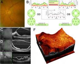
COEXISTENCE OF OPTIC NERVE HEAD DRUSEN
 COEXISTENCE OF OPTIC NERVE HEAD DRUSEN AND COMBINED HAMARTOMA OF THE RETINA AND RETINAL PIGMENT EPITHELIUM IN A TAIWANESE MALE Yo-Chen Chang 1 and Rong-Kung Tsai 2,3 1 Department of Ophthalmology, Kaohsiung
Retro-bulbar visual anatomy Optic nerves carry. Normal left ocular fundus. Retinal nerve fiber layer anatomy
 The Patient with Visual Loss: Localization of Neuropathologic Disease and Select Diseases of Neuropathologic Interest Steven A. Kane, M.D., Ph.D. The Edward S. Harkness Eye Institute Shared embryology
A Curious Case of Bilateral Optic Disc Edema Brittney Dautremont, DO, MPH
 A Curious Case of Bilateral Optic Disc Edema Brittney Dautremont, DO, MPH PGY2 Ophthalmology Resident Grandview Medical Center Dayton, OH CASE PRESENTATION 51 year old white female presenting with blurred
Anterior Ischemic Optic Neuropathy (AION)
") Anterior Ischemic Optic Neuropathy (AION) Your doctor thinks you have suffered an episode of anterior ischemic optic neuropathy (AION). This is the most common cause of sudden decreased vision in patients
Anterior Ischemic Optic Neuropathy (AION) Your doctor thinks you have suffered an episode of anterior ischemic optic neuropathy (AION). This is the most common cause of sudden decreased vision in patients
Chapter 2 Long Duration Flight Data
 Chapter 2 Long Duration Flight Data Astronaut s bodies suffer in microgravity. Without effective countermeasures, muscles atrophy, bones shed calcium, and eyesight deteriorates. We ve known about this
The Glaucoma Suspect. Evaluating the Suspect Disk. Dr Michael Forrest. ! the usual suspects: ! is it glaucoma? ! is it swollen?
 Evaluating the Suspect Disk Dr Michael Forrest Senior Lecturer, The University of Queensland Northside Eye Specialists, Nundah Visiting Ophthalmologist, Mater Hospital, Brisbane Australian Vision Convention
DEFINITION: DISCLOSURE: 7/25/2017 OPTIC NERVE GRAND ROUNDS: YOU VE GOT SOME NERVE. Joseph Sowka, OD, FAAO, Diplomate 28 YOF 28 YOF
 DISCLOSURE: OPTIC NERVE GRAND ROUNDS: YOU VE GOT SOME NERVE Dr. Joseph Sowka is a member of the Advisory Board for Novartis, Zeiss, B&L, and Allergan. He is a consultant for Novartis. Dr. Sowka has no
C19. Pediatric Neuro-ophthalmology: Dilemmas in clinical practice. 12 June, :15 15:45. Room 115 HAND-OUTS
 C19 Pediatric Neuro-ophthalmology: Dilemmas in clinical practice 12 June, 2017 14:15 15:45 Room 115 HAND-OUTS Is this strabismus really harmful? Karl Golnik, MD, MEd University of Cincinnati, USA Childhood
Interesting, unusual and eclectic cases from 2017 Robert A. Mittra, MD VitreoRetinal Surgery, P.A. Minneapolis, MN
 Fundus, SG Interesting, unusual and eclectic cases from 2017 Robert A. Mittra, MD VitreoRetinal Surgery, P.A. Minneapolis, MN Which is most likely? A) Age > 65, history of HTN B) Age 40 65, history of
Index. Note: Page numbers of article titles are in boldface type.
 Index Note: Page numbers of article titles are in boldface type. A Acetazolamide, in idiopathic intracranial hypertension, 49 52, 60 Angiography, computed tomography, in cranial nerve palsy, 103 107 digital
Interesting, unusual, eclectic cases from 2017 Robert A. Mittra, MD VitreoRetinal Surgery, P.A. Minneapolis, MN
 56 yo female, EW Presented to outside Ophthalmologist Diagnosed with viral conjunctivitis, but viral testing was negative. Also had pain around the eye and on the right side of her face Interesting, unusual,
Evaluation of ONH Pallor in Glaucoma Patients and Suspects. Leticia Rousso, O.D. Joseph Sowka, O.D
 Evaluation of ONH Pallor in Glaucoma Patients and Suspects Leticia Rousso, O.D Joseph Sowka, O.D I. Abstract This case report will evaluate a young glaucoma suspect with unilateral sectoral optic nerve
The Case: A 64 yo man with chronic back pain has elective multilevel lumbar spinal surgery
 The Case: A 64 yo man with chronic back pain has elective multilevel lumbar spinal surgery The Case: Upon awakening from anesthesia, he is blind in both eyes After Non-Ocular Surgeries Nancy J. Newman,
Evaluation of optic disc blood flow of intraconal orbital tumors using laser speckle flowgraphy.
 Research Article http://www.alliedacademies.org/ophthalmic-and-eye-research/ Evaluation of optic disc blood flow of intraconal orbital tumors using laser speckle flowgraphy. Hideki Chuman*, Takako Hidaka,
Optic Nerve Anomalies
 Optic Nerve Anomalies Raman Bhakhri, OD, FAAO Southern California College of Optometry Marshall B. Ketchum University Goals for today Review some of the optic nerve anomalies that can be seen in practice
Course # Flashes and Floaters and Curtains, Oh My!
 Course # 132 Flashes and Floaters and Curtains, Oh My! FLASHES and FLOATERS and CURTAINS, OH MY!!! FLASHES OF LIGHT Vitreous is the villain Retinal traction Retinal hole Retinal tear Migraine Classic migraine
Course # Flashes and Floaters and Curtains, Oh My!
 Course # 132 Flashes and Floaters and Curtains, Oh My! FLASHES and FLOATERS and CURTAINS, OH MY!!! FLASHES OF LIGHT Vitreous is the villain Retinal traction Retinal hole Retinal tear Migraine Classic migraine
OPTIC NERVE DISORDERS
 OPTIC NERVE DISORDERS OPTIC NEUROPATHIES INFLAMMATORY OPTIC NEUROPATHIES Cat scratch disease. Lyme disease. Viral infections of childhood (measles, mumps, chicken pox) with or without encephalitis Immun-
CHAPTER 13 CLINICAL CASES INTRODUCTION
 2 CHAPTER 3 CLINICAL CASES INTRODUCTION The previous chapters of this book have systematically presented various aspects of visual field testing and is now put into a clinical context. In this chapter,
Communicating with patients about alternative therapies: A case of optic nerve hypoplasia
 Communicating with patients about alternative therapies: A case of optic nerve hypoplasia Chief Complaint:14 year old male with poor vision since birth History of Present Illness: An otherwise healthy
Case #1: 68 M with floaters OS
 Case #1: 68 M with floaters OS Point-of-Care Ocular Sonography for the Emergency Department Nate Teismann MD Dept of Emergency Medicine, UCSF Topics in EM 2012 Acute onset of dark spots in L eye 2 days
IMAGE OF THE MOMENT PRACTICAL NEUROLOGY
 178 PRACTICAL NEUROLOGY IMAGE OF THE MOMENT Gawn G. McIlwaine*, James H. Vallance* and Christian J. Lueck *Princess Alexandra Eye Pavilion, Chalmers Street, Edinburgh UK; The Canberra Hospital, P.O. Box
53 year old woman attends your practice for routine exam. She has no past medical history or family history of note.
 Case 1 Normal Tension Glaucoma 53 year old woman attends your practice for routine exam. She has no past medical history or family history of note. Table 1. Right Eye Left Eye Visual acuity 6/6 6/6 Ishihara
Step 4: Ask permission to turn off lights or draw the curtains
 STEPS OF EYE EXAMINATION - FUNDUS Step 1: Approach the patient Read the instructions carefully for clues Shake hands, introduce yourself Ask permission to examine him I would like to examine your eyes,
Sahand Ensafi PA, CCPA, B.H.Sc.,Department of Emergency Medicine, University Health Network
 Sahand Ensafi PA, CCPA, B.H.Sc.,Department of Emergency Medicine, University Health Network No Disclosures Definitions Ophthalmologic Blindness Practical definition? WHO V/A less than 3/60 (snellen)
BMB Disclosures. Papilledema can be a. Neurological Emergency, Causing Preventable Blindness
 Reasonable Doubt: Can High Intracranial Pressure Occur Without Papilledema? 15 February 2013 Jonathan C. Horton hortonj@vision.ucsf.edu http://www.ucsf.edu/hortonlab BMB Disclosures Financial Disclosures
Sudden Vision Loss. Brendan Girschek, MD, FRCSC, FACS Vitreoretinal Surgery Cedar Valley Medical Specialists
 Sudden Vision Loss Brendan Girschek, MD, FRCSC, FACS Vitreoretinal Surgery Cedar Valley Medical Specialists My Credentials -Residency in Ophthalmology at the LSU Eye Center in New Orleans, LA -Fellowship
Blindness In An Elderly Woman
 Blindness In An Elderly Woman A 74 y/o woman with a chief complaint of: a cloud in front of my right eye and I can t t see through it Symptoms began 24 hours prior to examination. Visual loss was painless
OCULAR MANIFESTATIONS OF SYSTEMIC DISEASES THUCANH MULTERER, MD
 OCULAR MANIFESTATIONS OF SYSTEMIC DISEASES THUCANH MULTERER, MD UNDERGRADUATE: Philadelphia College of Pharmacy and Science 1996 MEDICAL SCHOOL: MCP Hahnemann School of Medicine, Philadelphia PA 2000 RESIDENCY:
This is the author's manuscript of the article published in final edited form as:
 Nonmydriatic Fundus Photography: A Practical Review for the Neurologist Devin D. Mackay, M.D.; Beau B. Bruce, M.D., Ph.D. From the Departments of Neurology, Ophthalmology, and Neurosurgery (DDM), Indiana
MRI Imaging of Neuromyelitis Optica
 July 2009 MRI Imaging of Neuromyelitis Optica Jenna Nolan, Harvard Medical School Year III Gillian Lieberman, MD Our Patient: Initial Presentation J.H. is a 29 year-old woman who presents with acute vision
No Financial Interest
 Pituitary Apoplexy Michael Vaphiades, D.O. Professor Department of Ophthalmology, Neurology, Neurosurgery University of Alabama at Birmingham, Birmingham, AL No Financial Interest N E U R O L O G I C
Is it Papilloedema? John Ross Ainsworth Orthoptic staff Birmingham Children s Hospital Birmingham and Midland Eye Centre University of Birmingham
 Is it Papilloedema? John Ross Ainsworth Orthoptic staff Birmingham Children s Hospital Birmingham and Midland Eye Centre University of Birmingham Aims Children/young people A bit about hypoplasia / NFL
Carotid Cavernous Fistula
 Chief Complaint: Double vision. Carotid Cavernous Fistula Alex W. Cohen, MD, PhD; Richard Allen, MD, PhD May 14, 2010 History of Present Illness: A 46 year old female patient presented to the Oculoplastics
Interferon-Associated Retinopathy: Communicating with Internal Medicine Ari Wes, Esther S. Hong, MD, and Thomas A. Oetting, MS, MD
 Interferon-Associated Retinopathy: Communicating with Internal Medicine Ari Wes, Esther S. Hong, MD, and Thomas A. Oetting, MS, MD July 26, 2010 Chief Complaint: New floaters in both eyes. History of Present
Unilateral Optic Nerve Hypoplasia in a patient desiring surgical treatment for exotropia
 Unilateral Optic Nerve Hypoplasia in a patient desiring surgical treatment for exotropia Michael S. Floyd, MD, Christy Benson, and Susannah Q. Longmuir, MD June 13, 2011 Chief Complaint: 17- year- old
Disclosures. Visual Pathways. Visual Pathways. Visual Loss Understanding the Patterns. I have no financial disclosures. Tabby A.
 Visual oss Understanding the Patterns Tabby A. Kennedy, MD University of Wisconsin Department of adiology I have no financial disclosures Acknowledgements: indell Gentry Greg Avey JP Yu Judy Chen Disclosures
Learn Connect Succeed. JCAHPO Regional Meetings 2017
 Learn Connect Succeed JCAHPO Regional Meetings 2017 You have some Nerve Asking Me to Work Up that Patient! What I Need to know about the Neuro- Ophthalmology Patient Financial Disclosures No relevant financial
Retina Conference. Janelle Fassbender, MD, PhD University of Louisville Department of Ophthalmology and Visual Sciences 09/04/2014
 Retina Conference Janelle Fassbender, MD, PhD University of Louisville Department of Ophthalmology and Visual Sciences 09/04/2014 Subjective CC/HPI: 64 year old Caucasian female referred by outside ophthalmologist
UC SF. g h. Eye Trauma. Martha Neighbor, MD Emergency Services San Francisco General Hospital University of California
 UC SF Eye Trauma sf g h Martha Neighbor, MD Emergency Services San Francisco General Hospital University of California Goals Recognize vision threatening eye emergencies Treat them when we can Know when
Recurrent transient visual loss in a middle aged woman
 Recurrent transient visual loss in a middle aged woman Chow SY, Draman N, Teh WM, Azhany Y Chow SY, Draman N, Teh WM, et al. Recurrent transient visual loss in a middle aged woman. Malays Fam Physician.
OCT : retinal layers. Extraocular muscles. History. Central vs Peripheral vision. History: Temporal course. Optical Coherence Tomography (OCT)
") Optical Coherence Tomography (OCT) OCT : retinal layers 7 Central vs Peripheral vision Extraocular muscles RPE E Peripheral Vision: Rods (95 million) 30% Ganglion cells Central Vision: Cones (5 million)
Optical Coherence Tomography (OCT) OCT : retinal layers 7 Central vs Peripheral vision Extraocular muscles RPE E Peripheral Vision: Rods (95 million) 30% Ganglion cells Central Vision: Cones (5 million)
Vascular Disease Ocular Manifestations of Systemic Hypertension
 Vascular Disease Ocular Manifestations of Systemic Hypertension Maynard L. Pohl, OD, FAAO Pacific Cataract & Laser Institute 10500 NE 8 th Street, Suite 1650 Bellevue, WA 98004 USA 425-462-7664 Cerebrovascular
THE EYE: RETINA AND GLOBE
 Neuroanatomy Suzanne Stensaas February 24, 2011, 10:00-12:00 p.m. Reading: Waxman Ch. 15. Your histology and gross anatomy books should be useful. Reading: Histology of the Eye from any histology book
Neuropathy (NAION) and Avastin. Clinical Assembly of the AOCOO-HNS Foundation May 9, 2013
 and Avastin. Clinical Assembly of the AOCOO-HNS Foundation May 9, 2013") Non Arteritic Ischemic Optic Neuropathy (NAION) and Avastin Shalom Kelman, MD Clinical Assembly of the AOCOO-HNS Foundation May 9, 2013 Anterior Ischemic Optic Neuropathy Acute, painless, visual loss,
Non Arteritic Ischemic Optic Neuropathy (NAION) and Avastin Shalom Kelman, MD Clinical Assembly of the AOCOO-HNS Foundation May 9, 2013 Anterior Ischemic Optic Neuropathy Acute, painless, visual loss,
Moncef Khairallah, MD
 Moncef Khairallah, MD Department of Ophthalmology, Fattouma Bourguiba University Hospital Faculty of Medicine, University of Monastir Monastir, Tunisia INTRODUCTION IU: anatomic form of uveitis involving
Syllabus-Ophthalmology Rotation Course: Objectives & Goals LOYOLA UNIVERSITY CHICAGO STRITCH SCHOOL OF MEDICINE
 Syllabus-Ophthalmology Rotation Course: Objectives & Goals LOYOLA UNIVERSITY CHICAGO STRITCH SCHOOL OF MEDICINE Department of Ophthalmology Course Objectives: By Core Competencies GENERAL INFORMATION:
Intro to Glaucoma/2006
 Intro to Glaucoma/2006 Managing Patients with Glaucoma is Exciting Interesting Challenging But can often be frustrating! Clinical Challenges To identify patients with risk factors for possible glaucoma.
Role Of Various Factors In The Treatment Of Optic Neuritis----A Study Abstract Aim: Materials & Methods Discussion: Conclusion: Key words
 IOSR Journal of Dental and Medical Sciences (IOSR-JDMS) e-issn: 2279-0853, p-issn: 2279-0861.Volume 15, Issue 9 Ver. X (September). 2016), PP 51-57 www.iosrjournals.org Role Of Various Factors In The Treatment
4/27/2010 INTRODUCTION TO RETINAL VASCULAR DISEASE VENOUS/VENULAR CENTRAL RETINAL VEIN OBSTRUCTION / CRVO ADDITIONAL FEATURES /COMPLICATIONS
 INTRODUCTION TO RETINAL VASCULAR DISEASE VENOUS/VENULAR Leo Semes, OD Professor, UAB Optometry 2 CENTRAL RETINAL VEIN OBSTRUCTION CENTRAL RETINAL VEIN OBSTRUCTION / OCCLUSION (CRVO) obstruction of the
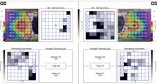
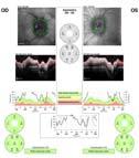
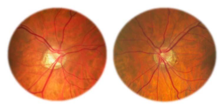
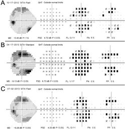
Question 1: Comment on the optic nerve appearance of each eye.
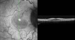
 Case 2 - Right Optic Nerve Head Drusen (ONHD) A 41 year old female was referred by her optometrist for a workup for unilateral optic disc drusen, OCT, and visual field changes. The patient was otherwise
Cases of visual impairment caused by cerebral venous sinus occlusion-induced intracranial hypertension in the absence of headache
 Zhao et al. BMC Neurology (2018) 18:159 https://doi.org/10.1186/s12883-018-1156-7 CASE REPORT Open Access Cases of visual impairment caused by cerebral venous sinus occlusion-induced intracranial hypertension
Neurology Case Presentation. Rawan Albadareen, MD 12/20/13
 Neurology Case Presentation Rawan Albadareen, MD 12/20/13 Case presentation A 49 y.o. female presented to the ED after an episode of zigzagging w a jagged bright light crossing through her Rt visual field
NEURO-OPHTHALMIC ASSESSMENT DR. B. C. UGWU
 CLINICAL VIGNETTE 2019; 5:1 NEURO-OPHTHALMIC ASSESSMENT DR. B. C. UGWU Editor-in-Chief: Prof Olufemi Idowu Neurological surgery Division, Department of Surgery, LASUCOM/LASUTH, Ikeja, Lagos, Nigeria. Copyright-
Optic Nerve Disorders
 Optic Nerve Disorders Optic Nerve Disorders Diagnosis and Management, MD Associate Professor of Ophthalmology and Neurology, University of Kentucky College of Medicine, Lexington, Kentucky, USA , MD Associate
Local Intra-arterial Fibrinolysis in Treatment of Incomplete Ophthalmic Artery Occlusion A Case Report
 CASE REPORT Local Intra-arterial Fibrinolysis in Treatment of Incomplete Ophthalmic Artery Occlusion A Case Report Shih-Ting Fang, Pao-Sheng Yen 1, Chien-Chung Chen, Yuan-Chieh Lee Department of Ophthalmology,
Topical Diagnosis of Chiasmal and Retrochiasmal Disorders
 Topical Diagnosis of Chiasmal and Retrochiasmal Disorders Leonard A. Levin CHAPTER 12 TOPICAL DIAGNOSIS OF OPTIC CHIASMAL LESIONS Visual Field Defects Etiologies of the Optic Chiasmal Syndrome Masqueraders
Supplementary Online Content
 Supplementary Online Content Park KH, Kim YK, Woo SJ, et al. Iatrogenic occlusion of the ophthalmic artery after cosmetic facial filler injections: a national survey by the Korean Retina Society. JAMA
Delayed Correction of Hypotony Maculopathy in a Patient with Glaucoma and Thyroid-Related Orbitopathy
 Published online: October 14, 2015 2015 The Author(s) Published by S. Karger AG, Basel 1663 2699/15/0063 0356$39.50/0 This article is licensed under the Creative Commons Attribution-NonCommercial 4.0 International
2009 REIMBURSEMENT GUIDE, VISUCAM and VISUCAM NM/FA
 2009 REIMBURSEMENT GUIDE FF 450 PLUS PRO NM, VISUCAM and VISUCAM NM/FA Zeiss Fundus Cameras INTRODUCTION The following guide provides an overview of billing and reimbursement for procedures performed with
Optic Nerve Disorders
 Optic Nerve Disorders Optic Nerve Disorders Diagnosis and Management Jane W. Chan, MD Associate Professor of Ophthalmology and Neurology, University of Kentucky College of Medicine, Lexington, Kentucky,
Mild NPDR. Moderate NPDR. Severe NPDR
 Diabetic retinopathy Diabetic retinopathy is the most common cause of blindness in adults aged 35-65 years-old. Hyperglycaemia is thought to cause increased retinal blood flow and abnormal metabolism in
Electrodiagnostics Alphabet Soup
 Nathan Lighthizer, O.D., F.A.A.O Assistant Professor, NSUOCO Chief of Specialty Care Clinics Chief of Electrodiagnostics Clinic What is electrodiagnostics testing? Visual Pathway Basic Understanding VEP
Optical Coherence Tomograpic Features in Idiopathic Retinitis, Vasculitis, Aneurysms and Neuroretinitis (IRVAN)
") Columbia International Publishing Journal of Ophthalmic Research (2014) Research Article Optical Coherence Tomograpic Features in Idiopathic Retinitis, Vasculitis, Aneurysms and Neuroretinitis (IRVAN)
Columbia International Publishing Journal of Ophthalmic Research (2014) Research Article Optical Coherence Tomograpic Features in Idiopathic Retinitis, Vasculitis, Aneurysms and Neuroretinitis (IRVAN)