Characterization of cerebral hemodynamic phases following severe head trauma: hypoperfusion, hyperemia, and vasospasm
|
|
|
- Judith Griffin
- 5 years ago
- Views:
Transcription
1 Characterization of cerebral hemodynamic phases following severe head trauma: hypoperfusion, hyperemia, and vasospasm Neil A. Martin, M.D., Ravish V. Patwardhan, M.D., Michael J. Alexander, M.D., Cynthia Zane Africk, M.D., Jae Hong Lee, M.D., Ehud Shalmon, M.D., David A. Hovda, Ph.D., and Donald P. Becker, M.D. University of California at Los Angeles Cerebral Blood Flow Laboratory, Brain Injury Research Center, and Division of Neurosurgery, University of California at Los Angeles School of Medicine, Los Angeles, California The extent and timing of posttraumatic cerebral hemodynamic disturbances have significant implications for the monitoring and treatment of patients with head injury. This prospective study of cerebral blood flow (CBF) (measured using 133 Xe clearance) and transcranial Doppler (TCD) measurements in 125 patients with severe head trauma has defined three distinct hemodynamic phases during the first 2 weeks after injury. The phases are further characterized by measurements of cerebral arteriovenous oxygen difference (AVDO 2 ) and cerebral metabolic rate of oxygen (CMRO 2 ). Phase I (hypoperfusion phase) occurs on the day of injury (Day 0) and is defined by a low CBF 15 calculated from cerebral clearance curves integrated to 15 minutes (mean CBF ± 2 ml/100 g/minute), normal middle cerebral artery (MCA) velocity (mean V MCA 56.7 ± 2.9 cm/second), normal hemispheric index (mean HI 1.67 ± 0.11), and normal AVDO 2 (mean AVDO ± 0.5 vol%). The CMRO 2 is approximately 50% of normal (mean CMRO ± 0.18 ml/100 g/minute) during this phase and remains depressed during the second and third phases. In Phase II (hyperemia phase, Days 1 3), CBF increases (46.8 ± 3 ml/100 g/minute), AVDO 2 falls (3.8 ± 0.1 vol%), V MCA velocity rises (86 ± 3.7 cm/second), and the HI remains less than 3 (2.41 ± 0.1). In Phase III (vasospasm phase, Days 4 15), there is a fall in CBF (35.7 ± 3.8 ml/100 g/minute), a further increase in V MCA (96.7 ± 6.3 cm/second), and a pronounced rise in the HI (2.87 ± 0.22). This is the first study in which CBF, metabolic, and TCD measurements are combined to define the characteristics and time courses of, and to suggest etiological factors for, the distinct cerebral hemodynamic phases that occur after severe craniocerebral trauma. This research is consistent with and builds on the findings of previous investigations and may provide a useful temporal framework for the organization of existing knowledge regarding posttraumatic cerebrovascular and metabolic pathophysiology. Key Words * cerebral blood flow * cerebral metabolism * head injury * hyperemia * ischemia * transcranial Doppler ultrasound * vasospasm
2 Disturbances of the cerebral circulation play a key role in the pathophysiology of head injury. Ischemic brain damage has been identified histologically in approximately 90% of patients who die following closed head injury.[21] Ischemia has been called the single most important secondary insult, and several clinical head trauma studies have linked low cerebral blood flow (CBF) to poor outcome.[8 10,26,42,46,47,49] Several factors may result in posttraumatic cerebral ischemia: increased intracranial pressure (ICP), systemic arterial hypotension, cerebral edema, focal tissue compression from hematomas, and microvascular pathology.[19,23,35,39,48,53] Another potential cause of cerebral ischemia is the posttraumatic spasm of the large cerebral arteries (vasospasm), which has been documented by angiographic and transcranial Doppler (TCD) studies.[33,36,37,57,60,63] Posttraumatic vasospasm has been associated with the finding of cerebral infarction on late computerized tomography (CT) scans and with poor outcome in some head-injured patients.[12,36,37,50] Cerebral hyperemia has also been observed following head injury and has been related to cerebral swelling, increased ICP, and poor outcome.[11,17,29,39,43] For several reasons it is critically important to understand the evolution and timing of these trauma-induced physiological derangements. First, the temporal correlation of cerebral circulatory changes with other physiological and clinical trends will provide new insights into the pathophysiology of brain injury. Second, clinical monitoring techniques that focus on ischemia, hyperemia, or vasospasm must be planned with the proper time courses in mind. Third, therapeutic strategies intended to counteract specific circulatory disturbances must be administered at the appropriate time. Although previously published studies have defined certain elements of the cerebral hemodynamic response to head injury, none has provided a comprehensive, day-to-day description of trauma-induced cerebral circulatory disturbances. The first study that combined an analysis of CBF and TCD measurements to delineate the time-dependent posttraumatic cerebral hemodynamic changes that occur after craniocerebral trauma was presented by this research group in 1992.[69] That pilot project defined three distinct phases: I, hypoperfusion during the first 24 hours (postinjury Day 0); II, hyperemia on postinjury Days 1 to 3; and III, vasospasm during Days 4 to 14. To validate these preliminary findings, the University of California at Los Angeles (UCLA) Brain Injury Research Center initiated a prospective study of trauma-induced cerebral circulatory disturbances in August We have analyzed the physiological measurements obtained in 125 severely head injured patients and have confirmed the existence of these three distinct hemodynamic phases. These results indicate that there is a stereotypical pattern of cerebral hemodynamic alterations that is generally found after severe traumatic brain injury. Recognition of this phasic pattern should prove useful in the future for the planning of monitoring in patients suffering from head trauma and for the design of time-dependent therapeutic strategies. Study Design and Patient Population CLINICAL MATERIAL AND METHODS The data presented in this report were collected prospectively during the evaluation and management of 125 patients with severe closed head injury who were admitted to UCLA or Harbor/UCLA Medical Centers between August 1992 and January This study included two subgroups. The first group included 91 patients who underwent serial CBF, cerebral metabolism, and/or TCD evaluations as part of their enrollment (with family consent) in an institutional review board approved prospective traumatic brain injury cohort study of epidemiological, clinical, and physiological predictors of long-term neuropsychological outcome (the UCLA Brain Injury Research Center [BIRC] project). The second group was composed of 34 patients who underwent CBF, cerebral metabolic, and/or TCD evaluations as
3 part of their clinical management. Because the clinical characteristics and the temporal pattern defined by the CBF and TCD studies in these two groups were similar, the groups were combined for the analyses reported in this paper. The mean age of the patients in this study was 33 ± 14.6 years (range years), and 83% of the patients were male. Admission Glasgow Coma Scale[58] (GCS) scores ranged from 3 to 8 (median 6).[35] Patients with multisystem trauma were not excluded. Twelve patients in whom CBF and TCD studies were obtained died during the course of their hospitalization. All studies, including those obtained as patients neared brain death, were included in this analysis. Management Protocol The patients were admitted directly to the neurosurgical intensive care unit (ICU), after an initial head CT scan was obtained, or postoperatively. Craniotomies were performed for removal of hematomas and/or other decompressive procedures in 60 patients. Intracranial pressure was measured in 106 patients by means of a ventriculostomy (Becker EDMS II drainage system; Pudenz Schulte Medical, Goleta, CA) or a fiberoptic ICP monitor (model V240; Camino Laboratories, San Diego, CA). The therapeutic protocol in the ICU included maintenance of ICP below 20 mm Hg and cerebral perfusion pressure (CPP) above 70 mm Hg. Intracranial hypertension (ICP > 20 mm Hg) was treated in a sequential fashion: positioning the patient with the head elevated to 30 and inducing mild hyperventilation (PaCO mm Hg), followed by ventricular CSF drainage, narcotic sedation, neuromuscular blockade, and bolus mannitol therapy as needed. The CPP was kept above 70 mm Hg by controlling the ICP and with the use of intravascular volume expansion and vasopressor therapy when necessary. In three of the study patients with intractably increased ICP (ICP > 30 mm Hg for 30 minutes), barbiturate coma (titrated to electroencephalogram burst suppression) was induced at some point during the hospital stay. The management protocol at UCLA and Harbor/UCLA Medical Centers includes intensive cerebral hemodynamic monitoring. Ideally, CBF and TCD studies were performed as soon as possible after admission and repeated regularly until the patient died, recovered, or was either transferred or discharged. Recently, jugular bulb venous blood sampling and calculation of brain arteriovenous oxygen difference (AVDO 2 ) have become a standard component of intensive monitoring to assess the adequacy of cerebral oxygen delivery. Significant abnormalities in CBF, TCD results, or cerebral AVDO 2 measurements prompted a careful assessment of the relevant physiological parameters (arterial blood pressure, ICP, PaCO 2, PaO 2 and oxygen saturation, and hematocrit). These parameters were optimized when the CBF and TCD measurements demonstrated a marked disturbance in the cerebral hemodynamic status (for example, hyperventilation was reduced and low PaCO 2 was corrected in patients with markedly reduced CBF). Beyond this, no specific treatment was administered for TCD-diagnosed vasospasm. Measurement Techniques Transcranial Doppler Ultrasound. The middle cerebral artery (MCA) and extracranial (EC) internal carotid artery (ICA) were insonated using a 2-MHz pulsed TCD device (Cerebrovascular Diagnostic System; Neurogard/Nicolet, Madison, WI) via transtemporal and submandibular methods respectively, as previously described by Aaslid, et al.[1] The flow parameters that were recorded and analyzed included the mean velocities (the time mean of the peak velocities over the course of four cardiac cycles) for the MCA (V MCA ) and extracranial ICA (V EC ICA ). The pulsatility index (PI = [systolic velocity diastolic velocity]/mean velocity) was also measured for each of these vessels; normal MCA values are 0.71 ±
4 0.13, whereas extracranial ICA values are 0.74 ± 0.13.[56] A direct relationship between the PI and increased peripheral vascular resistance has been demonstrated, if other hemodynamic factors, such as arterial pulse pressure, remain constant.[30] Because the extracranial ICA is not involved in vasospasm, velocity changes in this vessel result from changes in CBF rather than from changes in arterial diameter. Therefore V EC ICA can be used as an index of CBF and the ratio of V MCA to V EC ICA (V MCA /V EC ICA ) can be used to distinguish hyperemia from vasospasm. In hyperemia, both V MCA and V EC ICA are increased, and the ratio remains normal (1.7 ± 0.4).[31] In vasospasm, the V MCA is increased, the V EC ICA is unchanged or decreased, and thus the ratio is increased. Lindegaard, et al.,[31] have written that a V MCA /V EC ICA ratio greater than 3 corresponds to angiographically visible spasm. This ratio has been referred to as the "Lindegaard ratio" or the "hemispheric index" (HI). In this report the presence of vasospasm is determined by the simultaneous finding of V MCA greater than 120 cm/second, and V MCA /V EC ICA greater than 3. As a part of the routine clinical assessment, TCD studies were generally performed within 24 to 48 hours of admission and serial studies were performed for up to 3 weeks or more after admission. Patients enrolled in the BIRC project were studied, as much as possible, according to a prospectively established schedule (on Days 0, 1, 2, 3, 5, 7, 9, 10, 12, and 14). A total of 789 TCD studies were performed in 120 patients. Global CBF Measurements. Intravenous 133 Xe-CBF studies were performed using a portable apparatus (Cortexplorer 10; Ceretronix, Randers, Denmark) as previously described.[3,9,26,42 44] Approximately 20 to 30 mci of 133 Xe gas dissolved in saline was injected intravenously for each study. The portable CBF unit was used to estimate cerebral perfusion by analyzing 133 Xe wash-out curves measured over 11 minutes by five probes positioned over each cerebral hemisphere (primarily over the MCA territory). Analytic computer software was then used to extrapolate the data to 15 minutes, referencing the brain clearance curves to the clearance curve of exhaled end-tidal xenon-133 (which is used to estimate the arterial concentration of xenon-133). A modified height-over-area method was used to calculate CBF 15. This CBF index represents the mean flow of both fast- and slow-clearing compartments, is insensitive to "slippage," is more stable than individual compartment parameters in pathological conditions, correlates well with CBF measurements made using stable Xe-CT scanning, and has been used extensively in head trauma research.[3,8,9,26,42 44] Blood pressure, hematocrit, ICP, and PaCO 2 were recorded at the time of each study. The CBF 15 values for all 10 detectors were averaged and the mean was judged to represent the global CBF for that test. The CBF 15 values were corrected to a standard PaCO 2 of 34 mm Hg ("corrected CBF 15 "), assuming a 3% increase in CBF for each 1-mm Hg increase in PaCO 2 (following the convention of Obrist and colleagues[43]). A total of 313 CBF studies were performed in 69 of the 125 patients. Patients were missed during periods when a trained technologist was not available or when equipment malfunction precluded study, indicating that these patients were missed at random. The subgroup in which CBF studies were performed is representative of the group as a whole because the patients did not differ significantly in age (33.3 ± 15.8 years), gender (83% male), or median admission GCS score (6) from the entire study cohort or from the patients who underwent TCD testing. The first study on a given head-injured patient was typically performed within 48 hours of admission and
5 many were performed within 24 hours of admission (with three studies within 12 hours). For clinical assessment, serial studies were performed in patients over the subsequent weeks. Patients in the BIRC project were studied, as much as possible, according to a prospectively established schedule (on Days 0, 1, 3, 5, 10, and 14). Scheduled studies were missed or rescheduled for a variety of reasons such as delays in obtaining consent, patient unavailability during scheduled surgery, equipment malfunction, and radioisotope unavailability. Many CBF (and TCD) studies scheduled for the 2nd week after injury were not performed because stable patients were transferred to other medical or rehabilitation facilities (often at the request of managed health care organizations)--this is a source of bias that must be recognized. Jugular Bulb Catheter Studies. Jugular bulb catheterization was included as a part of routine management and the BIRC investigative protocol relatively recently. As soon as possible after admission to the ICU, retrograde catheterization of the internal jugular vein was accomplished using a No. 4 French fiberoptic intravascular oxygen saturation catheter (model U440; Abbott Laboratories, No. Chicago, IL). The catheter was introduced percutaneously and advanced to the jugular bulb at the base of the skull. The position was verified by lateral skull x-ray film. While the catheter was in place (usually for 5 7 days after insertion, occasionally up to postinjury Days 10 12), jugular venous and radial arterial blood samples were drawn every 8 hours, and values for AVDO 2 were calculated.[26,27] Simultaneous PaCO 2 readings were recorded. The AVDO 2 values were corrected to a PaCO 2 of 34 mm Hg, assuming an increase of 3% in AVDO 2 for every 1-mm Hg decrease in PaCO 2 (again after the convention of Obrist and colleagues[43]). The CMRO 2 was estimated from the product of AVDO 2 and the global value for CBF 15.[43]
6
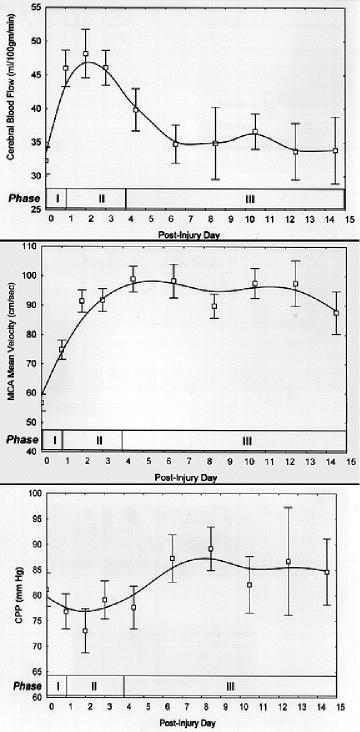
7 Fig. 1. Graphs displaying the time courses of posttraumatic CBF (adjusted to a PaCO 2 of 34 mm Hg), MCA velocity, and CPP observed after severe head trauma. Upper: The mean CBF values (± SEM) for all patients who were studied each day are represented. Center: The mean MCA velocity values (± SEM) were measured using TCD ultrasonography. Lower: The daily mean CPP values were arrived at using the following formula: CPP = mean arterial pressure mean ICP (± SEM). Postinjury Day 0 designates the first 24 hours after injury. The three cerebral hemodynamic phases are indicated at the bottom of the graph (Phase I, hypoperfusion; Phase II, hyperemia; and Phase III, vasospasm). A total of 369 measurements of AVDO 2 were made in 26 patients. This subgroup appears to be representative of the group as a whole (mean age 36.6 ± 17.7 years; median GCS score 5; 77% male). Measurements of ICP and CPP. Measurements of ICP were acquired during the time of CBF study. The CPP values were calculated from the ICP and the mean arterial pressure measurements recorded at the time of study. Definition of Timing of Posttraumatic Cerebral Hemodynamic Phases The daily values for CBF, TCD, and AVDO 2 measurements and physiological parameters were grouped into the three temporal phases as they were defined by our pilot project.[69] That preliminary study, based on CBF and TCD studies in 32 patients suffering from head trauma, identified three posttraumatic cerebral hemodynamic phases: Phase I, first 24 hours after injury (postinjury Day 0); Phase II, 24 to 96 hours after injury (postinjury Days 1 3); and Phase III (postinjury Days 4 14). The pilot study also included a fourth phase (postinjury Weeks 2 and 3) during which the CBF and TCD abnormalities began to resolve. Because of the paucity and irregular timing of CBF and TCD studies beyond 14 days postinjury, we have not included these late measurements in this report. Statistical Analysis The values for CBF, TCD, metabolic, and physiological measurements were sorted by postinjury day and the daily mean values (± standard error of the mean [SEM]) were calculated. Because fewer daily measurements were obtained after postinjury Day 3, from that point onward the measurements for 2 consecutive days were pooled and the mean values (± SEM) for that 2-day period were calculated. The daily mean values (± SEM) were used to generate the time-course graphs (Fig. 1). The curves were generated using a least-squares polynomial curve-fitting function. The values for the various parameters were also grouped according to phase, and the mean values (± SEM) for each phase were calculated (Tables 1 and 2). To describe the distribution of CBF and TCD measurements within the three phases, all studies performed during each phase were sorted into quantitatively stratified groups defined by the values indicated in Fig. 2. The relative frequency of studies in each group, for each phase, are illustrated in this figure. Two-sample, equal variance based t-tests with Bonferroni correction were used to confirm that the three hemodynamic phases, based on CO 2 -corrected CBF 15, V MCA, and HI, were separate and distinct. These tests were also used to assess any statistically significant differences in AVDO 2, CMRO 2, PI, ICP, and CPP values between phases.

8 Fig. 2. Bar graphs showing distribution of CBF values and MCA velocity values observed after severe head trauma. Upper: This figure presents, for each phase, the relative percentage of individual CBF studies in which CBF values were within designated ranges (hypoperfusion < 35 ml/100 g/minute; normal-to-relative hyperemia ml/100 g/minute; or absolute hyperemia > 55 ml/100 g/minute. Lower: This figure presents, for each phase, the relative percentage of individual TCD studies in which V MCA values were within the designated ranges (no vasospasm < 120 cm/second; mild-to-moderate vasospasm cm/second; or severe vasospasm > 160 cm/second). RESULTS Patterns of CBF and Blood Flow Velocity After Head Injury The daily mean values (± SEM) of the key parameters derived from CBF and TCD studies are illustrated graphically in Fig. 1 upper and center. For comparison, previous studies have described the normal adult mean V MCA as 34 to 86 cm/second and the mean V EC ICA as 28 to 46 cm/second.[2] Earlier studies
9 using the intravenous 133 Xe method for determining CBF have shown normal CBF 15 to be 44.1 ± 5.6 ml/100 g/minute at a PCO 2 of 34 mm Hg.[43] Simultaneous analysis of the daily mean CBF and MCA velocities reveals that the data conform to the pattern defined in our pilot study (Tables 1 3).[69] There are three distinct posttraumatic hemodynamic phases (Table 3). Phase I (the first 24 hours after injury) is referred to as the hypoperfusion phase and is defined by a low CBF (mean corrected CBF ± 2 ml/100 g/minute), a normal V MCA (mean V MCA 56.7 ± 2.9 cm/second), and a normal HI (mean HI 1.67 ± 0.11). In Phase II (hyperemia phase, Days 1 3) the CBF is transiently increased (mean corrected CBF ± 3 ml/100 g/minute), the V MCA is rapidly rising (mean V MCA 86 ± 3.7 cm/second), and the HI is normal (mean HI 2.40 ± 0.1). The third phase (vasospasm phase, Days 4 15) is characterized by a variable but gradually declining CBF (mean corrected CBF ± 3.8 ml/100 g/minute; lowest corrected CBF ± 6 ml/100 g/minute on Day 8). The V MCA increases significantly in this phase (mean V MCA 96.7 ± 6.3 cm/second; highest V MCA 111 ± 10.2 cm/second on Day 11), and the HI rises significantly (mean HI 2.87 ± 0.22). The phase of hypoperfusion (Phase I) was significantly distinguished from that of hyperemia (Phase II) in terms of lower CBF (p < 0.01); the vasospasm phase (Phase III) was significantly distinguished from both Phase II (based on a lower CBF, p < 0.01) and Phase I (based on a higher V MCA [p < 0.01] and HI [p < 0.01]).
10 Individual Variations in CBF and Blood Flow Velocity Figure 2 demonstrates the distributions of CBF values and V MCA values that were observed during each phase. Although these graphs demonstrate the prevalence of hypoperfusion during Phase I, hyperemia during Phase II, and vasospasm during Phase III, it is clear that not every patient followed the typical temporal pattern illustrated in Fig. 1 upper and center. A review of Fig. 2 demonstrates that approximately 50% of the patients have CBF profiles that are consistent with the "average" pattern. Approximately 40% of the patients appear to demonstrate a V MCA pattern consistent with both. Furthermore, when the subset of patients who have CBF and TCD measurements in each of the three phases are reviewed case by case, patterns similar to those presented in Fig. 1 upper and center prevail and are seen in 40 to 50% of the patients. Although a number of variations on the average temporal profile are possible, two other patterns are seen in a significant subgroup of patients: 1) persistence of hypoperfusion through all three phases and 2) early onset and persistence of hyperemia through Phases II and III (each seen in just under 15% of the patients). Measurements of AVDO 2 After Head Injury The AVDO 2 in normal subjects at a PaCO 2 of 34 mm Hg has been reported to be 5 to 9.8 vol%.[43] Jugular venous bulb sampling in our patients indicates that, in Phase I, there is a relatively normal AVDO 2 (mean PaCO 2 -corrected AVDO ± 0.5 vol%) (Table 1). However, in Phase II there is a dramatic, significant (p < 0.01) decrease in AVDO 2 (mean corrected AVDO ± 0.1 vol%) to well below normal values. The AVDO 2 measured during Phase III was only slightly higher than that for Phase II. The CMRO 2 was relatively stable through all three phases at a level approximately 50% below normal (normal CMRO ± 0.4 ml/100 g/minute) (Table 1).[43] Middle Cerebral Artery and Extracranial ICA PIs After Head Injury The PIs of the MCA and extracranial ICA wave forms (as measured by TCD ultrasonography) also show distinct phase-specific changes. Normal values for the MCA PI (PI MCA ) have been reported as 0.58 to 0.84 with the extracranial ICA PI (PI EC ICA ) slightly higher due to higher resistance in the ophthalmic distribution.[2] The PI of the MCA is similar to that of the extracranial ICA during Phases I and II: both are higher than normal during Phase I (PI MCA 1.13 ± 0.1; PI EC ICA 1.27 ± 0.07) and both fall toward normal values during Phase II (PI MCA 0.98 ± 0.06; PI EC ICA 1.05 ± 0.04) (Table 2). The PIs in these two vessels, however, diverge during Phase III (PI MCA 0.94 ± 0.06; PI EC ICA 1.11 ± 0.06) (Table 2).
11 Intracranial Pressure and CPP After Head Injury The mean ICP values were above 10 mm Hg all 15 days after injury (reaching a maximum on Day 10 of 18.2 ± 2.1 mm Hg) (Table 1). Although individual ICP recordings were often more than 20 mm Hg, the daily mean for the group as a whole never exceeded 20 mm Hg due to the aggressive ICP management that was a part of the standard treatment protocol. The mean daily CPP values for the group remained stable between 70 and 90 mm Hg throughout the entire course (Fig. 1 lower). Ischemia During the Three Phases None of the 21 patients in whom CBF measurements were obtained during Phase I demonstrated severe hypoperfusion (CBF 15, or PCO 2 -corrected CBF 15 ± 20 ml/100 g/minute). One patient had an (CO 2 -uncorrected) AVDO 2 measurement of 10.3 vol% when his PCO 2 was 22 mm Hg (no CBF measurement was made at the time). No other patient had a (CO 2 -uncorrected) AVDO 2 above 7 vol% during Phase I. It should be noted that only three of 21 CBF studies and no AVDO 2 measurements in this report were made before 12 hours postinjury. During Phase II, three of 100 (CO 2 -uncorrected) CBF measurements showed the CBF 15 to be less than 20 ml/100 g/minute. No (CO 2 -uncorrected) AVDO 2 measurements were above 8 vol% and only three of 95 values were over 7 vol% (PCO 2 ¾ 30 mm Hg in these three cases). During Phase III, 28 of 182 (CO 2 -uncorrected) CBF measurements demonstrated the CBF 15 to be less than 20 ml/100 g/minute. No (CO 2 -uncorrected) AVDO 2 values were over 8 vol% and only four of 269 measurements were above 7 vol% (all with PCO 2 ¾ 31 mm Hg). Hyperemia During the Three Phases The pattern defined by the daily CBF values showed a higher mean flow during postinjury Days 1 to 3, but there was considerable heterogeneity in the CBF measurements for postinjury Phases II and III (Figs. 1 upper and 2 upper). Phase I was the most homogeneous, with only 16.7% of the CBF values falling in the hyperemic range. Phase II had the widest variety of values, with CBF 15 values ranging from 13 to 99 ml/100 g/minute. Twenty-seven percent of the Phase II CBF values were in the range of absolute hyperemia (>= 55 ml/100 g/minute). Ten percent of the Phase II CBF values were greater than 70 ml/100 g/minute, which represented 75% of all of the very high CBF values recorded in the first 2 weeks. Overall, 38.8% of patients studied during this phase had at least one Day 1 to 3 CBF measurement above 55 ml/100 g/minute. Postinjury Days 4 and 5 represented a transition from Phase II to Phase III: almost all of the Phase III CBF values above 55 ml/100 g/minute were seen during these 2 days. Beyond Day 5, most of the CBF values were low. Vasospasm During the Three Phases Vasospasm of the MCA was defined as ipsilateral V MCA above 120 cm/second with an HI above 3. Using this criterion, we found that 50.4% of patients had at least one TCD study consistent with vasospasm during their hospital course. Overall, 37.6% of the patients were classified as having mild-to-moderate spasm (V MCA cm/second and HI 3 4), and 12.8% were classified as having severe spasm (V MCA >= 160 cm/second and HI >= 4). The time course of the spasm is apparent (Figs. 1 upper and 2 lower). Only one patient had a V MCA greater than 120 cm/second during the first 24 hours after injury (Fig. 2 lower). Although some patients
12 developed mild-to-moderate spasm during Phase II, almost all of the severe cases of spasm were seen during Phase III (Fig. 2 lower). However, it is important to note that during Phase III there were fewer TCD studies demonstrating evidence of spasm than CBF studies showing low blood flow (that is, many low CBF measurements during Phase III were not associated with vasospasm). DISCUSSION This prospective study has confirmed the findings of a pilot study that reported time-dependent changes in CBF, V MCA, and HI, which, considered together, defined three discrete hemodynamic phases (Table 3).[69] Phase I: Hypoperfusion (Day 0) During the first 24 hours following craniocerebral trauma, the uncorrected mean CBF was low (33.6 ± 1.6 ml/100 g/minute). Even when the CBF in these patients, who generally were hypocapnic because of hyperventilation, was corrected to a PaCO 2 of 34 mm Hg, the adjusted CBF was substantially lower than normal (normal 44.1 ± 5.6 ml/100 g/minute at a PCO 2 of 34 mm Hg).[43] Also using the 133 Xe clearance technique, Fieschi, et al.,[17] Overgaard and Tweed,[47] and others[9,13,15,26,44] have demonstrated that CBF is substantially below normal in the early hours after head injury. The quantitative global CBF values obtained by these investigators are remarkably similar to our results. Even using a different CBF-measuring technique (stable Xe-CT scanning), Marion and colleagues[34] and Bouma and associates[7,10] found levels of global CBF similar to those in the present study. Experimental studies in animal head-trauma models have also demonstrated an acute decrease in CBF to approximately 50% of baseline.[24,40,48,59,65,68] Extremely low, "ischemic" levels of CBF have been measured very early after the onset of injury. In a small number of 133 Xe-CBF studies conducted within 6 hours of injury, Bouma, et al.,[9] found the mean flow to be 22.5 ± 5.2 ml/100 g/minute, with a third of their patients demonstrating ischemic levels (CBF < 18 ml/100 g/minute). Using the Xe-CT technique, these investigators found very early evidence of either global or regional ischemia in approximately 30% of the patients and observed that these patients had a substantially worse outcome than patients without evidence of ischemia.[10] We did not observe such low levels of CBF in our patients, primarily because the first evaluation was not performed within 6 hours after patient admission. Our studies of cerebral oxygen metabolism rarely demonstrated the very high AVDO 2 levels that are the hallmark of ischemia. This is consistent with previously published results.[9,26,43,49,51] However, very early after injury when CBF is at its lowest, some cases display an AVDO 2 in the ischemic range.[9] These findings suggest that hyperacute posttraumatic cerebral ischemia may occur before measurements can generally be made, but indicate that the low global CBF seen in the first 24 hours is often matched to low CMRO 2 (Table 1). Blood flow velocity in the extracranial segment of the ICA and in the proximal MCA was in the normal or low-to-normal range during the first 24 hours after the onset of injury. The mean HI during the 1st postinjury day was also normal. Therefore, the results of the present study and those of other reported studies of posttraumatic blood flow velocity measurements refute the notion that a low CBF during the first 24 hours after head injury is due to acute vasospasm of the large basal cerebral arteries.[9,36,37,60]
13 What might be the cause of the low CBF seen in the first hours after head injury? The data presented in this report, supported by findings of other investigators, demonstrate that the impairment in CBF cannot be attributed to abnormally low CPP, excessive hyperventilation, or vasospasm.[9,36,37,44] The cerebral microcirculation appears to be the location of increased resistance causing early posttraumatic hypoperfusion. Our TCD studies have demonstrated that the PIs (which are increased by high distal vascular resistance) are elevated in both the extracranial ICA and the MCA. This strongly suggests the presence of high distal (microcirculatory) vascular resistance. The finding of subnormal cerebral blood volume during the first 24 hours postinjury also suggests narrowing of the microcirculation.[7] The cause of the narrowing in the microcirculation may be multifactorial. Extrinsic microvascular compression by damaged and edematous astrocytic processes has been observed in experimental models of head trauma and in specimens of damaged human brain tissue removed at surgery.[23,53] Active muscular constriction of the resistance arterioles might be caused by trauma-induced release of vasoactive substances such as calcium, catecholamines, prostaglandins, or neuropeptides (endothelin, neuropeptide Y).[18,22,25,38,55,61,62,66] Hemoglobin, released as a consequence of posttraumatic subarachnoid hemorrhage, has also been demonstrated to be a vasoconstrictor.[61] Additionally, intravascular thrombosis may play a role in trauma-induced microcirculatory obstruction.[23] Given the normal AVDO 2 values seen in most cases, posttraumatic hypoperfusion may also often represent appropriate coupling of low CBF to trauma-induced depression of cerebral oxidative metabolism. Phase II: Hyperemia (Days 1 3) The hypoperfusion found during the first 24 hours after head injury is followed by progressively increasing CBF (Fig. 1 upper). In fact, CBF probably begins rising within hours from the lowest levels immediately after injury. The increase in CBF reaches its maximum between 48 and 72 hours after the injury (postinjury Day 2 in this report). Overgaard and Tweed,[47] Obrist and colleagues,[42] Bouma and associates,[7 9] and others[14,42,44,50] have described a similar time course for posttraumatic hyperemia using the 133 Xe clearance technique, the Kety Schmidt (nitrous oxide saturation) technique, and stable Xe-CT scanning. The finding of increased CBF using these techniques is confirmed by the observation of increasing velocity in the extracranial segment of the ICA (Table 2). The increase in CBF elicits an increase in V MCA as well, although in some cases this may be due to the early onset of cerebral arterial spasm (manifested by an increase in the HI). In many cases the CBF rose from the subnormal levels of the first phase into a range that would be considered normal for a nontraumatized healthy adult. As stated by Obrist and colleagues,[43] a "normal" CBF can be considered "relative hyperemia" in a comatose trauma patient, in whom normal metabolic coupling would result in a level of CBF considerably below normal. However, almost 40% of the patients during postinjury Days 1 to 3 demonstrated "absolute hyperemia" (with at least one measurement of CBF above the upper limit of the "normal" CBF range). The hyperemia phase includes postinjury Days 1 through 3 (Hours 24 96). However, some hyperemic CBF measurements were recorded after Day 3 (Fig. 2 upper). The great majority of these few Phase III hyperemic CBF measurements were seen on postinjury Days 4 and 5, indicating that these days represented a transition between Phase II and Phase III. The clinical significance of hyperemia has not been fully defined. Clinical studies conducted by Bruce, et al.,[11] and Obrist and colleagues[43] suggest that hyperemia is the primary cause of increased ICP.
14 Other recent studies, however, have failed to confirm the correlation between elevated CBF and increased ICP.[41,49] The relationship of hyperemia to clinical outcome is also unclear.[26,41,49] Our group has recently completed an analysis of posttraumatic hyperemia and found that hyperemia-associated intracranial hypertension occurs in almost 15% of patients with moderate-to-severe head injury and is predictive of a poor outcome.[29] Early identification and aggressive treatment of these patients might improve their prognosis. Mild hyperemia without intracranial hypertension, conversely, is linked to favorable outcome in our experience.[29] The pathophysiological mechanisms that produce posttraumatic hyperemia remain obscure. The data presented in this report indicate that increases in mean arterial pressure or CPP are not responsible for this hemodynamic change. Hyperemia is not induced by an increase in the CMRO 2, as demonstrated by the results reported in this paper and in other work.[9,43] In fact, a certain degree of uncoupling of blood flow from oxygen metabolism seems to occur. However, the possibility exists that this hyperemia may occur in response to an increased cerebral metabolic rate of glucose. Posttraumatic hyperglycolysis has been identified in animal models of head trauma by Andersen and Marmarou,[4] Hovda, et al.,[24] and Yoshino and colleagues.[67] Recent positron emission tomography studies of glucose metabolism in patients with head trauma at UCLA have demonstrated global and regional hyperglycolysis in some patients during this time period.[5,6] The PIs in both the ICA and the MCA fall on postinjury Day 1 (Table 2), providing evidence that the hyperemic phase is associated with a drop in distal cerebrovascular resistance. The generation of vasodilatory metabolites (lactic acid, neuropeptides, and adenosine) has been associated with hyperemia, or "luxury perfusion," following ischemic damage to the brain and may play a role in posttraumatic hyperemia.[13,14,32,64] Alternatively, there may be a delayed onset of mechanically induced vascular smooth-muscle dysfunction (or "vasoparalysis") that results in a decrease in cerebrovascular resistance and an increase in blood flow.[20] The cause of the termination of the hyperemic phase is not entirely clear. This may result from the clearance of vasodilatory metabolites, recovery of vascular smooth-muscle function, or resolution of posttraumatic hyperglycolysis. However, examination of the MCA PIs indicates that this phase is not necessarily terminated by an increase in distal vascular resistance (Table 2). Our data indicate that the hyperemic phase may resolve, in part, because large artery spasm occurs. Phase III: Vasospasm (Days 4 14) In the third phase, the physiological findings include increasing V MCA and an increasing HI (Fig. 1 center and Table 2). These findings are consistent with the onset of vasospasm involving the proximal segment of the MCAs. The increase in the V MCA is clearly due to progressive arterial narrowing and not to a flow effect, because CBF falls during this phase.[12,20,36] Furthermore, an increasing PI found in the cervical segment of the ICA indicates an increase in downstream resistance, presumably due to the narrowing of the MCA. Approximately 50% of our patients developed significant MCA spasm, evidenced by MCA velocities greater than 120 cm/second and by a concomitant HI over 3. Although AVDO 2 recordings during Phase III rarely demonstrated the high levels associated with ischemia, focal vasospasm-induced ischemia may have been missed using this global measurement technique. Until recently, posttraumatic vasospasm had only been defined using angiography.[32,57,63] The largest angiographic study, by Suwanwela and Suwanwela,[57] found that 18.6% of patients with
15 moderate-to-severe head injury had narrowing of at least one intracranial artery. The study of posttraumatic vasospasm using TCD ultrasonography has allowed sequential noninvasive assessment of arterial narrowing following head injury. Weber, et al.,[60] found increased MCA velocities consistent with spasm in 40% of head-injured patients. Gomez and associates[20] used TCD ultrasonography in combination with angiography to confirm the finding of posttraumatic vasospasm. Our group's 1992 study[36] demonstrated TCD and CBF evidence of vasospasm in 27% of head-injured patients (with angiographic confirmation in three patients with severe spasm). This study demonstrated that MCA vasospasm began at approximately postinjury Day 4 and persisted for as long as 3 weeks after head injury. The degree of the spasm demonstrated in these studies has, in some cases, been as severe as that seen in patients with aneurysmal subarachnoid hemorrhage who develop delayed ischemic deficit. A more recent report published by our center, based on a larger group of patients, suggests that posttraumatic spasm is associated with poor clinical outcome.[37] However, less than 15% of patients have TCD findings that suggest severe vasospasm, whereas almost 50% of CBF measurements during Phase III show low flow. It is probable that low CBF during this phase is often coupled to persistent posttraumatic hypometabolism. The measurements of cerebral oxygen metabolism obtained during this time demonstrate that CMRO 2 remains approximately 50% of normal. Recent positron emission tomography studies (using [ 18 F]fluorodeoxyglucose) in head-injured patients have shown that the cerebral metabolic rate of glucose is also suppressed beyond the first 4 or 5 days after injury.[5,6] Therefore, this phase includes vasospasm in some cases, and low CBF coupled to low cerebral metabolism in others. Resolution of Posttraumatic Hemodynamic Changes This research study does not address the late resolution of trauma-induced hemodynamic disturbances because of the paucity of CBF or TCD evaluations performed beyond 2 weeks after injury (most patients have been discharged or transferred by this time). The few cases that had later measurements (reported in our pilot study) demonstrated gradual recoveries toward normal on the part of CBF and MCA velocities beginning 2 to 3 weeks after injury.[69] A previously published study of posttraumatic vasospasm also showed resolution of even the most severe cases after 3 to 4 weeks.[36] It seems that the fourth phase of the posttraumatic cerebral hemodynamic pattern should be termed the "recovery phase," beginning 2 to 3 weeks after onset and probably lasting weeks to months. Shortcomings, Flaws, and Sources of Bias Because of the logistical problems involved in performing TCD and CBF evaluations during the 1st clinically busy hours after admission to the emergency department, we have obtained few studies within 12 hours of injury. The earliest cerebral hemodynamic and metabolic alterations must be inferred from the few other clinical studies that have focused on the earliest hours and from laboratory investigations. The true prevalence of hyperacute ischemia (within the first 1 or 2 hours) in head-injured patients remains to be determined. The CBF and metabolic measurements reported in this paper represent global alterations. Focal disturbances in flow or metabolism caused by discrete traumatic lesions (hematomas or contusions) or related to isolated vascular pathology (focal spasm or dissection) will be "blurred" or may be missed entirely. Furthermore, subcortical, cerebellar, and brainstem CBF and metabolism are not specifically evaluated using the techniques discussed in this work. For regional flow and metabolism measurements
16 one must refer to autoradiographic studies conducted in experimental models of head trauma or to the few clinical studies performed with tomographic techniques.[7,10,34] Positron emission tomography (using 15 O-containing tracers for CBF and oxygen metabolism measurements and [ 18 F]fluorodeoxyglucose for glucose metabolism measurements) is the most promising, although logistically difficult, technique for defining pathophysiology on a regional scale. We have begun such studies and plan to extend these over the next several years.[5,6] One of the inherent flaws in this project is the inability to study patients who have been transferred to other medical facilities or rehabilitation units or discharged from the hospital. Therefore, it is likely that the data in later phases are skewed toward patients with more severe injuries and more complicated hospital stays. It is important to recognize that not every patient experienced the typical hemodynamic alterations during each postinjury phase (Fig. 2). Studies in progress will address CBF changes and vasospasm in clinically important subgroups (stratified by age, CT findings, presence or absence of intracranial hypertension, outcome, and so forth). In a fashion similar to that used by Robertson and colleagues,[49] future studies must address the importance of variations on the "average" temporal profile of hemodynamic disturbances. Potential Therapeutic Implications In Phase I, hypoperfusion may be a critical secondary insult to the traumatized brain, with ischemia and infarction compounding the initial mechanical injury. Phase I, therefore, requires rigorous maintenance of normal CPP, normal oxygenation, and a normal hematocrit level. In general, profound hyperventilation should be avoided because, as Sheinberg, et al.,[54] have demonstrated, hypocarbia may lead to more profound hypoperfusion and a concomitant poor outcome. Potential strategies for future therapy for the low blood flow of Phase I include induced arterial hypertension; pharmacological agents that might reverse the increase in small vessel resistance that appears to be present; or therapies (drugs or hypothermia) that protect neurons from ischemia-induced biochemical events. Recent work performed at our center and others has indicated that the hyperemia of Phase II may be associated with harmful intracranial hypertension.[11,29,43] Because this phase is not associated with MCA spasm or usually with a high AVDO 2 indicating inadequate oxygen delivery, hyperventilation should be safe during Phase II. During this phase, the prominence of hyperemia suggests that "CPP therapy" using pressor-induced arterial hypertension should be used cautiously and should, perhaps, be guided by CBF measurements.[52] Phase III appears to be a more important phase than generally appreciated. It is reasonable to propose that the vasospasm that occurs in some patients in Phase III should be monitored using TCD ultrasonography and treated in the same way as vasospasm that follows aneurysmal subarachnoid hemorrhage--with volume expansion and induced arterial hypertension.[28,45] Treatment with nimodipine (or yet-to-be developed drugs) may reduce the ischemic neurological damage that may occur during this phase. For particularly severe symptomatic cases, balloon angioplasty may be considered.[16] Acknowledgments The authors wish to thank Walter Obrist, Ph.D., David Stump, Ph.D., and David Beyda, M.D., for their advice and assistance with the CBF measurement technique. We would also like to acknowledge
17 Kathleen Thomas-Lukes, Brenda Rinsky, and Oscar Barcenas for performing the TCD studies presented in this report, and Eula Mckinney and Karen Einstein for secretarial assistance. References 1. Aaslid R, Markwalder TH, Nornes H: Noninvasive transcranial Doppler ultrasound recording of flow velocity in basal cerebral arteries. J Neurosurg 57: , Adams RJ, Nichols FT, Hess DC: Normal values and physiological variables, in Newell DW, Aaslid R (eds): Transcranial Doppler. New York: Raven Press, 1992, pp Agnoli A, Prencipe M, Priori AM, et al: Measurements of the rcbf by intravenous injection of 133 Xe. A comparative study with the intra-arterial injection method, in Brock M, Fieschi C, Ingvar DH, et al (eds): Cerebral Blood Flow. Clinical and Experimental Results. Berlin: Springer-Verlag, 1969, pp Andersen BJ, Marmarou A: Post-traumatic selective stimulation of glycolysis. Brain Res 585: , Bergsneider M, Hovda DA, Shalmon E, et al: Cerebral hyperglycolysis following severe traumatic brain injury in humans: a positron emission tomography study. J Neurosurg 86: , Bergsneider M, Kelly DF, Shalmon E, et al: Dynamic changes in cerebral glucose metabolism following severe head injury as measured by positron emission tomography. J Cereb Blood Flow Metab 15 (Suppl 1):S26, 1995 (Abstract) 7. Bouma GJ, Muizelaar JP: Cerebral blood flow, cerebral blood volume, and cerebrovascular reactivity after severe head injury. J Neurotrauma 9 (Suppl 1):S333 S348, Bouma GJ, Muizelaar JP: Evaluation of regional cerebral blood flow in acute head injury by stable xenon-enhanced computerized tomography. Acta Neurochir Suppl 59:34 40, Bouma GJ, Muizelaar JP, Choi SC, et al: Cerebral circulation and metabolism after severe traumatic brain injury: the elusive role of ischemia. J Neurosurg 75: , Bouma GJ, Muizelaar JP, Stringer WA, et al: Ultra-early evaluation of regional cerebral blood flow in severely head-injured patients using xenon-enhanced computerized tomography. J Neurosurg 77: , Bruce DA, Langfitt TW, Miller JD, et al: Regional cerebral blood flow, intracranial pressure, and brain metabolism in comatose patients. J Neurosurg 38: , Chan KH, Dearden NM, Miller JD: The significance of posttraumatic increase in cerebral blood flow velocity: a transcranial Doppler ultrasound study. Neurosurgery 30: , De Salles AAF, Muizelaar JP, Young HF: Hyperglycemia, cerebrospinal fluid lactic acidosis, and cerebral blood flow in severely head-injured patients. Neurosurgery 21:45 50, Enevoldsen EM, Cold G, Jensen FT, et al: Dynamic changes in regional CBF, intraventricular pressure, CSF ph and lactate levels during the acute phase of head injury. J Neurosurg 44: ,
18 Enevoldsen EM, Jensen FT: Compartmental analysis of regional cerebral blood flow in patients with acute severe head injuries. J Neurosurg 47: , Eskridge JM, Newell DW, Pendleton GA: Transluminal angioplasty for treatment of vasospasm. Neurosurg Clin North Am 1: , Fieschi C, Battistini N, Beduschi A, et al: Regional cerebral blood flow and intraventricular pressure in acute head injuries. J Neurol Neurosurg Psychiatry 37: , Fineman I, Hovda DA, Smith M, et al: Concussive brain injury is associated with a prolonged accumulation of calcium: a 45 Ca autoradiographic study. Brain Res 624:94 102, Gobiet W, Grote W, Bock WJ: The relation between intracranial pressure, mean arterial pressure and cerebral blood flow in patients with severe head injury. Acta Neurochir 32:13 24, Gomez CR, Backer RJ, Bucholz RD: Transcranial Doppler ultrasound following closed head injury: vasospasm or vasoparalysis? Surg Neurol 35:30 35, Graham DI, Adams JH, Doyle D: Ischaemic brain damage in fatal non-missile head injuries. J Neurol Sci 39: , Hayes RL, Jenkins LW, Lyeth BG: Neurotransmitter-mediated mechanisms of traumatic brain injury: acetylcholine and excitatory amino acids. J Neurotrauma 9 (Suppl 1):S173 S187, Hekmatpanah J, Hekmatpanah CR: Microvascular alterations following cerebral contusion in rats. Light, scanning, and electron microscope study. J Neurosurg 62: , Hovda DA, Lee SM, Smith ML, et al: The neurochemical and metabolic cascade following brain injury: moving from animal models to man. J Neurotrauma 12: , Huger F, Patrick G: Effect of concussive head injury on central catecholamine levels and synthesis rates in rat brain regions. J Neurochem 33:89 95, Jaggi JL, Obrist WD, Gennarelli TA, et al: Relationship of early cerebral blood flow and metabolism to outcome in acute head injury. J Neurosurg 72: , Jakobsen M, Enevoldsen E: Retrograde catheterization of the right internal jugular vein for serial measurements of cerebral venous oxygen content. J Cereb Blood Flow Metab 9: , Kassell NF, Peerless SJ, Durward QJ, et al: Treatment of ischemic deficits from vasospasm with intravascular volume expansion and induced arterial hypertension. Neurosurgery 11: , Kelly DF, Kordestani RK, Martin NA, et al: Hyperemia following traumatic brain injury: relationship to intracranial hypertension and outcome. J Neurosurg 85: , Legarth J, Thorup E: Characteristics of Doppler blood-velocity waveforms in a cardiovascular in vitro model. II: The influence of peripheral resistance, perfusion pressure and blood flow. Scand J Clin Lab Invest 49: , Lindegaard KF, Nornes H, Bakke SJ, et al: Cerebral vasospasm diagnosis by means of angiography
Stroke & Neurovascular Center of New Jersey. Jawad F. Kirmani, MD Director, Stroke and Neurovascular Center
 Stroke & Neurovascular Center of New Jersey Jawad F. Kirmani, MD Director, Stroke and Neurovascular Center Past, present and future Past, present and future Cerebral Blood Flow Past, present and future
Dynamic autoregulatory response after severe head injury
 J Neurosurg 97:1054 1061, 2002 Dynamic autoregulatory response after severe head injury ROMAN HLATKY, M.D., YU FURUYA, M.D., PH.D., ALEX B. VALADKA, M.D., JORGE GONZALEZ, M.D., ARI CHACKO, M.D., YASU MIZUTANI,
Cerebral Blood Flow and Metabolism during Mild Hypothermia in Patients with Severe Traumatic Brain Injury
 J Med Dent Sci 2010; 57: 133-138 Original Article Cerebral Blood Flow and Metabolism during Mild Hypothermia in Patients with Severe Traumatic Brain Injury Hiroyuki Masaoka Department of Neurosurgery,
Elevated jugular venous oxygen saturation after severe head injury
 J Neurosurg 90:9 15, 1999, Click here to return to Table of Contents Elevated jugular venous oxygen saturation after severe head injury MANUELA CORMIO, M.D., ALEX B. VALADKA, M.D., AND CLAUDIA S. ROBERTSON,
Medical Management of Intracranial Hypertension. Joao A. Gomes, MD FAHA Head, Neurointensive Care Unit Cerebrovascular Center
 Medical Management of Intracranial Hypertension Joao A. Gomes, MD FAHA Head, Neurointensive Care Unit Cerebrovascular Center Anatomic and Physiologic Principles Intracranial compartments Brain 80% (1,400
Diagnosis of Middle Cerebral Artery Occlusion with Transcranial Color-Coded Real-Time Sonography
 Diagnosis of Middle Cerebral Artery Occlusion with Transcranial Color-Coded Real-Time Sonography Kazumi Kimura, Yoichiro Hashimoto, Teruyuki Hirano, Makoto Uchino, and Masayuki Ando PURPOSE: To determine
PRACTICE GUIDELINE. DEFINITIONS: Mild head injury: Glasgow Coma Scale* (GCS) score Moderate head injury: GCS 9-12 Severe head injury: GCS 3-8
 score Moderate head injury: GCS 9-12 Severe head injury: GCS 3-8") PRACTICE GUIDELINE Effective Date: 9-1-2012 Manual Reference: Deaconess Trauma Services TITLE: TRAUMATIC BRAIN INJURY GUIDELINE OBJECTIVE: To provide practice management guidelines for traumatic brain
PRACTICE GUIDELINE Effective Date: 9-1-2012 Manual Reference: Deaconess Trauma Services TITLE: TRAUMATIC BRAIN INJURY GUIDELINE OBJECTIVE: To provide practice management guidelines for traumatic brain
UPSTATE Comprehensive Stroke Center. Neurosurgical Interventions Satish Krishnamurthy MD, MCh
 UPSTATE Comprehensive Stroke Center Neurosurgical Interventions Satish Krishnamurthy MD, MCh Regional cerebral blood flow is important Some essential facts Neurons are obligatory glucose users Under anerobic
Neuro Quiz 29 Transcranial Doppler Monitoring
 Verghese Cherian, MD, FFARCSI Penn State Hershey Medical Center, Hershey Quiz Team Shobana Rajan, M.D Suneeta Gollapudy, M.D Angele Marie Theard, M.D Neuro Quiz 29 Transcranial Doppler Monitoring This
Effect of stable xenon inhalation on intracranial pressure during measurement of cerebral blood flow in head injury
 J Neurosurg 81:822 828, 1994 Effect of stable xenon inhalation on intracranial pressure during measurement of cerebral blood flow in head injury JAN PLOUGMANN, M.D., JENS ASTRUP, M.D., JENS PEDERSEN, M.D.,
Lisa T. Hannegan, MS, CNS, ACNP. Department of Neurological Surgery University of California, San Francisco
 Lisa T. Hannegan, MS, CNS, ACNP Department of Neurological Surgery University of California, San Francisco Era of Clinical Neuro Monitoring Clinical Examination Heart rate Blood Pressure Body temperature
TRANSCRANIAL DOPPLER ULTRASOUND INTRODUCTION TO TCD INTERPRETATION
 TRANSCRANIAL DOPPLER ULTRASOUND INTRODUCTION TO TCD INTERPRETATION ---Rune Aaslid First TCD Publication 1982 WHAT IS TCD? Uses 2 MHz pulsed Doppler ultrasound Passes through cranial windows Provides information
TCD IN THE NICU, PICU AND OTHER APPLICATIONS. Dorothy Bulas M.D. Professor of Pediatrics & Radiology Children s National Washington D.C.
 TCD IN THE NICU, PICU AND OTHER APPLICATIONS Dorothy Bulas M.D. Professor of Pediatrics & Radiology Children s National Washington D.C. Objectives Recognize normal and abnormal cranial blood flow patterns
Monitoring of Regional Cerebral Blood Flow Using an Implanted Cerebral Thermal Perfusion Probe Archived Medical Policy
 Applies to all products administered or underwritten by Blue Cross and Blue Shield of Louisiana and its subsidiary, HMO Louisiana, Inc.(collectively referred to as the Company ), unless otherwise provided
Update on Guidelines for Traumatic Brain Injury
 Update on Guidelines for Traumatic Brain Injury Current TBI Guidelines Shirley I. Stiver MD, PhD Department of Neurosurgery Guidelines for the management of traumatic brain injury Journal of Neurotrauma
Traumatic Brain Injuries
 Traumatic Brain Injuries Scott P. Sherry, MS, PA-C, FCCM Assistant Professor Department of Surgery Division of Trauma, Critical Care and Acute Care Surgery DISCLOSURES Nothing to disclose Discussion of
Continuous monitoring of jugular venous oxygen saturation in head-injured patients
 J Neurosurg 76:212-217, 1992 Continuous monitoring of jugular venous oxygen saturation in head-injured patients MICHAEL SHEINBERG, B.S., MALCOLM,J. KANTER~ M.D., CLAUDIA S. ROBERTSON, M.D., CHARLES F.
TCD AND VASOSPASM SAH
 CURRENT TREATMENT FOR CEREBRAL ANEURYSMS TCD AND VASOSPASM SAH Michigan Sonographers Society 2 Nd Annual Fall Vascular Conference Larry N. Raber RVT-RDMS Clinical Manager General Ultrasound-Neurovascular
Cerebral autoregulation is a complex intrinsic control. Time course for autoregulation recovery following severe traumatic brain injury
 J Neurosurg 111:695 700, 2009 Time course for autoregulation recovery following severe traumatic brain injury Clinical article Gi l l E. Sv i r i, M.D., M.Sc., 1 Ru n e Aa s l i d, Ph.D., 2 Co l l e e
11/27/2017. Stroke Management in the Neurocritical Care Unit. Conflict of interest. Karel Fuentes MD Medical Director of Neurocritical Care
 Stroke Management in the Neurocritical Care Unit Karel Fuentes MD Medical Director of Neurocritical Care Conflict of interest None Introduction Reperfusion therapy remains the mainstay in the treatment
Positron Emission Tomography Imaging in Brain Injured Patients
 Positron Emission Tomography Imaging in Brain Injured Patients Paul Vespa, MD Professor Director of Neurocritical Care UCLA Brain Injury Research Center Outline Clinical Context of imaging Practical issues
Standardize comprehensive care of the patient with severe traumatic brain injury
 Trauma Center Practice Management Guideline Iowa Methodist Medical Center Des Moines Management of Patients with Severe Traumatic Brain Injury (GCS < 9) ADULT Practice Management Guideline Contact: Trauma
SAH READMISSIONS TO NCCU
 SAH READMISSIONS TO NCCU Are they preventable? João Amaral Rebecca Gorf Critical Care Outreach Team - NHNN 2015 Total admissions to NCCU =862 Total SAH admitted to NCCU= 104 (93e) (12.0%) Total SAH readmissions=
8/29/2011. Brain Injury Incidence: 200/100,000. Prehospital Brain Injury Mortality Incidence: 20/100,000
 Traumatic Brain Injury Almario G. Jabson MD Section Of Neurosurgery Asian Hospital And Medical Center Brain Injury Incidence: 200/100,000 Prehospital Brain Injury Mortality Incidence: 20/100,000 Hospital
Moron General Hospital Ciego de Avila Cuba. Department of Neurological Surgery
 Moron General Hospital Ciego de Avila Cuba Department of Neurological Surgery Early decompressive craniectomy in severe head injury with intracranial hypertension Angel J. Lacerda MD PhD, Daisy Abreu MD,
ISPUB.COM. Transcranial Doppler: An Overview of its Clinical Applications. A Alexandrov, M Joseph INTRODUCTION SICKLE CELL DISEASE
 ISPUB.COM The Internet Journal of Neuromonitoring Volume 1 Number 1 Transcranial Doppler: An Overview of its Clinical Applications A Alexandrov, M Joseph Citation A Alexandrov, M Joseph. Transcranial Doppler:
Neurointensive Care of Aneurysmal Subarachnoid Hemorrhage. Alejandro A. Rabinstein Department of Neurology Mayo Clinic, Rochester, USA
 Neurointensive Care of Aneurysmal Subarachnoid Hemorrhage Alejandro A. Rabinstein Department of Neurology Mayo Clinic, Rochester, USA The traditional view: asah is a bad disease Pre-hospital mortality
perfusion pressure: Definitions. Implication on management protocols. What happens when CPP is too low, and when it is too high? Non-invasive CPP?
 7. Cerebral perfusion pressure: Definitions. Implication on management protocols. What happens when CPP is too low, and when it is too high? Non-invasive CPP? Douglas J. Miller Miller JD, Stanek A, Langfitt
Linee guida sul trauma cranico: sempre attuali? Leonardo Bussolin AOU Meyer
 Linee guida sul trauma cranico: sempre attuali? Leonardo Bussolin AOU Meyer Vavilala MS, et al Retrospective multicenter cohort study Prehospital Arena ED OR - ICU Each 1% increase in adherence was associated
The predictive value of cerebral anaerobic metabolism with cerebral infarction after head injury
 J Neurosurg 67:361-368, 1987 The predictive value of cerebral anaerobic metabolism with cerebral infarction after head injury CLAUDIA S. ROBERTSON, M.D., ROBERT G. GROSSMAN, M.D.,,I. CLAY GOODMAN, M.D.,
Transcranial Doppler In Cerebral Vasospasm
 Cerebral Vasospasm 1042-3680/90 $0.00 +.20 Transcranial Doppler In Cerebral Vasospasm David W. Newell, MD,* and H. Richard Winn, MDt The confirmation of cerebral vasospasm following subarachnoid hemorrhage,
Any closer to evidence based practice? Asma Salloo Chris Hani Baragwantah Academic Hospital University of Witwatersrand
 Any closer to evidence based practice? Asma Salloo Chris Hani Baragwantah Academic Hospital University of Witwatersrand Evidence Pathophysiology Why? Management Non-degenerative, Non-congenital insult
DISCLOSURES. Specific TCD clinical applications for patients with traumatic brain injury 1/10/2015. FTE, Private Practice for profit TBI TBI: SCOPE
 DISCLOSURES Specific TCD clinical applications for patients with traumatic brain injury FTE, Private Practice for profit Alexander Razumovsky, PhD, FAHA 38 35 th Annual Meeting 38 35 th Annual Meeting
State of the Art Multimodal Monitoring
 State of the Art Multimodal Monitoring Baptist Neurological Institute Mohamad Chmayssani, MD Disclosures I have no financial relationships to disclose with makers of the products here discussed. Outline
11/23/2015. Disclosures. Stroke Management in the Neurocritical Care Unit. Karel Fuentes MD Medical Director of Neurocritical Care.
 Stroke Management in the Neurocritical Care Unit Karel Fuentes MD Medical Director of Neurocritical Care Disclosures I have no relevant commercial relationships to disclose, and my presentations will not
noninvasive, nonionizing, portable, inexpensive, safe for serial or prolonged studies
 TRANS CRANIAL DOPPLER Presented by : Anil Garg Transcranial Doppler 1982, Aaslid and colleagues introduced TCD as a non-invasive technique for monitoring blood flow velocity in basal cerebral arteries
Cerebral vasospasm evaluated by transcranial Doppler ultrasonography at different intracranial pressures
 J Neurosurg 75:752-758, 1991 Cerebral vasospasm evaluated by transcranial Doppler ultrasonography at different intracranial pressures J[)RGEN KLINGELH()FER, M.D., DIRK SANDER, M.D., MANFRED HOLZGRAEFE,
Bedside microdialysis for early detection of cerebral hypoxia in traumatic brain injury
 Neurosurg Focus 9 (5):E2, 2000 Bedside microdialysis for early detection of cerebral hypoxia in traumatic brain injury ASITA S. SARRAFZADEH, M.D., OLIVER W. SAKOWITZ, M.D., TIM A. CALLSEN, M.D., WOLFGANG
Case 1. Case 5/30/2013. Traumatic Brain Injury : Review, Update, and Controversies
 Case 1 Traumatic Brain Injury : Review, Update, and Controversies Shirley I. Stiver MD, PhD 32 year old male s/p high speed MVA Difficult extrication Intubated at scene Case BP 75 systolic / palp GCS 3
Ischemia cerebrale dopo emorragia subaracnoidea Vasospasmo e altri nemici
 Ischemia cerebrale dopo emorragia subaracnoidea Vasospasmo e altri nemici Nino Stocchetti Milan University Neuroscience ICU Ospedale Policlinico IRCCS Milano stocchet@policlinico.mi.it Macdonald RL et
7/18/2018. Cerebral Vasospasm: Current and Emerging Therapies. Disclosures. Objectives
 Cerebral : Current and Emerging Therapies Chad W. Washington MS, MD, MPHS Assistant Professor Department of Neurosurgery Disclosures None Objectives Brief Overview How we got here Review of Trials Meta-analysis
Head injuries. Severity of head injuries
 Head injuries ED Teaching day 23 rd October Severity of head injuries Minor GCS 14-15 Must not have any of the following: Amnesia 10min Neurological sign or symptom Skull fracture (clinically or radiologically)
Department of Clinical Neurosciences, University of Edinburgh, Western General Hospital, Edinburgh, Scotland
 J Neurosurg 77:55-61, 1992 The effect of changes in cerebral perfusion pressure upon middle cerebral artery blood flow velocity and jugular bulb venous oxygen saturation after severe brain injury KWAN-HON
Neurocritical Care Monitoring. Academic Half Day Critical Care Fellows
 Neurocritical Care Monitoring Academic Half Day Critical Care Fellows Clinical Scenarios for CNS monitoring No Universally accepted Guidelines Traumatic Brain Injury Intracerebral Hemorrhage Subarachnoid
Intracranial hypertension and cerebral perfusion pressure: influence on neurological deterioration and outcome in severe head injury*
 J Neurosurg 92:1 6, 2000, Click here to return to Table of Contents Intracranial hypertension and cerebral perfusion pressure: influence on neurological deterioration and outcome in severe head injury*
 www.yassermetwally.com MANAGEMENT OF CEREBRAL HAEMORRHAGE (ICH): A QUICK GUIDE Overview 10% of strokes is caused by ICH. Main Causes: Less than 40 years old: vascular malformations and illicit drug use.
Comparison of balloon angioplasty and papaverine infusion for the treatment of vasospasm following aneurysmal subarachnoid hemorrhage
 Neurosurg Focus 3 (4):Article 8, 1997 Comparison of balloon angioplasty and papaverine infusion for the treatment of vasospasm following aneurysmal subarachnoid hemorrhage J. Paul Elliott, M.D., David
THOMAS G. SAUL, M.D., THOMAS B. DUCKER, M.D., MICHAEL SALCMAN, M.D., AND ERIC CARRO, M.D.
 J Neurosurg 54:596-600, 1981 Steroids in severe head injury A prospective randomized clinical trial THOMAS G. SAUL, M.D., THOMAS B. DUCKER, M.D., MICHAEL SALCMAN, M.D., AND ERIC CARRO, M.D. Department
Brain SPECT Used to Evaluate Vasospasm After Subarachnoid Hemorrhage. Correlation with Angiography and Transcranial Doppler
 CLINICAL NUCLEAR MEDICINE Volume 26, Number 2, pp 125 130 2001, Lippincott Williams & Wilkins Brain SPECT Used to Evaluate Vasospasm After Subarachnoid Hemorrhage Correlation with Angiography and Transcranial
Perioperative Management of Traumatic Brain Injury. C. Werner
 Perioperative Management of Traumatic Brain Injury C. Werner Perioperative Management of TBI Pathophysiology Monitoring Oxygenation CPP Fluid Management Glycemic Control Temperature Management Surgical
Intra-arterial nimodipine for the treatment of vasospasm due to aneurysmal subarachnoid hemorrhage
 Romanian Neurosurgery (2016) XXX 4: 461 466 461 DOI: 10.1515/romneu-2016-0074 Intra-arterial nimodipine for the treatment of vasospasm due to aneurysmal subarachnoid hemorrhage A. Chiriac, Georgiana Ion*,
Role of Invasive ICP Monitoring in Patients with Traumatic Brain Injury: An Experience of 98 Cases
 31 Original Article Indian Journal of Neurotrauma (IJNT) 2006, Vol. 3, No. 1, pp. 31-36 Role of Invasive ICP Monitoring in Patients with Traumatic Brain Injury: An Experience of 98 Cases Deepak Kumar Gupta
The severity of neurologic deficits associated with
 Effect of Internal Carotid Artery Occlusion on Intracranial Hemodynamics Transcranial Doppler Evaluation and Clinical Correlation 589 Peter A. Schneider, MD, Mary E. Rossman, RVT, Eugene F. Bernstein,
WHITE PAPER: A GUIDE TO UNDERSTANDING SUBARACHNOID HEMORRHAGE
 WHITE PAPER: A GUIDE TO UNDERSTANDING SUBARACHNOID HEMORRHAGE Subarachnoid Hemorrhage is a serious, life-threatening type of hemorrhagic stroke caused by bleeding into the space surrounding the brain,
occlusions. Cerebral perfusion is driven fundamentally by regional cerebral
 Appendix Figures Figure A1. Hemodynamic changes that may occur in major anterior circulation occlusions. Cerebral perfusion is driven fundamentally by regional cerebral perfusion pressure (CPP). In response
Comparison of balloon angioplasty and papaverine infusion for the treatment of vasospasm following aneurysmal subarachnoid hemorrhage
 J Neurosurg 88:277 284, 1998 Comparison of balloon angioplasty and papaverine infusion for the treatment of vasospasm following aneurysmal subarachnoid hemorrhage J. PAUL ELLIOTT, M.D., DAVID W. NEWELL,
Continuous cerebral autoregulation monitoring
 Continuous cerebral autoregulation monitoring Dr Peter Smielewski ps10011@cam.ac.uk 20/10/2017 Division of Neurosurgery, Department of Clinical Neurosciences Determinants of cerebral blood flow Thanks
Doppler CO 2 Test as an Indicator of Cerebral Vasoreactivity and Prognosis in Severe Intracranial Hemorrhages
 962 Doppler CO 2 Test as an Indicator of Cerebral Vasoreactivity and Prognosis in Severe Intracranial Hemorrhages Jurgen Klingelhofer, MD, and Dirk Sander, MD Background and Purpose: Transcranial Doppler
WHI Form Report of Cardiovascular Outcome Ver (For items 1-11, each question specifies mark one or mark all that apply.
 WHI Form - Report of Cardiovascular Outcome Ver. 6. COMMENTS To be completed by Physician Adjudicator Date Completed: - - (M/D/Y) Adjudicator Code: OMB# 095-044 Exp: 4/06 -Affix label here- Clinical Center/ID:
Michael Avant, M.D. The Children s Hospital of GHS
 Michael Avant, M.D. The Children s Hospital of GHS OVERVIEW ER to ICU Transition Early Management Priorities the First 48 hours Organ System Support Complications THE FIRST 48 HOURS Communication Damage
Introduction to Neurosurgical Subspecialties:
 Introduction to Neurosurgical Subspecialties: Trauma and Critical Care Neurosurgery Brian L. Hoh, MD 1, Gregory J. Zipfel, MD 2 and Stacey Q. Wolfe, MD 3 1 University of Florida, 2 Washington University,
Management of Traumatic Brain Injury (and other neurosurgical emergencies)
") Management of Traumatic Brain Injury (and other neurosurgical emergencies) Laurel Moore, M.D. University of Michigan 22 nd Annual Review February 7, 2019 Greetings from Michigan! Objectives for Today s
Management of Traumatic Brain Injury (and other neurosurgical emergencies) Laurel Moore, M.D. University of Michigan 22 nd Annual Review February 7, 2019 Greetings from Michigan! Objectives for Today s
Comparison of Five Major Recent Endovascular Treatment Trials
 Comparison of Five Major Recent Endovascular Treatment Trials Sample size 500 # sites 70 (100 planned) 316 (500 planned) 196 (833 estimated) 206 (690 planned) 16 10 22 39 4 Treatment contrasts Baseline
ICP. A Stepwise Approach. Stephan A. Mayer, MD Professor, Neurology & Neurosurgery Director, Neurocritical Care, Mount Sinai Health System
 ICP A Stepwise Approach Stephan A. Mayer, MD Professor, Neurology & Neurosurgery Director, Neurocritical Care, Mount Sinai Health System ICP: Basic Concepts Monroe-Kellie doctrine: skull = fixed volume
Impact of traumatic subarachnoid hemorrhage on outcome in nonpenetrating head injury
 J Neurosurg 83:445 452, 1995 Impact of traumatic subarachnoid hemorrhage on outcome in nonpenetrating head injury Part I: A proposed computerized tomography grading scale KARL A. GREENE, M.D., PH.D., FREDERICK
Transcranial Doppler ultrasonography (TCD)
") 532 Transcranial Doppler Ultrasound Findings in Middle Cerebral Artery Occlusion M. Kaps, MD, M.S. Damian, MD, U. Teschendorf, and W. Dorndorf, MD We evaluated the efficacy of transcranial Doppler ultrasonography
532 Transcranial Doppler Ultrasound Findings in Middle Cerebral Artery Occlusion M. Kaps, MD, M.S. Damian, MD, U. Teschendorf, and W. Dorndorf, MD We evaluated the efficacy of transcranial Doppler ultrasonography
Shobana Rajan, M.D. Associate staff Anesthesiologist, Cleveland Clinic, Cleveland, Ohio
 Shobana Rajan, M.D. Associate staff Anesthesiologist, Cleveland Clinic, Cleveland, Ohio Shaheen Shaikh, M.D. Assistant Professor of Anesthesiology, University of Massachusetts Medical center, Worcester,
SUBJECT: Clinical Practice Guideline for the Management of Severe Traumatic Brain Injury
 ASPIRUS WAUSAU HOSPITAL, INC. Passion for excellence. Compassion for people. Effective Date: December 1, 2005 Proposed By: Samuel Picone III, MD, Trauma Medical Director Approval and Dates: Dr. Bunch,
Assessment of Vasospasm and Delayed Cerebral Ischemia after Subarachnoid Hemorrhage: Current concepts and Value of CT Perfusion and CT Angiography
 Assessment of Vasospasm and Delayed Cerebral Ischemia after Subarachnoid Hemorrhage: Current concepts and Value of CT Perfusion and CT Angiography Poster No.: C-2563 Congress: ECR 2012 Type: Educational
Vivek R. Deshmukh, MD Director, Cerebrovascular and Endovascular Neurosurgery Chairman, Department of Neurosurgery Providence Brain and Spine
 Vivek R. Deshmukh, MD Director, Cerebrovascular and Endovascular Neurosurgery Chairman, Department of Neurosurgery Providence Brain and Spine Institute The Oregon Clinic Disclosure I declare that neither
Hypervolemic Versus Normovolemic Therapy in Patients with Ruptured Cerebral Aneurysm. Sung Don Kang, M.D., Ph.D., Yo Sik Kim, M.D., Ph.D.
 원저 J Korean Neurol Assoc / Volume 24 / August, 2006 파열동맥류환자에서과혈량대정상혈량치료 원광대학교의과대학신경외과학교실, 신경과학교실 a 강성돈김요식 a Hypervolemic Versus Normovolemic Therapy in Patients with Ruptured Cerebral Aneurysm Sung Don
Mannitol for Resuscitation in Acute Head Injury: Effects on Cerebral Perfusion and Osmolality
 Original articles Mannitol for Resuscitation in Acute Head Injury: Effects on Cerebral Perfusion and Osmolality J. A. MYBURGH*, S. B. LEWIS *Intensive Care Unit, Royal Adelaide Hospital, Adelaide, SOUTH
Intracranial Cerebrovascular Evaluation Transcranial Doppler (Non-Imaging) and Transcranial Duplex Imaging (TCD-I)
 and Transcranial Duplex Imaging (TCD-I)") VASCULAR TECHNOLOGY PROFESSIONAL PERFORMANCE GUIDELINES Intracranial Cerebrovascular Evaluation Transcranial Doppler (Non-Imaging) and Transcranial Duplex Imaging (TCD-I) This Guideline was prepared by
VASCULAR TECHNOLOGY PROFESSIONAL PERFORMANCE GUIDELINES Intracranial Cerebrovascular Evaluation Transcranial Doppler (Non-Imaging) and Transcranial Duplex Imaging (TCD-I) This Guideline was prepared by
Complete Recovery of Perfusion Abnormalities in a Cardiac Arrest Patient Treated with Hypothermia: Results of Cerebral Perfusion MR Imaging
 pissn 2384-1095 eissn 2384-1109 imri 2018;22:56-60 https://doi.org/10.13104/imri.2018.22.1.56 Complete Recovery of Perfusion Abnormalities in a Cardiac Arrest Patient Treated with Hypothermia: Results
Regulation of Cerebral Blood Flow. Myogenic- pressure autoregulation Chemical: PaCO2, PaO2 Metabolic Neuronal
 Regulation of Cerebral Blood Flow Myogenic- pressure autoregulation Chemical: PaCO2, PaO2 Metabolic Neuronal The Autoregulation, Stupid! Drawing of her daughter (age 7) Flow through rigid tube Mogens Fog
HEAD INJURY. Dept Neurosurgery
 HEAD INJURY Dept Neurosurgery INTRODUCTION PATHOPHYSIOLOGY CLINICAL CLASSIFICATION MANAGEMENT - INVESTIGATIONS - TREATMENT INTRODUCTION Most head injuries are due to an impact between the head and another
Transcranial Doppler (TCD) is
 is") Role of transcranial Doppler in neurocritical care Maher Saqqur, MD, FRCPC; David Zygun, MD, MSc, FRCPC; Andrew Demchuk, MD, FRCPC Transcranial Doppler has several practical applications in neurocritical
Role of transcranial Doppler in neurocritical care Maher Saqqur, MD, FRCPC; David Zygun, MD, MSc, FRCPC; Andrew Demchuk, MD, FRCPC Transcranial Doppler has several practical applications in neurocritical
Transcranial Doppler (Basic Step) Dae-il Chang, M.D., Sung Sang Yoon, M.D. Department of Neurology, College of Medicine, Kyunghee university
 Dae-il Chang, M.D., Sung Sang Yoon, M.D. Department of Neurology, College of Medicine, Kyunghee university") Transcranial Doppler (Basic Step) Dae-il Chang, M.D., Sung Sang Yoon, M.D. Department of Neurology, College of Medicine, Kyunghee university Principles of Doppler Ultrasonography Major target Speed & direction
Transcranial Doppler (Basic Step) Dae-il Chang, M.D., Sung Sang Yoon, M.D. Department of Neurology, College of Medicine, Kyunghee university Principles of Doppler Ultrasonography Major target Speed & direction
INCREASED INTRACRANIAL PRESSURE
 INCREASED INTRACRANIAL PRESSURE Sheba Medical Center, Acute Medicine Department Irene Frantzis P-Year student SGUL 2013 Normal Values Normal intracranial volume: 1700 ml Volume of brain: 1200-1400 ml CSF:
Traumatic Brain Injury
 General Information Traumatic Brain Injury What you need to know Complicated condition with high variability in etiology, severity, distribution of injury, and pattern of functional impairment (Klyce,
Evaluation & Management of Elevated Intracranial Pressure in Adults. Dr. Tawfiq Almezeiny
 Evaluation & Management of Elevated Intracranial Pressure in Adults Dr. Tawfiq Almezeiny Objectives Pathophsiology of elevated intracranial pressure. Clinical features and sequences. Management : Investigations
Intracranial volume-pressure relationships during
 Journial of Neurology, Neurosurgery, and Psychiatry, 1974, 37, 115-1111 Intracranial volume-pressure relationships during experimental brain compression in primates 3. Effect of mannitol and hyperventilation
Recurring Extracranial Internal Carotid Artery Vasospasm Detected by Intravascular Ultrasound
 CSE EPOT ecurring Extracranial Internal Carotid rtery Vasospasm Detected by Intravascular Ultrasound Tomohisa Dembo 1,2 and Norio Tanahashi 2 bstract 24-year-old woman presented with headache and left-sided
Effect of early operation for ruptured aneurysms on prevention of delayed ischemic symptoms
 J Neurosurg 57:622-628, 1982 Effect of early operation for ruptured aneurysms on prevention of delayed ischemic symptoms MAMORU TANEDA, M.D. Department of Neurosurgery, Hanwa Memorial Hospital, Osaka,
Traumatic Brain Injury:
 Traumatic Brain Injury: Changes in Management Across the Spectrum of Age and Time Omaha 2018 Trauma Symposium June 15, 2018 Gail T. Tominaga, M.D., F.A.C.S. Scripps Memorial Hospital La Jolla Outline Background
PATHOPHYSIOLOGY OF ACUTE TRAUMATIC BRAIN INJURY. Dr Nick Taylor MBBS FACEM
 PATHOPHYSIOLOGY OF ACUTE TRAUMATIC BRAIN INJURY Dr Nick Taylor MBBS FACEM The Monro Kellie Doctrine CPP= MAP-ICP PRIMARY DAMAGE TBI is a heterogeneous disorder Brain damage results from external forces,
Introduction. Abstract. Michael Yannes 1, Jennifer V. Frabizzio, MD 1, and Qaisar A. Shah, MD 1 1
 Reversal of CT hypodensity after acute ischemic stroke Michael Yannes 1, Jennifer V. Frabizzio, MD 1, and Qaisar A. Shah, MD 1 1 Abington Memorial Hospital in Abington, Pennsylvania Abstract We report
Concurrent Subarachnoid Hemorrhage and Acute Myocardial Infarction: A Case Report
 Concurrent subarachnoid hemorrhage and AMI 155 Concurrent Subarachnoid Hemorrhage and Acute Myocardial Infarction: A Case Report Chen-Chuan Cheng 1, Wen-Shiann Wu 1, Chun-Yen Chiang 1, Tsuei-Yuang Huang
Noninvasive Methods of Neurovisualization in the Diagnostics of Secondary Ischemia at Nontraumatic Intracranial Hemorrhages
 American Journal of Medicine and Medical Sciences 2019, 9(1): 2-4 DOI: 10.592/j.ajmms.20190901.05 Noninvasive Methods of Neurovisualization in the Diagnostics of Secondary Ischemia at Nontraumatic Intracranial
ICP (Intracranial Pressure) Monitoring Brain Tissue Oxygen Monitoring Jugular Venous Bulb Oximetry
 Monitoring Brain Tissue Oxygen Monitoring Jugular Venous Bulb Oximetry") ICP (Intracranial Pressure) Monitoring Secondary brain injury may be a direct consequence of intracranial hypertension. Therefore monitoring of ICP and cerebral perfusion pressure (CPP) are immediate priority
ICP (Intracranial Pressure) Monitoring Secondary brain injury may be a direct consequence of intracranial hypertension. Therefore monitoring of ICP and cerebral perfusion pressure (CPP) are immediate priority
Management of head injury in the intensive-care unit
 Management of head injury in the intensive-care unit Keith Girling Key points Head injuries are common and have a major impact predominantly on young individuals. Key principles of head-injury management
Clinical Outcome of Borderline Subdural Hematoma with 5-9 mm Thickness and/or Midline Shift 2-5 mm
 Original Article Print ISSN: 2321-6379 Online ISSN: 2321-595X DOI: 10.17354/ijss/2017/300 Clinical Outcome of Borderline Subdural Hematoma with 5-9 mm Thickness and/or Midline Shift 2-5 mm Raja S Vignesh
Management of Severe Traumatic Brain Injury
 Guideline for North Bristol Trust Management of Severe Traumatic Brain Injury This guideline describes the following: Initial assessment and management of the patient with head injury Indications for CT
MICHAEL L. LEVY, M.D.
 Neurosurg Focus 8 (1):Article 2, 1999 Outcome prediction following penetrating craniocerebral injury in a civilian population: aggressive surgical management in patients with admission Glasgow Coma Scale
Intercenter variance in clinical trials of head trauma experience of the National Acute Brain Injury Study: Hypothermia
 See the Editorial and the Response in this issue, pp 733 734. J Neurosurg 95:751 755, 2001 Intercenter variance in clinical trials of head trauma experience of the National Acute Brain Injury Study: Hypothermia
Arterial Peaks in Regional Cerebral Blood Flow 133 Xenon Clearance Curves
 Arterial Peaks in Regional Cerebral Blood Flow 133 Xenon Clearance Curves BY STEPHEN G. ROSENBAUM, B.A., LINNETTE D. ILIFF, B.SC, M. PHIL., PH.D., J. W. D. BULL, M.D., F.R.C.P., F.F.R., G. H. DU BOULAY,
Index. Note: Page numbers of article titles are in bold face type.
 Neurosurg Clin N Am 13 (2002) 259 264 Index Note: Page numbers of article titles are in bold face type. A Abdominal injuries, in child abuse, 150, 159 Abrasions, in child abuse, 157 Abuse, child. See Child
Traumatic brain Injury- An open eye approach
 Traumatic brain Injury- An open eye approach Dr. Sunit Dr Sunit, Apollo children's hospital Blah blah Lots of head injury Lot of ill children Various methods of injury Various mechanisms of brain damage
Ovid: Effects of Neck Position and Head Elevation on Intracranial Press...
 Sida 1 av 5 Journal of Neurosurgical Anesthesiology Issue: Volume 12(1), January 2000, pp 10-14 Copyright: 2000 Lippincott Williams & Wilkins, Inc. Publication Type: [Clinical Reports] ISSN: 0898-4921