Conduction disorders
|
|
|
- Moris Reeves
- 5 years ago
- Views:
Transcription
1 Conduction disorders L.V. Bogun, N.I. Yabluchansky, F.M. Abdueva, O.Y. Bichkova, A.N. Fomich, P.A. Garkavyi, A.L. Kulik, N.V. Lysenko, N.V. Makienko, L.A. Martimyanova, I.V. Soldatenko, E.E. Tomina Department of Internal Medicine Faculty of Medicine Kharkiv V.N. Karazina National University Lecture for 5 course, update 2013
2 Cardiac Conduction Sinus Node Sinus Node (SA Node) The Heart s Natural Pacemaker Rate of bpm at rest
3 Cardiac Conduction AV Node Atrioventricular Node (AV Node) Receives impulses from SA node Delivers impulses to the His-Purkinje System Delivers rates between bpm if SA node fails to deliver impulses
4 Cardiac Conduction HIS Bundle Bundle of His Begins conduction to the ventricles AV Junctional Tissue: Rates between bpm

5 Cardiac Conduction Purkinje Fibers Purkinje Network Bundle Branches and Purkinje Fibers Moves the impulse through the ventricles for contraction Provides Escape Rhythm : Rates between bpm

6 ECGs Annotation Normal Ranges in Milliseconds: PR (Q) Interval ms QRS Complex ms QT Interval ms
7 Status Check Match the term on the left with the description on the right P-R Interval Click for Answer Escape rate is bpm AV Node Purkinje Network Bundle Branches Connect His bundle to Purkinje network Normally ms Depolarizes the Ventricles
8 Bradycardia Classifications Impulse Formation Disorders of Impulse Conduction
9 Bradycardia Classifications Impulse Formation Impulse Conduction Sinus Arrest Sinus Bradycardia Brady/Tachy Syndrome Slow or Blocked Conduction

10 Sinus Arrest Failure of sinus node discharge Absence of atrial depolarization Periods of ventricular asystole May be episodic as in vaso-vagal syncope, or carotid sinus hypersensitivity May require a pacemaker

11 Sinus Bradycardia Sinus Node depolarizes very slowly If the patient is symptomatic and the rhythm is persistent and irreversible, may require a pacemaker

12 Brady/Tachy Syndrome Intermittent episodes of slow and fast rates from the SA node or atria Brady < 60 bpm Tachy > 100 bpm AKA: Sinus Node Disease Patient may also have periods of AF and chronotropic incompetence 75-80% of pacemakers implanted for this diagnosis
13 Bradycardia Classifications Impulse Formation Impulse Conduction Sinus Arrest Sinus Bradycardia Brady/Tachy Syndrome Slow or Blocked Conduction

14 Mechanisms of Rhythm Disorders Slowed or Blocked Conduction Impulse generated normally Impulse slowed or blocked as it makes its way through the conduction system
15 Cardiac conduction block Block position: Sinoatrial; intra-atrial; atrioventricular; intra-ventricular Block degree 1. Type I: prolong the conductive time 2. Type II: partial block 3. Type III: complete block

16 Exit Block (Sinoatrial block) Transient block of impulses from the SA node Pacing is rare unless symptomatic, irreversible, and persistent
17 Atrioventricular (AV) Block AV block is a delay or failure in transmission of the cardiac impulse from atrium to ventricle. Etiology: Atherosclerotic heart disease; myocarditis; rheumatic fever; cardiomyopathy; drug toxicity; electrolyte disturbance, collagen disease
18 AV Block AV block is divided into three categories: 1. First-degree AV block 2. Second-degree AV block: further subdivided into Mobitz type I and Mobitz type II, or a high grade block (2:1, 3:1) 3. Third-degree AV block: complete block

19 First-Degree AV Block PR interval > 200 ms Delayed conduction through the AV Node - Not an indication for pacing Leave it alone!
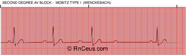
20 Second-Degree AV Block Mobitz I Progressive prolongation of the PR interval until there is failure to conduct and a ventricular beat is dropped AKA: Wenckebach block Usually not an indication for pacing

21 Second Degree AV Block Type 1 (Wenckebach) Increasing delay at AV node until a p wave is not conducted. Often comes post inferior MI with AV node ischemia Gradual prolongation of the PR interval before a skipped QRS. QRS are normal! No pacing as long as no bradycardia.
22 Second-Degree AV Block 2:1 block Regularly dropped ventricular beats A high grade block, Usually an indication for pacing May progress to third-degree, or Complete Heart block (CHB)

23 Second-degree AV block type II Sudden loss of a QRS wave because p wave was not transmitted beyond AV node.

24 2nd degree heart block (2:1)

25 Second Degree AV Block 3:1 block Sudden loss of a QRS wave because p wave was not transmitted beyond AV node. May be precursor to complete heart block and needs pacing.

26
 or narrow QRS if AV is pacemaker (proximal block)")
27 Third-Degree (Complete) AV Block No impulse conduction from the atria to the ventricles - atria and ventricles beat independently AND atria beat faster than ventricles: Complete A V disassociation Atrial rate is faster than Ventricular rate Usually a wide QRS as ventricular rate is idioventricular (distal block) or narrow QRS if AV is pacemaker (proximal block)
 heart")
28 Complete (3rd degree) heart block
29 AV Block Manifestations: First-degree AV block: almost no symptoms; Second degree AV block: palpitation, fatigue Third degree AV block: Dizziness, agina, heart failure, lightheadedness, and syncope may cause by slow heart rate, Adams-Stokes Syndrome may occurs in sever case. First heart sound varies in intensity, will appear booming first sound
30 AV Block Treatment: 1. I or II degree I type AV block needn t antibradycardia agent therapy 2. II degree II type and III degree AV block need antibradycardia agent therapy 3. Implant Pace Maker
31 Intraventricular Block Intraventricular conduction system: 1. Right bundle branch 2. Left bundle branch 3. Left anterior fascicular 4. Left posterior fascicular
32 Intraventricular Block Etiology: Myocarditis, valve disease, cardiomyopathy, CAD, hypertension, pulmonary heart disease, drug toxicity, Lenegre disease, Lev s disease et al. Manifestation: Single fascicular or bifascicular block is asymptom; tri-fascicular block may have dizziness; palpitation, syncope and Adams-stokes syndrome

33 Intraventricular Block

34
 in V1, and a slurred S-wave in V5 - V6. 3. The T wave should be deflected opposite the terminal deflection of the QRS complex.")
35 Right Bundle Branch Block (RBBB) Right ventricle gets a delayed impulse 1. Depolarization spreads from the left ventricle to the right ventricle. 2. This creates a second R-wave (R ) in V1, and a slurred S-wave in V5 - V6. 3. The T wave should be deflected opposite the terminal deflection of the QRS complex. This is known as appropriate T wave discordance with bundle branch block. A concordant T wave may suggest ischemia or myocardial infarction. 4. Pacemaker if syncope occurs. QRS is widened V1 and V2 have rsr

36 Left Bundle Branch Block (LBBB) Left ventricle gets a delayed impulse 1. Depolarization enters the right side of the right ventricle first and simultaneously depolarizes the septum from right to left. This creates a QS or rs complex in lead V1 and a monophasic or notched R wave in lead V6. 2. The T wave should be deflected opposite the terminal deflection of the QRS complex. This is known as appropriate T wave discordance with bundle branch block. A concordant T wave may suggest ischemia or myocardial infarction. QRS is widened V5 and V6 have RR (rabbit ears) 3. Pacemaker if syncope occurs
37 Therapy: Intraventricular Block 1. Treat underlying disease 2. If the patient is asymptom; no treat, 3. If progress to complete block, may need implant pace maker if the patient with syncope

38
39 Pacemaker Permanent- battery under skin Temporary- battery outside body Types Transvenous Epicardial- bypass surgery Transcutaneous- emergency Modes Asynchronous- at preset time without fail Synchronous or demand- when HR goes below set rate

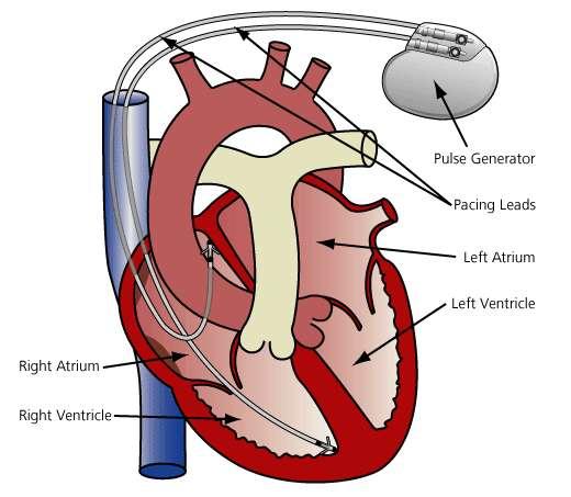
40 Pacemaker


41 Pacemaker Problems: Failure to sense Failure to capture
42 Sudden cardiac death(scd) Sudden cardiac death (SCD) is used to describe cardiac arrest with cessation of cardiac function, whether or not resuscitation or spontaneous reversion occurs Patients who do not die after cardiac arrest should be said to have experienced aborted SCD
43 Sudden cardiac death(scd) Definition by WHO Sudden collapse of cardiac function occurring within one hour of symptoms
44 Sudden cardiac death(scd) Pathophysiology The vast majority of cases of SCD are due to ventricular arrhythmias Ventricular tachycardia (VT) or ventricular fibrillation (VF) account for the majority of episodes This almost always occurs in the setting of underlying myocardial disease More than 80% of SCD events occur in individuals with coronary artery disease (CAD)
45 Sudden cardiac death(scd) Symptoms & signs Chest pain Dyspnea Fatigue Palpitations Syncope

46

47

48
49 SCD & ischemic heart disease Incidence of VT and VF after ST-elevation MI VF:4.2 % VT:3.5 % Both VF and VT:2.7 % 80 to 85 % of these arrhythmias occurred in the first 48 hours.
50 Hypertrophic cardiomyopathy Most common cause of SCD in young(age 35 y/o) SCD is primarily related to VT or VF. The mechanism of arrhythmia in this setting is not clear Autosomal-dominant inherited disease
51 Valvular disease Aortic stenosis (predominate) The mechanism of sudden death is unclear, and both malignant ventricular arrhythmia and bradyarrhythmia have been documented
52 Absence of structural heart disease Long QT syndrome Wolff-Parkinson-White (WPW) syndrome Commotio cordis
53 Long QT syndrome Prolonged QT interval Polymorphic ventricular tachycardia (VT) called torsade de pointes

54

55 QTc = QT interval square root of the RR interval (in msec)

56
57 Wolff Parkinson White (WPW) syndrome The type of pre-excitation syndrome: existence of an atrioventricular accessory pathway bundle of Kent) Atrial fibrillation (AF) with a rapid ventricular response was the most common VT/VF may occur

58 Wolff Parkinson White syndrome
59 Wolff Parkinson White (WPW) A supraventricular rhythm originating in the SA node with normal & regular P-waves PR interval is abnormally short (< 0.12 sec) QRS is wide with a slurred upstroke (AKA the delta-wave) Delta-waves are due to the accessory conduction pathway (bundle of Kent) from the atria to the ventricles, that bypasses the AV node Must manifest a tacchycardia at some point in time Treatment: III class, Procainamide, radiofrequency ablation

60 WPW syndrome
61 Commotio cordis Refers to SCD that most often occurs in young athletes who have been struck in the precordium with a projectile object such as a baseball, hockey puck, or fist The most common arrhythmia is VF
62 4 rhythms that produce pulseless arrest: pulseless ventricular tachycardia (VT) ventricular fibrillation (VF) asystole pulseless electrical activity (PEA)

63
64 Asystole Asystole is defined as a cardiac arrest rhythm in which there is no discernible electrical activity on the ECG monitor. Asystole is sometimes referred to as a flat line. Confirmation that a flat line is truly asystole is an important step. Ensure that asystole is not another rhythm that looks like a flat line. Fine VF can appear to be asystole, and a flat line on a monitor can be due to operator error or equipment failure The following are common causes of an isoelectric line that is not asystole: 1. loose or disconnected leads; 2. loss of power to the ECG monitor; 3. low signal gain on the ECG monitor. Asystole for many patients is the result of a prolonged illness or cardiac arrest, and prognosis is very poor. Few patients will likely have a positive outcome and successful treatment of cardiac arrest with asystole will usually involve identification and correction of an underlying cause of the asystole.

65 Agonal rhythm/asystole Ventricular rhythm Two ventricular complexes to none Ventricular rate None Atrial rhythm None Atrial rate None PRI: None QRS: 0.14 sec to none

66 Asystole Ventricular rhythm Ventricular rate Atrial rhythm Atrial rate PRI: QRS: None None None None None None

67 Rhythm Strip During Episode of Sudden Death

68
69 There are several important points that should be considered when initiating the pulseless arrest algorithm: High-quality CPR should be performed until the defibrillator is attached the patient. Interruptions in chest compressions should be kept to a minimum. Rapid use of the defibrillator should be emphasized. If possible, use a manual defibrillator over an AED since the use of the AED can result in prolonged interruptions in chest compressions for rhythm analysis and shock administration.


70
71 Defibrillation and the Shock Most defibrillators used today are biphasic. Biphasic means that the electrical current travels from one paddle to the other paddle and then back in the other direction. The biphasic shock also requires less energy to restore normal heart rhythm and is believed reduce skin burns and cellular damage to the heart. When using a biphasic defibrillator in VF and/or pulseless VT, you will use a dose of Joules to shock. Start with 120J and increase the dosing in a stepwise fashion up to 200 Joules as needed.

72 To ensure safety during the shock To ensure safety during the shock, providers should always announce the following statement, I am going to shock on three. One, I m clear Two, you re clear Three, everybody is clear.
73 Status Check Click for Answer What is the most likely rhythm disorder that might result in a patient getting a pacemaker? Sinus node disease What are some symptoms a patient might complain of? Fatigue, shortness of breath, palpitations, inability to perform activities of daily living, vertigo, syncope, racing heart at rest, slow pulse rate
74 Status Check What are some simple diagnostic tests used to make this diagnosis? 12-lead ECG, Ambulatory ECG (Holter) Click for Answer
75 Status Check Identify the Rhythm Ventricular Tachycardia Sinus Bradycardia Complete Heart Block Atrial Fibrillation Ventricular Fibrillation Click for Answer
76 Status Check Identify the Rhythm Ventricular Tachycardia Sinus Bradycardia Complete Heart Block Atrial Fibrillation Ventricular Fibrillation Click for Answer
77 Status Check Identify the Rhythm Ventricular Tachycardia Sinus Bradycardia Complete Heart Block Atrial Fibrillation Ventricular Fibrillation Click for Answer
78 Status Check Ventricular Tachycardia Sinus Bradycardia Complete Heart Block Atrial Fibrillation Ventricular Fibrillation Click for Answer
UNDERSTANDING YOUR ECG: A REVIEW
 UNDERSTANDING YOUR ECG: A REVIEW Health professionals use the electrocardiograph (ECG) rhythm strip to systematically analyse the cardiac rhythm. Before the systematic process of ECG analysis is described
Cardiac Implanted Electronic Devices Pacemakers, Defibrillators, Cardiac Resynchronization Devices, Loop Recorders, etc.
 Cardiac Implanted Electronic Devices Pacemakers, Defibrillators, Cardiac Resynchronization Devices, Loop Recorders, etc. The Miracle of Living February 21, 2018 Matthew Ostrom MD,FACC,FHRS Division of
REtrive. REpeat. RElearn Design by. Test-Enhanced Learning based ECG practice E-book
 Test-Enhanced Learning Test-Enhanced Learning Test-Enhanced Learning Test-Enhanced Learning based ECG practice E-book REtrive REpeat RElearn Design by S I T T I N U N T H A N G J U I P E E R I Y A W A
Conduction Problems / Arrhythmias. Conduction
 Conduction Problems / Arrhythmias Conduction Wolf-Parkinson White Syndrome (WPW) and Lown-Ganong-Levine (LGL): Atrial impulses bypass the AV node through an accessory pathway or bypass tract (bundle of
PATIENT WITH ARRHYTHMIA IN DENTIST S OFFICE. Małgorzata Kurpesa, MD., PhD. Chair&Department of Cardiology
 PATIENT WITH ARRHYTHMIA IN DENTIST S OFFICE Małgorzata Kurpesa, MD., PhD. Chair&Department of Cardiology Medical University of Łódź The heart is made up of four chambers Left Atrium Right Atrium Left Ventricle
-RHYTHM PRACTICE- By Dr.moanes Msc.cardiology Assistant Lecturer of Cardiology Al Azhar University. OBHG Education Subcommittee
 -RHYTHM PRACTICE- By Dr.moanes Msc.cardiology Assistant Lecturer of Cardiology Al Azhar University The Normal Conduction System Sinus Node Normal Sinus Rhythm (NSR) Sinus Bradycardia Sinus Tachycardia
Emergency Medical Training Services Emergency Medical Technician Paramedic Program Outlines Outline Topic: WPW Revised: 11/2013
 Emergency Medical Training Services Emergency Medical Technician Paramedic Program Outlines Outline Topic: WPW Revised: 11/2013 Wolff-Parkinson-White syndrome (WPW) is a syndrome of pre-excitation of the
Electrocardiography Abnormalities (Arrhythmias) 7. Faisal I. Mohammed, MD, PhD
 7. Faisal I. Mohammed, MD, PhD") Electrocardiography Abnormalities (Arrhythmias) 7 Faisal I. Mohammed, MD, PhD 1 Causes of Cardiac Arrythmias Abnormal rhythmicity of the pacemaker Shift of pacemaker from sinus node Blocks at different
Electrocardiography Abnormalities (Arrhythmias) 7 Faisal I. Mohammed, MD, PhD 1 Causes of Cardiac Arrythmias Abnormal rhythmicity of the pacemaker Shift of pacemaker from sinus node Blocks at different
SIMPLY ECGs. Dr William Dooley
 SIMPLY ECGs Dr William Dooley Content Basic ECG interpretation pattern Some common (examined) abnormalities Presenting ECGs in context Setting up an ECG Setting up an ECG 1 V1-4 th Right intercostal space
Arrhythmic Complications of MI. Teferi Mitiku, MD Assistant Clinical Professor of Medicine University of California Irvine
 Arrhythmic Complications of MI Teferi Mitiku, MD Assistant Clinical Professor of Medicine University of California Irvine Objectives Brief overview -Pathophysiology of Arrhythmia ECG review of typical
CRC 431 ECG Basics. Bill Pruitt, MBA, RRT, CPFT, AE-C
 CRC 431 ECG Basics Bill Pruitt, MBA, RRT, CPFT, AE-C Resources White s 5 th ed. Ch 6 Electrocardiography Einthoven s Triangle Chest leads and limb leads Egan s 10 th ed. Ch 17 Interpreting the Electrocardiogram
Sudden cardiac death: Primary and secondary prevention
 Sudden cardiac death: Primary and secondary prevention By Kai Chi Chan Penultimate Year Medical Student St George s University of London at UNic Sheba Medical Centre Definition Sudden cardiac arrest (SCA)
CORONARY ARTERIES. LAD Anterior wall of the left vent Lateral wall of left vent Anterior 2/3 of interventricluar septum R & L bundle branches
 CORONARY ARTERIES RCA Right atrium Right ventricle SA node 55% AV node 90% Posterior wall of left ventricle in 90% Posterior third of interventricular septum 90% LAD Anterior wall of the left vent Lateral
Paroxysmal Supraventricular Tachycardia PSVT.
 Atrial Tachycardia; is the name for an arrhythmia caused by a disorder of the impulse generation in the atrium or the AV node. An area in the atrium sends out rapid signals, which are faster than those
ECG ABNORMALITIES D R. T AM A R A AL Q U D AH
 ECG ABNORMALITIES D R. T AM A R A AL Q U D AH When we interpret an ECG we compare it instantaneously with the normal ECG and normal variants stored in our memory; these memories are stored visually in
Cardiology Flash Cards
 Cardiology Flash Cards EKG in a nut shell www.brain101.info Conduction System www.brain101.info 2 Analyzing EKG Step by step Steps in Analyzing ECG'S 1. Rhythm: - Regular _ Sinus, Junctional or Ventricular.
EKG Abnormalities. Adapted from:
 EKG Abnormalities Adapted from: http://www.bem.fi/book/19/19.htm Some key terms: Arrhythmia-an abnormal rhythm or sequence of events in the EKG Flutter-rapid depolarizations (and therefore contractions)
SIMPLY ECGs. Dr William Dooley
 SIMPLY ECGs Dr William Dooley 1 No anatomy just interpretation 2 Setting up an ECG 3 Setting up an ECG 1 V1-4 th Right intercostal space at sternal border 2 V2-4 th Left intercostal space at sternal border
Study methodology for screening candidates to athletes risk
 1. Periodical Evaluations: each 2 years. Study methodology for screening candidates to athletes risk 2. Personal history: Personal history of murmur in childhood; dizziness, syncope, palpitations, intolerance
Chapter 16: Arrhythmias and Conduction Disturbances
 Complete the following. Chapter 16: Arrhythmias and Conduction Disturbances 1. Cardiac arrhythmias result from abnormal impulse, abnormal impulse, or both mechanisms together. 2. is the ability of certain
ECG S: A CASE-BASED APPROACH December 6,
 ECG S: A CASE-BASED APPROACH December 6, 2018 1 Faculty Disclosure Faculty: Lorne Gula MD, FRCPC Professor, Western University Cardiologist, Hearth Rhythm Specialist Director, Electrophysiology Laboratory,
InterQual Care Planning SIM plus Criteria 2014 Clinical Revisions
 InterQual Care Planning SIM plus Criteria 2014 Clinical Revisions The Clinical Revisions provide details of changes to InterQual Clinical Criteria. They do not provide information on changes made to CareEnhance
ECG Interpretation Made Easy
 ECG Interpretation Made Easy Dr. A Tageldien Abdellah, MSc MD EBSC Lecturer of Cardiology- Hull University Hull York Medical School 2007-2008 ECG Interpretation Made Easy Synopsis Benefits Objectives Process
The ECG Course. Boone County Fire Protection District EMS Education
 The ECG Course Level I G rated material AV Blocks What Causes AV Block? Long list of bad things that includes ischemia and.. Old age / disease Medications or drugs Electrolyte imbalances Physiologic Blocks
Huseng Vefali MD St. Luke s University Health Network Department of Cardiology
 Huseng Vefali MD St. Luke s University Health Network Department of Cardiology Learning Objectives Establish Consistent Approach to Interpreting ECGs Review Essential Cases for Paramedics and first responders
Abnormalities Caused by Left Bundle Branch Block
 Marquette University e-publications@marquette Physician Assistant Studies Faculty Research and Publications Physician Assistant Studies, Department 12-17-2010 Abnormalities Caused by Left Bundle Branch
Arrhythmia Management Joshua M. Cooper, MD, FHRS, FACC
 Arrhythmia Management Joshua M. Cooper, MD, FHRS, FACC Professor of Medicine Director of Cardiac Electrophysiology Temple University Health System Plumbing Electrical System Bradyarrhythmias Sinus Node
Cardiac Arrhythmia How to approach นพ.พ น จ แกวส วรรณะ หน วยโรคห วใจและหลอดเล อด
 Cardiac Arrhythmia How to approach นพ.พ น จ แกวส วรรณะ หน วยโรคห วใจและหลอดเล อด EKG paper is a grid where time is measured along the horizontal axis. Each small square is 1 mm in length and represents
ECG Interpretation Cat Williams, DVM DACVIM (Cardiology)
") ECG Interpretation Cat Williams, DVM DACVIM (Cardiology) Providing the best quality care and service for the patient, the client, and the referring veterinarian. GOAL: Reduce Anxiety about ECGs Back to
ECG Interpretation Cat Williams, DVM DACVIM (Cardiology) Providing the best quality care and service for the patient, the client, and the referring veterinarian. GOAL: Reduce Anxiety about ECGs Back to
Chapter 9. Learning Objectives. Learning Objectives 9/11/2012. Cardiac Arrhythmias. Define electrical therapy
 Chapter 9 Cardiac Arrhythmias Learning Objectives Define electrical therapy Explain why electrical therapy is preferred initial therapy over drug administration for cardiac arrest and some arrhythmias
CSI Skills Lab #5: Arrhythmia Interpretation and Treatment
 CSI 202 - Skills Lab #5: Arrhythmia Interpretation and Treatment Origins of the ACLS Approach: CSI 202 - Skills Lab 5 Notes ACLS training originated in Nebraska in the early 1970 s. Its purpose was to
Step by step approach to EKG rhythm interpretation:
 Sinus Rhythms Normal sinus arrhythmia Small, slow variation of the R-R interval i.e. variation of the normal sinus heart rate with respiration, etc. Sinus Tachycardia Defined as sinus rhythm with a rate
a lecture series by SWESEMJR
 Arrhythmias Automaticity- the ability to depolarize spontaneously. Pacemakers: Sinoatrial node: 70 bpm AV-nodal area: 40 bpm His-Purkinje: 20-40 bpm it Mechanisms of arrhythmias 1. Increased automaticity
Northwest Community Healthcare Paramedic Education Program AV Conduction Defects/AV Blocks Connie J. Mattera, M.S., R.N., EMT-P
 Northwest Community Healthcare Paramedic Education Program AV Conduction Defects/ Connie J. Mattera, M.S., R.N., EMT-P Reading assignments: Bledsoe Vol. 3: pp. 88-93; 120-121 (atropine, norepinephrine,
Return to Basics. ECG Rate and Rhythm. Management of the Hospitalized Patient September 25, 2009
 Management of the Hospitalized Patient September 25, 2009 ECG Refresher and Update 2009 Return to Basics Determine rate and rhythm Determine intervals and axes Define morphology of P-QRS-T-U Compare with
12-Lead ECG Interpretation. Kathy Kuznar, RN, ANP
 12-Lead ECG Interpretation Kathy Kuznar, RN, ANP The 12-Lead ECG Objectives Identify the normal morphology and features of the 12- lead ECG. Perform systematic analysis of the 12-lead ECG. Recognize abnormalities
Please check your answers with correct statements in answer pages after the ECG cases.
 ECG Cases ECG Case 1 Springer International Publishing AG, part of Springer Nature 2018 S. Okutucu, A. Oto, Interpreting ECGs in Clinical Practice, In Clinical Practice, https://doi.org/10.1007/978-3-319-90557-0
2) Heart Arrhythmias 2 - Dr. Abdullah Sharif
 Heart Arrhythmias 2 - Dr. Abdullah Sharif") 2) Heart Arrhythmias 2 - Dr. Abdullah Sharif Rhythms from the Sinus Node Sinus Tachycardia: HR > 100 b/m Causes: o Withdrawal of vagal tone & Sympathetic stimulation (exercise, fight or flight) o Fever
2) Heart Arrhythmias 2 - Dr. Abdullah Sharif Rhythms from the Sinus Node Sinus Tachycardia: HR > 100 b/m Causes: o Withdrawal of vagal tone & Sympathetic stimulation (exercise, fight or flight) o Fever
ACLS Study Guide for Precourse Self-Assessment
 20 rhythm strips on Precourse Self-Assessment with the following matching choices: Agonal Rhythm/Asystole Atrial Fibrillation Flutter Ventricular Fibrillation Monomorphic Ventricular Normal Sinus Rhythm
The Electrocardiogram
 The Electrocardiogram Chapters 11 and 13 AUTUMN WEDAN AND NATASHA MCDOUGAL The Normal Electrocardiogram P-wave Generated when the atria depolarizes QRS-Complex Ventricles depolarizing before a contraction
Junctional Premature Contraction (JPC)
") Where s the PAC? Junctional Premature Contraction (JPC) A junctional premature contraction (JPC) is a beat that originates prematurely in the AV node. It can occur sporadically or in a grouped pattern.
Where s the PAC? Junctional Premature Contraction (JPC) A junctional premature contraction (JPC) is a beat that originates prematurely in the AV node. It can occur sporadically or in a grouped pattern.
1 Cardiology Acute Care Day 22 April 2013 Arrhythmia Tutorial Course Material
 1 Cardiology Acute Care Day 22 April 2013 Arrhythmia Tutorial Course Material Arrhythmia recognition This tutorial builds on the ECG lecture and provides a framework for approaching any ECG to allow the
Return to Basics. Normal Intervals & Axes. ECG Rate and Rhythm
 Return to Basics Management of the Hospitalized Patient October 15, 2010 ECG Refresher and Update 2010 Determine rate and rhythm Determine intervals and axes Define morphology of P-QRS-T-U Compare with
15 16 September Seminar W10O. ECG for General Practice
 15 16 September 2012 Seminar W10O ECG for General Practice Speaker: Ms Natasha Eaton ECG for General Practice Speaker: Natasha Eaton Cardiac CNC Executive Representative Electrocardiography The graphic
Pennsylvania Academy of Family Physicians Foundation & UPMC 43rd Refresher Course in Family Medicine CME Conference March 10-13, 2016
 Pennsylvania Academy of Family Physicians Foundation & UPMC 43rd Refresher Course in Family Medicine CME Conference March 10-13, 2016 Disclosures: EKG Workshop Louis Mancano, MD Speaker has no disclosures
Figure 2. Normal ECG tracing. Table 1.
 Figure 2. Normal ECG tracing that navigates through the left ventricle. Following these bundle branches the impulse finally passes to the terminal points called Purkinje fibers. These Purkinje fibers are
ABCs of ECGs. Shelby L. Durler
 ABCs of ECGs Shelby L. Durler Objectives Review the A&P of the cardiac conduction system Placement and obtaining 4-lead and 12-lead ECGs Overview of the basics of ECG rhythm interpretation Intrinsic
ECG CONVENTIONS AND INTERVALS
 1 ECG Waveforms and Intervals ECG waveforms labeled alphabetically P wave== represents atrial depolarization QRS complex=ventricular depolarization ST-T-U complex (ST segment, T wave, and U wave)== V repolarization.
ECG Cases and Questions. Ashish Sadhu, MD, FHRS, FACC Electrophysiology/Cardiology
 ECG Cases and Questions Ashish Sadhu, MD, FHRS, FACC Electrophysiology/Cardiology 32 yo female Life Insurance Physical 56 yo male with chest pain Terminology Injury ST elevation Ischemia T wave inversion
Indications for Permanent Pacing Joe Gallinghouse, M.D. Texas Cardiac Arrhythmia Austin, Texas
 Indications for Permanent Pacing Joe Gallinghouse, M.D. Texas Cardiac Arrhythmia Austin, Texas Remember the Suture! Impulse Formation and Conduction Disturbances Cardiac Electrical Anatomy Sinoatrial Node
Course Objectives. Proper Lead Placements. Review the ECG print paper. Review the mechanics of the Myocardium. Review basics of ECG Rhythms
 ECG Interpretations Course Objectives Proper Lead Placements Review the ECG print paper Review the mechanics of the Myocardium Review basics of ECG Rhythms How Leads Work The ECG Leads we use are Bipolar
2017 EKG Workshop Advanced. Family Medicine Review Course Lou Mancano, MD, FAAFP Reading Health System Family and Community Medicine Reading, PA
 2017 EKG Workshop Advanced Family Medicine Review Course Lou Mancano, MD, FAAFP Reading Health System Family and Community Medicine Reading, PA Part II - Objective Describe a useful approach to interpreting
ECG Basics Sonia Samtani 7/2017 UCI Resident Lecture Series
 ECG Basics Sonia Samtani 7/2017 UCI Resident Lecture Series Agenda I. Introduction II.The Conduction System III.ECG Basics IV.Cardiac Emergencies V.Summary The Conduction System Lead Placement avf Precordial
Return to Basics. ECG Rate and Rhythm. Management of the Hospitalized Patient October 4, 2007
 Management of the Hospitalized Patient October 4, 2007 ECG Refresher for the Hospitalists Return to Basics Determine rate and rhythm Determine intervals and axes Define morphology of P-QRS-T-U Compare
Anti arrhythmic drugs. Hilal Al Saffar College of medicine Baghdad University
 Anti arrhythmic drugs Hilal Al Saffar College of medicine Baghdad University Mechanism of Arrhythmia Abnormal heart pulse formation Abnormal heart pulse conduction Classification of Arrhythmia Abnormal
EHRA Accreditation Exam - Sample MCQs Cardiac Pacing and ICDs
 EHRA Accreditation Exam - Sample MCQs Cardiac Pacing and ICDs Dear EHRA Member, Dear Colleague, As you know, the EHRA Accreditation Process is becoming increasingly recognised as an important step for
Cardiac Pacing. Learning outcomes. Introduction. The cardiac impulse - its formation and its failure CHAPTER. To understand:
 Cardiac Pacing CHAPTER 10 Learning outcomes To understand: The indications for cardiac pacing in the peri-arrest setting How to perform percussion pacing How to apply non-invasive, transcutaneous electrical
Hatim Al Lawati. MD, FRCPC, DABIM(CV), FACC
, FACC") Hatim Al Lawati. MD, FRCPC, DABIM(CV), FACC Consultant Interventional Cardiology & Structural Heart Disease Department of Medicine Sultan Qaboos University Hospital hatim.al.lawati@gmail.com April 2017
Hatim Al Lawati. MD, FRCPC, DABIM(CV), FACC Consultant Interventional Cardiology & Structural Heart Disease Department of Medicine Sultan Qaboos University Hospital hatim.al.lawati@gmail.com April 2017
TEST BANK FOR ECGS MADE EASY 5TH EDITION BY AEHLERT
 Link download full: http://testbankair.com/download/test-bank-for-ecgs-made-easy-5thedition-by-aehlert/ TEST BANK FOR ECGS MADE EASY 5TH EDITION BY AEHLERT Chapter 5 TRUE/FALSE 1. The AV junction consists
ECGs and Arrhythmias: Family Medicine Board Review 2009
 Rate Rhythm Intervals Hypertrophy ECGs and Arrhythmias: Family Medicine Board Review 2009 Axis Jess (Fogler) Waldura, MD University of California, San Francisco walduraj@nccc.ucsf.edu Ischemia Overview
Electrical System Overview Electrocardiograms Action Potentials 12-Lead Positioning Values To Memorize Calculating Rates
 Electrocardiograms Electrical System Overview James Lamberg 2/ 74 Action Potentials 12-Lead Positioning 3/ 74 4/ 74 Values To Memorize Inherent Rates SA: 60 to 100 AV: 40 to 60 Ventricles: 20 to 40 Normal
Supplementary Online Content
 Supplementary Online Content Tseng ZH, Hayward RM, Clark NM, et al. Sudden death in patients with cardiac implantable electronic devices. JAMA Intern Med. Published online June 22, 2015. doi:10.1001/jamainternmed.2015.2641.
physiology 6 Mohammed Jaafer Turquoise team
 15 physiology 6 Mohammed Jaafer 22-3-2016 Turquoise team Cardiac Arrhythmias and Their Electrocardiographic Interpretation Today, we are going to talk about the abnormal excitation. As we said before,
Dr. Schroeder has no financial relationships to disclose
 Valerie A Schroeder MD MS Assistant Professor University of Kansas Medical Center READING THE WAVES- THE HEART S ELECTRICAL MESSAGE FINANCIAL DISCLOSURE Dr. Schroeder has no financial relationships to
Cardiac Telemetry Self Study: Part One Cardiovascular Review 2017 THINGS TO REMEMBER
 Please review the above anatomy of the heart. THINGS TO REMEMBER There are 3 electrolytes that affect cardiac function o Sodium, Potassium, and Calcium When any of these electrolytes are out of the normal
Electrocardiography for Healthcare Professionals
 Electrocardiography for Healthcare Professionals Kathryn A. Booth Thomas O Brien Chapter 10: Pacemaker Rhythms and Bundle Branch Block Learning Outcomes 10.1 Describe the various pacemaker rhythms. 10.2
Cardiovascular Nursing Practice: A Comprehensive Resource Manual and Study Guide for Clinical Nurses 2 nd Edition
 Cardiovascular Nursing Practice: A Comprehensive Resource Manual and Study Guide for Clinical Nurses 2 nd Edition Table of Contents Volume 1 Chapter 1: Cardiovascular Anatomy and Physiology Basic Cardiac
ECGs: Everything a finalist needs to know. Dr Amy Coulden As part of the Simply Finals series
 ECGs: Everything a finalist needs to know Dr Amy Coulden As part of the Simply Finals series Aims and objectives To be able to interpret basic ECG abnormalities To be able to recognise commonly tested
Family Medicine for English language students of Medical University of Lodz ECG. Jakub Dorożyński
 Family Medicine for English language students of Medical University of Lodz ECG Jakub Dorożyński Parts of an ECG The standard ECG has 12 leads: six of them are considered limb leads because they are placed
BEDSIDE ECG INTERPRETATION
 BEDSIDE ECG INTERPRETATION Presented by: Ryan Dean, RN, MSN, CCRN, CCNS, CFRN Flight Nurse 2017 Based on presentations originally by Gennifer DePaoli, RN Objectives Hospital policies Electrical conduction
Blocks & Dissociations. Reading Assignment (p47-52 in Outline )
") Blocks & Dissociations Reading Assignment (p47-52 in Outline ) Objectives Who are Wenckebach and Mobitz? Review SA and AV Blocks AV Dissociations: learning who s the boss and why 2 nd degree SA Block:
Blocks & Dissociations Reading Assignment (p47-52 in Outline ) Objectives Who are Wenckebach and Mobitz? Review SA and AV Blocks AV Dissociations: learning who s the boss and why 2 nd degree SA Block:
The Efficient and Smart Methods for Diagnosis of SVT 대구파티마병원순환기내과정병천
 The Efficient and Smart Methods for Diagnosis of SVT 대구파티마병원순환기내과정병천 Differentiation Supraventricular Origin from Ventricular Origin on ECG. QRS-Complex Width. 1. Narrow QRS-Complex Tachycardia (
Unit 6: Circulatory System. 6.2 Heart
 Unit 6: Circulatory System 6.2 Heart Functions of Circulatory System 1. The heart is the pump necessary to circulate blood to all parts of the body 2. Arteries, veins and capillaries are the structures
Nathan Cade, MD Brandon Fainstad, MD Andrew Prouse, MD
 Nathan Cade, MD Brandon Fainstad, MD Andrew Prouse, MD OBJECTIVES 1. Identify the basic electrophysiology of the four causes of wide complex tachycardia. 2. Develop a simple framework for acute management
Cardiac arrhythmias. Janusz Witowski. Department of Pathophysiology Poznan University of Medical Sciences. J. Witowski
 Cardiac arrhythmias Janusz Witowski Department of Pathophysiology Poznan University of Medical Sciences A 68-year old man presents to the emergency department late one evening complaining of increasing
HR: 50 bpm (Sinus) PR: 280 ms QRS: 120 ms QT: 490 ms Axis: -70. Sinus bradycardia with one ventricular escape (*)
 PR: 280 ms QRS: 120 ms QT: 490 ms Axis: -70. Sinus bradycardia with one ventricular escape (*)") 1? HR: 50 bpm (Sinus) PR: 280 ms QRS: 120 ms QT: 490 ms Axis: -70 1 Sinus P waves? 2 sinus cycles The pause (2 sinus cycles) suggests that the sinus fired (?) but did not conduct to the atria (i.e., missing
1? HR: 50 bpm (Sinus) PR: 280 ms QRS: 120 ms QT: 490 ms Axis: -70 1 Sinus P waves? 2 sinus cycles The pause (2 sinus cycles) suggests that the sinus fired (?) but did not conduct to the atria (i.e., missing
Temporary pacemaker 삼성서울병원 심장혈관센터심장검사실 박정왜 RN, CCDS
 Temporary pacemaker 삼성서울병원 심장혈관센터심장검사실 박정왜 RN, CCDS NBG Codes 1st Letter 2nd Letter 3rd Letter A V D Chamber(s) Paced = atrium = ventricle = dual (both atrium and ventricle) Chamber(s) Sensed A = atrium
EKG Competency for Agency
 EKG Competency for Agency Name: Date: Agency: 1. The upper chambers of the heart are known as the: a. Atria b. Ventricles c. Mitral Valve d. Aortic Valve 2. The lower chambers of the heart are known as
6/19/2018. Background Athlete s heart. Ultimate question. Applying the International Criteria for ECG
 Applying the International Criteria for ECG Interpretation in Athletes to a preparticipation screening program DAVE SIEBERT, MD, CAQSM ASSISTANT PROFESSOR DEPARTMENT OF FAMILY MEDICINE UNIVERSITY OF WASHINGTON
Introduction to ECG Gary Martin, M.D.
 Brief review of basic concepts Introduction to ECG Gary Martin, M.D. The electrical activity of the heart is caused by a sequence of rapid ionic movements across cell membranes resulting first in depolarization
This presentation will deal with the basics of ECG description as well as the physiological basics of
 Snímka 1 Electrocardiography basics This presentation will deal with the basics of ECG description as well as the physiological basics of Snímka 2 Lecture overview 1. Cardiac conduction system functional
Understanding the 12-lead ECG, part II
 Bundle-branch blocks Understanding the 12-lead ECG, part II Most common electrocardiogram (ECG) abnormality Appears as a wider than normal S complex Occurs when one of the two bundle branches can t conduct
Review Packet EKG Competency This packet is a review of the information you will need to know for the proctored EKG competency test.
 Review Packet EKG Competency 2015 This packet is a review of the information you will need to know for the proctored EKG competency test. Normal Sinus Rhythm Rhythm: Regular Ventricular Rate: 60-100 bpm
ECG Interpretation. Introduction to Cardiac Telemetry. Michael Peters, RN, CCRN, CFRN CALSTAR Air Medical Services
 ECG Interpretation Introduction to Cardiac Telemetry Michael Peters, RN, CCRN, CFRN CALSTAR Air Medical Services Disclosures Nothing to disclose Objectives Describe the electrical conduction pathway in
Appendix D Output Code and Interpretation of Analysis
 Appendix D Output Code and Interpretation of Analysis 8 Arrhythmia Code No. Description 8002 Marked rhythm irregularity 8110 Sinus rhythm 8102 Sinus arrhythmia 8108 Marked sinus arrhythmia 8120 Sinus tachycardia
Arrhythmias. Pulmonary Artery
 Arrhythmias Introduction Cardiac arrhythmia is an irregularity of the heart beat that causes the heart to beat too slowly, too fast, or irregularly. There are different types of arrhythmias. Most arrhythmias
Bradydysrhythmias and Atrioventricular Conduction Blocks
 Emerg Med Clin N Am 24 (2006) 1 9 Bradydysrhythmias and Atrioventricular Conduction Blocks Jacob W. Ufberg, MD*, Jennifer S. Clark, MD Department of Emergency Medicine, Temple University School of Medicine,
ECG interpretation basics
 ECG interpretation basics Michał Walczewski, MD Krzysztof Ozierański, MD 21.03.18 Electrical conduction system of the heart Limb leads Precordial leads 21.03.18 Precordial leads Precordial leads 21.03.18
Ass. Prof. Tomon Thongsri, MD Buddhachinaraj Phitsanuloke Hospital
 Treatment Bradyarrhythmia Ass. Prof. Tomon Thongsri, MD Buddhachinaraj Phitsanuloke Hospital 1 What is the rhythm? Sinus Bradycardia What s rhythm Sinus Bradycardia Treatment Asymptomatic No treatment
If the P wave > 0.12 sec( 3 mm) usually in any lead. Notched P wave usually in lead I,aVl may be lead II Negative terminal portion of P wave in V1, 1
 usually in any lead. Notched P wave usually in lead I,aVl may be lead II Negative terminal portion of P wave in V1, 1") If the P wave > 0.12 sec( 3 mm) usually in any lead. Notched P wave usually in lead I,aVl may be lead II Negative terminal portion of P wave in V1, 1 mm depth and 3 mm width( most specific) Since Mitral
If the P wave > 0.12 sec( 3 mm) usually in any lead. Notched P wave usually in lead I,aVl may be lead II Negative terminal portion of P wave in V1, 1 mm depth and 3 mm width( most specific) Since Mitral
DR QAZI IMTIAZ RASOOL OBJECTIVES
 PRACTICAL ELECTROCARDIOGRAPHY DR QAZI IMTIAZ RASOOL OBJECTIVES Recording of electrical events in heart Established electrode pattern results in specific tracing pattern Health of heart i. e. Anatomical
Dr.Binoy Skaria 13/07/15
 Dr.Binoy Skaria binoyskaria@hotmail.com binoy.skaria@heartofengland.nhs.uk 13/07/15 Acknowledgement Medtronic, Google images & Elsevier for slides Natalie Ryan, Events Manager, HEFT- for organising the
December 2018 Tracings
 Tracings Tracing 1 Tracing 4 Tracing 1 Answer Tracing 4 Answer Tracing 2 Tracing 5 Tracing 2 Answer Tracing 5 Answer Tracing 3 Tracing 6 Tracing 3 Answer Tracing 6 Answer Questions? Contact Dr. Nelson
Basic electrocardiography reading. R3 lee wei-chieh
 Basic electrocardiography reading R3 lee wei-chieh The Normal Conduction System Lead Placement avf Limb Leads Precordial Leads Interpretation Rate Rhythm Interval Axis Chamber abnormality QRST change What
Chapter 20 (2) The Heart
 The Heart") Chapter 20 (2) The Heart ----------------------------------------------------------------------------------------------------------------------------------------- Describe the component and function of
Chapter 20 (2) The Heart ----------------------------------------------------------------------------------------------------------------------------------------- Describe the component and function of
Arrhythmia Study Guide 3 Junctional and Ventricular Rhythms
 Arrhythmia Study Guide 3 Junctional and Ventricular Rhythms JUNCTIONAL RHYTHMS The AV Junction (Bundle of His and surrounding cells) only acts as pacemaker of the heart when the SA Node is not firing normally
1 st Degree Block Prolonged P-R interval caused by first degree heart block (lead II)
") AV Heart Blocks 1 st degree A condition of a rhythm, not a true rhythm Need to always state underlying rhythm 2 nd degree Type I - Wenckebach Type II Classic dangerous to the patient Can be variable (periodic)
AV Heart Blocks 1 st degree A condition of a rhythm, not a true rhythm Need to always state underlying rhythm 2 nd degree Type I - Wenckebach Type II Classic dangerous to the patient Can be variable (periodic)
HTEC 91. Performing ECGs: Procedure. Normal Sinus Rhythm (NSR) Topic for Today: Sinus Rhythms. Characteristics of NSR. Conduction Pathway
 Topic for Today: Sinus Rhythms. Characteristics of NSR. Conduction Pathway") HTEC 91 Medical Office Diagnostic Tests Week 3 Performing ECGs: Procedure o ECG protocol: you may NOT do ECG if you have not signed up! If you are signed up and the room is occupied with people who did
HTEC 91 Medical Office Diagnostic Tests Week 3 Performing ECGs: Procedure o ECG protocol: you may NOT do ECG if you have not signed up! If you are signed up and the room is occupied with people who did
Conduction System of the Heart 4. Faisal I. Mohammed, MD, PhD
 Conduction System of the Heart 4 Faisal I. Mohammed, MD, PhD 1 Objectives List the parts that comprise the conduction system Explain the mechanism of slow response action potential (pacemaker potential)
Paediatric ECG Interpretation
 Paediatric ECG Interpretation Dr Sanj Fernando (thanks to http://lifeinthefastlane.com/ecg-library/paediatric-ecginterpretation/) 3 yo boy complaining of abdominal pain and chest pain Child ECG vs Adult