TAMPONADE CARDIAQUE. Dr Cédrick Zaouter TUSAR 15 décembre 2015
|
|
|
- Catherine Adams
- 5 years ago
- Views:
Transcription


1 TAMPONADE CARDIAQUE Dr Cédrick Zaouter TUSAR 15 décembre 2015
2 OUTLINE History Incidence Definition Pathophysiology Aetiologies Investigations - Echocardiography Treatment of cardiac tamponade
3 Pericardial effusion & Cardiac Tamponade Claudius Galen ( AD) first description Richard Lowe (1669): Physiology Edmund Rose: cardiac tamponade
4 Incidence of postoperative pericardial effusions The reported incidence of postoperative pericardial effusions ranges between 1% and 77% References: 1.Tsang TS, Barnes ME, Hayes SN, et al.. Chest 1999;116: Ikäheimo MJ, Huikuri HV, Airaksinen KE, et al.. Am Heart J 1988;116(1 pt 1): Pepi M, Muratori M, Barbier P, et al. Br Heart J 1994;72: Malouf JF, Alam S, Gharzeddine W, Stefadouros MA.. Eur Heart J 1993;14: Cheung EW, Ho SA, Tang KK, Chau AK, Chiu CS, Cheung YF. Heart 2003;89: Kuvin JT, Harati NA, Pandian NG, Bojar RM, Khabbaz KR. Ann Thorac Surg 2002;74:
5 Study design Incidence of postop pericardial effusions: 1% and 77% Small prospective studies: higher incidences reported > frequent echocardiographic examinations most effusions were clinically insignificant 77% Retrospective studies of clinically important pericardial effusions the reported incidence is 1% to 2% References: 1.Tsang TS, Barnes ME, Hayes SN, et al.. Chest 1999;116: Ikäheimo MJ, Huikuri HV, Airaksinen KE, et al.. Am Heart J 1988;116(1 pt 1): Pepi M, Muratori M, Barbier P, et al. Br Heart J 1994;72: Malouf JF, Alam S, Gharzeddine W, Stefadouros MA.. Eur Heart J 1993;14: Cheung EW, Ho SA, Tang KK, Chau AK, Chiu CS, Cheung YF. Heart 2003;89: Kuvin JT, Harati NA, Pandian NG, Bojar RM, Khabbaz KR. Ann Thorac Surg 2002;74:
6 Definition Fluid in the pericardial sac, creating an increased pressure within the pericardial space that impairs the ability of the heart to fill and to pump Abnormally large volumes of fluid within the pericardial sac does not always necessitate the development of tamponade


7 ANATOMY OF THE PERICARDIUM Fusing where the great vessels exit 1. Mesothelium cells volume of ml 2. Buffer the heart from external impact 3. Reduce resistance during motion 4. Barrier against infection
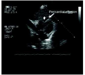
8 Pericardial effusion does not mean cardiac tamponade 50 ml 2000 ml Volume exceeds limit of parietal pericardial stretch Pericardial reserve volume Pericardial effusion: anatomical diagnosis Cardiac tamponade: physiological diagnosis

9 In the absence of other pathology pericardiocentesis will normalize pressures and improve the cardiac output, confirming the diagnosis

10 Pericardium compliance reduced TUMOR
11 Pericardium compliance reduced NATURE OF THE FLUID Acute effusions > transudates blood. Chronic effusions: > fibrosed and more viscous
12 Aetiologies The most common diagnoses were: 1- Acute idiopathic pericarditis (20%) 2- Iatrogenic effusion(16%) 3- Malignancy (13%), 4- Chronic idiopathic effusion (9%), 5- Acute myocardial infarction (8%), 6- End-stage renal disease (6%) 7- Congestive heart failure (5%), 8- Collagen vascular disease (5%) 9- Tuberculosis & Bacterial infection (4%). Effusions are extremely common after cardiac surgery BUT large effusions causing tamponade are rare in this setting LeRoy C. JAMA, April 25, 2007 Vol 297, No. 16

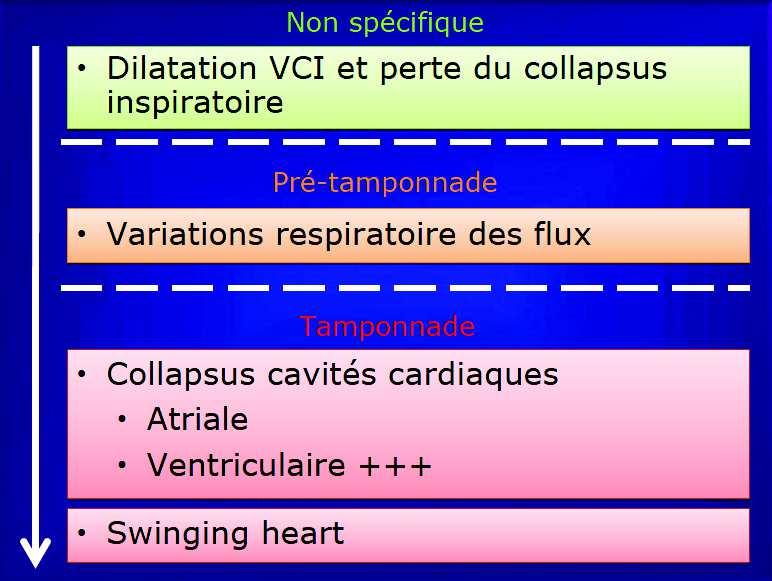
13 Pathophysiology Stages of cardiac tamponade Early stages R side < L side Reduced venous return R sides are compressed CVP rises Impaired RV filling Under-loaded Ventricle Low contractility Low SV

14 Pathophysiology Stages of cardiac tamponade Late stages Pericardial pressure > L side Reduced CO Obstructive shock Cardiac arrest Increased fluid retention


15 Suspicion: Clinical Features Dyspnea Tachycardia Hypotension poor discriminating Beck s triad EJVP Reduced heart sounds poor discriminating Hypotension Pulsus paradoxus > 10mmHg Ventricular interdependence
16 Suspicion: Clinical Features PAST MEDICAL HISTORY PM lead insertion Chest pain infarction/ pericarditis Renal failure > Ureamic P.Eff Tubercolosis > Inflammatory P.Eff Symptoms of pericardial effusion are nonspecific Few patients have the classic presentation of tamponade Many patients with pericardial effusion report minimal problems


17 Investigations Simple investigations ECG - Variation of QRS: electrical alternans CXR - Cardiomegaly
18 Investigations Simple investigations ECG The pooled sensitivity of low QRS voltage was only 42% CI: 32%-53% 2 studies reported that electrical alternans, sensitivity of 16% to 21%. Atrial arrhythmias are infrequent in cardiac tamponade ATOTW 283 Cardiac Tamponade March 2013 P. Odor A. Bailey; St. George s Hospital, London, UK
19 Investigations Advanced investigation: Echocardiography TTE demonstrates tamponade by RA collapse sensitivity of % and specificity %. TEE better views of the posterior aspects of the pericardium > essential: retroatrial ATOTW 283 Cardiac Tamponade March 2013 P. Odor A. Bailey; St. George s Hospital, London, UK

20 Quantification of Pericardial Effusions by Echocardiography and Computed Tomography Echocardiographically R2: 0,74 Small underestimation CT R2 : ml David Leibowitz et al Amer J of Cardiol 2011; 107:331
21 Investigations Echocardiography TTE The European Society of Cardiology RECOMMENDS A) 2D - Imaging: A. Collapse B) DOPPLER B. Septal-shift C. IVC dilatation
22 Investigations Echocardiography TTE The European Society of Cardiology RECOMMENDS A) 2D - Imaging: A. Collapse of 1- RA in systole 1-1 Partial: pretamponade= early stage Apical 4-chambers / Subcostal 1-2 Complete: Atrial transmural pressure is negligible = tamponade Apical 4-chambers / Subcostal 2- Anterior RV free wall in diastole Subcostal view / parasternal short axis view

23 Partial RA collapse: pretamponade = early stage

24 Partial RA collapse: pretamponade = early stage
25

26 Cardiac tamponade
27 Investigations Echocardiography TTE The European Society of Cardiology RECOMMENDS 2D - Imaging: A. Collapse of RA, RV, LA & LV Apical 4-chambers / Subcostal B. Septal shift-ventricular interdependency Apical 4-chambers view

28 Pulsus paradoxus Ventricular interdependence
29
30 Investigations Echocardiography TTE The European Society of Cardiology RECOMMENDS 2D - Imaging: A. Collapse of RA, RV, LA & LV Apical 4-chambers / Subcostal B. Septal shift-ventricular interdependency Apical four-chambers view C. IVC dilatation, without respiratory variation Subcostal view
31
32
33 Echocardiography TTE Investigations The European Society of Cardiology RECOMMENDS 2-Pulse wave Doppler A. An inspiratory rise of the right flows of more than 40 50% B. An inspiratory decrease of the left flows above 25 40% C. An inverted E/A ratio at the tricuspid and the mitral level > relaxation impairment of both ventricles The intrathoracic pressure regimen is inverted during mechanical ventilation and study of flows using Doppler is totally inaccurate.


34 Tricuspid flow Rise of the right flows of more than 40 50%

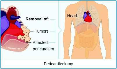
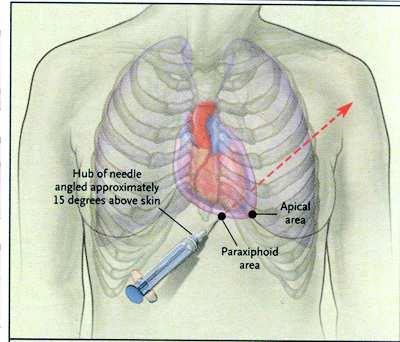
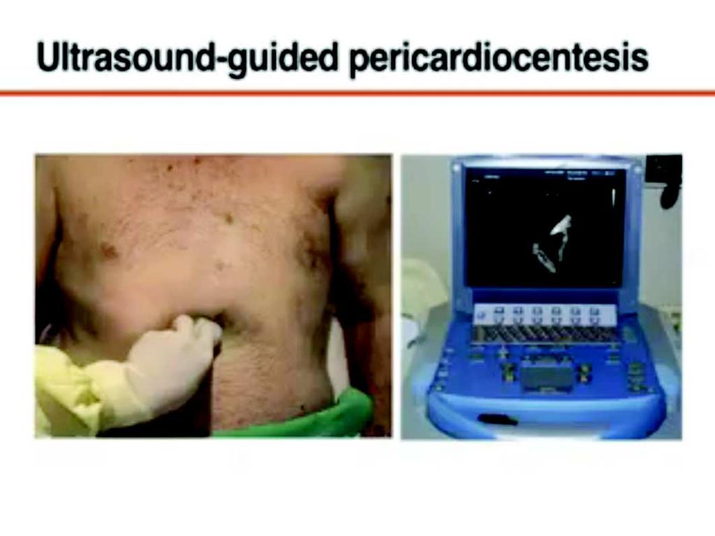
35 Mitral flow Reduction of the left flows above than 25-40%
36

37 Pattern of diastole in the sub hepatic veins
38

39 Differential diagnosis Pleural effusion

40 Differential diagnosis Pleural effusion
41 Pitfalls Hypovolemia RVH Wrapped Epicardial effusion Especially after cardiac surgery
42 Treatment Large pericardial effusions require drainage in most cases Open drainage or pericardiocentesis Location and character of the effusion Physician preference and experience Echocardiography-guided pericardiocentesis is safe. 1. Tsang TS, Barnes ME, Hayes SN, et al.. Chest 1999;116: Tsang TS, Enriquez-Sarano M, Freeman WK, et al. Mayo Clin Proc 2002;77: Susini G, Pepi M, Sisillo E, et al.. J Cardio- thorac Vasc Anesth 1993;7:
43
44
45 Merci
Pericardial Diseases. Smonporn Boonyaratavej, MD. Division of Cardiology, Department of Medicine Chulalongkorn University
 Pericardial Diseases Smonporn Boonyaratavej, MD Division of Cardiology, Department of Medicine Chulalongkorn University Cardiac Center, King Chulalongkorn Memorial Hospital 21 AUGUST 2016 Pericardial
THE PERICARDIUM: LOOKING OUTSIDE THE HEART
 THE PERICARDIUM: LOOKING OUTSIDE THE HEART DISCLOSURE Relevant relationships with commercial entities none Potential for conflicts of interest within this presentation none Steps taken to review and mitigate
Adel Hasanin Ahmed 1
 Adel Hasanin Ahmed 1 PERICARDIAL DISEASE The pericardial effusion ends anteriorly to the descending aorta and is best visualised in the PLAX. PSAX is actually very useful sometimes for looking at posterior
The role of bedside ultrasound in the diagnosis of pericardial effusion and cardiac tamponade
 Symposium The role of bedside ultrasound in the diagnosis of pericardial effusion and cardiac tamponade Adam Goodman, Phillips Perera, Thomas Mailhot, Diku Mandavia Department of Emergency Medicine, Los
Outline. Echocardiographic Assessment of Pericardial Effusion/Tamponade: The Essentials
 Echocardiographic Assessment of Pericardial Effusion/Tamponade: The Essentials John R Schairer DO FACC Henry Ford Heart and Vascular Institute No Disclosures Outline Normal Anatomy and Physiology Pathophysiology
Echocardiography as a diagnostic and management tool in medical emergencies
 Echocardiography as a diagnostic and management tool in medical emergencies Frank van der Heusen MD Department of Anesthesia and perioperative Care UCSF Medical Center Objective of this presentation Indications
PERICARDIAL DIAESE. Kaijun Cui Associated professor Sichuan University
 PERICARDIAL DIAESE Kaijun Cui Associated professor Sichuan University CLASSIFICATION acute pericarditis pericardial effusion cardiac tamponade constrictive pericarditis congenitally absent pericardium
We are now going to review the diagnosis and management of pericardial collections and tamponade
 We are now going to review the diagnosis and management of pericardial collections and tamponade FEEL COURSE PAGE 1 Paying particular attention to the difference between a collection and cardiac tamponade
Index. K Knobology, TTE artifact, image resolution, ultrasound, 14
 A Acute aortic regurgitation (AR), 124 128 Acute aortic syndrome (AAS) classic aortic dissection diagnosis, 251 263 evolutive patterns, 253 255 pathology, 250 251 classifications, 247 248 incomplete aortic
Department of Cardiac, Thoracic and Vascular Sciences University of Padua Cardiac Tamponade. Echocardiography in Diagnosis and Management
 Department of Cardiac, Thoracic and Vascular Sciences University of Padua Cardiac Tamponade. Echocardiography in Diagnosis and Management Luigi P. Badano, MD, FESC, FACC Declaration of interest **Dr. Badano
Constrictive Pericarditis Pitfalls in MR Diagnosis Cylen Javidan-Nejad Associate Professor Mallinckrodt Institute of Radiology Washington University
 Constrictive Pericarditis Pitfalls in MR Diagnosis Cylen Javidan-Nejad Associate Professor Mallinckrodt Institute of Radiology Washington University in St. Louis Goal o To review the imaging criteria of
Pericardial Disease: Case Examples. Echo Fiesta 2017
 Pericardial Disease: Case Examples Echo Fiesta 2017 2014 2014 MFMER MFMER 3346252-1 slide-1 Objectives Have a systematic approach to evaluation of constriction 2014 MFMER 3346252-2 CASE 1 2013 MFMER 3248567-3
Disclosures. Cardiac Ultrasound. Introductory Case. 80 y/o male Syncope at home Emesis x 3 in ambulance Looks sick. No pain.
 Disclosures Cardiac Ultrasound Justin A Davis, MD MPH RDMS Subchief for Emergency Ultrasound Kaiser Permanente East Bay Medical Center I have nothing to disclose. Introductory Case HR 118 BP 65/43 RR 27
Transthoracic Echocardiography:
 Transthoracic Echocardiography: An essential tool for the obstetric anaesthetist? Brendan Carvalho MBBCh, FRCA Department of Anesthesiology Stanford University, California Focused TTE Stethoscope of the
The right heart: the Cinderella of heart failure
 The right heart: the Cinderella of heart failure Piotr Ponikowski, MD, PhD, FESC Medical University, Centre for Heart Disease Clinical Military Hospital Wroclaw, Poland none Disclosure Look into the Heart
Echocardiography Conference
 Echocardiography Conference David Stultz, MD Cardiology Fellow, PGY-6 September 20, 2005 Atrial Septal Aneurysm Bulging of Fossa Ovalis Associated commonly with Atrial septal defect or small perforations
ΚΑΡΔΙΟΛΟΓΟΣ EUROPEAN ACCREDITATION IN TRANSTHORACIC AND TRANSESOPHAGEAL ECHOCARDIOGRAPHY
 1 ΚΑΡΔΙΟΛΟΓΟΣ EUROPEAN ACCREDITATION IN TRANSTHORACIC AND TRANSESOPHAGEAL ECHOCARDIOGRAPHY 2 Constrictive pericarditis (CP) is characterized by impaired ventricular filling due to a stiffened or noncompliant
Pericardial Effusion After Cardiac Surgery: Risk Factors, Patient Profiles, and Contemporary Management
 Pericardial Effusion After Cardiac Surgery: Risk Factors, Patient Profiles, and Contemporary Management Elena A. Ashikhmina, MD, Hartzell V. Schaff, MD, Lawrence J. Sinak, MD, Zhuo Li, MS, Joseph A. Dearani,
Essentials of Pericardial Diseases
 Essentials of Pericardial Diseases 1 Nikolaos Skubas MD, 2 Manuel Fontes MD The pericardial diseases result in cardiovascular perturbations ranging from asymptomatic electrocardiographic findings (in pericarditis
Normal Pericardial Physiology
 Normal Pericardial Physiology Normal pericardium contains 20-30 ml of lymphoid fluid lubricating function that facilitates normal myocardial rotation and translation during each cardiac cycle in that the
Pericardial disease. Se-Jung Yoon Cardiology division NHIS Ilsan hospital
 Pericardial disease Se-Jung Yoon Cardiology division NHIS Ilsan hospital Normal pericardial effusion Normal pericardium Normal pericardium Pericardial Layers: Visceral layer Parietal layer Fibrous pericardium
Pericardial effusion, Cardiac Tamponade, and echo guided pericardiocentesis
 KSC 2017 Echo5- Myocardial and Pericardial disease Pericardial effusion, Cardiac Tamponade, and echo guided pericardiocentesis Ji-Hyun Jung Division of Cardiology Sejong Hospital KSC 2017 The 61 th Annual
Adult Echocardiography Examination Content Outline
 Adult Echocardiography Examination Content Outline (Outline Summary) # Domain Subdomain Percentage 1 2 3 4 5 Anatomy and Physiology Pathology Clinical Care and Safety Measurement Techniques, Maneuvers,
Palpable Pulsus Paradoxus in the Setting of Malignant Pericardial Effusion and Tamponade Akshay Pendyal, MD
 Palpable Pulsus Paradoxus in the Setting of Malignant Pericardial Effusion and Tamponade Akshay Pendyal, MD University of Colorado Department of Internal Medicine None Conflicts of Interest Objectives
Certificate in Clinician Performed Ultrasound (CCPU) Syllabus. Rapid Cardiac Echo (RCE)
 Syllabus. Rapid Cardiac Echo (RCE)") Certificate in Clinician Performed Ultrasound (CCPU) Syllabus Rapid Cardiac Echo (RCE) Purpose: Rapid Cardiac Echocardiography (RCE) This unit is designed to cover the theoretical and practical curriculum
Certificate in Clinician Performed Ultrasound (CCPU) Syllabus Rapid Cardiac Echo (RCE) Purpose: Rapid Cardiac Echocardiography (RCE) This unit is designed to cover the theoretical and practical curriculum
Πνευμονική υπέρταση και περικαρδιακή συλλογή. Τρόποι αντιμετώπισης
 Πνευμονική υπέρταση και περικαρδιακή συλλογή. Τρόποι αντιμετώπισης Γεώργιος Λάζαρος Καρδιολόγος, Διευθυντής ΕΣΥ Α Πανεπιστημιακή Καρδιολογική Κλινική Ιπποκράτειο Γ.Ν. Αθηνών Pericardial syndromes o Acute
Index. Note: Page numbers of article titles are in boldface type.
 Index Note: Page numbers of article titles are in boldface type. A Acute coronary syndrome(s), anticoagulant therapy in, 706, 707 antiplatelet therapy in, 702 ß-blockers in, 703 cardiac biomarkers in,
Pericardial diseases
 Pericardial diseases Anatomy of the pericardium Consists of parietal and visceral membranes. The space between them(pericardial space is normally filled by a lymph like fluid. The fluid s normal quantity
Cath Lab Essentials: Pericardial effusion & tamponade
 Cath Lab Essentials: Pericardial effusion & tamponade Pranav M. Patel, MD, FACC, FSCAI Chief, Division of Cardiology Director, Cardiac Cath Lab & CCU University of California, Irvine Division of Cardiology
Lab 16. The Cardiovascular System Heart and Blood Vessels. Laboratory Objectives
 Lab 16 The Cardiovascular System Heart and Blood Vessels Laboratory Objectives Describe the anatomical structures of the heart to include the pericardium, chambers, valves, and major vessels. Describe
Copyright 2017 American College of Emergency Physicians. All rights reserved.
 POLICY Approved April 2017 Guidelines for the Use of Transesophageal Echocardiography (TEE) in the ED for Cardiac Arrest Approved by the ACEP Board of Directors April 2017 1. Introduction The American
Constrictive/Restrictive Cardiomyopathies: Diagnosis and Management Update; Radiation Induced Heart Disease. Alexander (Sandy) Dick, MD
 Dick, MD") Constrictive/Restrictive Cardiomyopathies: Diagnosis and Management Update; Radiation Induced Heart Disease Alexander (Sandy) Dick, MD Outline Pericardial Constriction Diagnosis: Imaging, Hemodynamics
Constrictive/Restrictive Cardiomyopathies: Diagnosis and Management Update; Radiation Induced Heart Disease Alexander (Sandy) Dick, MD Outline Pericardial Constriction Diagnosis: Imaging, Hemodynamics
Das recht Ventrikel ist auch noch da! RV function The RV operates as. Physiology Not very sensitive to preload Good compliance of the free wall
 Das recht Ventrikel ist auch noch da! I.Michaux Intensive Care Medicine University Hospital CHU UCL Namur Mont-Godinne Belgium RV function The RV operates as a low pressure, volume pump, moving the blood
The Doppler Examination. Katie Twomley, MD Wake Forest Baptist Health - Lexington
 The Doppler Examination Katie Twomley, MD Wake Forest Baptist Health - Lexington OUTLINE Principles/Physics Use in valvular assessment Aortic stenosis (continuity equation) Aortic regurgitation (pressure
Heart Failure. Cardiac Anatomy. Functions of the Heart. Cardiac Cycle/Hemodynamics. Determinants of Cardiac Output. Cardiac Output
 Cardiac Anatomy Heart Failure Professor Qing ZHANG Department of Cardiology, West China Hospital www.blaufuss.org Cardiac Cycle/Hemodynamics Functions of the Heart Essential functions of the heart to cover
Choose the grading of diastolic function in 82 yo woman
 Question #1 Choose the grading of diastolic function in 82 yo woman E= 80 cm/s A= 70 cm/s LAVI < 34 ml/m 2 1= Grade 1 2= Grade 2 3= Grade 3 4= Normal 5= Indeterminate 2018 MFMER 3712003-1 Choose the grading
Hypotension in the intensive care unit
 IM BOARD REVIEW CME CREDIT AHMED AL-HAZZOURI, MD Department of Internal Medicine, Cleveland Clinic JAMES K. STOLLER, MD, EDITOR PETER MAZZONE, MD, MPH Department of Pulmonary, Allergy, and Critical Care
A Case of Impending Cardiac Tamponade Caused by Effusive Constrictive Pericarditis
 Archives of Clinical and Medical Case Reports doi: 10.26502/acmcr.96550038 Volume 2, Issue 5 Case Report A Case of Impending Cardiac Tamponade Caused by Effusive Constrictive Pericarditis Catalina Sanchez-Alvarez
A classic case of amyloid cardiomyopathy
 Images in... A classic case of amyloid cardiomyopathy Hayan Jouni, 1 William G Morice, 2 S Vincent Rajkumar, 3 Joerg Herrmann 4 1 Department of Internal Medicine, Mayo Clinic, Rochester, Minnesota, USA
Integrative Clinical Hemodyamics
 Integrative Clinical Hemodyamics James A. Goldstein, MD Director, Research & Education Division of Cardiology William Beaumont Hospital Disclosure Information Integrative Clinical Hemodyamics James A.
Pericardial Diseases/Tamponade Illustrative Cases
 Pericardial Diseases/Tamponade Illustrative Cases Jae K. Oh, MD Echo Hawaii 2017 2012 MFMER 3200268v3(2010)-1 Case #1 47 year old man Chest pain Not exertional Normal Examination 2016 MFMER slide-2 1 47
Evaluation of the Right Ventricle in Candidates for Right Ventricular Assist Device Implantation.
 Evaluation of the Right Ventricle in Candidates for Right Ventricular Assist Device Implantation. Evaluation of RVAD Function. Ioannis A Paraskevaidis Attikon University Hospital Historical Perspective
Constrictive Pericarditis
 Constrictive Pericarditis Never Confused with Anything Else Jae K. Oh, MD 2018 MFMER 3712003-1 ARS #1 CP Which of following patients has constrictive pericarditis? 1 2 3 Medial e 13 cm/s Medial e 3 cm/s
Rotation: Echocardiography: Transthoracic Echocardiography (TTE)
") Rotation: Echocardiography: Transthoracic Echocardiography (TTE) Rotation Format and Responsibilities: Fellows rotate in the echocardiography laboratory in each clinical year. Rotations during the first
Rotation: Echocardiography: Transthoracic Echocardiography (TTE) Rotation Format and Responsibilities: Fellows rotate in the echocardiography laboratory in each clinical year. Rotations during the first
ASCeXAM / ReASCE. Practice Board Exam Questions Monday Morning
 ASCeXAM / ReASCE Practice Board Exam Questions Monday Morning Ultrasound Physics Artifacts Doppler Physics Imaging, Knobology, and Artifacts Echocardiographic Evaluation of the RV Tricuspid and Pulmonary
M-Mode Echocardiography Is it still Alive? Itzhak Kronzon, MD,FASE. Sampling Rate M-Mode: 1800 / sec 2D: 30 / sec
 M-Mode Echocardiography Is it still Alive? Itzhak Kronzon, MD,FASE Honoraria: Philips Classical M-mode Echocardiography M-Mode offers better time and image resolution. Sampling Rate M-Mode: 1800 / sec
Right-Sided Congestive Heart Failure Basics
 Right-Sided Congestive Heart Failure Basics OVERVIEW Failure of the right side of the heart to pump blood at a sufficient rate to meet the needs of the body or to prevent blood from pooling within the
Cardiology. Objectives. Chapter
 1:44 M age 1121 Chapter Cardiology Objectives art 1: Cardiovascular natomy and hysiology, ECG Monitoring, and Dysrhythmia nalysis (begins on p. 1127) fter reading art 1 of this chapter, you should be able
Etiology, Classification & Management. Sheba Medical Center Cardiology Department Matthew Wright St. George s University of London
 Etiology, Classification & Management Sheba Medical Center Cardiology Department Matthew Wright St. George s University of London Introduction World Health Organization (1995): Diseases of myocardium (heart
Intro Case. Outline What We ll Cover. What we won t cover. Cardiac Ultrasound and The RUSH Exam: Bedside Ultrasound in Resuscitation and Shock
 Cardiac Ultrasound and The RUSH Exam: Bedside Ultrasound in Resuscitation and Shock Justin Davis, MD, MPH, RDMS Associate Physician Subchief for Emergency Ultrasound Services Kaiser Oakland Medical Center
Transient Constrictive Pericarditis: Causes and Natural History
 Journal of the American College of Cardiology Vol. 43, No. 2, 2004 2004 by the American College of Cardiology Foundation ISSN 0735-1097/04/$30.00 Published by Elsevier Inc. doi:10.1016/j.jacc.2003.08.032
Left atrial function. Aliakbar Arvandi MD
 In the clinic Left atrial function Abstract The left atrium (LA) is a left posterior cardiac chamber which is located adjacent to the esophagus. It is separated from the right atrium by the inter-atrial
Cardiovascular Nursing Practice: A Comprehensive Resource Manual and Study Guide for Clinical Nurses 2 nd Edition
 Cardiovascular Nursing Practice: A Comprehensive Resource Manual and Study Guide for Clinical Nurses 2 nd Edition Table of Contents Volume 1 Chapter 1: Cardiovascular Anatomy and Physiology Basic Cardiac
Imaging in Heart Failure: A Multimodality Approach. Thomas Ryan, MD
 Imaging in Heart Failure: A Multimodality Approach Thomas Ryan, MD Heart Failure HFrEF HFpEF EF50% Lifetime risk 20% Prevalence 6M Americans Societal costs - $30B 50% 5-year survival 1 Systolic
The Cardiovascular System
 The Cardiovascular System The Manila Times College of Subic Prepared by: Stevens B. Badar, RN, MANc THE HEART Anatomy of the Heart Location and Size approx. the size of a person s fist, hollow and cone-shaped,
Tricuspid and Pulmonary Valve Disease
 Tricuspid and Pulmonary Valve Disease Lawrence Rudski MD FRCPC FACC FASE Professor of Medicine Director, Division of Cardiology Jewish General Hospital McGill University Question 1 All of the following
COMPREHENSIVE EVALUATION OF FETAL HEART R. GOWDAMARAJAN MD
 COMPREHENSIVE EVALUATION OF FETAL HEART R. GOWDAMARAJAN MD Disclosure No Relevant Financial Relationships with Commercial Interests Fetal Echo: How to do it? Timing of Study -optimally between 22-24 weeks
Case # 1. Page: 8. DUKE: Adams
 Case # 1 Page: 8 1. The cardiac output in this patient is reduced because of: O a) tamponade physiology O b) restrictive physiology O c) coronary artery disease O d) left bundle branch block Page: 8 1.
Localized Cardiac Tamponade after Open-Heart Surgery
 Ann Thorac Cardiovasc Surg 2012; 18: 524 529 Original Article Localized Cardiac Tamponade after Open-Heart Surgery doi: 10.5761/atcs.oa.11.01855 Anna Grumann, 1 Leonel Baretto, 1 Anthony Dugard, 1,2 Pierre
2/4/2011. Nathan Kerner, M.D.
 Nathan Kerner, M.D. Definition Elevated pressures - cut off usually >40 mmhg pulmonary artery systolic pressure (PASP) Usually associated with elevated pulmonary vascular resistance (PVR) measured in dynessec/cm
Pericardial Diseases 2015 Update
 Pericardial Diseases 2015 Update BRUCE W. USHER, MD PROFESSOR OF MEDICINE CARDIOLOGY DIVISION MEDICAL UNIVERSITY SOUTH CAROLINA CHARLESTON, SOUTH CAROLINA GUIDELINES: PERICARDIAL DISEASES Ø European Society
An Uncommon Cardiac Etiology of Liver Cirrhosis, Recurrent Ascites, Atrial Fibrillation and Congestive Heart Failure
 Cronicon OPEN ACCESS EC CARDIOLOGY Case Report An Uncommon Cardiac Etiology of Liver Cirrhosis, Recurrent Ascites, Atrial Fibrillation and Congestive Heart Failure Montaser Y Ismail 1 *, Mohammed I Nassar
TAVR: Echo Measurements Pre, Post And Intra Procedure
 2017 ASE Florida, Orlando, FL October 10, 2017 8:00 8:25 AM 25 min TAVR: Echo Measurements Pre, Post And Intra Procedure Muhamed Sarić MD, PhD, MPA Director of Noninvasive Cardiology Echo Lab Associate
Cardiovascular emergencies. 05/March/2014 László Rudas Szeged
 Cardiovascular emergencies 05/March/2014 László Rudas Szeged Acute chest pain Acute heart failure Sudden cardiac death Acute chest pain What is the etiology? Chest pain signals emergency: - ACS - Pulmonary
Learning Objectives. Denver Health Medical Center. Nothing to Disclose... Advanced Topics in Anesthesia
 Nothing to Disclose... Learning Objectives 1. Describe which clinical situations are appropriate for TEE monitoring in noncardiac surgery including indications / contraindications for TEE placement. 2.
Low-pressure cardiac tamponade has been described as a
 Pericardial Disease Low-Pressure Cardiac Tamponade Clinical and Hemodynamic Profile Jaume Sagristà-Sauleda, MD; Juan Angel, MD; Antonia Sambola, MD; Joan Alguersuari, MD; Gaietà Permanyer-Miralda, MD;
Value of echocardiography in chronic dyspnea
 Value of echocardiography in chronic dyspnea Jahrestagung Schweizerische Gesellschaft für /Schweizerische Gesellschaft für Pneumologie B. Kaufmann 16.06.2016 Chronic dyspnea Shortness of breath lasting
Cardiac tamponade and Pericardiocentesis Made Easy
 Cardiac tamponade and Pericardiocentesis Made Easy www.cardiconcept.com Etiology of pericardial diseases. Non Infectious cause Infectious cause European Heart Journal (2015) 36, 2921 2964 Recommendations
PRELIMINARY STUDIES OF LEFT VENTRICULAR WALL THICKNESS AND MASS OF NORMOTENSIVE AND HYPERTENSIVE SUBJECTS USING M-MODE ECHOCARDIOGRAPHY
 Malaysian Journal of Medical Sciences, Vol. 9, No. 1, January 22 (28-33) ORIGINAL ARTICLE PRELIMINARY STUDIES OF LEFT VENTRICULAR WALL THICKNESS AND MASS OF NORMOTENSIVE AND HYPERTENSIVE SUBJECTS USING
Proceedings of the 34th World Small Animal Veterinary Congress WSAVA 2009
 www.ivis.org Proceedings of the 34th World Small Animal Veterinary Congress WSAVA 2009 São Paulo, Brazil - 2009 Next WSAVA Congress : Reprinted in IVIS with the permission of the Congress Organizers MANAGEMENT
Echocardiographic Cardiovascular Risk Stratification: Beyond Ejection Fraction
 Echocardiographic Cardiovascular Risk Stratification: Beyond Ejection Fraction October 4, 2014 James S. Lee, M.D., F.A.C.C. Associates in Cardiology, P.A. Silver Spring, M.D. Disclosures Financial none
Atrial Septal Defects
 Supplementary ACHD Echo Acquisition Protocol for Atrial Septal Defects The following protocol for echo in adult patients with atrial septal defects (ASDs) is a guide for performing a comprehensive assessment
Squeeze, Squeeze, Squeeze: The Importance of Right Ventricular Function and PH
 Squeeze, Squeeze, Squeeze: The Importance of Right Ventricular Function and PH Javier Jimenez MD PhD FACC Director, Advanced Heart Failure and Pulmonary Hypertension Miami Cardiac & Vascular Institute
Echo in the Emergency Room: Who Does It and To Whom?
 Echo in the Emergency Room: Who Does It and To Whom? Vera H. Rigolin, MD Professor of Medicine Northwestern University Feinberg School of Medicine Medical Director, Echocardiography Laboratory Northwestern
LV FUNCTION ASSESSMENT: WHAT IS BEYOND EJECTION FRACTION
 LV FUNCTION ASSESSMENT: WHAT IS BEYOND EJECTION FRACTION Jamilah S AlRahimi Assistant Professor, KSU-HS Consultant Noninvasive Cardiology KFCC, MNGHA-WR Introduction LV function assessment in Heart Failure:
POCUS for the Internist: Lungs & Pericardial Effusions
 POCUS for the Internist: Lungs & Pericardial Effusions Jeremy S. Boyd, MD, FACEP Asst. Professor of Emergency Medicine Vanderbilt University Medical Illustrations courtesy of Robinson Ferre, MD, FACEP
Cardiac Mass in a 15-Year-Old Boy
 Cardiac Mass in a 15-Year-Old Boy Echocardiographic Case Report Hortensia Vuçini Department of Cardiology and Cardiac Surgery UHC Mother Theresa Tirana, Albania October 20, 2007 Case Presentation 15 year-old
Echocardiography in the diagnosis and management of pericardial disease Mauro Pepi and Manuela Muratori
 Echocardiography in the diagnosis and management of pericardial disease Mauro Pepi and Manuela Muratori This review covers the role of echocardiography in the diagnosis and management of the main pericardial
Pulmonary Hypertension. Echocardiography: Pearls & Pitfalls
 Pulmonary Hypertension Echocardiography: Pearls & Pitfalls Αθανάσιος Γ. Κουτσάκης Ειδικευόμενος Καρδιολογίας Α Καρδιολογική Κλινική ΑΠΘ Σεμινάρια Ομάδων Εργασίας Ελληνικής Καρδιολογικής Εταιρείας Ιωάννινα,
Evaluation of the Right Ventricle and Risk Stratification for Sudden Cardiac Death
 Evaluation of the Right Ventricle and Risk Stratification for Sudden Cardiac Death Presenters: Sabrina Phillips, MD FACC FASE Director, Adult Congenital Heart Disease Services The University of Oklahoma
Shock, Monitoring Invasive Vs. Non Invasive
 Shock, Monitoring Invasive Vs. Non Invasive Paula Ferrada MD Assistant Professor Trauma, Critical Care and Emergency Surgery Virginia Commonwealth University Shock Fluid Pressors Ionotrope Intervention
Echo Emergencies. Outline. Michael H. Picard, MD Massachusetts General Hospital Harvard Medical School No disclosures
 Echo Emergencies Michael H. Picard, MD Massachusetts General Hospital Harvard Medical School No disclosures Outline Common emergency / on call scenarios Tamponade Pulmonary embolism/rv strain Cardiogenic
CMS Limitations Guide - Radiology Services
 CMS Limitations Guide - Radiology Services Starting October 1, 2015, CMS will update their existing medical necessity limitations on tests and procedures to correspond to ICD-10 codes. This limitations
Patrick C. Cullinan, DO, NBPNS, FCCM, FACOEP, FACOI Associate Clinical Professor, UIWSOM, San Antonio, Texas Adjunct Assistant Professor, University
 Patrick C. Cullinan, DO, NBPNS, FCCM, FACOEP, FACOI Associate Clinical Professor, UIWSOM, San Antonio, Texas Adjunct Assistant Professor, University of Texas Health Science Center, Department of Emergency
The Cardiovascular System. Chapter 15. Cardiovascular System FYI. Cardiology Closed systemof the heart & blood vessels. Functions
 Chapter 15 Cardiovascular System FYI The heart pumps 7,000 liters (4000 gallons) of blood through the body each day The heart contracts 2.5 billion times in an avg. lifetime The heart & all blood vessels
Decompensated cardiac tamponade is a medical emergency
 Heart Failure Hemodynamic Effects of Volume Expansion in Patients With Cardiac Tamponade Jaume Sagristà-Sauleda, MD; Juan Angel, MD; Antonia Sambola, MD; G. Permanyer-Miralda, MD Background Volume expansion
Certificate in Clinician Performed Ultrasound (CCPU) Syllabus. Basic Echocardiography in Life Support
 Syllabus. Basic Echocardiography in Life Support") Certificate in Clinician Performed Ultrasound (CCPU) Syllabus Basic Echocardiography in Life Support Page 1 of 7 05/18 ACN 001 679 161 ABN 64 001 679 Basic Echocardiography in Life Support (BELS) Syllabus
Certificate in Clinician Performed Ultrasound (CCPU) Syllabus Basic Echocardiography in Life Support Page 1 of 7 05/18 ACN 001 679 161 ABN 64 001 679 Basic Echocardiography in Life Support (BELS) Syllabus
Case Report Sinus Venosus Atrial Septal Defect as a Cause of Palpitations and Dyspnea in an Adult: A Diagnostic Imaging Challenge
 Case Reports in Medicine Volume 2015, Article ID 128462, 4 pages http://dx.doi.org/10.1155/2015/128462 Case Report Sinus Venosus Atrial Septal Defect as a Cause of Palpitations and Dyspnea in an Adult:
University of Florida Department of Surgery. CardioThoracic Surgery VA Learning Objectives
 University of Florida Department of Surgery CardioThoracic Surgery VA Learning Objectives This service performs coronary revascularization, valve replacement and lung cancer resections. There are 2 faculty
Hemodynamic Monitoring
 Perform Procedure And Interpret Results Hemodynamic Monitoring Tracheal Tube Cuff Pressure Dean R. Hess PhD RRT FAARC Hemodynamic Monitoring Cardiac Rate and Rhythm Arterial Blood Pressure Central Venous
4. The two inferior chambers of the heart are known as the atria. the superior and inferior vena cava, which empty into the left atrium.
 Answer each statement true or false. If the statement is false, change the underlined word to make it true. 1. The heart is located approximately between the second and fifth ribs and posterior to the
Diastolic Function: What the Sonographer Needs to Know. Echocardiographic Assessment of Diastolic Function: Basic Concepts 2/8/2012
 Diastolic Function: What the Sonographer Needs to Know Pat Bailey, RDCS, FASE Technical Director Beaumont Health System Echocardiographic Assessment of Diastolic Function: Basic Concepts Practical Hints
Delayed cardiac tamponade after open heart surgery - is supplemental CT imaging reasonable?
 Floerchinger et al. Journal of Cardiothoracic Surgery 2013, 8:158 RESEARCH ARTICLE Open Access Delayed cardiac tamponade after open heart surgery - is supplemental CT imaging reasonable? Bernhard Floerchinger
efferent fibers from t.. Heart Surface anatomy and heart sounds -Dry lecture -Gray s 169,
 A patient is diagnosed with ischemia (i.e., lack of blood flow) in a left lobar pulmonary vein. The attending physician determines that the ischemia is due to a vasospastic episode. Constriction of this
The impacts of pericardial effusion on the heart function of infants and young children with respiratory syncytial virus infection
 The impacts of pericardial effusion on the heart function of infants and young children with respiratory syncytial virus infection Author(s): Muslim M. Al Saadi, Abdullah S. Al Jarallah Vol. 13, No. 1
Right Heart Hemodynamics: Echo-Cath Discrepancies
 Department of cardiac, thoracic and vascular sciences University of Padua, School of Medicine Padua, Italy Right Heart Hemodynamics: Echo-Cath Discrepancies Luigi P. Badano, MD, PhD, FESC, FACC **Dr. Badano
Relax and Learn At the Farm 2012
 Relax and Learn At the Farm Session 9: Invasive Hemodynamic Assessment and What to Do with the Data Carol Jacobson RN, MN Cardiovascular Nursing Education Associates Function of CV system is to deliver
Breakout Session: Transesophageal Echocardiography
 Breakout Session: Transesophageal Echocardiography Doris Ockert, MD Andrew Schroeder, MD University of Wisconsin School of Medicine and Public Health Jutta Novalija, MD, PhD Medical College of Wisconsin
Cardiovascular System- Heart. Miss Wheeler Unit 8
 Cardiovascular System- Heart Miss Wheeler Unit 8 Overview CARDIOVASCULAR SYSTEM heart vessels Made up of heart, blood vessels, and blood Functions Heart- pump blood Vessels- (veins, arteries, capillaries)