Pericardial effusion, Cardiac Tamponade, and echo guided pericardiocentesis
|
|
|
- Dwain Morgan
- 5 years ago
- Views:
Transcription

1 KSC 2017 Echo5- Myocardial and Pericardial disease Pericardial effusion, Cardiac Tamponade, and echo guided pericardiocentesis Ji-Hyun Jung Division of Cardiology Sejong Hospital KSC 2017 The 61 th Annual Scientific Meeting of the Korean Society of Cardiology
2 The Korean Society of Cardiology COI Disclosure Name of First Author: Ji-Hyun Jung The authors have no financial conflicts of interest to disclose concerning the presentation KSC 2017 The 61 th Annual Scientific Meeting of the Korean Society of Cardiology


3 Anatomy of normal pericardium Visceral pericardium a single layer of mesothelial cells Parietal pericardium a fibrous structure that is <2 mm thick The 2 layers of the pericardium are separated by a potential space (15~35ml)

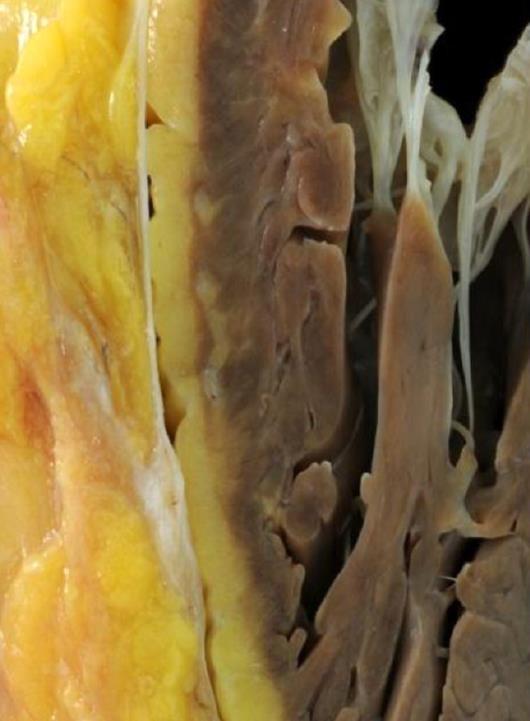
4 Anatomy of normal pericardium
5 Mechanical Functions of the pericardium Effects on chambers - Limits short-term cardiac distention - Facilitates cardiac chamber coupling and interaction - Maintains pressure-volume relation of the cardiac chambers and output from them - Maintains LV geometry Effects on whole heart - Lubricates, minimizes friction - Equalizes gravitation and inertial, hydrostatic forces - Mechanical barrier to infection Immunologic Vasomotor Fibrinolytic Modulation of myocyte structure and function and gene expression Vehicle for drug delivery and gene therapy

6 Pericardial effusion Abnormal accumulation of fluid in the pericardial space
7 Idiopathic Etiology of Pericardial disease - Acute idiopathic pericarditis* (probably viral or post-viral) - Chronic idiopathic effusion Infectious - Viral, Bacterial infection (Tuberculosis), Fungal Inflammatory - Associated connective tissue disease (RA, SLE, Others) Post-myocardial infarction (ASD, Free wall rupture, Dressler syndrome ) Associated with systemic disease (Uremia, Hypothyroidism, Cirrhosis, Amyloidosis) Malignancy (Direct tumor involvement, Effusion due to lymphatic obstruction) Miscellaneous - Posttrauma, Postsurgical, Radiation induced - Congestive heart failure, Severe PAH, RV Failure - Down syndrome - Pregnancy
8 Clinical presentation Asymptomatic Shortness of breath or difficulty breathing (dyspnea) Discomfort when breathing while lying down (orthopnea) Chest pain, usually behind the breastbone or on the left side of the chest Chest fullness
9 2015 ESC guideline Diagnosis of Pericardial effusion Transthoracic echocardiography is recommended in all patients with suspected pericardial effusion (I C) Chest X-ray is recommended in patients with a suspicion of pericardial effusion or pleuropulmonary involvement (I C) Assessment of markers of inflammation (i.e. CRP) are recommended in patients with pericardial effusion (I C) CT or CMR should be considered in suspected cases of loculated pericardial effusion, pericardial thickening and masses, as well as associated chest Abnormalities (IIa C) Echo-free space DDX) mediastinal fat, fibrosis, thymus, or other tissue
10 2015 ESC guideline Diagnosis of Pericardial effusion Transthoracic echocardiography is recommended in all patients with suspected pericardial effusion (I C) Chest X-ray is recommended in patients with a suspicion of pericardial effusion or pleuropulmonary involvement (I C) Assessment of markers of inflammation (i.e. CRP) are recommended in patients with pericardial effusion (I C) CT or CMR should be considered in suspected cases of loculated pericardial effusion, pericardial thickening and masses, as well as associated chest Abnormalities (IIa C) Echo-free space DDX) mediastinal fat, fibrosis, thymus, or other tissue
11 Amount of pericardial effusion 다른 reference 에서는어떻게정의하는지. seen only in systole <10 mm mm >20 mm Klein et al ASE guideline


12 Amount of pericardial effusion 다른 reference 에서는어떻게정의하는지. seen only in systole <10 mm mm >20 mm
 Pericardial reflections surround the pulmonary")

 Note the nodular")
13 Differentiation of Pericardial from Pleural Effusion 1) Pericardial reflections surround the pulmonary veins and tend to limit the potential space behind the LA. 2) Descending thoracic aorta 3) Frequently identify the parietal pericardium 4) Note the nodular densities overlying on the visceral aspect of the pericardium


14 Pericardial effusion on CT Localization and quantitation of pericardial fluid Tissue characterization on the basis of computed tomographic attenuation (> 60 Hounsfield units suggest hemorrhage) Differentiation of pericardial thickening from fluid Feasibility of surgery vs percutaneous drainage of complex effusions 2015 ESC guideline
15 2015 ESC guideline Diagnosis of Pericardial effusion Transthoracic echocardiography is recommended in all patients with suspected pericardial effusion (I C) Chest X-ray is recommended in patients with a suspicion of pericardial effusion or pleuropulmonary involvement (I C) Assessment of markers of inflammation (i.e. CRP) are recommended in patients with pericardial effusion (I C) CT or CMR should be considered in suspected cases of loculated pericardial effusion, pericardial thickening and masses, as well as associated chest Abnormalities (IIa C) Echo-free space DDX) mediastinal fat, fibrosis, thymus, or other tissue

16 2015 ESC guideline Sx (Pericarditis) Treatment of pericardial effusion Amount, Hemodynamic significance Inflammatory marker Large idiopathic chronic effusions have a 30 35% risk of progression to cardiac tamponade
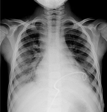
17 70/M Chief Complaint) Diarrhea, vomiting (2WA) DOE (8WA) V/S) 120/ PEx) Friction rub+ Lab) CBC K, CRP 3.4
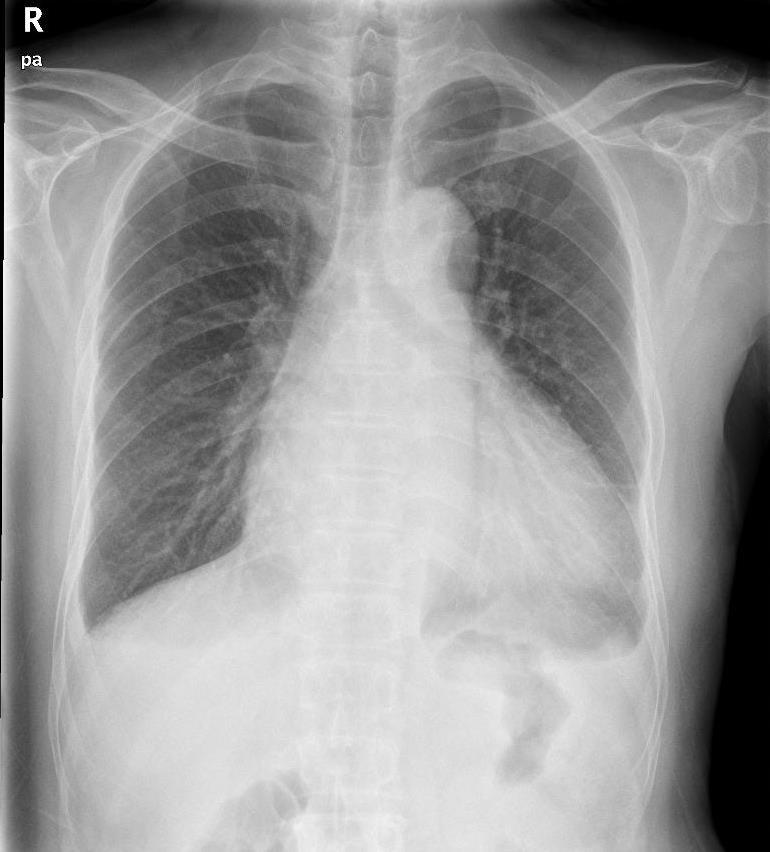
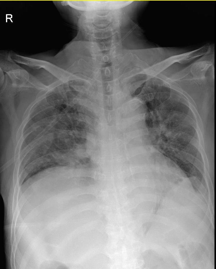
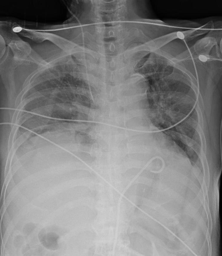
18 Chest PA
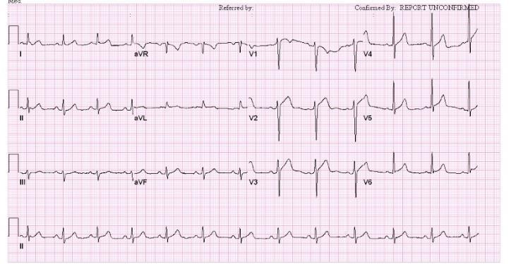
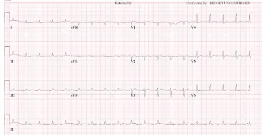
19 ECG
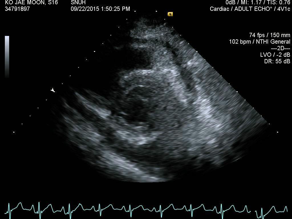
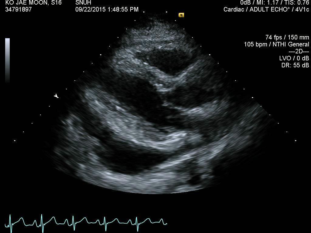
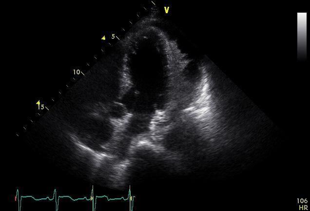
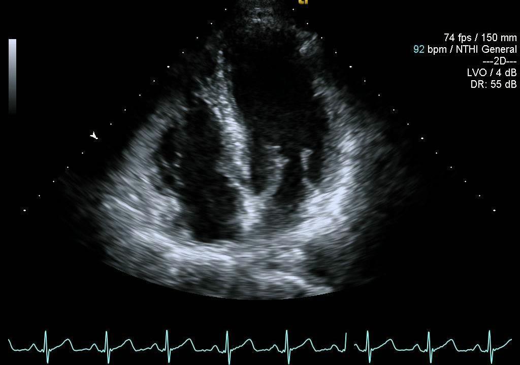
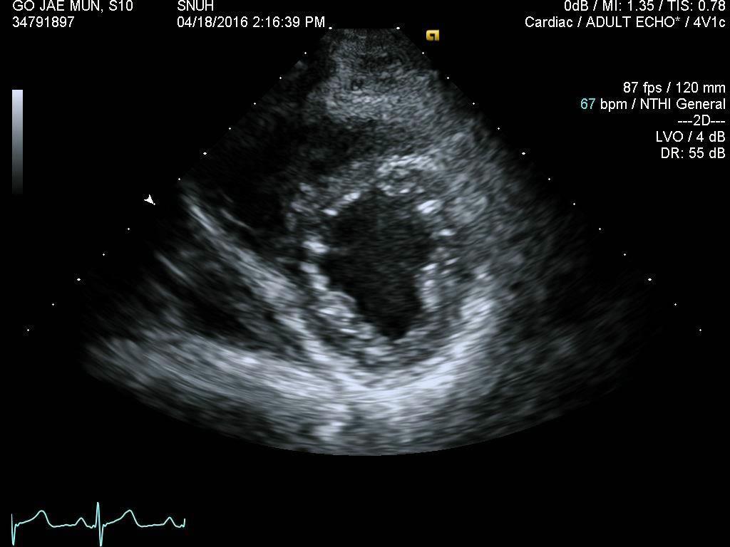
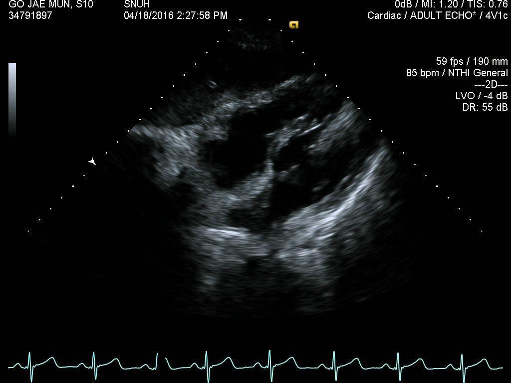
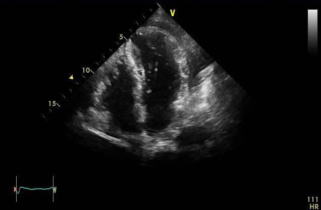
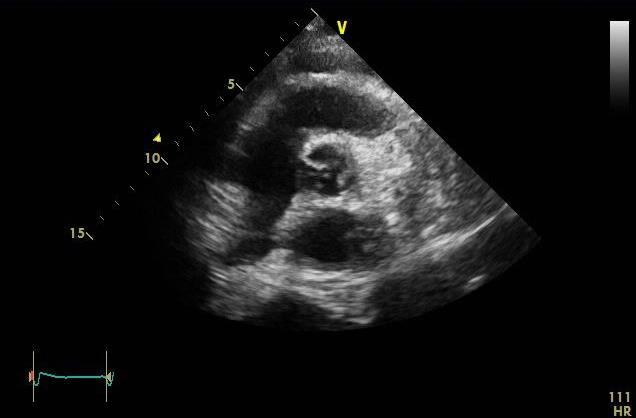
20 Echocardiography


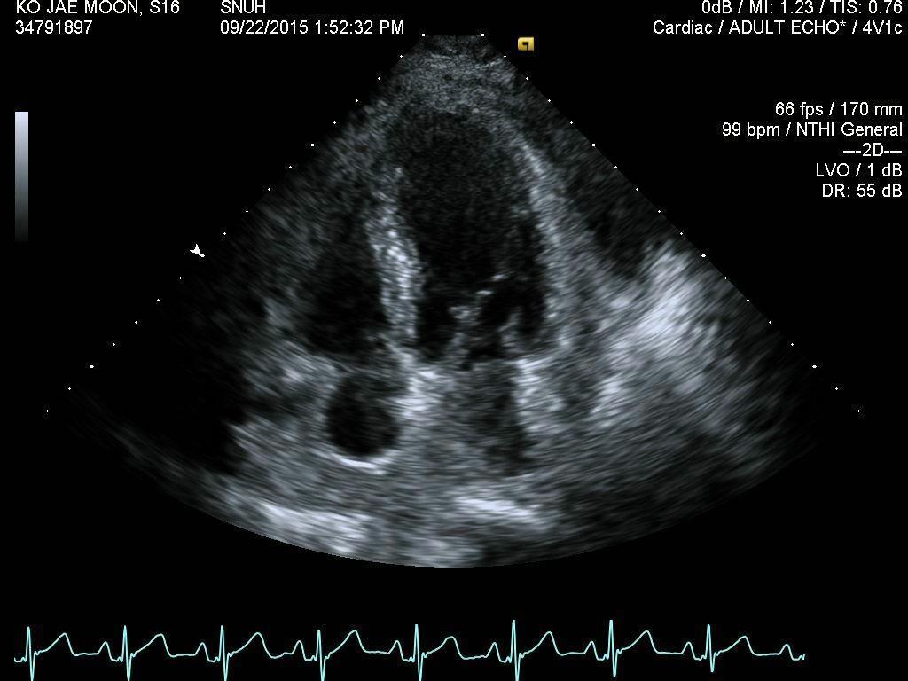
21 Carol-F 처방후 2 주뒤 Echo f/u
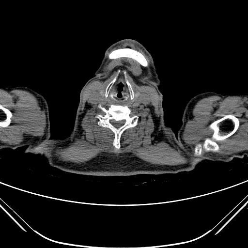
22 Chest CT

23
24 Pericardial window operation Pericardium, biopsy Fibrinous pericarditis with chronic granulomatous inflammation and caseous necrosis Pericardial analysis Lymphodominant exudate (Poly 23%, Lymph 55%) ADA 33, LD 199, Protein 4.1 c/w TB pericarditis
25 Cardiac Tamponade and Pericardiocentesis

26 Pathophysiology of cardiac tamponade Intrapericardial Pressure equal end-diastolic pressures in RV or LV Decreased CO Roy CL, et al. JAMA. 2007;297:
27 Ralph Shabetai. Heart Mar; 90(3): Cardiac tamponade life-threatening, slow or rapid compression of the heart due to the pericardial accumulation in the pericardial sac. Fatal!!! 1. amount of pericardial contents 2. rate of accumulation 속도에따른그래프 200cc >2000cc

28 Diagnosis of cardiac tamponade Chest PA ECG Enlarged cardiac silhouette (>250ml) Water bottle sign Low voltage Electrical alternans ST - T changes due to pericarditis

 Sternbach G. Claude Beck. J Emerg MEd.")
29 Diagnosis of cardiac tamponade Beck s triad Hypotension Soft or absent heart sounds Jugular venous distention (with a prominent x descent, absent y descent) Sternbach G. Claude Beck. J Emerg MEd Sep 6(5):417-9.
30 Diagnosis of cardiac tamponade Clinical diagnosis is important! Beck s triad Tachycardia Hypotension Pulsus paradoxus A key diagnostic finding
31 Pulsus paradoxus BP drop (>10mmHg) during inspiration Expiration Inspiration Expiration Inspiration
 and tachycardia (77%) have good sensitivity. - Hypotension and distant heart sounds are insensitive physical findings. JAMA 2007;297:1810-1818.")
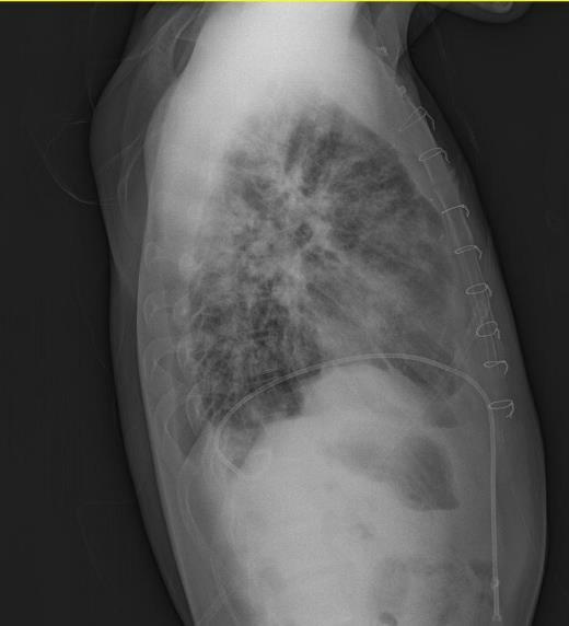
32 Pulsus paradoxus in pericardial tamponade - A paradoxical pulse is the most helpful clinical test for cardiac tamponade (sensitivity of 98%, specificity of 83%) - Elevated jugular venous pressure (76%) and tachycardia (77%) have good sensitivity. - Hypotension and distant heart sounds are insensitive physical findings. JAMA 2007;297:

33 Pulsus paradoxus in pericardial tamponade N Engl J med 2003:349:
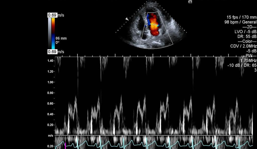
34 Echocardiographic findings in cardiac tamponade 2D and M-mode Diastolic RV collapse RA collapse/inversion Swinging heart Doppler Exaggerated respiratory variation in inflow velocity Exaggerated respiratory variation in inferior vena cava flow Phasic variation in RVOT/LVOT

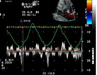
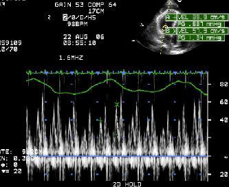
 : MV E-wave >30% :")

35 Echocardiographic findings in cardiac tamponade Exaggerated respiratory variation in inflow velocity (E wave) : MV E-wave >30% : TV E-wave (earliest finding) Phasic variation in RVOT/LVOT Klein et al ASE guideline

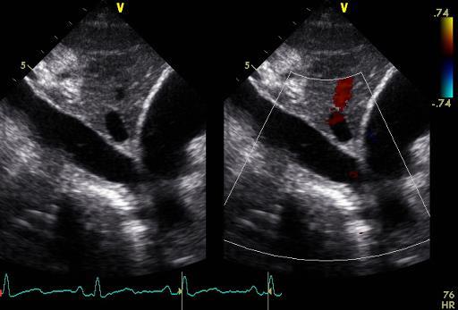
 IVC plethora Klein et al.")
36 Echocardiographic findings in cardiac tamponade -Diastolic flow reversal after expiration -Velocities in tamponade are markedly reduced, reflecting reduced cardiac filling (normal 50 ->20~40cm/s) IVC plethora Klein et al ASE guideline

37 Echocardiographic findings in cardiac tamponade RV, RA collapse at early diastolic phase (AV, PV closing) Duration of RA >1/3 of cardiac cycle Klein et al ASE guideline Feigenbaum s Echocardiography, 7 th ed
 Klein et al.")
38 Echocardiographic findings in cardiac tamponade RV, RA collapse (M-mode) Klein et al ASE guideline Feigenbaum s Echocardiography, 7 th ed

39 Echocardiographic findings swinging of the heart
40 Echocardiographic findings in cardiac tamponade 2D and M-mode Diastolic RV collapse RA collapse/inversion Swinging heart Doppler Exaggerated respiratory variation in inflow velocity Exaggerated respiratory variation in inferior vena cava flow Phasic variation in RVOT/LVOT Cardiac tamponade is a clinical diagnosis
41 85/M Dyspnea 으로타병원에서치료받던중, CBC abnormality (lymphocytosis) 관찰되어 CLL 의심하내원함.
42 ECG
43 Chest PA
44 Echocardiography
45 Echocardiography

46
47 Chest PA
48 Treatment of cardiac tamponade EM) Pericardiocentesis Medical treatment Hypotensive patients with hypovolemia : A low volume ( ml) of normal saline to improve haemodynamic parameters Infusion of higher volumes may increase wedge pressure and intrapericardial pressure, and reduce cardiac output
49 Indication of Pericardiocentesis (PCC) Hemodynamically unstable patients emergent procedure!! Patients without hemodynamic compromise Symptomatic moderate to large effusion non-responsive to medical therapy Small effusion when tuberculous, bacterial or neoplastic pericarditis is suspected Chronic large effusion (>20mm in diastole) Diagnostic purpose without large effusion Low diagnostic power & High procedural risk Viral pericarditis self-limiting
50 Contraindication of PCC No absolute contraindications Relative contraindications Aortic dissection Post-infarction rupture of the free wall If hemodynamically unstable, very small amounts of the hemopericardium can be attempted To maintain blood pressure at around 90 mmhg Uncorrected coagulopathy, anticoagulant therapy, thrombocytopaenia (PLT <50,000/mm³)
51 Puncture site 1. Apical approach 1-2 cm lateral to the apex beat within the fifth, sixth or seventh intercostal space. Advance the needle over the superior border of the rib Risk of ventricular puncture/ pneumothorax The thicker left ventricle wall is more likely to self-seal after puncture The path to reach the pericardium is shorter Caterina CC. Cardiology Practice ESC, 2017 Nov;15
 Low risk of pneumothorax Caterina CC.")
52 Puncture site 2. Subxiphoid the fifth left intercostal space, close to the sternal margin Risk of pneumothorax and puncture of the internal thoracic vessels 3. Subxiphoid approach Between the xiphisternum and left costal margin 15 to 30 angle (deeper angle may enter peritoneal cavity) Low risk of pneumothorax Caterina CC. Cardiology Practice ESC, 2017 Nov;15
 1%")
 scalpel J shaped")
53 PCC set dilator Needle (or epidural needle) 1% lidocaine (contrast agent) scalpel J shaped guide wires

 Check the")

54 Fluoroscopy-guided technique First imaging system Subxiphoid approach a needle containing a contrast medium Directed toward the left shoulder (30 ) Check the guidewire position in at least two angiographic projections SW Han, Korean Circ J Oct;40(10):


55 Echo-guided technique Safe and simple technique Semi-reclining position (angle of 30 ) and slightly rotated leftwards The extracardiac position of the tip or injecting 5 ml of agitated saline infusion real-time echo-monitored procedure Caterina CC. Cardiology Practice ESC, 2017 Nov;15
56 Surgical drainage Recurrent tamponade, when it is necessary to remove loculated effusions and/or when it is necessary to obtain tissue for diagnosis
57 After PCC Check Chest PA!! Pericardial drainage Immediately after PCC: remove enough fluid to normalize the CVP and BP (not >1 L) repeated every 4 to 6 hours Maintenance : 24 to 72 hours is sufficient Removal drainage < 25~30ml/day The recurrence rate after the initial procedure 27~55%
58 After PCC
59 Complications Minor complication (0.4-20%) Transient vasovagal hypotension, bradycardia Arrhythmia (SVT) Pneumothorax Pleuropericardial fistulas Major complication ( %) Death Injury of the cardiac chambers, coronary arteries or intercostal vessels Puncture of the abdominal viscera or peritoneal cavity Pneumothorax requiring chest tube placement Pneumopericardium Ventricular arrhythmias Pericardial decompression syndrome After a successful pericardial drainage, from a few hours to days later mechanism : unknown explanation is an acute left ventricular overload due to an increased right-sided preload associated with a persistent catecholaminergic peripheral vasoconstriction. prevention: remove enough fluid to normalize the central venous and systemic blood pressure (not >1 L) and to complete the removal in the subsequent few hours

60 Pneumothorax after PCC
61 Conclusion All patients with pericardial effusion or tamponade should undergo TTE to assess for the extent of effusion and hemodynamic compromise. CT and/or CMR should be done for those patients with complex effusion with subacute tamponade with the need for drainage. CT and/or CMR should be done for those with suspected hemopericardium or pericardial clot and to assess the source of effusion as in malignancy or inflammation.
62 Conclusion All patients with clinically suspected cardiac tamponade should undergo TTE with Doppler echocardiography as the initial imaging test, which can provide a definite diagnosis in most patients. Cardiac tamponade is a medical emergency. Pericardiocentesis is a life saving procedure in cardiac tamponade.
63 감사합니다.
THE PERICARDIUM: LOOKING OUTSIDE THE HEART
 THE PERICARDIUM: LOOKING OUTSIDE THE HEART DISCLOSURE Relevant relationships with commercial entities none Potential for conflicts of interest within this presentation none Steps taken to review and mitigate
Cardiac tamponade and Pericardiocentesis Made Easy
 Cardiac tamponade and Pericardiocentesis Made Easy www.cardiconcept.com Etiology of pericardial diseases. Non Infectious cause Infectious cause European Heart Journal (2015) 36, 2921 2964 Recommendations
Pericardial Diseases. Smonporn Boonyaratavej, MD. Division of Cardiology, Department of Medicine Chulalongkorn University
 Pericardial Diseases Smonporn Boonyaratavej, MD Division of Cardiology, Department of Medicine Chulalongkorn University Cardiac Center, King Chulalongkorn Memorial Hospital 21 AUGUST 2016 Pericardial
Outline. Echocardiographic Assessment of Pericardial Effusion/Tamponade: The Essentials
 Echocardiographic Assessment of Pericardial Effusion/Tamponade: The Essentials John R Schairer DO FACC Henry Ford Heart and Vascular Institute No Disclosures Outline Normal Anatomy and Physiology Pathophysiology
Echocardiography as a diagnostic and management tool in medical emergencies
 Echocardiography as a diagnostic and management tool in medical emergencies Frank van der Heusen MD Department of Anesthesia and perioperative Care UCSF Medical Center Objective of this presentation Indications
Adel Hasanin Ahmed 1
 Adel Hasanin Ahmed 1 PERICARDIAL DISEASE The pericardial effusion ends anteriorly to the descending aorta and is best visualised in the PLAX. PSAX is actually very useful sometimes for looking at posterior
We are now going to review the diagnosis and management of pericardial collections and tamponade
 We are now going to review the diagnosis and management of pericardial collections and tamponade FEEL COURSE PAGE 1 Paying particular attention to the difference between a collection and cardiac tamponade
PERICARDIAL DIAESE. Kaijun Cui Associated professor Sichuan University
 PERICARDIAL DIAESE Kaijun Cui Associated professor Sichuan University CLASSIFICATION acute pericarditis pericardial effusion cardiac tamponade constrictive pericarditis congenitally absent pericardium
Pericardial diseases
 Pericardial diseases Anatomy of the pericardium Consists of parietal and visceral membranes. The space between them(pericardial space is normally filled by a lymph like fluid. The fluid s normal quantity
Department of Cardiac, Thoracic and Vascular Sciences University of Padua Cardiac Tamponade. Echocardiography in Diagnosis and Management
 Department of Cardiac, Thoracic and Vascular Sciences University of Padua Cardiac Tamponade. Echocardiography in Diagnosis and Management Luigi P. Badano, MD, FESC, FACC Declaration of interest **Dr. Badano
Pericardial disease. Se-Jung Yoon Cardiology division NHIS Ilsan hospital
 Pericardial disease Se-Jung Yoon Cardiology division NHIS Ilsan hospital Normal pericardial effusion Normal pericardium Normal pericardium Pericardial Layers: Visceral layer Parietal layer Fibrous pericardium
Normal Pericardial Physiology
 Normal Pericardial Physiology Normal pericardium contains 20-30 ml of lymphoid fluid lubricating function that facilitates normal myocardial rotation and translation during each cardiac cycle in that the
Palpable Pulsus Paradoxus in the Setting of Malignant Pericardial Effusion and Tamponade Akshay Pendyal, MD
 Palpable Pulsus Paradoxus in the Setting of Malignant Pericardial Effusion and Tamponade Akshay Pendyal, MD University of Colorado Department of Internal Medicine None Conflicts of Interest Objectives
TAMPONADE CARDIAQUE. Dr Cédrick Zaouter TUSAR 15 décembre 2015
 TAMPONADE CARDIAQUE Dr Cédrick Zaouter TUSAR 15 décembre 2015 OUTLINE History Incidence Definition Pathophysiology Aetiologies Investigations - Echocardiography Treatment of cardiac tamponade Pericardial
Cath Lab Essentials: Pericardial effusion & tamponade
 Cath Lab Essentials: Pericardial effusion & tamponade Pranav M. Patel, MD, FACC, FSCAI Chief, Division of Cardiology Director, Cardiac Cath Lab & CCU University of California, Irvine Division of Cardiology
The role of bedside ultrasound in the diagnosis of pericardial effusion and cardiac tamponade
 Symposium The role of bedside ultrasound in the diagnosis of pericardial effusion and cardiac tamponade Adam Goodman, Phillips Perera, Thomas Mailhot, Diku Mandavia Department of Emergency Medicine, Los
Πνευμονική υπέρταση και περικαρδιακή συλλογή. Τρόποι αντιμετώπισης
 Πνευμονική υπέρταση και περικαρδιακή συλλογή. Τρόποι αντιμετώπισης Γεώργιος Λάζαρος Καρδιολόγος, Διευθυντής ΕΣΥ Α Πανεπιστημιακή Καρδιολογική Κλινική Ιπποκράτειο Γ.Ν. Αθηνών Pericardial syndromes o Acute
A Case of Impending Cardiac Tamponade Caused by Effusive Constrictive Pericarditis
 Archives of Clinical and Medical Case Reports doi: 10.26502/acmcr.96550038 Volume 2, Issue 5 Case Report A Case of Impending Cardiac Tamponade Caused by Effusive Constrictive Pericarditis Catalina Sanchez-Alvarez
Index. Note: Page numbers of article titles are in boldface type.
 Index Note: Page numbers of article titles are in boldface type. A Acute coronary syndrome(s), anticoagulant therapy in, 706, 707 antiplatelet therapy in, 702 ß-blockers in, 703 cardiac biomarkers in,
Pericardial Diseases/Tamponade Illustrative Cases
 Pericardial Diseases/Tamponade Illustrative Cases Jae K. Oh, MD Echo Hawaii 2017 2012 MFMER 3200268v3(2010)-1 Case #1 47 year old man Chest pain Not exertional Normal Examination 2016 MFMER slide-2 1 47
Integrative Clinical Hemodyamics
 Integrative Clinical Hemodyamics James A. Goldstein, MD Director, Research & Education Division of Cardiology William Beaumont Hospital Disclosure Information Integrative Clinical Hemodyamics James A.
Background & Indications Probe Selection
 Teresa S. Wu, MD, FACEP Director, EM Ultrasound Program & Fellowship Co-Director, Simulation Based Training Program & Fellowship Associate Program Director, EM Residency Program Maricopa Medical Center
ΚΑΡΔΙΟΛΟΓΟΣ EUROPEAN ACCREDITATION IN TRANSTHORACIC AND TRANSESOPHAGEAL ECHOCARDIOGRAPHY
 1 ΚΑΡΔΙΟΛΟΓΟΣ EUROPEAN ACCREDITATION IN TRANSTHORACIC AND TRANSESOPHAGEAL ECHOCARDIOGRAPHY 2 Constrictive pericarditis (CP) is characterized by impaired ventricular filling due to a stiffened or noncompliant
10/16/2014. CCRN Review - Cardiovascular. CCRN Review - Cardiovascular. CCRN Review - Cardiovascular
 Hypertrophic (IHSS) Diagnosis Chest x ray cardiomegaly Electrocardiography LV hypertrophy, ST segment T was changes, Q waves in inferior & precordial leads Atrial & ventricular dysrhythmias Hypertrophic
Cardiac Mass in a 15-Year-Old Boy
 Cardiac Mass in a 15-Year-Old Boy Echocardiographic Case Report Hortensia Vuçini Department of Cardiology and Cardiac Surgery UHC Mother Theresa Tirana, Albania October 20, 2007 Case Presentation 15 year-old
Pericardial Diseases 2015 Update
 Pericardial Diseases 2015 Update BRUCE W. USHER, MD PROFESSOR OF MEDICINE CARDIOLOGY DIVISION MEDICAL UNIVERSITY SOUTH CAROLINA CHARLESTON, SOUTH CAROLINA GUIDELINES: PERICARDIAL DISEASES Ø European Society
Right-Sided Congestive Heart Failure Basics
 Right-Sided Congestive Heart Failure Basics OVERVIEW Failure of the right side of the heart to pump blood at a sufficient rate to meet the needs of the body or to prevent blood from pooling within the
An Uncommon Cardiac Etiology of Liver Cirrhosis, Recurrent Ascites, Atrial Fibrillation and Congestive Heart Failure
 Cronicon OPEN ACCESS EC CARDIOLOGY Case Report An Uncommon Cardiac Etiology of Liver Cirrhosis, Recurrent Ascites, Atrial Fibrillation and Congestive Heart Failure Montaser Y Ismail 1 *, Mohammed I Nassar
Pericardial Effusion
 Pericardial Effusion How does the heart work? The heart is the organ responsible for pumping blood to and from all tissues of the body. The heart is divided into right and left sides. The job of the right
Pericardial Disease: Case Examples. Echo Fiesta 2017
 Pericardial Disease: Case Examples Echo Fiesta 2017 2014 2014 MFMER MFMER 3346252-1 slide-1 Objectives Have a systematic approach to evaluation of constriction 2014 MFMER 3346252-2 CASE 1 2013 MFMER 3248567-3
Constrictive Pericarditis Pitfalls in MR Diagnosis Cylen Javidan-Nejad Associate Professor Mallinckrodt Institute of Radiology Washington University
 Constrictive Pericarditis Pitfalls in MR Diagnosis Cylen Javidan-Nejad Associate Professor Mallinckrodt Institute of Radiology Washington University in St. Louis Goal o To review the imaging criteria of
Essentials of Pericardial Diseases
 Essentials of Pericardial Diseases 1 Nikolaos Skubas MD, 2 Manuel Fontes MD The pericardial diseases result in cardiovascular perturbations ranging from asymptomatic electrocardiographic findings (in pericarditis
Unit 6: Circulatory System. 6.2 Heart
 Unit 6: Circulatory System 6.2 Heart Functions of Circulatory System 1. The heart is the pump necessary to circulate blood to all parts of the body 2. Arteries, veins and capillaries are the structures
Medical NREMT-PTE. NREMT Paramedic Trauma Exam.
 Medical NREMT-PTE NREMT Paramedic Trauma Exam https://killexams.com/pass4sure/exam-detail/nremt-pte Question: 41 Which of the following most accurately describes the finding of jugular venous distension
Heart Anatomy. 7/5/02 Stephen G Davenport 1
 Heart Anatomy Copyright 1999, Stephen G. Davenport, No part of this publication may be reproduced, stored in a retrieval system, or transmitted, in any form without prior written permission. 7/5/02 Stephen
Hemodynamic Monitoring
 Perform Procedure And Interpret Results Hemodynamic Monitoring Tracheal Tube Cuff Pressure Dean R. Hess PhD RRT FAARC Hemodynamic Monitoring Cardiac Rate and Rhythm Arterial Blood Pressure Central Venous
Shock, Monitoring Invasive Vs. Non Invasive
 Shock, Monitoring Invasive Vs. Non Invasive Paula Ferrada MD Assistant Professor Trauma, Critical Care and Emergency Surgery Virginia Commonwealth University Shock Fluid Pressors Ionotrope Intervention
Constrictive Pericarditis
 Constrictive Pericarditis Never Confused with Anything Else Jae K. Oh, MD 2018 MFMER 3712003-1 ARS #1 CP Which of following patients has constrictive pericarditis? 1 2 3 Medial e 13 cm/s Medial e 3 cm/s
POCUS for the Internist: Lungs & Pericardial Effusions
 POCUS for the Internist: Lungs & Pericardial Effusions Jeremy S. Boyd, MD, FACEP Asst. Professor of Emergency Medicine Vanderbilt University Medical Illustrations courtesy of Robinson Ferre, MD, FACEP
Disclosures. Cardiac Ultrasound. Introductory Case. 80 y/o male Syncope at home Emesis x 3 in ambulance Looks sick. No pain.
 Disclosures Cardiac Ultrasound Justin A Davis, MD MPH RDMS Subchief for Emergency Ultrasound Kaiser Permanente East Bay Medical Center I have nothing to disclose. Introductory Case HR 118 BP 65/43 RR 27
Rotation: Echocardiography: Transthoracic Echocardiography (TTE)
") Rotation: Echocardiography: Transthoracic Echocardiography (TTE) Rotation Format and Responsibilities: Fellows rotate in the echocardiography laboratory in each clinical year. Rotations during the first
Rotation: Echocardiography: Transthoracic Echocardiography (TTE) Rotation Format and Responsibilities: Fellows rotate in the echocardiography laboratory in each clinical year. Rotations during the first
The Cardiovascular System. Chapter 15. Cardiovascular System FYI. Cardiology Closed systemof the heart & blood vessels. Functions
 Chapter 15 Cardiovascular System FYI The heart pumps 7,000 liters (4000 gallons) of blood through the body each day The heart contracts 2.5 billion times in an avg. lifetime The heart & all blood vessels
efferent fibers from t.. Heart Surface anatomy and heart sounds -Dry lecture -Gray s 169,
 A patient is diagnosed with ischemia (i.e., lack of blood flow) in a left lobar pulmonary vein. The attending physician determines that the ischemia is due to a vasospastic episode. Constriction of this
AP2 Lab 1 - Blood & Heart
 AP2 Lab 1 - Blood & Heart Project 1 - Formed Elements Identification & Recognition See fig. 17.10 and Table 17.2. Instructor may also provide other images. Note: See Fig. 17.11 All formed elements are
Certificate in Clinician Performed Ultrasound (CCPU) Syllabus. Rapid Cardiac Echo (RCE)
 Syllabus. Rapid Cardiac Echo (RCE)") Certificate in Clinician Performed Ultrasound (CCPU) Syllabus Rapid Cardiac Echo (RCE) Purpose: Rapid Cardiac Echocardiography (RCE) This unit is designed to cover the theoretical and practical curriculum
Certificate in Clinician Performed Ultrasound (CCPU) Syllabus Rapid Cardiac Echo (RCE) Purpose: Rapid Cardiac Echocardiography (RCE) This unit is designed to cover the theoretical and practical curriculum
Case 5 15-year-old male
 Case 5 15-year-old male Present illness: Six months ago, abnormality of ECG was incidentally detected by annual health check. His blood level of γ-gtp, HbA1c and norepinephrine were elevated; however,
Miscellaneous Cardiology Topics pregnancy - congenital - myocarditis - pericardial disease. Pregnancy and Cardiovascular Disease MCQ
 Miscellaneous Cardiology Topics pregnancy - congenital - myocarditis - pericardial disease Maan Jokhadar, MD, FACC Emory Center for Advanced Heart Failure Therapy Emory Adult Congenital Heart Center Pregnancy
Constrictive pericarditis: Morphological, functional and haemodynamic evaluation
 Constrictive pericarditis: Morphological, functional and haemodynamic evaluation Poster No.: C-0743 Congress: ECR 2010 Type: Educational Exhibit Topic: Cardiac Authors: B. Graca, P. Donato, M. Ferreira,
Cardiology. Objectives. Chapter
 1:44 M age 1121 Chapter Cardiology Objectives art 1: Cardiovascular natomy and hysiology, ECG Monitoring, and Dysrhythmia nalysis (begins on p. 1127) fter reading art 1 of this chapter, you should be able
10/1/2016. Constrictive Pericarditis Unique Hemodynamics. What s New in Pericardial Disease? Case-based Discussion
 Mayo Clinic Department of Cardiovascular Diseases Mayo Clinic Echocardiography Review Course for Boards and Recertification What s New in Pericardial Disease? Case-based Discussion Jae K. Oh, MD Samsung
Diagnostic Approach to Pleural Effusion
 Diagnostic Approach to Pleural Effusion Objectives Define the leading causes of pleural effusion Classify the type of effusion Identify procedures and tests associated with diagnosis 2 Agenda Basic anatomy
Objectives. Highlight typical feature of TB pericarditis. How to make a diagnosis. How to treat TB pericarditis
 Dr. Conteh Objectives Highlight typical feature of TB pericarditis How to make a diagnosis How to treat TB pericarditis New evidence for adjunctive corticosteroid Introduction TB pericarditis occurs in
Constrictive/Restrictive Cardiomyopathies: Diagnosis and Management Update; Radiation Induced Heart Disease. Alexander (Sandy) Dick, MD
 Dick, MD") Constrictive/Restrictive Cardiomyopathies: Diagnosis and Management Update; Radiation Induced Heart Disease Alexander (Sandy) Dick, MD Outline Pericardial Constriction Diagnosis: Imaging, Hemodynamics
Constrictive/Restrictive Cardiomyopathies: Diagnosis and Management Update; Radiation Induced Heart Disease Alexander (Sandy) Dick, MD Outline Pericardial Constriction Diagnosis: Imaging, Hemodynamics
Right lung. -fissures:
 -Right lung is shorter and wider because it is compressed by the right copula of the diaphragm by the live.. 2 fissure, 3 lobes.. hilum : 2 bronchi ( ep-arterial, hyp-arterial ), one artery mediastinal
Adult Echocardiography Examination Content Outline
 Adult Echocardiography Examination Content Outline (Outline Summary) # Domain Subdomain Percentage 1 2 3 4 5 Anatomy and Physiology Pathology Clinical Care and Safety Measurement Techniques, Maneuvers,
Index. K Knobology, TTE artifact, image resolution, ultrasound, 14
 A Acute aortic regurgitation (AR), 124 128 Acute aortic syndrome (AAS) classic aortic dissection diagnosis, 251 263 evolutive patterns, 253 255 pathology, 250 251 classifications, 247 248 incomplete aortic
Looking Outside the Box: Incidental Extracardiac Finding in Echo
 Looking Outside the Box: Incidental Extracardiac Finding in Echo Dr. Aijaz Shah Head of Division, Adult Echocardiography Laboratory Prince Sultan Cardiac Centre Riyadh Case 1 17 year old boy presented
Acute Pericardial Tamponade
 Acute Pericardial Tamponade Marx: Rosen's Emergency Medicine: Concepts and Clinical Practice, 5th ed. Epidemiology The reported incidence of acute pericardial tamponade is approximately 2% in patients
Transient Constrictive Pericarditis: Causes and Natural History
 Journal of the American College of Cardiology Vol. 43, No. 2, 2004 2004 by the American College of Cardiology Foundation ISSN 0735-1097/04/$30.00 Published by Elsevier Inc. doi:10.1016/j.jacc.2003.08.032
The Cardiovascular System
 The Cardiovascular System The Manila Times College of Subic Prepared by: Stevens B. Badar, RN, MANc THE HEART Anatomy of the Heart Location and Size approx. the size of a person s fist, hollow and cone-shaped,
For more information about how to cite these materials visit
 Project: Ghana Emergency Medicine Collaborative Document Title: Case Presentation- Pericarditis Author(s): Kwaku Nyame License: Unless otherwise noted, this material is made available under the terms of
TAVR: Echo Measurements Pre, Post And Intra Procedure
 2017 ASE Florida, Orlando, FL October 10, 2017 8:00 8:25 AM 25 min TAVR: Echo Measurements Pre, Post And Intra Procedure Muhamed Sarić MD, PhD, MPA Director of Noninvasive Cardiology Echo Lab Associate
Lecture 2: Clinical anatomy of thoracic cage and cavity II
 Lecture 2: Clinical anatomy of thoracic cage and cavity II Dr. Rehan Asad At the end of this session, the student should be able to: Identify and discuss clinical anatomy of mediastinum such as its deflection,
THORACIC AORTIC DISSECTION
 The Essence of Aortic Dissection THORACIC AORTIC DISSECTION Aortic dissection can be classified as acute if it s onset has been less than 14 days or chronic if its onset has been more than 14 days. Mortality
Large veins of the thorax Brachiocephalic veins
 Large veins of the thorax Brachiocephalic veins Right brachiocephalic vein: formed at the root of the neck by the union of the right subclavian & the right internal jugular veins. Left brachiocephalic
Cor pulmonale. Dr hamid reza javadi
 1 Cor pulmonale Dr hamid reza javadi 2 Definition Cor pulmonale ;pulmonary heart disease; is defined as dilation and hypertrophy of the right ventricle (RV) in response to diseases of the pulmonary vasculature
PLEURAE and PLEURAL RECESSES
 PLEURAE and PLEURAL RECESSES By Dr Farooq Aman Ullah Khan PMC 26 th April 2018 Introduction When sectioned transversely, it is apparent that the thoracic cavity is kidney shaped: a transversely ovoid space
Resolution of a respiratory failure due to massive chronic pericardial effusion with a pericardial window: the simplest is the best
 Case Report Page 1 of 5 Resolution of a respiratory failure due to massive chronic pericardial effusion with a pericardial window: the simplest is the best Barbara Bonfanti 1, Luca Bertolaccini 2, Piercamillo
Dr. Rami M. Adil Al-Hayali Assistant Professor in Medicine
 Dr. Rami M. Adil Al-Hayali Assistant Professor in Medicine Venous thromboembolism: pulmonary embolism (PE) deep vein thrombosis (DVT) 1% of all patients admitted to hospital 5% of in-hospital mortality
The pericardial sac is composed of the outer fibrous pericardium
 Pericardiectomy for Constrictive or Recurrent Inflammatory Pericarditis Mauricio A. Villavicencio, MD, Joseph A. Dearani, MD, and Thoralf M. Sundt, III, MD Anatomy and Preoperative Considerations The pericardial
The Cardiovascular System Part I: Heart Outline of class lecture After studying part I of this chapter you should be able to:
 The Cardiovascular System Part I: Heart Outline of class lecture After studying part I of this chapter you should be able to: 1. Describe the functions of the heart 2. Describe the location of the heart,
Clinical Indications for Echocardiography
 Clinical Indications for Echocardiography Echocardiography is widely utilised and potential applications are increasing with advances in technology. The aim of this document is two-fold: 1) To define clinical
CASE DISCUSSION. Dr JAYASREE VEERABOINA 2nd yr PG MS OBG
 CASE DISCUSSION Dr JAYASREE VEERABOINA 2nd yr PG MS OBG Normal Cardiovascular changes in Pregnancy CARDIAC OUTPUT 5 th wk -- starts 12 wks -- 30-35% 30-32 wks -- 40% During labour -- 50% After delivery
Pericardiocentesis and Drainage by a Silicon Rubber Line. without Echocardiographic Guidance. Experience in 55 Consecutive Patients
 Pericardiocentesis and Drainage by a Silicon Rubber Line without Echocardiographic Guidance Experience in 55 Consecutive Patients Kunshen LIU, M.D., Wenling LIU, M.D., Xiaotao LI, M.D., Yue XIA, M.D.,
HISTORY. Question: What category of heart disease is suggested by the fact that a murmur was heard at birth?
 HISTORY 23-year-old man. CHIEF COMPLAINT: Decreasing exercise tolerance of several years duration. PRESENT ILLNESS: The patient is the product of an uncomplicated term pregnancy. A heart murmur was discovered
Changes in the Venous Pulse Waveform in Pericardial Effusion Revealed by Doppler. Benoy N Shah MD(Res) MRCP FESC & Dhrubo J Rakhit PhD FRCP FACC
 MRCP FESC & Dhrubo J Rakhit PhD FRCP FACC") Page 1 of 5 Title of image and video article Changes in the Venous Pulse Waveform in Pericardial Effusion Revealed by Doppler Echocardiography of the Superior Vena Cava Authors Benoy N Shah MD(Res) MRCP
Page 1 of 5 Title of image and video article Changes in the Venous Pulse Waveform in Pericardial Effusion Revealed by Doppler Echocardiography of the Superior Vena Cava Authors Benoy N Shah MD(Res) MRCP
Acute Coronary Syndromes Unstable Angina Non ST segment Elevation MI (NSTEMI) ST segment Elevation MI (STEMI)
 ST segment Elevation MI (STEMI)") Leanna R. Miller, RN, MN, CCRN-CSC, PCCN-CMC, CEN, CNRN, CMSRN, NP Education Specialist LRM Consulting Nashville, TN Objectives Evaluate common abnormalities that mimic myocardial infarction. Identify
Leanna R. Miller, RN, MN, CCRN-CSC, PCCN-CMC, CEN, CNRN, CMSRN, NP Education Specialist LRM Consulting Nashville, TN Objectives Evaluate common abnormalities that mimic myocardial infarction. Identify
SPOTLIGHT ON PERICARDIAL DISEASE
 Vet Times The website for the veterinary profession https://www.vettimes.co.uk SPOTLIGHT ON PERICARDIAL DISEASE Author : DAN FORSTER Categories : Vets Date : February 16, 2009 DAN FORSTER discusses the
Anatomy of the Heart. Figure 20 2c
 Anatomy of the Heart Figure 20 2c Pericardium & Myocardium Remember, the heart sits in it s own cavity, known as the mediastinum. The heart is surrounded by the Pericardium, a double lining of the pericardial
Περικαρδίτιδα στην καρδιακή ανεπάρκεια
 ΣΕΜΙΝΑΡΙΑ ΟΜΑΔΩΝ ΕΡΓΑΣΙΑΣ, ΙΩΑΝΝΙΝΑ 2015 Περικαρδίτιδα στην καρδιακή ανεπάρκεια Γεώργιος Λάζαρος Α Πανεπιστηµιακή Καρδιολογική Κλινική Ιπποκράτειο Γ.Ν. Αθηνών I declare that I have no conflict of interest
10. Thick deposits of lipids on the walls of blood vessels, called, can lead to serious circulatory issues. A. aneurysm B. atherosclerosis C.
 Heart Student: 1. carry blood away from the heart. A. Arteries B. Veins C. Capillaries 2. What is the leading cause of heart attack and stroke in North America? A. alcohol B. smoking C. arteriosclerosis
Dana Alrafaiah. - Moayyad Al-Shafei. -Mohammad H. Al-Mohtaseb. 1 P a g e
 - 6 - Dana Alrafaiah - Moayyad Al-Shafei -Mohammad H. Al-Mohtaseb 1 P a g e Quick recap: Both lungs have an apex, base, mediastinal and costal surfaces, anterior and posterior borders. The right lung,
Oncologic Emergencies
 Oncologic Emergencies Peter Bjerkerot RN, OCN 1339 Normandy Drive Atlanta, GA 30306-2574 404.754.5952 WebPage http://boyrn.com peter.bjerkerot@mindspring.com Full Disclosure Statement Celgene Nurse Advisory
HISTORY. Question: How do you interpret the patient s history? CHIEF COMPLAINT: Dyspnea of two days duration. PRESENT ILLNESS: 45-year-old man.
 HISTORY 45-year-old man. CHIEF COMPLAINT: Dyspnea of two days duration. PRESENT ILLNESS: His dyspnea began suddenly and has been associated with orthopnea, but no chest pain. For two months he has felt
UPDATE ON CONSTRICTIVE PERICARDITIS ECHOCARDIOGRAPHY AND CARDIAC CATHETERISATION
 Arsen D. Ristić, MD, PhD, FESC (no conflicts of interest to disclose regarding this presentation) UPDATE ON CONSTRICTIVE PERICARDITIS ECHOCARDIOGRAPHY AND CARDIAC CATHETERISATION Department of Cardiology,
Serous fluids. Dr. Mohamed Saad Daoud
 Serous fluids 1 Reference Books: Urinanalysis and body fluids (Susan King Strasinger- Marjorie Schaub De Lorenzo) Fifth edition 2 The closed cavities of the body namely, the pleural, pericardial, and peritoneal
Heart Dissection. 5. Locate the tip of the heart or the apex. Only the left ventricle extends all the way to the apex.
 Heart Dissection Page 1 of 6 Background: The heart is a four-chambered, hollow organ composed primarily of cardiac muscle tissue. It is located in the center of the chest in between the lungs. It is the
In ESH we usually see blunt chest trauma but penetrating injuries also treated here (usually as single injuries, like stab wound)
") Chest Trauma Dr Csaba Dioszeghy MD PhD FRCEM FFICM FERC East Surrey Hospital Emergency Department Scope Thoracic injuries are common and can be life threatening In ESH we usually see blunt chest trauma
Chest Trauma Dr Csaba Dioszeghy MD PhD FRCEM FFICM FERC East Surrey Hospital Emergency Department Scope Thoracic injuries are common and can be life threatening In ESH we usually see blunt chest trauma
suggested by Katz and Gauchat (3) for the ex- diaphragm during inspiration, traction is applied Dornhorst, Howard, and Leathart (2), using an
 for the ex- diaphragm during inspiration, traction is applied Dornhorst, Howard, and Leathart (2), using an") Journal of Clinical Investigation Vol. 42, No. 2, 1963 THE MECHANISM OF PULSUS PARADOXUS DURING ACUTE PERICARDIAL TAMPONADE * By RICHARD J. GOLINKO,t NEVILLE KAPLAN, AND ABRAHAM M. RUDOLPH t (From the
Journal of Clinical Investigation Vol. 42, No. 2, 1963 THE MECHANISM OF PULSUS PARADOXUS DURING ACUTE PERICARDIAL TAMPONADE * By RICHARD J. GOLINKO,t NEVILLE KAPLAN, AND ABRAHAM M. RUDOLPH t (From the
The Management of Chest Trauma. Tom Scaletta, MD FAAEM Immediate Past President, AAEM
 The Management of Chest Trauma Tom Scaletta, MD FAAEM Immediate Past President, AAEM Trichotomizing Rib Fractures Upper 1-3 vascular injuries Middle 4-9 Lower 10-12 12 liver/spleen injuries Management
Echocardiographic Cardiovascular Risk Stratification: Beyond Ejection Fraction
 Echocardiographic Cardiovascular Risk Stratification: Beyond Ejection Fraction October 4, 2014 James S. Lee, M.D., F.A.C.C. Associates in Cardiology, P.A. Silver Spring, M.D. Disclosures Financial none
10/14/2018 Dr. Shatarat
 2018 Objectives To discuss mediastina and its boundaries To discuss and explain the contents of the superior mediastinum To describe the great veins of the superior mediastinum To describe the Arch of
CARDIOVASCULAR SYSTEM
 CARDIOVASCULAR SYSTEM Overview Heart and Vessels 2 Major Divisions Pulmonary Circuit Systemic Circuit Closed and Continuous Loop Location Aorta Superior vena cava Right lung Pulmonary trunk Base of heart
CMS Limitations Guide - Radiology Services
 CMS Limitations Guide - Radiology Services Starting October 1, 2015, CMS will update their existing medical necessity limitations on tests and procedures to correspond to ICD-10 codes. This limitations
ΔΙΑΧΕΙΡΙΣΗ ΑΣΘΕΝΩΝ ΜΕ ΜΕΣΟΚΟΛΠΙΚΗ ΕΠΙΚΟΙΝΩΝΙΑ ΖΑΧΑΡΑΚΗ ΑΓΓΕΛΙΚΗ ΚΑΡΔΙΟΛΟΓΟΣ ΗΡΑΚΛΕΙΟ - ΚΡΗΤΗ
 ΔΙΑΧΕΙΡΙΣΗ ΑΣΘΕΝΩΝ ΜΕ ΜΕΣΟΚΟΛΠΙΚΗ ΕΠΙΚΟΙΝΩΝΙΑ ΖΑΧΑΡΑΚΗ ΑΓΓΕΛΙΚΗ ΚΑΡΔΙΟΛΟΓΟΣ ΗΡΑΚΛΕΙΟ - ΚΡΗΤΗ European Accreditation in TTE, TEE and CHD Echocardiography NOTHING TO DECLARE ATRIAL SEPTAL DEFECT TYPES SECUNDUM
ASCeXAM / ReASCE. Practice Board Exam Questions Monday Morning
 ASCeXAM / ReASCE Practice Board Exam Questions Monday Morning Ultrasound Physics Artifacts Doppler Physics Imaging, Knobology, and Artifacts Echocardiographic Evaluation of the RV Tricuspid and Pulmonary
State of the Art of Percutaneous Procedures for the Management of Tamponade
 Session: Diagnosis and Management of Cardiac Tamponade State of the Art of Percutaneous Procedures for the Management of Tamponade Prof. Dr. Bernhard Maisch Director of Internal Medicine-Cardiology the
M-Mode Echocardiography Is it still Alive? Itzhak Kronzon, MD,FASE. Sampling Rate M-Mode: 1800 / sec 2D: 30 / sec
 M-Mode Echocardiography Is it still Alive? Itzhak Kronzon, MD,FASE Honoraria: Philips Classical M-mode Echocardiography M-Mode offers better time and image resolution. Sampling Rate M-Mode: 1800 / sec
Echo Emergencies. Outline. Michael H. Picard, MD Massachusetts General Hospital Harvard Medical School No disclosures
 Echo Emergencies Michael H. Picard, MD Massachusetts General Hospital Harvard Medical School No disclosures Outline Common emergency / on call scenarios Tamponade Pulmonary embolism/rv strain Cardiogenic
Heart Failure Syndromes related to Unusual Cardiomyopathies
 Heart Failure Syndromes related to Unusual Cardiomyopathies Juan M. Aranda Jr., M.D. Professor of Medicine Medical Director of Heart Failure/ Transplant Program University of Florida College of Medicine