Basic Fetal Cardiac Evaluation
|
|
|
- Hilary Cannon
- 5 years ago
- Views:
Transcription


1 Basic Fetal Cardiac Evaluation Mert Ozan Bahtiyar, MD Director, Fetal Care Center Division of Maternal Fetal Medicine Department of Obstetrics, Gynecology and Reproductive Sciences S L I D E 1
2 Background CHD is a leading cause of infant mortality Prenatal detection may improve outcomes TGA, HLHS, coarctation Society guidelines Screening exam vs. echocardiogram S L I D E 2

3 S L I D E 3
4 Examination Levels Basic Ultrasound (76805) 4 chamber view RVOT LVOT Detailed Ultrasound (76811) Basic + Aortic arch SVC/IVC 3VV Fetal echocardiogram 3V&T S L I D E 6
5 Fetal Echo - Some Fetal Indications Abnormal cardiac screening exam First degree relative of fetus with CHD Abnormal heart rate or rhythm Fetal chromosomal anomaly Extracardiac anomaly Hydrops Increased NT Monochorionic twins S L I D E 7
6 Congenital Heart Defects in Monochorionic Twin Gestation Bahtiyar et al. JUM 26(11): S L I D E 8
7 Fetal Echo - Some Maternal Indications Autoimmune antibodies (SSA/Ro, SSB/La) Familial inherited disorders (e.g. 22q11.2 del) Metabolic disease (e.g. DM, PKU) Teratogen exposure (e.g. retinoids, lithium) IVF S L I D E 9
8 Bahtiyar MO. J Ultrasound Med 2010; S L I D E 10
9 Timing/Technique Usually weeks Technical limitations (obesity, position, late gestation) Optimization of equipment (zoom, frequency, harmonics, narrow field, high frame rate, etc) AIUM: Because the heart is a dynamic structure, a complete evaluation can only be made if real-time imaging with acquisition of analog recordings or digital video clips is used a standard part of every fetal echocardiogram. Clips of (at least): 4 chamber, LVOT, RVOT, 3VTV, sag AA/DA with and without Color S L I D E 11
10 Parameters Visceral/abdominal situs Atria Ventricles Great arteries Atrioventricular junction Ventriculoarterial junction Heart rate/rhythm Cardiac biometry (optional) Cardiac function assessment (optional) S L I D E 12
11 Specific Views Grayscale 4 chamber view LVOT RVOT 3 vessel and trachea view Short-axis low for ventricles, high for outflow Long-axis view Aortic arch view Ductal arch view SVC/IVC S L I D E 13

12 S L I D E 14
13 Specific Views Color Systemic veins SVC/IVC, DV Pulmonary veins Foramen ovale AV valves Atrial and ventricular septa Semilunar valves Ductal arch Aortic arch Umb vein/artery (optional) S L I D E 15
14 Specific Views Pulsed Doppler AV valves Semilunar valves DV Umb vein/artery (optional) Cardiac rhythm disturbance Any structure in which an abnormality on Color Doppler is detected S L I D E 16

15 Upper Abdomen Stomach Aorta on left IVC on the right and more ventral Umbilical vein to the left portal sinus L R S L I D E 17


16 Abdominal Situs Inversus S L I D E 18

17 S L I D E 19
18 Four Chamber View Heart area ~1/3 of chest area Hypoechogenic rim Long axis to the left, 45 ± 20 Abnormal axis a/w CHD, esp outflow tract anomalies Abnormal axis a/w chromosomal anomaly Left deviation with gastroschisis/omphalocele Position Displacement with CDH, space-occupying lesions (CPAM, etc), lung hypoplasia/ageneis RV LV, RA LA Visualize the crux of the heart Examine the interventricular septum S L I D E 20

19 Four Chamber View S L I D E 21

20 Interventricular Septum S L I D E 22
21 Interventricular septum S L I D E 23
22 Differentiating the Ventricles Right Shape square Trabeculated Moderator band apical Papillary muscles attach to interventricular septum Tricuspid valve belongs to RV Left Shape oval Smooth LV forms apex of heart Papillary muscles attach to free wall Mitral valve belongs to LV S L I D E 24

23 Four Chamber View L R Comstock CH. Obstet Gynecol 1987 S L I D E 25

24 Ventricles Single ventricle HLHS Pulmonary atresia Double inlet AV valve attachment AV discordance Dextrocardia Heterotaxy Allen JUM 2005 S L I D E 26

25 AV valves S L I D E 27

26 AV valve attachment S L I D E 28
27 Differentiating the Atria Right Anteriorly located Receives IVC, SVC, coronary sinus Appendage is pyramidal in shape with broad base Posterior portion is smooth, anterior portion is trabeculated Left Posteriorly located, over the spine Receives 4 pulmonary veins Appendage is narrow, fingerlike with coarse walls Foramen ovale flap into LA Anterior and posterior portions are smooth S L I D E 29

28 Left atrium S L I D E 30

29 Left atrium Pulmonary Veins S L I D E 31

30 Right atrium S L I D E 32


31 AV discordance d-tga cc-tga McEwing & Chaoui, UOG 2004 S L I D E 33
32 4 chamber view Normal TOF DORV dtga Truncus arteriosus VSD (malalign, outlet) AV, PV stenosis AV, PV atresia Hypoplast or interrup AA Abnormal Single ventricle variants Complete AVCD cctga HLHS VSD (membr) TV, MV atresia Ebsteins RV disproporation (TAPVR, coarct) S L I D E 34

33 S L I D E 35
34 Outflow tract views RVOT LVOT Cross at right angles Connection to appropriate vessels Opening of valves Relationship of great arteries dtga: Ao ant/rt of PA cctga: Ao ant/lt of PA DORV: side by side (or other) S L I D E 36

35 LVOT Vessel arising from the LV Aorta Continuity of ventricular septum and aorta Post wall of AAo contiguous with ant cusp of MV Valve moves freely, not thickened 3 head vessels Outlet VSDs, conotruncal anomalies S L I D E 37

36 Long axis view MV/AV share fibrous continuity LV is bullet shaped PV/AV not seen in same plane S L I D E 38


37 S L I D E 39
38 RVOT Vessel arising from the RV Pulmonary artery PA is slightly larger than Ao Crosses ascending Ao at ~right angle just above origin Branches into RPA (1 st ), then LPA S L I D E 40

39 Valve integrity S L I D E 41
40 Overriding aorta S L I D E 42
41 DDx VSD with great vessel override TOF Pulm atresia w VSD Absent pulm valve Truncus arteriosus DORV Diagnostic clue Patent, narrow PA Antegrade flow in PA Very narrow PA No antegrade flow in PA Very large PA To-and-fro blood flow in PA PA arises from the overriding aorta PA is overriding and aorta courses in parallel Additional signs Antegrade or retrograde flow in DA DA tortuous with retrograde flow No DA generally Aortic root is more narrow than PA Valve of the overriding vessel may show regurg Mimics TGA with VSD Aorta or PA may be of normal size or narrow Abuhamad & Chaoui, 2010 S L I D E 43
42 3 Vessel View Number of vessels = 3 Vessel arrangement (relative position) Left Right = PA, Ao, SVC Vessel size PA> Ao > SVC Vessel alignment Anterior Posterior = PA, Ao, SVC S L I D E 44

43 PA Ao SVC S L I D E 45

44 L R PA Ao SVC S L I D E 47

45 3 Vessel Trachea View Ductal & aortic arches : are to the LEFT of the trachea form a V as they join the descending aorta Nl 4 chamber/abnl 3V ctga TOF Pulmonary atresia w VSD Abnormal 3VT Coarctation Right aortic arch Double aortic arch Vascular rings S L I D E 48

46 Azygous vein S L I D E 49

47 Thymus Li L. Ultrasound Obstet Gynecol 2011;37: S L I D E 50

48 S L I D E 51

49 S L I D E 52

50 S L I D E 53


51 Persistent LSVC with interr IVC L LSVC PA Ao SVC R S L I D E 54

52 S L I D E 55

53 d-tga S L I D E 56

54 S L I D E 57


55 Short axis view S L I D E 59

56 The 4 second echo S L I D E 60
57 Bahtiyar MO. Obstet Gynecol Clin N Am 42 (2015) S L I D E 61
58 Bahtiyar MO. Obstet Gynecol Clin N Am 42 (2015) S L I D E 62
59 Bahtiyar MO. Obstet Gynecol Clin N Am 42 (2015) S L I D E 63
60 Conclusions Levels of examination Systematic approach Color Doppler Referral as indicated S L I D E 64
61 A systematic approach to fetal heart examination, regular feedback, and implementation of training programs could improve detection rates and in turn neonatal outcome. In utero detection of congenital heart disease (CHD) allows possible prenatal interventions. In utero detection of CHD improves postnatal outcome. S L I D E 65
62 Thank you S L I D E 66
ISUOG Basic Training. Obtaining & Interpreting Heart Views Correctly Alfred Abuhamad, USA. Basic training. Editable text here
 ISUOG Basic Training Obtaining & Interpreting Heart Views Correctly Alfred Abuhamad, USA Learning Objectives 6, 7 & 8 At the end of the lecture you will be able to: describe how to assess cardiac situs
ULTRASOUND OF THE FETAL HEART
 ULTRASOUND OF THE FETAL HEART Cameron A. Manbeian, MD Disclosure Statement Today s faculty: Cameron Manbeian, MD does not have any relevant financial relationships with commercial interests or affiliations
Heart and Soul Evaluation of the Fetal Heart
 Heart and Soul Evaluation of the Fetal Heart Ivana M. Vettraino, M.D., M.B.A. Clinical Associate Professor, Michigan State University College of Human Medicine Objectives Review the embryology of the formation
Disclosures. Outline. Learning Objectives. Introduction. Introduction. Sonographic Screening Examination of the Fetal Heart
 Sonographic Screening Examination of the Fetal Heart Lami Yeo, MD Director of Fetal Cardiology Perinatology Research Branch of NICHD / NIH / DHHS Bethesda, MD and Detroit, Michigan, USA Professor, Division
September 28-30, 2018
 September 28-30, 2018 Course Director Optimizing Detection of Congenital Heart Disease: Important Anatomic Cardiac Regions The Top 5 Critical Anatomic Regions in Fetal Cardiac Imaging Alfred Abuhamad,
Most common fetal cardiac anomalies
 Most common fetal cardiac anomalies Common congenital heart defects CHD % of cardiac defects Chromosomal Infants Fetuses anomaly (%) 22q11 deletion (%) VSD 30 5~10 20~40 10 PS 9 5 (PA w/ VSD) HLHS 7~9
Making Sense of Cardiac Views and Imaging Characteristics for 13 Congenital Heart Defects (CHDs)
") Making Sense of Cardiac Views and Imaging Characteristics for 13 Congenital Heart Defects (CHDs) Manny Gaziano, MD, FACOG obimages.net obimages.net@gmail.com Acknowledgements: Krista Wald, RDMS, sonographer,
Making Sense of Cardiac Views and Imaging Characteristics for 13 Congenital Heart Defects (CHDs) Manny Gaziano, MD, FACOG obimages.net obimages.net@gmail.com Acknowledgements: Krista Wald, RDMS, sonographer,
Fetal Echocardiography and the Routine Obstetric Sonogram
 JDMS 23:143 149 May/June 2007 143 Fetal Echocardiography and the Routine Obstetric Sonogram SHELLY ZIMBELMAN, RT(R)(CT), RDMS, RDCS ASAD SHEIKH, MD, RDCS Congenital heart disease (CHD) is the most common
Outflow Tracts Anomalies
 Diagnosis of Outflow Tract Anomalies in the Fetus General Framing D.Paladini Fetal Medicine & Surgery Unit Gasllini Children s Hospital - Genoa dariopaladini@ospedale-gaslini.ge.it Outflow Tracts Anomalies
Heart and Lungs. LUNG Coronal section demonstrates relationship of pulmonary parenchyma to heart and chest wall.
 Heart and Lungs Normal Sonographic Anatomy THORAX Axial and coronal sections demonstrate integrity of thorax, fetal breathing movements, and overall size and shape. LUNG Coronal section demonstrates relationship
Disclosures 5/2/17. None
 Joshua A. Copel, MD Professor, Ob-Gyn & Pediatrics Yale University School of Medicine New Haven, CT None Disclosures 1 Infant mortality, USA, 2006 # Rate* % Congenital anomalies 5,769 133.3 19.7 Premat,
PRACTICAL GUIDE TO FETAL ECHOCARDIOGRAPHY IC Huggon and LD Allan
 PRACTICAL GUIDE TO FETAL ECHOCARDIOGRAPHY IC Huggon and LD Allan Fetal Cardiology Unit, Harris Birthright Research Centre for Fetal Medicine, King's College Hospital, London, UK IMPORTANCE OF PRENATAL
Systematic approach to Fetal Echocardiography. Objectives. Introduction 11/2/2015
 Systematic approach to Fetal Echocardiography. Pediatric Echocardiography Conference, JCMCH November 7, 2015 Rajani Anand Objectives Fetal cardiology pre-test Introduction Embryology and Physiology of
Congenital Heart Defects
 Normal Heart Congenital Heart Defects 1. Patent Ductus Arteriosus The ductus arteriosus connects the main pulmonary artery to the aorta. In utero, it allows the blood leaving the right ventricle to bypass
ECHOCARDIOGRAPHIC APPROACH TO CONGENITAL HEART DISEASE: THE UNOPERATED ADULT
 ECHOCARDIOGRAPHIC APPROACH TO CONGENITAL HEART DISEASE: THE UNOPERATED ADULT Karen Stout, MD, FACC Divisions of Cardiology University of Washington Medical Center Seattle Children s Hospital NO DISCLOSURES
Segmental Analysis. Gautam K. Singh, M.D. Washington University School of Medicine St. Louis
 Segmental Analysis Gautam K. Singh, M.D. Washington University School of Medicine St. Louis Segmental Analysis Segmental Analysis: From Veins to Ventricles Segmental Approach to Evaluation of Congenital
An update on technique of fetal echocardiography with emphasis on anomalies detectable in four chambered view.
 An update on technique of fetal echocardiography with emphasis on anomalies detectable in four chambered view. Dr. Ranjitha.G Specialist Radiologist NMC-SH Al ain, UAE Fetal echocardiography is an essential
COMPREHENSIVE EVALUATION OF FETAL HEART R. GOWDAMARAJAN MD
 COMPREHENSIVE EVALUATION OF FETAL HEART R. GOWDAMARAJAN MD Disclosure No Relevant Financial Relationships with Commercial Interests Fetal Echo: How to do it? Timing of Study -optimally between 22-24 weeks
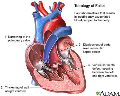
Fetal Tetralogy of Fallot
 36 Fetal Tetralogy of Fallot E.D. Bespalova, R.M. Gasanova, O.A.Pitirimova National Scientific and Practical Center of Cardiovascular Surgery, Moscow Elena D. Bespalova, MD Professor, Director Rena M,
CMR for Congenital Heart Disease
 CMR for Congenital Heart Disease * Second-line tool after TTE * Strengths of CMR : tissue characterisation, comprehensive access and coverage, relatively accurate measurements of biventricular function/
Segmental approach to normal and abnormal situs arrangement - Echocardiography -
 Segmental approach to normal and abnormal situs arrangement - Echocardiography - Jan Marek Great Ormond Street Hospital & Institute of Cardiovascular Sciences, University College London No disclosures
List of Videos. Video 1.1
 Video 1.1 Video 1.2 Video 1.3 Video 1.4 Video 1.5 Video 1.6 Video 1.7 Video 1.8 The parasternal long-axis view of the left ventricle shows the left ventricular inflow and outflow tract. The left atrium
All You Need to Know About Situs and Looping Disorders: Embryology, Anatomy, and Echocardiography
 All You Need to Know About Situs and Looping Disorders: Embryology, Anatomy, and Echocardiography Helena Gardiner Co-Director of Fetal Cardiology, The Fetal Center, University of Texas at Houston Situs
Screening for Critical Congenital Heart Disease
 Screening for Critical Congenital Heart Disease Caroline K. Lee, MD Pediatric Cardiology Disclosures I have no relevant financial relationships or conflicts of interest 1 Most Common Birth Defect Most
Fetal Cardiac Anomaly
 89 Symposium: OB/GY US (Room B) 12 : 10 1 2 : 30 Fetal Cardiac Anomaly 1. One third of all congenital anomalies 2. 6 10/1,000 live births 3. Related with more than 50% of childhood deaths and 20-30% of
Anomalous Systemic Venous Connection Systemic venous anomaly
 World Database for Pediatric and Congenital Heart Surgery Appendix B: Diagnosis (International Paediatric and Congenital Cardiac Codes (IPCCC) and definitions) Anomalous Systemic Venous Connection Systemic
"Lecture Index. 1) Heart Progenitors. 2) Cardiac Tube Formation. 3) Valvulogenesis and Chamber Formation. 4) Epicardium Development.
 Heart Progenitors. 2) Cardiac Tube Formation. 3) Valvulogenesis and Chamber Formation. 4) Epicardium Development.") "Lecture Index 1) Heart Progenitors. 2) Cardiac Tube Formation. 3) Valvulogenesis and Chamber Formation. 4) Epicardium Development. 5) Septation and Maturation. 6) Changes in Blood Flow during Development.
"Lecture Index 1) Heart Progenitors. 2) Cardiac Tube Formation. 3) Valvulogenesis and Chamber Formation. 4) Epicardium Development. 5) Septation and Maturation. 6) Changes in Blood Flow during Development.
DEVELOPMENT OF THE CIRCULATORY SYSTEM L E C T U R E 5
 DEVELOPMENT OF THE CIRCULATORY SYSTEM L E C T U R E 5 REVIEW OF CARDIAC ANATOMY Heart 4 chambers Base and apex Valves Pericardial sac 3 layers: epi, myo, endo cardium Major blood vessels Aorta and its
Giovanni Di Salvo MD, PhD, FESC Second University of Naples Monaldi Hospital
 Giovanni Di Salvo MD, PhD, FESC Second University of Naples Monaldi Hospital VSD is one of the most common congenital cardiac abnormalities in the newborn. It can occur as an isolated finding or in combination
Cardiac ultrasound protocols
 Cardiac ultrasound protocols IDEXX Telemedicine Consultants Two-dimensional and M-mode imaging planes Right parasternal long axis four chamber Obtained from the right side Displays the relative proportions
Before we are Born: Fetal Diagnosis of Congenital Heart Disease
 Before we are Born: Fetal Diagnosis of Congenital Heart Disease Mohamed Sulaiman, MD Pediatric cardiologist Kidsheart: American Fetal & Children's Heart Center Dubai Healthcare City, Dubai-UAE First Pediatric
Cardiac Catheterization Cases Primary Cardiac Diagnoses Facility 12 month period from to PRIMARY DIAGNOSES (one per patient)
") PRIMARY DIAGNOSES (one per patient) Septal Defects ASD (Atrial Septal Defect) PFO (Patent Foramen Ovale) ASD, Secundum ASD, Sinus venosus ASD, Coronary sinus ASD, Common atrium (single atrium) VSD (Ventricular
PRIMARY DIAGNOSES (one per patient) Septal Defects ASD (Atrial Septal Defect) PFO (Patent Foramen Ovale) ASD, Secundum ASD, Sinus venosus ASD, Coronary sinus ASD, Common atrium (single atrium) VSD (Ventricular
Heart Development and Congenital Heart Disease
 Heart Development and Congenital Heart Disease Sally Dunwoodie s.dunwoodie@victorchang.edu.au Developmental and Stem Cell Biology Division Victor Chang Cardiac Research Institute for the heart of Australia...
Transposition of the Great Arteries Preoperative Diagnostic Considerations. John Simpson Evelina Children s Hospital London, UK
 Transposition of the Great Arteries Preoperative Diagnostic Considerations John Simpson Evelina Children s Hospital London, UK Areas to be covered Definitions Scope of occurrence of transposition of the
Foetal Cardiology: How to predict perinatal problems. Prof. I.Witters Prof.M.Gewillig UZ Leuven
 Foetal Cardiology: How to predict perinatal problems Prof. I.Witters Prof.M.Gewillig UZ Leuven Cardiopathies Incidence : 8-12 / 1000 births ( 1% ) Most frequent - Ventricle Septum Defect 20% - Atrium Septum
Adult Congenital Heart Disease: What All Echocardiographers Should Know Sharon L. Roble, MD, FACC Echo Hawaii 2016
 1 Adult Congenital Heart Disease: What All Echocardiographers Should Know Sharon L. Roble, MD, FACC Echo Hawaii 2016 DISCLOSURES I have no disclosures relevant to today s talk 2 Why should all echocardiographers
UPDATE FETAL ECHO REVIEW
 UPDATE 1 FETAL ECHO REVIEW Study Alert for RDCS Candidates D A V I E S P U B L I S H I N G I N C. Fetal Echo Review Study Alert U P D A T E D A U G U S T 1, 2 0 1 2 Nikki Stahl, RT(R)(M)(CT), RDMS, RVT
Congenital Heart Disease An Approach for Simple and Complex Anomalies
 Congenital Heart Disease An Approach for Simple and Complex Anomalies Michael D. Pettersen, MD Director, Echocardiography Rocky Mountain Hospital for Children Denver, CO None Disclosures 1 ASCeXAM Contains
Fetal echocardiography. Ahmeabad. to neonatal series due to high SB rate. Prenatal detection can improve the fetal outcome. (4, 5) 60% 40% 20%
 60% 40% 20%") Guidelines for Fetal Echocardiography 1 Fetal echocardiography Introduction Dr Jayprakash Shah MD; FICOG Chairman Imaging science committee FOGSI Fetal Medicine expert Rajni Hospital, Ahmedabad, Ex sonologist
Guidelines for Fetal Echocardiography 1 Fetal echocardiography Introduction Dr Jayprakash Shah MD; FICOG Chairman Imaging science committee FOGSI Fetal Medicine expert Rajni Hospital, Ahmedabad, Ex sonologist
Pediatric Echocardiography Examination Content Outline
 Pediatric Echocardiography Examination Content Outline (Outline Summary) # Domain Subdomain Percentage 1 Anatomy and Physiology Normal Anatomy and Physiology 10% 2 Abnormal Pathology and Pathophysiology
NASCI 2012 Segmental Analysis
 NASCI 2012 Segmental Analysis Frandics Chan, M.D., Ph.D. Stanford University Medical Center Lucile Packard Department Children s of Radiology Hospital Menagerie of Congenital Cardiac Lesions 1. Absent
Case 47 Clinical Presentation
 93 Case 47 C Clinical Presentation 45-year-old man presents with chest pain and new onset of a murmur. Echocardiography shows severe aortic insufficiency. 94 RadCases Cardiac Imaging Imaging Findings C
Echocardiographic assessment in Adult Patients with Congenital Heart Diseases
 Echocardiographic assessment in Adult Patients with Congenital Heart Diseases Athanasios Koutsakis Cardiologist, Cl. Research Fellow George Giannakoulas Ass. Professor in Cardiology 1st Cardiology Department,
Hypoplastic Left Heart Syndrome: Echocardiographic Assessment
 Hypoplastic Left Heart Syndrome: Echocardiographic Assessment Craig E Fleishman, MD, FACC, FASE Director, Non-invasive Cardiac Imaging The Hear Center at Arnold Palmer Hospital for Children, Orlando SCAI
Congenital Heart Disease: Physiology and Common Defects
 Congenital Heart Disease: Physiology and Common Defects Jamie S. Sutherell, M.D, M.Ed. Associate Professor, Pediatrics Division of Cardiology Director, Medical Student Education in Pediatrics Director,
cardiac imaging planes planning basic cardiac & aortic views for MR
 cardiac imaging planes planning basic cardiac & aortic views for MR Dianna M. E. Bardo, M. D. Assistant Professor of Radiology & Cardiovascular Medicine Director of Cardiac Imaging cardiac imaging planes
CONGENITAL HEART DISEASE (CHD)
") CONGENITAL HEART DISEASE (CHD) DEFINITION It is the result of a structural or functional abnormality of the cardiovascular system at birth GENERAL FEATURES OF CHD Structural defects due to specific disturbance
CONGENITAL HEART DISEASE (CHD) DEFINITION It is the result of a structural or functional abnormality of the cardiovascular system at birth GENERAL FEATURES OF CHD Structural defects due to specific disturbance
Notes: 1)Membranous part contribute in the formation of small portion in the septal cusp.
Membranous part contribute in the formation of small portion in the septal cusp.") Embryology 9 : Slide 16 : There is a sulcus between primitive ventricular and bulbis cordis that will disappear gradually and lead to the formation of one chamber which is called bulboventricular chamber.
Embryology 9 : Slide 16 : There is a sulcus between primitive ventricular and bulbis cordis that will disappear gradually and lead to the formation of one chamber which is called bulboventricular chamber.
3/14/2011 MANAGEMENT OF NEWBORNS CARDIAC INTENSIVE CARE CONFERENCE FOR HEALTH PROFESSIONALS IRVINE, CA. MARCH 7, 2011 WITH HEART DEFECTS
 CONFERENCE FOR HEALTH PROFESSIONALS IRVINE, CA. MARCH 7, 2011 MANAGEMENT OF NEWBORNS WITH HEART DEFECTS A NTHONY C. CHANG, MD, MBA, MPH M E D I C AL D I RE C T OR, HEART I N S T I T U T E C H I LDRE N
Atrial Septal Defects
 Supplementary ACHD Echo Acquisition Protocol for Atrial Septal Defects The following protocol for echo in adult patients with atrial septal defects (ASDs) is a guide for performing a comprehensive assessment
Chapter 2 Cardiac Interpretation of Pediatric Chest X-Ray
 Chapter 2 Cardiac Interpretation of Pediatric Chest X-Ray Ra-id Abdulla and Douglas M. Luxenberg Key Facts The cardiac silhouette occupies 50 55% of the chest width on an anterior posterior chest X-ray
Paediatric Cardiology. Acyanotic CHD. Prof F F Takawira
 Paediatric Cardiology Acyanotic CHD Prof F F Takawira Aetiology Chromosomal Down syndrome, T13, T18 Genetic syndromes (gene defects) Velo-Cardio-facial (22 del) Genetic syndromes (undefined aetiology)
Data Collected: June 17, Reported: June 30, Survey Dates 05/24/ /07/2010
 Job Task Analysis for ARDMS Pediatric Echocardiography Data Collected: June 17, 2010 Reported: Analysis Summary For: Pediatric Echocardiography Exam Survey Dates 05/24/2010-06/07/2010 Invited Respondents
Assessment of fetal heart function and rhythm
 Assessment of fetal heart function and rhythm The fetal myocardium Early Gestation Myofibrils 30% of myocytes Less sarcoplasmic reticula Late Gestation Myofibrils 60% of myocytes Increased force per unit
Common Defects With Expected Adult Survival:
 Common Defects With Expected Adult Survival: Bicuspid aortic valve :Acyanotic Mitral valve prolapse Coarctation of aorta Pulmonary valve stenosis Atrial septal defect Patent ductus arteriosus (V.S.D.)
Congenital Heart Disease Systematic Interpretation of CT Suhny Abbara, MD
 Congenital Heart Disease Systematic Interpretation of CT Suhny Abbara, MD Chief, Cardiothoracic Imaging Division Professor of Radiology UT Southwestern Medical Center, Dallas, TX Suhny.Abbara@UTSouthwestern.edu
The Physiology of the Fetal Cardiovascular System
 The Physiology of the Fetal Cardiovascular System Jeff Vergales, MD, MS Department of Pediatrics Division of Pediatric Cardiology jvergales@virginia.edu Disclosures I serve as the medical director for
CARDIAC AND CORONARY ARTERY ANATOMY NO DISCLOSURES. Axial Anatomy of Heart. Axial Anatomy of Heart. Axial Anatomy of Heart
 CARDIAC AND CORONARY ARTERY ANATOMY NO DISCLOSURES NASCI MEETING, ORLANDO FLORIDA 2009 KOSTAKI G. BIS, MD, FACR DEPARTMENT OF RADIOLOGY WILLIAM BEAUMONT HOSPITAL Royal Oak, Michigan OBJECTIVES CARDIAC
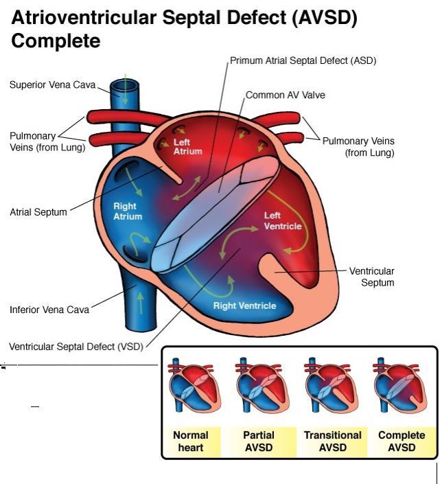
Anatomy of Atrioventricular Septal Defect (AVSD)
") Surgical challenges in atrio-ventricular septal defect in grown-up congenital heart disease Anatomy of Atrioventricular Septal Defect (AVSD) S. Yen Ho Professor of Cardiac Morphology Royal Brompton and
Surgical challenges in atrio-ventricular septal defect in grown-up congenital heart disease Anatomy of Atrioventricular Septal Defect (AVSD) S. Yen Ho Professor of Cardiac Morphology Royal Brompton and
Accuracy of prenatal diagnosis of fetal congenital heart disease by different
 Accuracy of prenatal diagnosis of fetal congenital heart disease by different methods with echocardiography Ying Zhang 1* * Corresponding author Email: baogoubei@hotmail.com Ai-Lu Cai 1 Email: caial_us@hotmail.com
Cardiovascular Pathophysiology: Right to Left Shunts aka Cyanotic Lesions
 Cardiovascular Pathophysiology: Right to Left Shunts aka Cyanotic Lesions Ismee A. Williams, MD, MS iib6@columbia.edu Pediatric Cardiology Learning Objectives To discuss the hemodynamic significance of
Cardiovascular Pathophysiology: Right to Left Shunts aka Cyanotic Lesions Ismee A. Williams, MD, MS Pediatric Cardiology
 Cardiovascular Pathophysiology: Right to Left Shunts aka Cyanotic Lesions Ismee A. Williams, MD, MS iib6@columbia.edu Pediatric Cardiology Learning Objectives To discuss the hemodynamic significance of
British Society of Echocardiography
 British Society of Echocardiography Affiliated to the British Cardiac Society A Minimum Dataset for a Standard Adult Transthoracic Echocardiogram From the British Society of Echocardiography Education
Transposition of the great arteries in the fetus: assessment of the spatial relationships of the arterial trunks by four-dimensional echocardiography
 Ultrasound Obstet Gynecol 2008; 31: 271 276 Published online in Wiley InterScience (www.interscience.wiley.com). DOI: 10.1002/uog.5276 Transposition of the great arteries in the fetus: assessment of the
Congenital Heart Disease: a Pictorial Illustration of Putting Segmental Approach into Practice
 pissn 2384-1095 eissn 2384-1109 imri 2015;19:205-211 http://dx.doi.org/10.13104/imri.2015.19.4.205 Congenital Heart Disease: a Pictorial Illustration of Putting Segmental Approach into Practice Tse Hang
Diagnosis of Congenital Cardiac Defects Between 11 and 14 Weeks Gestation in High-Risk Patients
 Article Diagnosis of Congenital Cardiac Defects Between 11 and 14 Weeks Gestation in High-Risk Patients Zeev Weiner, MD, Abraham Lorber, MD, Eliezer Shalev, MD Objective. To examine the feasibility of
ISUOG Basic Training Distinguishing Between Normal and Abnormal Appearances of the Fetal Anatomy
 ISUOG Basic Training Distinguishing Between Normal and Abnormal Appearances of the Fetal Anatomy Reem S. Abu-Rustum, Lebanon Learning Objective At the end of the lecture you will be able to: Compare the
Fetal Rhythm and Blues
 Fetal Rhythm and Blues John Cotton, MD Professor of Pediatrics Division of Pediatric Cardiology Director, Fetal Cardiology Program UNC Chapel Hill, School of Medicine Objectives To review methods used
HDlive Silhouette Mode With Spatiotemporal Image Correlation for Assessment of the Fetal Heart
 ORIGINAL RESEARCH HDlive Silhouette Mode With Spatiotemporal Image Correlation for Assessment of the Fetal Heart Toshiyuki Hata, MD, PhD, Mohamed Ahmed Mostafa AboEllail, MD, Suraphan Sajapala, MD, Mari
ISUOG Basic Training Distinguishing Between Normal and Abnormal Appearances of the Fetal Anatomy. Basic Training
 ISUOG Distinguishing Between Normal and Abnormal Appearances of the Fetal Anatomy Learning Objective At the end of the lecture you will be able to: Compare the differences between the ultrasound appearances
Introduction to Fetal Medicine. Lloyd R. Feit M.D. Associate Professor of Pediatrics Warren Alpert Medical School Brown University
 Associate Professor of Pediatrics Warren Alpert Medical School Brown University Fetal Cardiology Important in evaluation of high risk pregnancies. Information obtainable in > 95% of patients attempted.
Identification of congenital cardiac malformations by echocardiography in midtrimester fetus*
 Br Heart J 1981; 46: 358-62 Identification of congenital cardiac malformations by echocardiography in midtrimester fetus* LINDSEY D ALLAN, MICHAEL TYNAN, STUART CAMPBELL, ROBERT H ANDERSON From Guy's Hospital;
the Cardiovascular System I
 the Cardiovascular System I By: Dr. Nabil A Khouri MD, MsC, Ph.D MEDIASTINUM 1. Superior Mediastinum 2. inferior Mediastinum Anterior mediastinum. Middle mediastinum. Posterior mediastinum Anatomy of
Preoperative Echocardiographic Assessment of Uni-ventricular Repair
 Preoperative Echocardiographic Assessment of Uni-ventricular Repair Salem Deraz, MD Pediatric Cardiologist, Aswan Heart Centre Magdi Yacoub Heart Foundation Uni-ventricular repair A single or series of
The Fetal Cardiology Program
 The Fetal Cardiology Program at Texas Children s Fetal Center About the program Since the 1980s, Texas Children s Fetal Cardiology Program has provided comprehensive fetal cardiac care to expecting families
ADULT CONGENITAL HEART DISEASE. Stuart Lilley
 ADULT CONGENITAL HEART DISEASE Stuart Lilley More adults than children have congenital heart disease Huge variety of congenital lesions from minor to major Heart failure, re-operation and arrhythmia are
Anatomy & Physiology
 1 Anatomy & Physiology Heart is divided into four chambers, two atrias & two ventricles. Atrioventricular valves (tricuspid & mitral) separate the atria from ventricles. they open & close to control flow
9/8/2009 < 1 1,2 3,4 5,6 7,8 9,10 11,12 13,14 15,16 17,18 > 18. Tetralogy of Fallot. Complex Congenital Heart Disease.
 Current Indications for Pediatric CTA S Bruce Greenberg Professor of Radiology Arkansas Children s Hospital University of Arkansas for Medical Sciences greenbergsbruce@uams.edu 45 40 35 30 25 20 15 10
AbnormalThree-VesselView on Sonography: A Clue to the Diagnosis of Congenital Heart Disease in the Fetus
 rt Pictorial Essay bnormalthree-vesselview on Sonography: Clue to the Diagnosis of Congenital Heart Disease in the Fetus screening tool for major congenital heart diseases [I. 2J. However, anomalies of
Anatomy of the Heart. Figure 20 2c
 Anatomy of the Heart Figure 20 2c Pericardium & Myocardium Remember, the heart sits in it s own cavity, known as the mediastinum. The heart is surrounded by the Pericardium, a double lining of the pericardial
Three-dimensional (3D) and 4D color Doppler fetal echocardiography using spatio-temporal image correlation (STIC)
 and 4D color Doppler fetal echocardiography using spatio-temporal image correlation (STIC)") Ultrasound Obstet Gynecol 2004; 23: 535 545 Published online 6 May 2004 in Wiley InterScience (www.interscience.wiley.com). DOI: 10.1002/uog.1075 Three-dimensional (3D) and 4D color Doppler fetal echocardiography
Ultrasound Obstet Gynecol 2004; 23: 535 545 Published online 6 May 2004 in Wiley InterScience (www.interscience.wiley.com). DOI: 10.1002/uog.1075 Three-dimensional (3D) and 4D color Doppler fetal echocardiography
MEDICAL MANAGEMENT WITH CAVEATS 1. In one study of 50 CHARGE patients with CHD, 75% required surgery. 2. Children with CHARGE may be resistant to chlo
 CARDIOLOGY IN CHARGE SYNDROME: FOR THE PHYSICIAN Angela E. Lin, M.D. Teratology Program/Active Malformation Surveillance, Brigham and Women's Hospital, Old PBBH-B501, 75 Francis St., Boston, MA 02115 alin@partners.org
Accuracy of the Fetal Echocardiogram in Double-outlet Right Ventricle
 Blackwell Publishing IncMalden, USACHDCongenital Heart Disease 2006 The Authors; Journal compilation 2006 Blackwell Publishing, Inc.? 200723237Original ArticleFetal Echocardiogram in Double-outlet Right
Ch.15 Cardiovascular System Pgs {15-12} {15-13}
 Ch.15 Cardiovascular System Pgs {15-12} {15-13} E. Skeleton of the Heart 1. The skeleton of the heart is composed of rings of dense connective tissue and other masses of connective tissue in the interventricular
Surgical Procedures. Direct suture of small ASDs Patch repair Transcatheter closure with a prosthetic device called occluder
 PEDIATRIC Review Surgical Procedures Atrial Septal Defect repair: Direct suture of small ASDs Patch repair Transcatheter closure with a prosthetic device called occluder Balloon atrial septostomy (Rashkind)
Communication of Mitral Valve with Both Ventricles Associated with Double Outlet Right Ventricle
 Communication of Mitral Valve with Both Ventricles Associated with Double Outlet Right Ventricle By RAJENTDRA TANDON, M.D., JAMES H. MOLLR, MD, AND JESSE E. EDWARDS, M.D. SUMMARY A rare case of an infant
human anatomy 2016 lecture thirteen Dr meethak ali ahmed neurosurgeon
 Heart The heart is a hollow muscular organ that is somewhat pyramid shaped and lies within the pericardium in the mediastinum. It is connected at its base to the great blood vessels but otherwise lies
Normal TTE Examination, Doppler Echocardiography and Normal Antegrade Flow Patterns
 Normal TTE Examination, Doppler Echocardiography and Normal Antegrade Flow Patterns Pravin Patil, MD FACC FASE Associate Professor of Medicine Director, Cardiovascular Disease Training Program Lewis Katz
Chapter 14. Circulatory System Images. VT-122 Anatomy & Physiology II
 Chapter 14 Circulatory System Images VT-122 Anatomy & Physiology II The mediastinum Dog heart Dog heart Cat heart Dog heart ultrasound Can see pericardium as distinct bright line Pericardial effusion Fluid
Appendix A.1: Tier 1 Surgical Procedure Terms and Definitions
 Appendix A.1: Tier 1 Surgical Procedure Terms and Definitions Tier 1 surgeries AV Canal Atrioventricular Septal Repair, Complete Repair of complete AV canal (AVSD) using one- or two-patch or other technique,
Improvement in the antenatal detection rate of CHD and its effect on survival in Wales: How can we improve our results further?
 Improvement in the antenatal detection rate of CHD and its effect on survival in Wales: How can we improve our results further? Dr Orhan Uzun Consultant Paediatric Cardiologist UHW Welcome Improvement!
Summary. HVRA s Cardio Vascular Genetic Detailed L2 Obstetrical Ultrasound. CPT 76811, 76825, _ 90% CHD detection. _ 90% DS detection.
 What is the role of fetal echocardiography (2D 76825, cardiovascular color flow mapping 93325) as performed in conjunction with detailed fetal anatomy scan (CPT 76811) now that AIUM requires limited outflow
Anatomy lab -1- Imp note: papillary muscle Trabeculae Carneae chordae tendineae
 Anatomy lab -1- Imp note: the arrangement of this sheet is different than the lab recording, it has been arranged in a certain way to make it easier to study. When you open the left ventricle you can see
Echocardiographic and anatomical correlates in the fetus*
 Br Heart J 1980; : 51 Echocardiographic and anatomical correlates in the fetus* LINDSEY D ALLAN, MICHAEL J TYNAN, STUART CAMPBELL, JAMES L WILKINSON, ROBERT H ANDERSON From King's College Hospital, and
Early fetal echocardiography: congenital heart disease detection and diagnostic accuracy in the hands of an experienced fetal cardiology program
 DOI: 10.1002/pd.4372 ORIGINAL ARTICLE Early fetal echocardiography: congenital heart disease detection and diagnostic accuracy in the hands of an experienced fetal cardiology program Jodi I. Pike, Anita
Lab Activity 23. Cardiac Anatomy. Portland Community College BI 232
 Lab Activity 23 Cardiac Anatomy Portland Community College BI 232 Cardiac Muscle Histology Branching cells Intercalated disc: contains many gap junctions connecting the adjacent cell cytoplasm, creates
Blood supply of the Heart & Conduction System. Dr. Nabil Khouri
 Blood supply of the Heart & Conduction System Dr. Nabil Khouri Arterial supply of Heart Right coronary artery Left coronary artery 3 Introduction: Coronary arteries - VASAVASORUM arising from aortic sinuses
By Dickens ATURWANAHO & ORIBA DAN LANGOYA MAKchs, MBchB CONGENTAL HEART DISEASE
 By Dickens ATURWANAHO & ORIBA DAN LANGOYA MAKchs, MBchB CONGENTAL HEART DISEASE Introduction CHDs are abnormalities of the heart or great vessels that are present at birth. Common type of heart disease
D. PALADINI, M. VASSALLO, G. SGLAVO, C. LAPADULA and P. MARTINELLI
 Ultrasound Obstet Gynecol 2006; 27: 555 561 Published online in Wiley InterScience (www.interscience.wiley.com). DOI: 10.1002/uog.2749 The role of spatio-temporal image correlation (STIC) with tomographic
Case # 1. Page: 8. DUKE: Adams
 Case # 1 Page: 8 1. The cardiac output in this patient is reduced because of: O a) tamponade physiology O b) restrictive physiology O c) coronary artery disease O d) left bundle branch block Page: 8 1.