Transducer Selection. Renal Artery Duplex Exam. Renal Scan. Renal Scan Echogenicity. How to Perform an Optimal Renal Artery Doppler Examination
|
|
|
- Kristian Summers
- 6 years ago
- Views:
Transcription


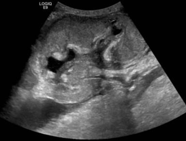
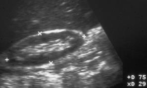
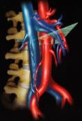
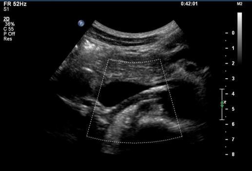
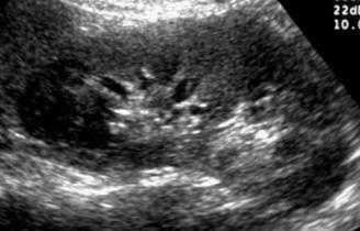
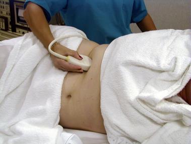
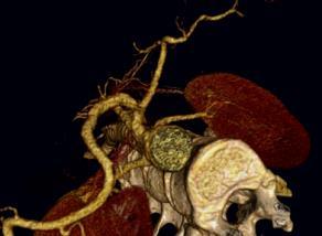
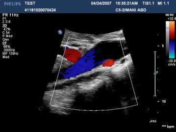
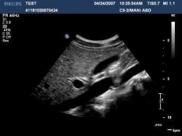
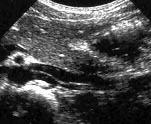
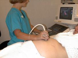
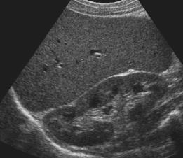
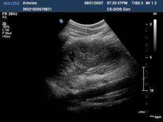
1 How to Perform an Optimal Renal Artery Doppler Examination Director of Ultrasound Education & Quality Assurance Baylor College of Medicine Division of Maternal-Fetal Medicine Maternal Fetal Center Imaging Manager Texas Children s Hospital, Pavilion for Women Houston Texas & Clinical Instructor Thomas Jefferson University Hospital - Radiology Department Philadelphia, Pennsylvania Transducer Selection Curved linear 3-6 MHz Sector 2-4 MHz Exam Renal B-scan should be performed before Doppler assesment Size Cortical Thinning Cortical Echogenicity Hydronephrosis Calcification Masses Renal Scan Renal Scan Echogenicity Glomerulonephritis Glomerulosclerosis Collagen vascular diseases Diabetic nephropathy Chronic pyelonephritis Sickle cell disease Interstitial nephritis AIDS nephropathy 1


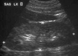
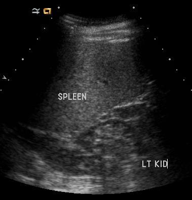
2 Urothelial Thickening Renal Scan Central Collecting System Emphysematous Pyelonephritis Renal Scan Small kidney is irreparable Concentrate Doppler on normal sized kidney 2


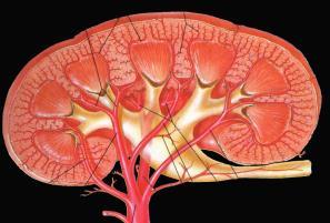
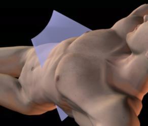
3 Know Your Landmarks Which Windows to Use? Which Windows to Use? Which Windows to Use? 3


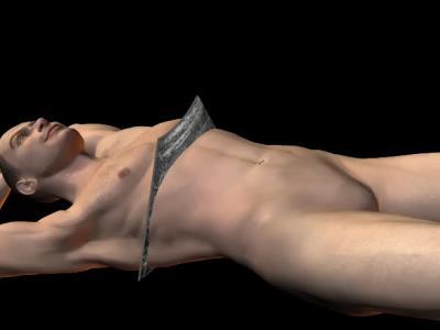
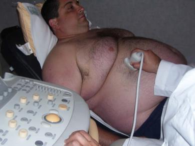
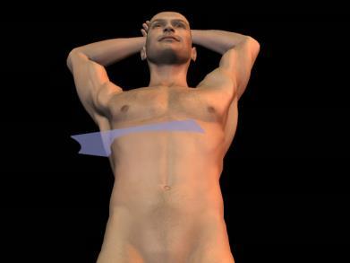
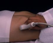
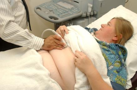
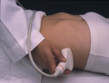
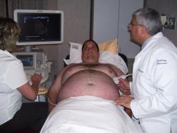
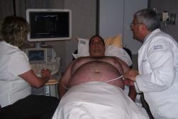
4 Which Positions to Use? Tips on How to Find the Renal Arteries Supine Supine Scanning Difficult Patients 4

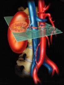
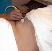
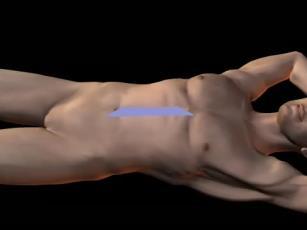
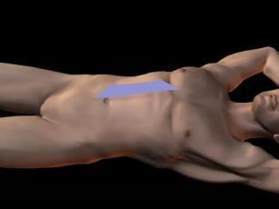
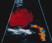
5 Use Multiple Approaches Right Coronal supine / decubitus Use Multiple Approaches Right Coronal supine / decubitus Oblique Oblique SAG TRV Oblique SAG TRV 5


6 Oblique SAG TRV Oblique SAG Tilt Oblique SAG Tilt Oblique SAG Tilt Oblique SAG Tilt Supine 6


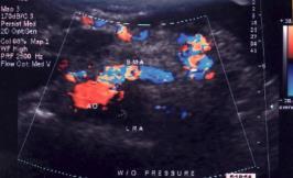
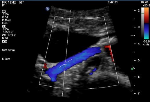
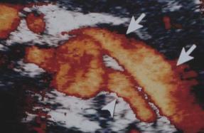
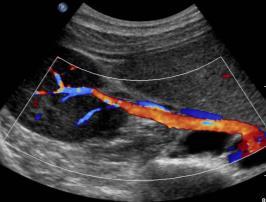
7 Supine Supine SMA A LRV IVC PC LRV SMA A SV LRA Supine Supine Supine Without Any Push 7


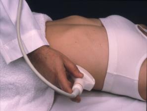
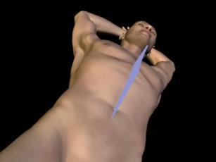
8 With Push Left Coronal supine / decubitus Left Coronal supine / decubitus Left Coronal Left Coronal supine / decubitus 8


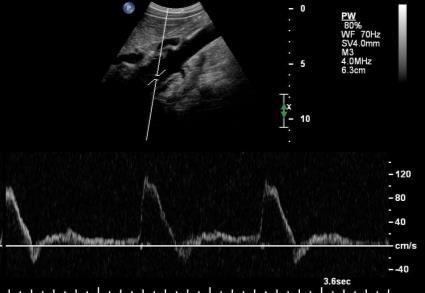
9 Left Coronal Renal Aortic Ratio PSV in MRA jet PSV in or just above the level of renal arteries Renal Aortic Ratio Renal Aortic Ratio 9


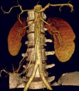
10 Renal Artery Stenosis Renal aortic ratio greater than 3.5 suggests > 60% stenosis Note: presence of aortic atherosclerosis and AAA may falsely lower or elevate the RAR Renal Artery Disease Two most common primary diseases: Atherosclerosis FMD Hypertension & ischemic nephropathy Age > Male Origin of MRA Proximal 1/3 of MRA Progression common Occlusion common Diabetes, aorto-iliac disease, CAD Atherosclerosis Fibromuscular Dysplasia (FMD) Age % female Distal 2/3 of MRA & branches Progression rare Occlusion rare Cause unknown Renal Artery Stenosis - FMD Treatment Percutaneous transluminal angioplasty or stent placement Surgical repair/bypass Cure hypertension Salvage renal function 10


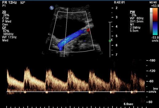
11 Renal artery PSV > 200 cm/s (constitutes > 60% stenosis) Renal artery EDV > 150 cm/s (constitutes > 80% stenosis) 11

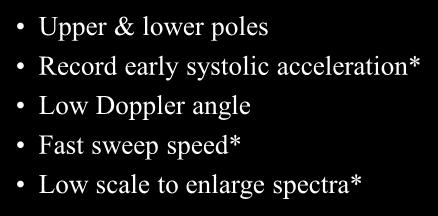
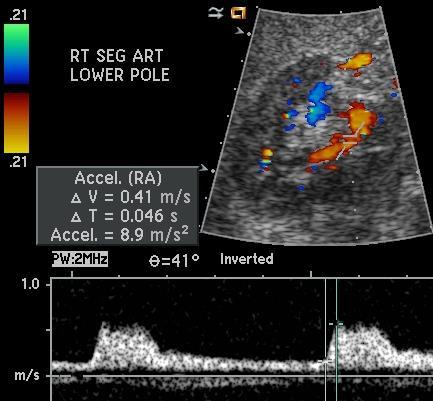
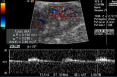
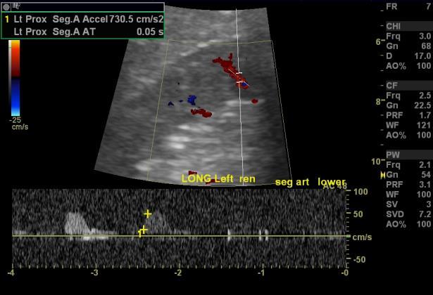
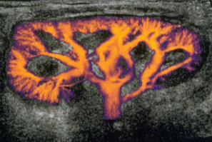
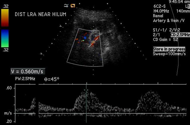
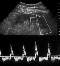
12 Segmental Artery Normal Acceleration Index > 300 cm/s 2 Intrarenal Evaluation Upper & lower poles Record early systolic acceleration* Low Doppler angle Fast sweep speed* Low scale to enlarge spectra* 12


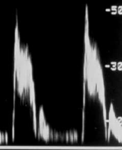
13 Rate of Systolic Acceleration Acceleration time Acceleration index Length of time from onset of systole to ESP Acceleration Time Acceleration Index Slope of line from onset of systole to ESP Stavros, Radiology: 1992 Stavros, Radiology: 1992 Onset of systole ESP SP Segmental Arteries * * Incorrect * Correct 13


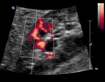
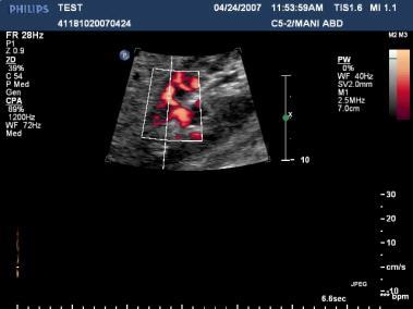
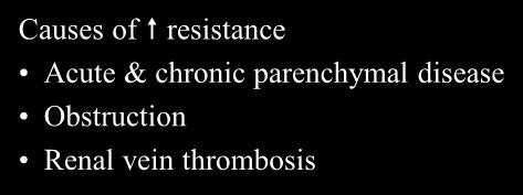
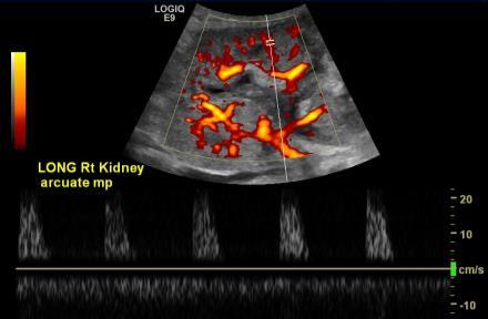
14 Segmental Arteries Segmental Arteries AI is decreased to < 300 cm/s 2 Pre Post Angioplasty Arcuate Artery Arcuate Artery RI < 0.75 Arcuate Artery Causes of resistance Acute & chronic parenchymal disease Obstruction Renal vein thrombosis 14


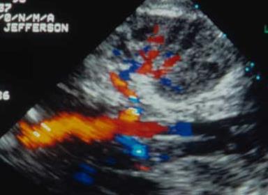
15 Caution Rely on Multiple Parameters for the Diagnosis of RAS Renal Artery Doppler 1. Renal artery to aortic ratio (RAR) 2. Increased peak systolic velocity 3. Acceleration time or systolic rise time + B-mode assessment Follow renal artery from aorta to hilum sample every cm, any area of color aliasing Narrowing by color Doppler Disturbed flow downstream Spectral Doppler Low PRF before aliasing Lowest possible wall filter Small sample volume Obtain spectral Doppler from proximal, mid & distal main renal artery as well as segmental & arcuate arteries Diagnostic Criteria Renal aortic ratio > 3.5 suggests > 60% stenosis Thank You Renal artery PSV > 200 cm/s constitutes > 60% stenosis Renal artery EDV > 150 cm/s constitutes 80% stenosis Acceleration index < 300 cm/s 2 15
What effects will proximal or distal disease have on an waveform?
 Spectral Doppler Interpretation Director Director of of Ultrasound Ultrasound Education Education & & Quality Quality Assurance Assurance Baylor Baylor College College of of Medicine Medicine Division
What effects will proximal or distal disease have on a waveform?
 Spectral Doppler Interpretation Director of Ultrasound Education & Quality Assurance Baylor College of Medicine Division of Maternal-Fetal Medicine Maternal Fetal Center Imaging Manager Texas Children
Duplex Ultrasound of the Renal Arteries. Duplex Ultrasound. In the Beginning
 Duplex Ultrasound of the Renal Arteries DIMENSIONS IN HEART AND VASCULAR CARE 2013 PENN STATE HEART AND VASCULAR INSTITUTE ROBERT G. ATNIP MD PROFESSOR OF SURGERY AND RADIOLOGY Duplex Ultrasound Developed
Ultrasound of the Renal Arteries
 Ultrasound of the Renal Arteries Greg Curry Vascular Ultrasound Workshop Aug 2017 The Examination Technique Pathophysiology Role of US then and now Background Live Scanning Ultrasound Population: 20% Hypertensive
11 TH ANNUAL VASCULAR NONINVASIVE TESTING SYMPOSIUM NOVEMBER 10, 2018
 11 TH ANNUAL VASCULAR NONINVASIVE TESTING SYMPOSIUM NOVEMBER 10, 2018 RENAL ARTERY DISEASE AND RENOVASCULAR HYPERTENSION 1 WHAT IS RENOVASCULAR HYPERTENSION? https://my.clevelandclinic.org/health/diseases/16459-renovascular-hypertension
Abdominal Doppler Mastering the next level of vascular anatomy in the belly. Cindy A. Owen, RDMS, RVT
 Abdominal Doppler Mastering the next level of vascular anatomy in the belly Cindy A. Owen, RDMS, RVT Introduction Abdominal Doppler is a tough exam Success is dependent on: Patient body habitus Patient
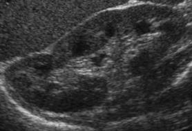
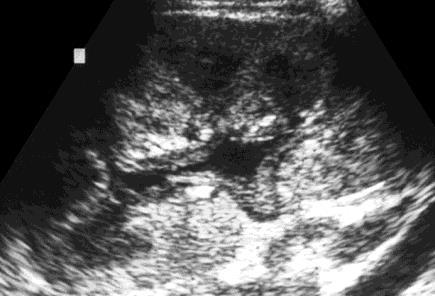
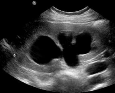
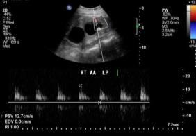
Case 8038 Renal allograft complicated with renal artery stenosis
 Case 8038 Renal allograft complicated with renal artery stenosis Santiago I, Canelas A, Pinto AP Section: Cardiovascular Published: 2009, Nov. 30 Patient: 61 year(s), male Clinical History A 61-year-old
What Do We Know? Disclosure Statement: 3/11/2015. Deep abdominal imaging
 Marsha M. Neumyer, BS, RVT, FSVU, FSDMS, FAIUM International Director Vascular Diagnostic Educational Services Vascular Resource Associates Harrisburg, PA Disclosure Statement: CME Calendar QR Code Marsha
No financial or commercial relationships to disclose
 Deanna New, RVT No financial or commercial relationships to disclose IAC REQUIREMENTS: The main duty of a sonographer is to make the physician or radiologists job easier by capturing images and doing
MESENTERIC ISCHEMIA. Phillip J Bendick, PhD
 MESENTERIC ISCHEMIA Phillip J Bendick, PhD Arterial Celiac - Hepatic - Splenic Superior Mesenteric Artery Inferior Mesenteric Artery Venous Mesenteric system Porto - hepatic system Inferior Vena Cava Acute
Section II: Patient Interview Grade: 5
 Only written competency completed with this EXACT form will be accepted for grading. No modifications to the LAYOUT of the form will be accepted for a written competency. Failure to comply will result
US of Renovascular Hypertension. Jonathan R. Dillman, MD, MSc Associate Professor Director, Thoracoabdominal Imaging
 US of Renovascular Hypertension Jonathan R. Dillman, MD, MSc Associate Professor Director, Thoracoabdominal Imaging Disclosures Nothing Relevant Unrelated grant funding Siemens US Toshiba US Objectives
Renal Artery Ultrasound
 Ask the Expert Renal Artery Ultrasound Looking for windows of opportunity... by Peter Coombs AMS Peter Coombs is Co-ordinator of the Graduate Diploma of Ultrasound at Monash University, Melbourne. Dear
Vascular Ultrasound: Current state, current needs, future directions
 Vascular Ultrasound: Current state, current needs, future directions Laurence Needleman, MD Thomas Jefferson University Hospitals Sidney Kimmel Medical College of Thomas Jefferson University Disclosures
GUNDERSEN/LUTHERAN ULTRASOUND DEPARTMENT POLICY AND PROCEDURE MANUAL
 GUNDERSEN/LUTHERAN ULTRASOUND DEPARTMENT POLICY AND PROCEDURE MANUAL SUBJECT: Carotid Duplex Ultrasound SECTION: Vascular Ultrasound ORIGINATOR: Deborah L. Richert, BSVT, RDMS, RVT DATE: October 15, 2015
Visceral Vascular Ultrasound. Joel Thompson, MD, MPH Borg & Ide Imaging
 Visceral Vascular Ultrasound Joel Thompson, MD, MPH Borg & Ide Imaging Objectives: Review major abdominal vascular structures Identify normal peak systolic velocity (PSV) for major abdominal arteries.
Recommendations for Follow-up After Vascular Surgery Arterial Procedures SVS Practice Guidelines
 Recommendations for Follow-up After Vascular Surgery Arterial Procedures 2018 SVS Practice Guidelines vsweb.org/svsguidelines About the guidelines Published in the July 2018 issue of Journal of Vascular
Vascular Sonography Examination
 Vascular Sonography Examination The purpose of The American Registry of Radiologic Technologists (ARRT ) Vascular Sonography Examination is to assess the knowledge and cognitive skills underlying the intelligent
Goals. Access flow and renal artery stenosis evaluation by Doppler ultrasound. Reimbursement. WHY use of Doppler Ultrasound
 Access flow and renal artery stenosis evaluation by Doppler ultrasound Adina Voiculescu, MD Interventional Nephrology Brigham and Women s Hospital Boston Instructor at Harvard Medical School Understand
Optimising your Doppler settings for an accurate PI. Alison McGuinness Mid Yorks Hospitals
 Optimising your Doppler settings for an accurate PI Alison McGuinness Mid Yorks Hospitals Applications Both maternal uterine and fetal circulations can be studied with doppler sonography Uterine arteries
NON-ATHEROSCLEROTIC PATHOLOGY OF THE CAROTID ARTERIES
 NON-ATHEROSCLEROTIC PATHOLOGY OF THE CAROTID ARTERIES Leslie M. Scoutt, MD, FACR Professor of Diagnostic Radiology & Surgery Vice Chair, Dept of Radiology & Biomedical Imaging Chief, Ultrasound Section
Carotid Abnormalities Coils, Kinks and Tortuosity David Lorelli M.D., RVT, FACS Michigan Vascular Association Conference Saturday, October 20, 2012
 Carotid Abnormalities Coils, Kinks and Tortuosity David Lorelli M.D., RVT, FACS Michigan Vascular Association Conference Saturday, October 20, 2012 Page 1 Table of Contents Carotid Anatomy Carotid Duplex
STRUCTURED EDUCATION REQUIREMENTS IMPLEMENTATION DATE: JULY 1, 2016
 STRUCTURED EDUCATION REQUIREMENTS Vascular Sonography The purpose of structured education is to provide the opportunity for individuals to develop mastery of discipline-specific knowledge that, when coupled
Deb Coghlan AMS (Vascular and General ) Brisbane, Australia
 Brisbane, Australia") Deb Coghlan AMS (Vascular and General ) Brisbane, Australia ANEURYSMAL DIISEASE The infrarenal aorta enlarges with age, and is the commonest site for arterial aneurysms. An aneurysm is a permanent focal
Deb Coghlan AMS (Vascular and General ) Brisbane, Australia ANEURYSMAL DIISEASE The infrarenal aorta enlarges with age, and is the commonest site for arterial aneurysms. An aneurysm is a permanent focal
Image Formation (10) 2 Evaluation and Selection of Representative Images (10)
 2 Evaluation and Selection of Representative Images (10)") STRUCTURED SELF ASSESSMENT CONTENT SPECIFICATIONS SSA LAUNCH DATE: JANUARY 1, 2018 Vascular Sonography The purpose of continuing qualifications requirements (CQR) is to assist registered technologists
STRUCTURED SELF ASSESSMENT CONTENT SPECIFICATIONS SSA LAUNCH DATE: JANUARY 1, 2018 Vascular Sonography The purpose of continuing qualifications requirements (CQR) is to assist registered technologists
Renal artery stenosis
 Renal artery stenosis Dr. Alexander Woywodt Consultant Renal Physician, Royal Preston Hospital Preston, 31.10.2007 Menu anatomy of the renal arteries diseases of the large renal arteries atherosclerotic
Renal Artery Stenosis With Severe Hypertension: A Case Report
 CASE REPORT Renal Artery Stenosis With Severe Hypertension: A Case Report Suwaid MA ABSTRACT Background: Renal artery stenosis (RAS) is found in 77% of hypertensive patients and is responsible for 1-2%
1. Long images of aorta (prox, mid, and dist) with AP measurements. 2. Trans images of aorta (prox, mid, and dist) with R/L measurements.
 with AP measurements. 2. Trans images of aorta (prox, mid, and dist) with R/L measurements.") Aorta 1. Long images of aorta (prox, mid, and dist) with AP measurements. 2. Trans images of aorta (prox, mid, and dist) with R/L measurements. 3. Long images of R/L common iliac arteries with AP measurements.
Aorta 1. Long images of aorta (prox, mid, and dist) with AP measurements. 2. Trans images of aorta (prox, mid, and dist) with R/L measurements. 3. Long images of R/L common iliac arteries with AP measurements.
Scanning Mesenteric and Hypogastric Artery Aneurysms
 Scanning Mesenteric and Hypogastric Artery Aneurysms Marsha M. Neumyer, BS, RVT, FSVU, FSDMS, FAIUM International Director Vascular Diagnostic Education Services Vascular Resource Associates Harrisburg,
Abdominal Ultrasound : Aorta, Kidneys, Bladder
 Abdominal Ultrasound : Aorta, Kidneys, Bladder Nilam J. Soni, MD, MSc Associate Professor of Medicine Divisions of Hospital Medicine and Pulmonary/Critical Care Medicine Department of Medicine University
Beyond Stenosis Severity: Top 5 Important Duplex Characteristics to Identify in a Patient with Carotid Disease
 Beyond Stenosis Severity: Top 5 Important Duplex Characteristics to Identify in a Patient with Carotid Disease Jan M. Sloves RVT, RCS, FASE Technical Director New York Cardiovascular Associates Disclosures
Abdomen and Retroperitoneum Ultrasound Protocols
 Abdomen and Retroperitoneum Ultrasound Protocols Reviewed By: Anna Ellermeier, MD Last Reviewed: March 2018 Contact: (866) 761-4200, Option 1 **NOTE for all examinations: 1. If documenting possible flow
Treatment of choice for end stage renal disease Imaging to establish baseline and diagnosis of potential complications Review common surgical
 Treatment of choice for end stage renal disease Imaging to establish baseline and diagnosis of potential complications Review common surgical techniques Review normal appearance Discuss US diagnosis of
TAVR TTE INTERROGATION BY ALAN MATTHEWS
 TAVR TTE INTERROGATION BY ALAN MATTHEWS KEYS TO ACCURATE ASSESSMENT EDWARDS SAPIEN VALVE 3 PHASES OF TAVR TTE Evaluation (Qualifying) Placement (Intraoperative) Follow-up (Post-Op) GOALS High quality TTE
Vascular Technology Examination Content Outline
 Vascular Technology Examination Content Outline (Outline Summary) # Domain Subdomain Percentage 1 Normal Anatomy, Perfusion, and Function Evaluate normal anatomy, perfusion, function 2 Pathology, Perfusion,
HD Scanning: Velocities and Volume Flow
 HD Scanning: Velocities and Volume Flow Non-Invasive Lab Symposium West Orange, NJ April 27, 2018 Volume Flow Cindy Sturt, MD, FACS, RVT 500,000 Americans on dialysis 20-25% annual mortality 65% 5 year
Vascular Imaging in the Pediatric Abdomen. Jonathan Swanson, MD
 Vascular Imaging in the Pediatric Abdomen Jonathan Swanson, MD Goals and Objectives To understand the imaging approach, appearance, and clinical manifestations of the common pediatric abdominal vascular
Case Report 1. CTA head. (c) Tele3D Advantage, LLC
 Tele3D Advantage, LLC") Case Report 1 CTA head 1 History 82 YEAR OLD woman with signs and symptoms of increased intra cranial pressure in setting of SAH. CT Brain was performed followed by CT Angiography of head. 2 CT brain Extensive
Case Report 1 CTA head 1 History 82 YEAR OLD woman with signs and symptoms of increased intra cranial pressure in setting of SAH. CT Brain was performed followed by CT Angiography of head. 2 CT brain Extensive
OPTIMIZING ECHO ACQUISTION FOR STRAIN AND DIASTOLOGY
 OPTIMIZING ECHO ACQUISTION FOR STRAIN AND DIASTOLOGY October 8, 2017 Deborah Agler, ACS, RDCS, FASE Coordinator of Education and Training Cleveland Clinic General Principles Diastology Clinical Data Heart
Guidelines, Policies and Statements D5 Statement on Abdominal Scanning
 Guidelines, Policies and Statements D5 Statement on Abdominal Scanning Disclaimer and Copyright The ASUM Standards of Practice Board have made every effort to ensure that this Guideline/Policy/Statement
Abdominal Ultrasonography
 Abdominal Ultrasonography David A. Masneri, DO, FACEP, FAAEM Assistant Professor of Emergency Medicine Assistant Director, Emergency Medicine Residency Medical Director, Operational Medicine Division Center
Abdominal Aortic Aneurysm (AAA)
") Abdominal Aortic Aneurysm (AAA) Vascular Workshop: Objectives Anatomy Keith VanHaltren Indications Technique Cases Abdominal Aorta: Normal Size Abdominal aortic aneurysm: Definition Normal diameter of
Abdominal Aortic Aneurysm (AAA) Vascular Workshop: Objectives Anatomy Keith VanHaltren Indications Technique Cases Abdominal Aorta: Normal Size Abdominal aortic aneurysm: Definition Normal diameter of
Fibromuscular Dysplasia. Miranda Forrest Baker College
 Fibromuscular Dysplasia Miranda Forrest Baker College Overview Case Study Patient Information Exam Images Findings FMD Types Signs and Symptoms Treatment Case Study Patient Information Female 57 years
Indications: following: embolization. artery that has diseases 5. The evaluation. of suspected. such entities. a cold hand. biopsy
 Peripheral Arterial Ultrasound Protocol Using Color and Spectral Doppler Reviewed by: Mark Yuhasz, MD Last Review Date: January 2015 Contact: (866) 761 4200, Option 1 Indications: The indications for peripheral
Carotid Artery Doppler
 Carotid Artery Doppler Patient Position supine or semisupine head slightly hyper extended rotated 45 away from the side being examined. Higher frequency linear transducers (7 MHz) Vessels should be imaged
Peripheral Vascular Disease
 Peripheral artery disease (PAD) results from the buildup of plaque (atherosclerosis) in the arteries of the legs. For people with PAD, symptoms may be mild, requiring no treatment except modification of
Mesenteric/Splanchnic Artery Duplex Imaging
 VASCULAR TECHNOLOGY PROFESSIONAL PERFORMANCE GUIDELINES Mesenteric/Splanchnic Artery Duplex Imaging This Guideline was prepared by members of the Society for Vascular Ultrasound (SVU) as a template to
Introduction History Preceded by Arterial Doppler and ABI Indications
 Elise Brady, RVT, RDMS Introduction History Preceded by Arterial Doppler and ABI Indications 1) Abnormal ABI (within 2weeks of duplex) 2) Abnormal Doppler waveforms 3) Claudication 4) History of PVD 5)
Lower Extremity Artery: Physiologic Testing
 Master Title Ultrasound for Initial Evaluation of Lower Extremity Arterial Occlusive Disease: WHY? Gregory L. Moneta MD Professor and Chief Knight Cardiovascular Institute Division of Vascular Surgery
Imaging Strategy For Claudication
 Who are the Debators? Imaging Strategy For Claudication Duplex Ultrasound Alone is Adequate to Select Patients for Endovascular Intervention - Pro: Dennis Bandyk MD No Disclosures PRO - Vascular Surgeon
Ultrasound 10/1/2014. Basic Echocardiography for the Internist. Mechanical (sector) transducer Piezoelectric crystal moved through a sector sweep
 transducer Piezoelectric crystal moved through a sector sweep") Ultrasound Basic Echocardiography for the Internist Carol Gruver, MD, FACC UT Erlanger Cardiology Mechanical wave of compression and rarefaction Requires a medium for transmission Ultrasound frequency
Ultrasound Basic Echocardiography for the Internist Carol Gruver, MD, FACC UT Erlanger Cardiology Mechanical wave of compression and rarefaction Requires a medium for transmission Ultrasound frequency
Pitfalls in the evaluation of carotid artery stenosis. Serge Kownator «Centre Cardiologique et Vasculaire» Thionville, Fr
 Pitfalls in the evaluation of carotid artery stenosis Serge Kownator «Centre Cardiologique et Vasculaire» Thionville, Fr Disclosure Statement of Financial Interest I currently have, or have had over the
Basic Abdominal Sonography
 24S Basic Abdominal Sonography Procedural Overview JOHN FATCHETT II, RDMS is provided. Patient preparation (i.e., fasting) scanning techniques, spleen, transducer. evaluation of abdominal anatomy in the
What is Ultrasound? What is Ultrasound? B A. Basic Principles of Ultrasound. Basic Principles of Ultrasound. Basic Principles of Ultrasound
 Introduction to Ultrasound Principles Mani Montazemi, RDMS Baylor College of Medicine Division of Maternal-Fetal Medicine Department of Obstetrics and Gynecology Manager, Maternal Fetal Center Imaging
Hemodynamic Assessment. Assessment of Systolic Function Doppler Hemodynamics
 Hemodynamic Assessment Matt M. Umland, RDCS, FASE Aurora Medical Group Milwaukee, WI Assessment of Systolic Function Doppler Hemodynamics Stroke Volume Cardiac Output Cardiac Index Tei Index/Index of myocardial
Basic Ultrasound Physics Board Review Questions
 Basic Ultrasound Physics Board Review Questions Sidney K. Edelman, PhD ESP Ultrasound The Woodlands, TX Question 1 What is the wavelength of 2 MHz sound in soft tissue? 1. 1.54 mm 2. 0.75 mm 3. 0.75 cm
Evaluation of Colour Duplex Ultrasound Scanning in Diagnosis of Renal Artery Stenosis, Compared to Angiography: A Prospective Study on 53 Patients
 Eur J Vasc Endovasc Surg 14, 305-309 (1997) Evaluation of Colour Duplex Ultrasound Scanning in Diagnosis of Renal Artery Stenosis, Compared to Angiography: A Prospective Study on 53 Patients M. Mollo,
Duplex ultrasound is first-line imaging for all
 Our Protocol for Transabdominal Pelvic Vein Duplex Ultrasound A summary of s protocol for pelvic vein duplex ultrasonography, including equipment, patient positioning, ultrasound settings, and technique.
Current Role of Renal Artery Stenting in Patients with Renal Artery Stenosis
 Current Role of Renal Artery Stenting in Patients with Renal Artery Stenosis Young-Guk Ko, M.D. Severance Cardiovascular Hospital, Yonsei University College of Medicine, Seoul, Korea Etiology Fibromuscular
Radiologic Importance of a High- Resistive Vertebral Artery Doppler Waveform on Carotid Duplex Ultrasonography
 CME Article Radiologic Importance of a High- Resistive Vertebral Artery Doppler Waveform on Carotid Duplex Ultrasonography Esther S. H. Kim, MD, MPH, Megan Thompson, Kristine M. Nacion, BA, Carmel Celestin,
Subclavian artery Stenting
 Subclavian artery Stenting Etiology Atherosclerosis Takayasu s arteritis Fibromuscular dysplasia Giant Cell Arteritis Radiation-induced Vascular Injury Thoracic Outlet Syndrome Neurofibromatosis Incidence
Diagnosis of Renal Artery Stenosis (RAS)
") May 2001 Diagnosis of Renal Artery Stenosis (RAS) Kurt Fink, Harvard Medical School, Year III Epidemiology Hypertension -Affects 60 million Americans Essential HTN >95% of cases Secondary HTN 1-5% of cases
May 2001 Diagnosis of Renal Artery Stenosis (RAS) Kurt Fink, Harvard Medical School, Year III Epidemiology Hypertension -Affects 60 million Americans Essential HTN >95% of cases Secondary HTN 1-5% of cases
The role of ultrasound duplex in endovenous procedures
 The role of ultrasound duplex in endovenous procedures Neophytos A. Zambas MD, PhD Vascular Surgeon Polyclinic Ygia, Limassol, Cyprus ΚΕΑΕΧ ΚΥΠΡΙΑΚΗ ΕΤΑΙΡΕΙΑ ΑΓΓΕΙΑΚΗΣ ΚΑΙ ΕΝΔΑΓΓΕΙΑΚΗΣ ΧΕΙΡΟΥΡΓΙΚΗΣ Pre
Carotid Doppler: Doppler wave forms obtained from the common, external and internal carotid arteries. As well as the vertebral and subclavian
 Competency Carotid Doppler: Doppler wave forms obtained from the common, external and internal carotid arteries. As well as the vertebral and subclavian arteries. Preferred angle is 60 degrees or less.
8/20/18. The Doppler Effect. Objectives. What is the Doppler Effect. Doppler principles. Spectral Waveform. Image recognition. Vascular Ultrasound
 Vascular Ultrasound: Physics and Haemodynamics Objectives Doppler principles Spectral Waveform Key factors Haemodynamics: Stenosis Waveforms Image recognition Vascular Ultrasound: A flawed paradigm What
An Overview of Post-EVAR Endoleaks: Imaging Findings and Management. Ravi Shergill BSc Sean A. Kennedy MD Mark O. Baerlocher MD FRCPC
 An Overview of Post-EVAR Endoleaks: Imaging Findings and Management Ravi Shergill BSc Sean A. Kennedy MD Mark O. Baerlocher MD FRCPC Disclosure Slide Mark O. Baerlocher: Current: Consultant for Boston
Abdominal Exam: The examination of the abdomen used by physicians to detect an abdominal aortic aneurysm.
 Glossary of Terms Abdominal Exam: The examination of the abdomen used by physicians to detect an abdominal aortic aneurysm. Angiogram: A diagnostic test requiring the insertion of a catheter into an artery
AIUM Practice Guideline for the Performance of Renal Artery Duplex Sonography
 AIUM Practice Guideline for the Performance of Renal Artery Duplex Sonography 2008 by the American Institute of Ultrasound in Medicine The American Institute of Ultrasound in Medicine (AIUM) is a multidisciplinary
Multislice CTA for Renal Artery Stenting
 Multislice CT for Renal rtery Stenting How CT can be a useful modality for diagnosing and managing renal artery stenosis for stent placement. Y MICHEL WHOLEY, MD, M; JMES WU, ; WILLIM C.L. WU, MD, FCC;
Carotid Duplex: Beyond Stenosis Ido Weinberg, MD Vascular Medicine Massachusetts General Hospital Assistant Professor of Medicine Harvard Medical
 Carotid Duplex: Beyond Stenosis Ido Weinberg, MD Vascular Medicine Massachusetts General Hospital Assistant Professor of Medicine Harvard Medical School Boston, Massachusetts Disclosures I do not have
Abdominal Aortic Doppler Waveform in Patients with Aorto-iliac Disease
 Eur J Vasc Endovasc Surg (2010) 39, 714e718 Abdominal Aortic Doppler Waveform in Patients with Aorto-iliac Disease G. Styczynski a, *, C. Szmigielski a, J. Leszczynski b, A. Kuch-Wocial a, M. Szulc a a
Renal Doppler Hospital Based Study and Review of Literature
 ORIGINAL RESEARCH www.ijcmr.com Hospital Based Study and Review of Literature Jahangeer Ahmad Bhat 1, Murassa Shamshad 2, Aresalan Malik 1, Iqbal Bhat 3, Iqbal Dar 3 ABSTRACT Introduction: Study was done
Radiologic Evaluation of Peripheral Arterial Disease
 January 2003 Radiologic Evaluation of Peripheral Arterial Disease Grace Tye, Harvard Medical School Year III Patient D.M. CC: 44 y/o male with pain in his buttocks Occurs after walking 2 blocks. Pain is
RADIOLOGY HEAD & NECK IMAGING. Iranian Journal of. Hadi Rokni Yazdi 1*, Safoura Faraji 2, Farokhlegha Ahmadi 3, Reza Shahmirzae 4
 HEAD & NECK IMAGING Iran J Radiol. 2012;9(1):12-16. DOI: 10.5812/iranjradiol.6730 Iranian Journal of RADIOLOGY RADIOLOGYwww.iranjradiol.com Color Doppler Indices of Orbital Arterial Flow in End-Stage Renal
Vikram Dogra, M.D. Professor of Radiology, Urology & BME Department of Imaging Sciences University Of Rochester Medical Center
 Ultrasound of the Scrotum Vikram Dogra, M.D. Professor of Radiology, Urology & BME Department of Imaging Sciences University Of Rochester Medical Center Etiologies of Acute Scrotal Pain Epididymitis/Orchitis
Postoperative AV Fistula Evaluation. Postoperative examination protocol. Postoperative AVF Protocol. Hemodialysis Access Surveillance
 Hemodialysis Access Surveillance Postoperative AV Fistula Evaluation Failure of maturation Stenosis Perigraft mass/fluid collection Joseph L. Mills, Sr., M.D. Professor of Surgery Chief, Division of Vascular
Pre-and Post Procedure Non-Invasive Evaluation of the Patient with Carotid Disease
 Pre-and Post Procedure Non-Invasive Evaluation of the Patient with Carotid Disease Michael R. Jaff, D.O., F.A.C.P., F.A.C.C. Assistant Professor of Medicine Harvard Medical School Director, Vascular Medicine
Enhancement of Cranial US: Utility of Supplementary Acoustic Windows and Doppler Harriet J. Paltiel, MD
 Enhancement of Cranial US: Utility of Supplementary Acoustic Windows and Doppler Harriet J. Paltiel, MD Boston Children s Hospital Harvard Medical School None Disclosures Conventional US Anterior fontanelle
US in non-traumatic acute abdomen. Lalita, M.D. Radiologist Department of radiology Faculty of Medicine ChiangMai university
 US in non-traumatic acute abdomen Lalita, M.D. Radiologist Department of radiology Faculty of Medicine ChiangMai university Sagittal Orientation Transverse (Axial) Orientation Coronal Orientation Intercostal
Abdominal ultrasound:
 Abdominal ultrasound: Non-traumatic acute abdomen Wittanee Na-ChiangMai, MD Department of Radiology ChiangMai University 26/04/2017 Contents Technique of examination Normal anatomy Emergency conditions
RENAL AND MESENTERIC ARTERY STENTS Are There Standard Velocity Criteria for Restenosis?
 RENAL AND MESENTERIC ARTERY STENTS Are There Standard Velocity Criteria for Restenosis? R. Eugene Zierler, M.D. The D. E. Strandness, Jr. Vascular Laboratory University of Washington Medical Center Division
Ultrasound Evaluation after EVAR: (Trying to) Let the CAT Scan Out of the Bag
 Let the CAT Scan Out of the Bag") Ultrasound Evaluation after EVAR: (Trying to) Let the CAT Scan Out of the Bag Joseph-Vincent V. Blas, MD Division of Vascular Surgery Department of Surgery Greenville Health System University of South
Ultrasound Evaluation after EVAR: (Trying to) Let the CAT Scan Out of the Bag Joseph-Vincent V. Blas, MD Division of Vascular Surgery Department of Surgery Greenville Health System University of South
Physician s Vascular Interpretation Examination Content Outline
 Physician s Vascular Interpretation Examination Content Outline (Outline Summary) # Domain Subdomain Percentage 1 2 3 4 5 6 Cerebrovascular Abdominal Peripheral Arterial - Duplex Imaging Peripheral Arterial
Disclosure Statement:
 Marsha M. Neumyer, BS, RVT, FSVU, FSDMS, FAIUM International Director Vascular Diagnostic Educational Services Vascular Resource Associates Harrisburg, PA Disclosure Statement: CME Calendar QR Code Marsha
Vascular Surgery Cases: Detours. Brian F. Stull, RDMS, RVT UNC REX Healthcare Vascular Specialists
 Vascular Surgery Cases: Detours Brian F. Stull, RDMS, RVT UNC REX Healthcare Vascular Specialists Brian.Stull@Unchealth.unc.edu Objectives Anatomy of a bypass graft Where does it connect, where does it
RPVI Exam Review ecourse
 RPVI Exam Review ecourse The RPVI Exam Review ecourse consists of ten Vascular Physics Modules and fourteen Vascular Specialty Modules. Detailed descriptions of module content are listed below. During
Acute Type B dissection. Closure of the infra diaphragmatic tear: how and when?
 Acute Type B dissection. Closure of the infra diaphragmatic tear: how and when? Prof. Olgierd Rowiński II Department of Clinical Radiology Medical University of Warsaw Disclosure Speaker name: Olgierd
Sonography in Renovascular Hypertension
 Review rticle Sonography in Renovascular Hypertension Hsin-Yi Lee, MD, Edward G. Grant, MD Objective. To familiarize practitioners with different sonographic manifestations of renal artery compromise and
Abdominal Ultrasound
 Abdominal Ultrasound Imaging Control Buttons Depth The organ imaged should take up 3/4 of the screen Frequency = Penetration Use high frequencies (harmonics) for fluid filled and superficial structures
CT abdomen and pelvis
 CT abdomen and pelvis General indications: Assessment of vague abdominal symptoms (pain, colics,distenstion,...) Varifecation of a lesion discovered by other diagnostic modalities as US, barium,ivp, Staging
Practical Point in Holistic Diabetic Foot Care 3 March 2016
 Diabetic Foot Ulcer : Vascular Management Practical Point in Holistic Diabetic Foot Care 3 March 2016 Supapong Arworn, MD Division of Vascular and Endovascular Surgery Department of Surgery, Chiang Mai
Ultrasound Imaging of The Posterior Circulation
 Ultrasound Imaging of The Posterior Circulation Michigan Sonographers Society 2 Nd Annual Fall Vascular Conference Larry N. Raber RDMS-RVT Clinical Manager General Ultrasound/Neurovascular Laboratory Cleveland
RadRx Your Prescription for Accurate Coding & Reimbursement Copyright All Rights Reserved.
 Interventional Radiology Coding Case Studies Prepared by Stacie L. Buck, RHIA, CCS-P, RCC, CIRCC, AAPC Fellow President & Senior Consultant Week of October 29, 2018 Mesenteric Arteriogram & Thrombectomy/Thrombolysis
Original Article. Direct Duplex Scanning Parameters in the Diagnosis of Renal Artery Stenosis: A Study to Validate and Optimize Cut-off Points
 Article Original Article Direct Duplex Scanning Parameters in the Diagnosis of Renal Artery Stenosis: A Study to Validate and Optimize Cut-off Points Carla Motta Cardoso 1,2, Sérgio Salles Xavier 1, Gaudencio
Supplementary Online Content
 Supplementary Online Content Smith D, Chudgar A, Daly B, Cooper M. Evaluation of potential renal transplant recipients with computed tomography angiography. Arch Intern Med. doi: 10.1001/archsurg.2012.1466.
WHI Form Report of Cardiovascular Outcome Ver (For items 1-11, each question specifies mark one or mark all that apply.
 WHI Form - Report of Cardiovascular Outcome Ver. 6. COMMENTS To be completed by Physician Adjudicator Date Completed: - - (M/D/Y) Adjudicator Code: OMB# 095-044 Exp: 4/06 -Affix label here- Clinical Center/ID:
Natural history of atherosclerotic renal artery stenosis: A prospective study with duplex ultrasonography
 Natural history of atherosclerotic renal artery stenosis: A prospective study with duplex ultrasonography R. Eugene Zierler, MD, Robert o. Bergelin, MS, Janette A. Isaacson, RVT, and D. Eugene Strandness,
Case 9799 Stanford type A aortic dissection: US and CT findings
 Case 9799 Stanford type A aortic dissection: US and CT findings Accogli S, Aringhieri G, Scalise P, Angelini G, Pancrazi F, Bemi P, Bartolozzi C Department of Diagnostic and Interventional Radiology, University
Carotid Ultrasound: Improving Ultrasound
 Carotid Ultrasound: Improving Ultrasound Edward I. Bluth, M.D., F.A.C.R. Chairman Emeritus, Department of Radiology, Ochsner Clinic Foundation, New Orleans, Louisiana Professor, Ochsner Clinical School,
Lower Extremity Arterial Duplex Evaluation
 VASCULAR TECHNOLOGY PROFESSIONAL PERFORMANCE GUIDELINES Lower Extremity Arterial Duplex Evaluation This Guideline was prepared by the Professional Guidelines Subcommittee of the Society for Vascular Ultrasound