The Cardiovascular System
|
|
|
- Felix Douglas
- 5 years ago
- Views:
Transcription

1 PowerPoint Lecture Slide Presentation by Patty Bostwick-Taylor, Florence-Darlington Technical College The Cardiovascular System 11PART A
2 The Cardiovascular System A closed system of the heart and blood vessels The heart pumps blood Blood vessels allow blood to circulate to all parts of the body The function of the cardiovascular system is to deliver oxygen and nutrients and to remove carbon dioxide and other waste products
3 The Heart Location Thorax between the lungs in the inferior mediastinum Orientation Pointed apex directed toward left hip Base points toward right shoulder About the size of your fist

4 The Heart Figure 11.1a b

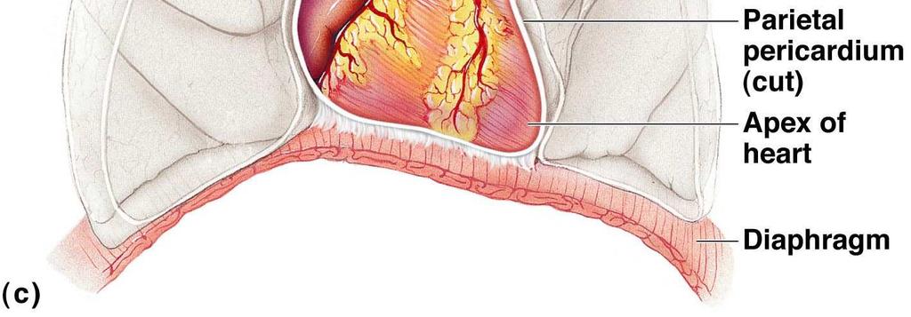
5 The Heart Figure 11.1c

6 The Heart Figure 11.2a
7 The Heart: Coverings Pericardium a double-walled sac Fibrous pericardium is loose and superficial Serous membrane is deep to the fibrous pericardium and composed of two layers Visceral pericardium Next to heart; also known as the epicardium Parietal pericardium Outside layer that lines the inner surface of the fibrous pericardium Serous fluid fills the space between the layers of pericardium
8 The Heart: Heart Wall Figure 11.2b
9 The Heart: Heart Wall Three layers Epicardium Outside layer This layer is the visceral pericardium Connective tissue layer Myocardium Middle layer Mostly cardiac muscle Endocardium Inner layer Endothelium

10 The Heart: Heart Wall Figure 11.2b
11 The Heart: Heart Wall Figure 11.2c
12 The Heart: Chambers Right and left side act as separate pumps Four chambers Atria Receiving chambers Right atrium Left atrium Ventricles Discharging chambers Right ventricle Left ventricle
13 The Heart: Chambers Figure 11.2c

14 Differences in Right and Left Ventricles Figure 11.4
15 The Heart: Septa Interventricular septum Separates the two ventricles Interatrial septum Separates the two atria
16 The Heart: Chambers Figure 11.2c
17 The Heart: Valves Allow blood to flow in only one direction to prevent backflow Four valves Atrioventricular (AV) valves between atria and ventricles Bicuspid (mitral) valve (left side of heart) Tricuspid valve (right side of heart) Semilunar valves between ventricle and artery Pulmonary semilunar valve Aortic semilunar valve
18 The Heart: Valves Figure 11.2c
19 The Heart: Valves AV valves Anchored in place by chordae tendineae ( heart strings ) Open during heart relaxation and closed during ventricular contraction Semilunar valves Closed during heart relaxation but open during ventricular contraction Notice these valves operate opposite of one another to force a one-way path of blood through the heart
 Figure 11.")
20 Operation of the AV valves Blood returning to the atria, puts pressure against AV valves; the AV valves are forced open Ventricles AV valves open (a) Figure 11.5a, step 1
21 Operation of the AV valves Blood returning to the atria, puts pressure against AV valves; the AV valves are forced open As the ventricles fill, AV valve flaps hang limply into ventricles Ventricles AV valves open (a) Figure 11.5a, step 2
22 Operation of the AV valves Blood returning to the atria, puts pressure against AV valves; the AV valves are forced open As the ventricles fill, AV valve flaps hang limply into ventricles Atria contract, forcing additional blood into ventricles Ventricles AV valves open (a) Figure 11.5a, step 3
 Figure 11.")
23 Ventricles contract, forcing blood against AV valve flaps (a) Figure 11.5a, step 4
 AV valves closed Figure 11.5a, step 5")
24 Ventricles contract, forcing blood against AV valve flaps AV valves close (a) AV valves closed Figure 11.5a, step 5
 AV valves closed Figure 11.")
25 Ventricles contract, forcing blood against AV valve flaps AV valves close Chordae tendineae tighten, preventing valve flaps from everting into atria (a) AV valves closed Figure 11.5a, step 6

 Semilunar valve open Figure 11.")
26 Operation of the semilunar valves Aorta Pulmonary trunk As ventricles contract and intraventricular pressure rises, blood is pushed up against semilunar valves, forcing them open (b) Semilunar valve open Figure 11.5b, step 1
 Semilunar valve open Semilunar valve closed Figure 11.")
27 Aorta Pulmonary trunk Operation of the semilunar valves As ventricles contract and intraventricular pressure rises, blood is pushed up against semilunar valves, forcing them open As ventricles relax, and intraventricular pressure falls, blood flows back from arteries, filling the leaflets of semilunar valves and forcing them to close (b) Semilunar valve open Semilunar valve closed Figure 11.5b, step 2
28 Systemic and Pulmonary Circulations Systemic circulation Blood flows from the left side of the heart through the body tissues and back to the right side of the heart Pulmonary circulation Blood flows from the right side of the heart to the lungs and back to the left side of the heart
29 Systemic and Pulmonary Circulations Figure 11.3
30 The Heart: Associated Great Vessels Arteries Aorta Leaves left ventricle Pulmonary arteries Leave right ventricle
31 The Heart: Associated Great Vessels Veins Superior and inferior venae cavae Enter right atrium Pulmonary veins (four) Enter left atrium


32 The Heart: Associated Great Vessels Figure 11.2c
33 The Heart
34 Blood Flow Through the Heart Superior and inferior venae cavae dump blood into the right atrium From right atrium, through the tricuspid valve, blood travels to the right ventricle From the right ventricle, blood leaves the heart as it passes through the pulmonary semilunar valve into the pulmonary trunk Pulmonary trunk splits into right and left pulmonary arteries that carry blood to the lungs
35 Blood Flow Through the Heart Oxygen is picked up and carbon dioxide is dropped off by blood in the lungs Oxygen-rich blood returns to the heart through the four pulmonary veins Blood enters the left atrium and travels through the bicuspid valve into the left ventricle From the left ventricle, blood leaves the heart via the aortic semilunar valve and aorta

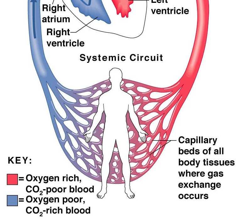
36 Systemic and Pulmonary Circulations Figure 11.3
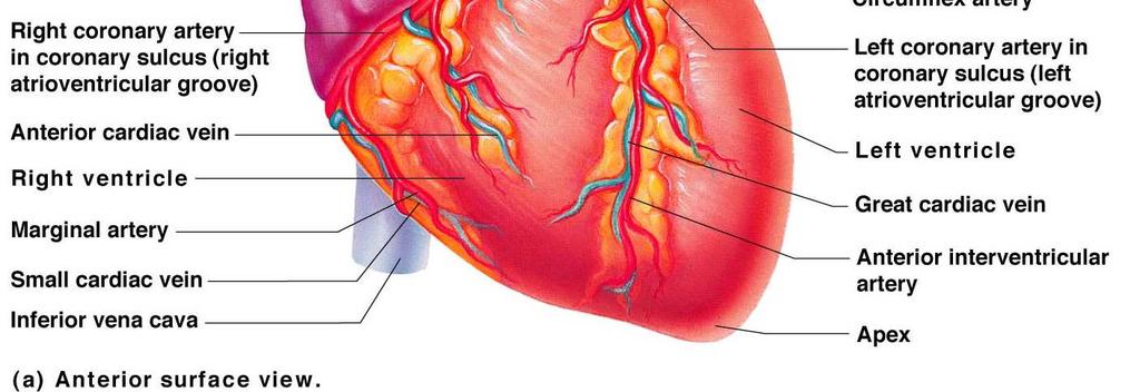
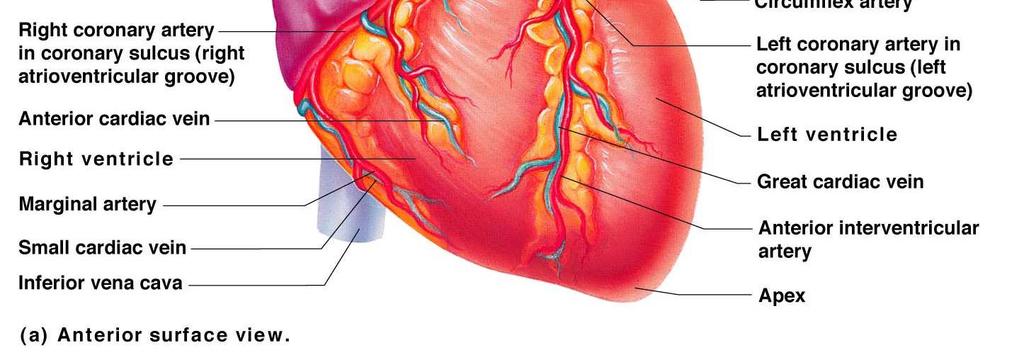
37 Coronary Circulation Blood in the heart chambers does not nourish the myocardium The heart has its own nourishing circulatory system consisting of Coronary arteries branch from the aorta to supply the heart muscle with oxygenated blood Cardiac veins drain the myocardium of blood Coronary sinus a large vein on the posterior of the heart, receives blood from cardiac veins Blood empties into the right atrium via the coronary sinus
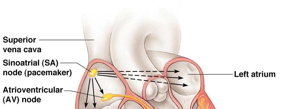
38 The Heart: Conduction System Intrinsic conduction system (nodal system) Heart muscle cells contract, without nerve impulses, in a regular, continuous way
39 The Heart: Conduction System Special tissue sets the pace Sinoatrial node = SA node ( pacemaker ), is in the right atrium Atrioventricular node = AV node, is at the junction of the atria and ventricles Atrioventricular bundle = AV bundle (bundle of His), is in the interventricular septum Bundle branches are in the interventricular septum Purkinje fibers spread within the ventricle wall muscles
40 Heart Contractions Figure 11.6
41 Heart Contractions Contraction is initiated by the sinoatrial node (SA node) Sequential stimulation occurs at other autorhythmic cells Force cardiac muscle depolarization in one direction from atria to ventricles
42 Heart Contractions Once SA node starts the heartbeat Impulse spreads to the AV node Then the atria contract At the AV node, the impulse passes through the AV bundle, bundle branches, and Purkinje fibers Blood is ejected from the ventricles to the aorta and pulmonary trunk as the ventricles contract
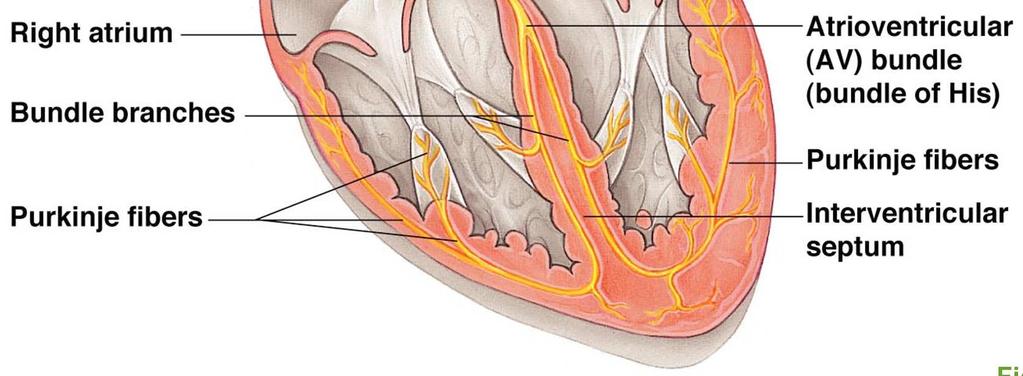
43 Heart Contractions Figure 11.6
44 Heart Contractions Tachycardia rapid heart rate over 100 beats per minute Bradycardia slow heart rate less than 60 beats per minutes
45 The Heart: Cardiac Cycle Atria contract simultaneously Atria relax, then ventricles contract Systole = contraction Diastole = relaxation
46 Filling Heart Chambers: Cardiac Cycle Left atrium Right atrium Left ventricle Right ventricle Ventricular filling Atrial contraction Isovolumetric contraction phase Ventricular ejection phase Isovolumetric relaxation Mid-to-late diastole (ventricular filling) Ventricular systole (atria in diastole) Early diastole Figure 11.7
47 Filling Heart Chambers: Cardiac Cycle Left atrium Right atrium Left ventricle Right ventricle Ventricular filling Mid-to-late diastole (ventricular filling) Figure 11.7, step 1a
48 Filling Heart Chambers: Cardiac Cycle Left atrium Right atrium Left ventricle Right ventricle Ventricular filling Atrial contraction Mid-to-late diastole (ventricular filling) Figure 11.7, step 1b
49 Filling Heart Chambers: Cardiac Cycle Left atrium Right atrium Left ventricle Right ventricle Ventricular filling Atrial contraction Isovolumetric contraction phase Mid-to-late diastole (ventricular filling) Ventricular systole (atria in diastole) Figure 11.7, step 2a
50 Filling Heart Chambers: Cardiac Cycle Left atrium Right atrium Left ventricle Right ventricle Ventricular filling Atrial contraction Isovolumetric contraction phase Ventricular ejection phase Mid-to-late diastole (ventricular filling) Ventricular systole (atria in diastole) Figure 11.7, step 2b
51 Filling Heart Chambers: Cardiac Cycle Left atrium Right atrium Left ventricle Right ventricle Ventricular filling Atrial contraction Isovolumetric contraction phase Ventricular ejection phase Isovolumetric relaxation Mid-to-late diastole (ventricular filling) Ventricular systole (atria in diastole) Early diastole Figure 11.7, step 3
52 The Heart: Cardiac Cycle Cardiac cycle events of one complete heart beat Mid-to-late diastole blood flows from atria into ventricles Ventricular systole blood pressure builds before ventricle contracts, pushing out blood Early diastole atria finish refilling, ventricular pressure is low
53 PowerPoint Lecture Slide Presentation by Patty Bostwick-Taylor, Florence-Darlington Technical College The Cardiovascular System 11PART A
54 The Heart: Cardiac Output Cardiac output (CO) Amount of blood pumped by each side (ventricle) of the heart in one minute Stroke volume (SV) Volume of blood pumped by each ventricle in one contraction (each heartbeat) Usually remains relatively constant About 70 ml of blood is pumped out of the left ventricle with each heartbeat Heart rate (HR) Typically 75 beats per minute
55 The Heart: Cardiac Output CO = HR SV CO = HR (75 beats/min) SV (70 ml/beat) CO = 5250 ml/min Starling s law of the heart the more the cardiac muscle is stretched, the stronger the contraction Changing heart rate is the most common way to change cardiac output
56 The Heart: Regulation of Heart Rate Increased heart rate Sympathetic nervous system Crisis Low blood pressure Hormones Epinephrine Thyroxine Exercise Decreased blood volume
57 The Heart: Regulation of Heart Rate Decreased heart rate Parasympathetic nervous system High blood pressure or blood volume Decreased venous return

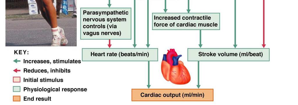
58 Cardiac Output Regulation Figure 11.8
59 Blood Vessels: The Vascular System Transport blood to the tissues and back Carry blood away from the heart Arteries Arterioles Exchanges between tissues and blood Capillary beds Return blood toward the heart Venules Veins
60 Blood Vessels: The Vascular System Figure 11.9a
61 Blood Vessels: Microscopic Anatomy Three layers (tunics) Tunic intima Endothelium Tunic media Smooth muscle Controlled by sympathetic nervous system Tunic externa Mostly fibrous connective tissue


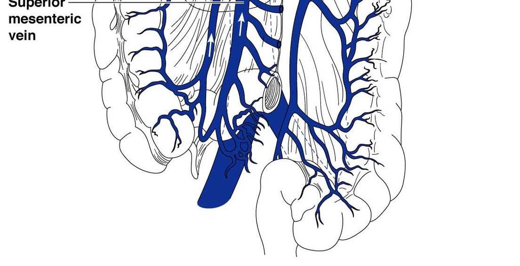
62 Blood Vessels: The Vascular System Figure 11.9b
63 Differences Between Blood Vessels Walls of arteries are the thickest Lumens of veins are larger Larger veins have valves to prevent backflow Skeletal muscle milks blood in veins toward the heart Walls of capillaries are only one cell layer thick to allow for exchanges between blood and tissue

64 Blood Vessels: The Vascular System Figure 11.9a

65 Blood Vessels: The Vascular System Figure 11.10
66 Movement of Blood Through Vessels Most arterial blood is pumped by the heart Veins use the milking action of muscles to help move blood
67 Capillary Beds Capillary beds consist of two types of vessels Vascular shunt vessel directly connecting an arteriole to a venule True capillaries exchange vessels Oxygen and nutrients cross to cells Carbon dioxide and metabolic waste products cross into blood


68 Capillary Beds Figure 11.11a


69 Capillary Beds Figure 11.11b
70 Major Arteries of System Circulation Aorta Largest artery in the body Leaves from the left ventricle of the heart Regions Ascending aorta leaves the left ventricle Aortic arch arches to the left Thoracic aorta travels downward through the thorax Abdominal aorta passes through the diaphragm into the abdominopelvic cavity
71 Major Arteries of System Circulation Arterial branches of the ascending aorta Right and left coronary arteries serve the heart

72 The Heart Figure 11.2a
73 Major Arteries of Systemic Circulation Arterial branches of the aortia arch (BCS) Brachiocephalic trunk splits into the Right common carotid artery Right subclavian artery Left common carotid artery splits into the Left internal and external carotid arteries Left subclavian artery branches into the Vertebral artery In the axilla, the subclavian artery becomes the axillary artery brachial artery radial and ulnar arteries
74 Major Arteries of Systemic Circulation Arterial branches of the thoracic aorta Intercostal arteries supply the muscles of the thorax wall Other branches of the thoracic aorta supply the Lungs (bronchial arteries) Esophagus (esophageal arteries) Diaphragm (phrenic arteries)
75 Major Arteries of Systemic Circulation Arterial branches of the abdominal aorta Celiac trunk is the first branch of the abdominal aorta. Three branches are Left gastric artery (stomach) Splenic artery (spleen) Common hepatic artery (liver) Superior mesenteric artery supplies most of the small intestine and first half of the large intestine
76 Major Arteries of Systemic Circulation Arterial branches of the abdominal aorta Left and right renal arteries (kidney) Left and right gonadal arteries Ovarian arteries in females serve the ovaries Testicular arteries in males serve the testes Lumbar arteries serve muscles of the abdomen and trunk
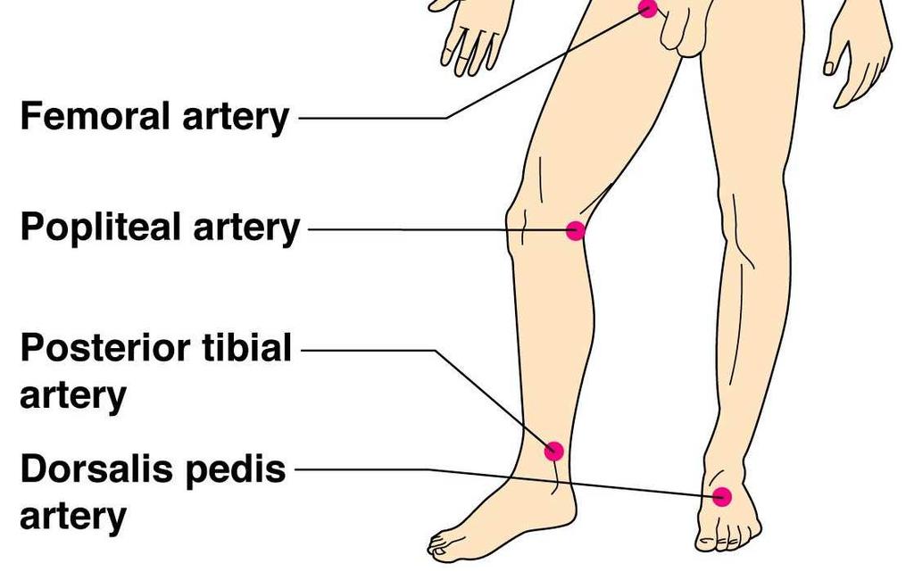
77 Major Arteries of Systemic Circulation Arterial branches of the abdominal aorta Inferior mesenteric artery serves the second half of the large intestine Left and right common iliac arteries are the final branches of the aorta Internal iliac arteries serve the pelvic organs External iliac arteries enter the thigh femoral artery popliteal artery anterior and posterior tibial arteries


78 Major Arteries of Systemic Circulation Figure 11.12
79 Major Veins of Systemic Circulation Superior and inferior vena cava enter the right atrium of the heart Superior vena cava drains the head and arms Inferior vena cava drains the lower body
80 The Heart Figure 11.2b
81 Major Veins of Systemic Circulation Veins draining into the superior vena cava Radial and ulnar veins brachial vein axillary vein These veins drain the arms Cephalic vein drains the lateral aspect of the arm and empties into the axillary vein Basilic vein drains the medial aspect of the arm and empties into the brachial vein Basilic and cephalic veins are jointed at the median cubital vein (elbow area)
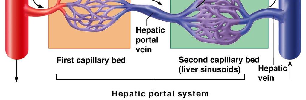
82 Major Veins of Systemic Circulation Veins draining into the superior vena cava Subclavian vein receives Venous blood from the arm via the axillary vein Venous blood from skin and muscles via external jugular vein Vertebral vein drains the posterior part of the head Internal jugular vein drains the dural sinuses of the brain
83 Major Veins of Systemic Circulation Veins draining into the superior vena cava Left and right brachiocephalic veins receive venous blood from the Subclavian veins Vertebral veins Internal jugular veins Brachiocephalic veins join to form the superior vena cava right atrium of heart Azygous vein drains the thorax
84 Major Veins of Systemic Circulation Veins draining into the inferior vena cava Anterior and posterior tibial veins and fibial veins drain the legs Posterior tibial vein popliteal vein femoral vein external iliac vein Great saphenous veins (longest veins of the body) receive superficial drainage of the legs Each common iliac vein (left and right) is formed by the union of the internal and external iliac vein on its own side
85 Major Veins of Systemic Circulation Veins draining into the inferior vena cava Right gonadal vein drains the right ovary in females and right testicle in males Left gonadal vein empties into the left renal vein Left and right renal veins drain the kidneys Hepatic portal vein drains the digestive organs and travels through the liver before it enters systemic circulation
86 Major Veins of Systemic Circulation Veins draining into the inferior vena cava Left and right hepatic veins drain the liver


87 Major Veins of Systemic Circulation Figure 11.13
88 Arterial Supply of the Brain Internal carotid arteries divide into Anterior and middle cerebral arteries These arteries supply most of the cerebrum Vertebral arteries join once within the skull to form the basilar artery Basilar artery serves the brain stem and cerebellum
89 Arterial Supply of the Brain Posterior cerebral arteries form from the division of the basilar artery These arteries supply the posterior cerebrum
90 Circle of Willis Anterior and posterior blood supplies are united by small communicating arterial branches Result complete circle of connecting blood vessels called cerebral arterial circle or circle of Willis

91 Arterial Supply of the Brain Figure 11.14
92 Fetal Circulation Fetus receives exchanges of gases, nutrients, and wastes through the placenta Umbilical cord contains three vessels Umbilical vein carries blood rich in nutrients and oxygen to the fetus Umbilical arteries (2) carry carbon dioxide and debris-laden blood from fetus to placenta
93 Fetal Circulation Blood flow bypasses the liver through the ductus venosus and enters the inferior vena cava right atrium of heart Blood flow bypasses the lungs Blood entering right atrium is shunted directly into the left atrium through the foramen ovale Ductus arteriosus connects the aorta and pulmonary trunk (becomes ligamentum arteriosum at birth)

94 Fetal Circulation Figure 11.15
95 Hepatic Portal Circulation Veins of hepatic portal circulation drain Digestive organs Spleen Pancreas Hepatic portal vein carries this blood to the liver Liver helps maintain proper glucose, fat, and protein concentrations in blood
96 Hepatic Portal Circulation Major vessels of hepatic portal circulation Inferior and superior mesenteric veins Splenic vein Left gastric vein

97 Hepatic Portal Circulation Figure 11.16

98 Hepatic Portal Circulation Figure 11.17
99 Pulse Pulse Pressure wave of blood Monitored at pressure points in arteries where pulse is easily palpated Pulse averages beats per minute at rest

100 Pulse Figure 11.18
101 Blood Pressure Measurements by health professionals are made on the pressure in large arteries Systolic pressure at the peak of ventricular contraction Diastolic pressure when ventricles relax Write systolic pressure first and diastolic last (120/80 mm Hg) Pressure in blood vessels decreases as distance from the heart increases


102 Comparison of Blood Pressures in Different Vessels Figure 11.19

103 Measuring Arterial Blood Pressure Figure 11.20a

104 Measuring Arterial Blood Pressure Figure 11.20b

105 Measuring Arterial Blood Pressure Figure 11.20c

106 Measuring Arterial Blood Pressure Figure 11.20d
107 Blood Pressure: Effects of Factors BP is blood pressure BP is affected by age, weight, time of day, exercise, body position, emotional state CO is the amount of blood pumped out of the left ventricle per minute PR is peripheral resistance, or the amount of friction blood encounters as it flows through vessels Narrowing of blood vessels and increased blood volume increases PR BP = CO PR
108 Blood Pressure: Effects of Factors Neural factors Autonomic nervous system adjustments (sympathetic division) Renal factors Regulation by altering blood volume Renin hormonal control
109 Blood Pressure: Effects of Factors Temperature Heat has a vasodilating effect Cold has a vasoconstricting effect Chemicals Diet Various substances can cause increases or decreases


110 Factors Determining Blood Pressure Figure 11.21
111 Variations in Blood Pressure Normal human range is variable Normal mm Hg systolic mm Hg diastolic Hypotension Low systolic (below 110 mm HG) Often associated with illness Hypertension High systolic (above 140 mm HG) Can be dangerous if it is chronic
112 Capillary Exchange Substances exchanged due to concentration gradients Oxygen and nutrients leave the blood Carbon dioxide and other wastes leave the cells
113 Capillary Exchange: Mechanisms Direct diffusion across plasma membranes Endocytosis or exocytosis Some capillaries have gaps (intercellular clefts) Plasma membrane not joined by tight junctions Fenestrations (pores) of some capillaries


114 Capillary Exchange: Mechanisms Figure 11.22
115 Fluid Movements at Capillary Beds Blood pressure forces fluid and solutes out of capillaries Osmotic pressure draws fluid into capillaries Blood pressure is higher than osmotic pressure at the arterial end of the capillary bed Blood pressure is lower than osmotic pressure at the venous end of the capillary bed

116 Fluid Movements at Capillary Beds Figure 11.23
117 Developmental Aspects of the Cardiovascular System A simple tube heart develops in the embryo and pumps by the fourth week The heart becomes a four-chambered organ by the end of seven weeks Few structural changes occur after the seventh week
118 Developmental Aspects of the Cardiovascular System Aging problems associated with the cardiovascular system include Venous valves weaken Varicose veins Progressive atherosclerosis Loss of elasticity of vessels leads to hypertension Coronary artery disease results from vessels filled with fatty, calcified deposits
The Cardiovascular System
 PowerPoint Lecture Slide Presentation by Patty Bostwick-Taylor, Florence-Darlington Technical College The Cardiovascular System 11PART B The Heart: Cardiac Output Cardiac output (CO) Amount of blood pumped
Unit 11 - The Cardiovascular System 1
 Unit 11 - The Cardiovascular System 1 I. Unit 11: The Cardiovascular System A. The Cardiovascular System 1. A closed system of the heart and blood vessels a) The heart pumps blood b) Blood vessels allow
Unit 11: The Cardiovascular System
 Unit 11: The Cardiovascular System I. The Cardiovascular System A. A closed system of the heart and blood vessels 1. The heart pumps blood 2. Blood vessels allow blood to circulate to all parts of the
Figure ) The specific chamber of the heart that is indicated by letter A is called the. Diff: 1 Page Ref: 364
 The specific chamber of the heart that is indicated by letter A is called the. Diff: 1 Page Ref: 364") Essentials of Anatomy and Physiology, 9e (Marieb) Chapter 11 The Cardiovascular System Short Answer Figure 11.1 Using Figure 11.1, identify the following: 1) The Purkinje fibers are indicated by label.
Essentials of Anatomy and Physiology, 9e (Marieb) Chapter 11 The Cardiovascular System Short Answer Figure 11.1 Using Figure 11.1, identify the following: 1) The Purkinje fibers are indicated by label.
The Cardiovascular System
 The Cardiovascular System The Cardiovascular System A closed system of the heart and blood vessels The heart pumps blood Blood vessels allow blood to circulate to all parts of the body The function of
The Cardiovascular System
 Essentials of Human Anatomy & Physiology Elaine N. Marieb Seventh Edition Chapter 11 The Cardiovascular System Slides 11.1 11.19 Lecture Slides in PowerPoint by Jerry L. Cook The Cardiovascular System
A closed system of the heart/blood. Function: The heart pumps blood. Blood vessels allow blood to circulate throughout the body
 A closed system of the heart/blood The heart pumps blood It is no more than a transportation pump Blood vessels allow blood to circulate throughout the body MILES of blood vessels intricate network At
CARDIOVASCULAR SYSTEM
 CARDIOVASCULAR SYSTEM CARDIAC SYSTEM TWO TYPES OF CIRCULATION Systemic system delivers blood to ALL body cells and carries away waste. The red blood cells use hemoglobin to carry oxygen to the cells Pulmonary
The Cardiovascular System
 11 PART A The Cardiovascular System PowerPoint Lecture Slide Presentation by Jerry L. Cook, Sam Houston University ESSENTIALS OF HUMAN ANATOMY & PHYSIOLOGY EIGHTH EDITION ELAINE N. MARIEB The Cardiovascular
The Cardiovascular System
 11 The Cardiovascular System Yong Jeong, MD, PhD Department of Bio and Brain Engineering The Cardiovascular System A closed system of the heart and blood vessels The heart pumps blood Blood vessels allow
Major Function of the Cardiovascular System. Transportation. Structures of the Cardiovascular System. Heart - muscular pump
 Structures of the Cardiovascular System Heart - muscular pump Blood vessels - network of tubes Blood - liquid transport vehicle brachiocephalic trunk superior vena cava right pulmonary arteries right pulmonary
The Cardiovascular System
 PowerPoint Lecture Slide Presentation by Patty Bostwick-Taylor, Florence-Darlington Technical College The Cardiovascular System 11 PART A The Cardiovascular System A closed system of the heart and blood
Chapter 11. The Cardiovascular System
 Chapter 11 The Cardiovascular System The Cardiovascular System The Cardiovascular System A closed system of the heart and blood vessels Heart pumps blood Blood vessels circulate blood to all parts of the
HUMAN HEART. Learn the following structures on the heart models.
 HUMAN HEART Learn the following structures on the heart models. The human heart has four chambers that consist of the right atrium, left atrium, right ventricle, and left ventricle. The atria are smaller
Function: Transportation of. Oxygen Nutrients Waste Hormones gases
 Function: Transportation of Oxygen Nutrients Waste Hormones gases Pericardium: double sac of serous membrane filled with fluid (pericardial fluid to be exact) that surrounds the heart. Parietal pericardium:
Cardiovascular System
 Cardiovascular System I. Structure of the Heart A. Average adult heart is 14 cm long and 9 cm wide. B. Lies in the mediastinum. C. Enclosed in the pericardium. 1. Fibrous pericardium- Outer, tough connective
10. Thick deposits of lipids on the walls of blood vessels, called, can lead to serious circulatory issues. A. aneurysm B. atherosclerosis C.
 Heart Student: 1. carry blood away from the heart. A. Arteries B. Veins C. Capillaries 2. What is the leading cause of heart attack and stroke in North America? A. alcohol B. smoking C. arteriosclerosis
Chapter 14. The Cardiovascular System
 Chapter 14 The Cardiovascular System Introduction Cardiovascular system - heart, blood and blood vessels Cardiac muscle makes up bulk of heart provides force to pump blood Function - transports blood 2
The Cardiovascular System (Heart)
") The Cardiovascular System The Cardiovascular System (Heart) A closed system of the heart and blood vessels The heart pumps blood Blood vessels allow blood to circulate to all parts of the body The function
The Cardiovascular System The Cardiovascular System (Heart) A closed system of the heart and blood vessels The heart pumps blood Blood vessels allow blood to circulate to all parts of the body The function
1. Which of the following blood vessels has a thin elastic layer? A. Aorta. B. Pulmonary artery. C. Posterior vena cava. D. Mesenteric capillary.
 CIRCULATORY SYSTEM 1. Which of the following blood vessels has a thin elastic layer? A. Aorta. B. Pulmonary artery. C. Posterior vena cava. D. Mesenteric capillary. 2. Capillary beds are equipped with
Circulation. Sinoatrial (SA) Node. Atrioventricular (AV) Node. Cardiac Conduction System. Cardiac Conduction System. Linked to the nervous system
 Node. Atrioventricular (AV) Node. Cardiac Conduction System. Cardiac Conduction System. Linked to the nervous system") Circulation Cardiac Conduction System AHS A H S Your body resembles a large roadmap. There are routes or arteries that take you downtown to the heart of the city and veins that take you to the outskirts
Circulation Cardiac Conduction System AHS A H S Your body resembles a large roadmap. There are routes or arteries that take you downtown to the heart of the city and veins that take you to the outskirts
The Heart. The Heart A muscular double pump. The Pulmonary and Systemic Circuits
 C H A P T E R 19 The Heart The Heart A muscular double pump circuit takes blood to and from the lungs Systemic circuit vessels transport blood to and from body tissues Atria receive blood from the pulmonary
MODULE 2: CARDIOVASCULAR SYSTEM ANTOMY An Introduction to the Anatomy of the Heart and Blood vessels
 MODULE 2: CARDIOVASCULAR SYSTEM ANTOMY An Introduction to the Anatomy of the Heart and Blood vessels The cardiovascular system includes a pump (the heart) and the vessels that carry blood from the heart
The Cardiovascular System
 Essentials of Human Anatomy & Physiology Elaine N. Marieb Slides 11.1 11.19 Seventh Edition Chapter 11 The Cardiovascular System Functions of the Cardiovascular system Function of the heart: to pump blood
The Cardiovascular System. Preview of Heart Action. The CV system provides oxygen & nutrients to tissues-removes wastes.
 The Cardiovascular System BIO 250 Human Anatomy & Physiology Preview of Heart Action http://www.youtube.com/watch?v=d3zdj gfddk0&nr=1 The CV system provides oxygen & nutrients to tissues-removes wastes.
Chapter 13. Cardiovascular System
 Chapter 13 Cardiovascular System 1 Introduction A. The cardiovascular system consists of the heart and vessels (arteries, capillaries and veins.) B. A functional cardiovascular system is vital for supplying
The Cardiovascular System
 C H A P T E R 1 4 The Cardiovascular System OBJECTIVES After studying this chapter, you should be able to: 1. Describe how the heart is positioned in the thoracic cavity. 2. List and describe the layers
THE CARDIOVASCULAR SYSTEM. Part 1
 THE CARDIOVASCULAR SYSTEM Part 1 CARDIOVASCULAR SYSTEM Blood Heart Blood vessels What is the function of this system? What other systems does it affect? CARDIOVASCULAR SYSTEM Functions Transport gases,
3 Circulatory Pathways
 40 Chapter 3 Circulatory Pathways Systemic Arteries -Arteries carry blood away from the heart to the various organs of the body. -The aorta is the longest artery in the body; it branches to give rise to
The HEART. What is it???? Pericardium. Heart Facts. This muscle never stops working It works when you are asleep
 This muscle never stops working It works when you are asleep The HEART It works when you eat It really works when you exercise. What is it???? Located between the lungs in the mid thoracic region Apex
Cardiovascular System
 Cardiovascular System Purpose Transport oxygen and nutrients Take waste products away from tissues & organs Things we learned Blood pressure: the force of blood pushing against the walls of blood vessels
Anatomy and Physiology, Spring 2015 Exam II: Form A April 9, Name Student Number
 Anatomy and Physiology, Spring 2015 Exam II: Form A April 9, 2015 Name Student Number For Questions 1 2 refer to the following table. 1 Ventricular pressure is greater than aortic 6 AV valve is open 2
Cardiovascular System. Biology 105 Lecture 15 Chapter 12
 Cardiovascular System Biology 105 Lecture 15 Chapter 12 Outline I. Functions of cardiovascular system II. Components of the cardiovascular system: I. Blood vessels II. Heart III. Regulation of the heartbeat
Cardiovascular System
 Hole s Essentials of Human Anatomy & Physiology David Shier Jackie Butler Ricki Lewis Created by Dr. Melissa Eisenhauer Head Athletic Trainer/Assistant Professor Trevecca Nazarene University Chapter 13
THE HEART OBJECTIVES: LOCATION OF THE HEART IN THE THORACIC CAVITY CARDIOVASCULAR SYSTEM
 BIOLOGY II CARDIOVASCULAR SYSTEM ACTIVITY #3 NAME DATE HOUR THE HEART OBJECTIVES: Describe the anatomy of the heart and identify and give the functions of all parts. (pp. 356 363) Trace the flow of blood
REVIEW SHEET Anatomy of Blood Vessels
 REVIEW SHEET Anatomy of Blood Vessels Name LabTime/Date Microscopic Structure of the Blood Vessels 1. Cross-sectional views of an aftery of a vein are shown here. ldentify each; on the lines to the sides,
Blood Vessels. Types of Blood Vessels Arteries carry blood away from the heart Capillaries smallest blood vessels. Veins carry blood toward the heart
 C H A P T E R Blood Vessels 20 Types of Blood Vessels Arteries carry blood away from the heart Capillaries smallest blood vessels The site of exchange of molecules between blood and tissue fluid Veins
Chapter 12 Lecture Outline
 Chapter 12 Lecture Outline See separate PowerPoint slides for all figures and tables preinserted into PowerPoint without notes. Copyright The McGraw-Hill Companies, Inc. Permission required for reproduction
Cardiovascular. Function of the cardiovascular system is to transport blood containing: Nutrients Waste Hormones Immune cells Oxygen
 Cardiovascular The Cardiovascular System - Arteries Arteries Cardiovascular System Function of the cardiovascular system is to transport blood containing: Carry blood away from heart Carotid arteries Deliver
Pearson's Comprehensive Medical Assisting Administrative and Clinical Competencies
 Pearson's Comprehensive Medical Assisting Administrative and Clinical Competencies THIRD EDITION CHAPTER 27 The Cardiovascular System Lesson 1: Overview of the Cardiovascular System Lesson Objectives Upon
Cardiovascular Anatomy Dr. Gary Mumaugh
 Cardiovascular Anatomy Dr. Gary Mumaugh Location of Heart Approximately the size of your fist Location o Superior surface of diaphragm o Left of the midline in mediastinum o Anterior to the vertebral column,
Circulatory System Review
 Circulatory System Review 1. Know the diagrams of the heart, internal and external. a) What is the pericardium? What is myocardium? What is the septum? b) Explain the 4 valves of the heart. What is their
1. What kind of blood is found in the rt. atrium? (oxygenated or deoxygenated)
") Carl Christennsen, PhD Chap. 19, 20, & 21 - Circulatory System Bio. 2304 Human Anatomy HEART 1. What kind of blood is found in the rt. atrium? (oxygenated or deoxygenated) Where does this blood come from?
Carl Christennsen, PhD Chap. 19, 20, & 21 - Circulatory System Bio. 2304 Human Anatomy HEART 1. What kind of blood is found in the rt. atrium? (oxygenated or deoxygenated) Where does this blood come from?
Human Anatomy, First Edition
 Human Anatomy, First Edition McKinley & O'Loughlin Chapter 22 : Heart 1 Functions of the Heart Center of the cardiovascular system, the heart. Connects to blood vessels that transport blood between the
VESSELS: GROSS ANATOMY
 ACTIVITY 10: VESSELS AND CIRCULATION OBJECTIVES: 1) How to get ready: Read Chapter 23, McKinley et al., Human Anatomy, 4e. All text references are for this textbook. 2) Observe and sketch histology slide
Circulatory System Notes
 Circulatory System Notes Functions of Circulatory System A. Transports B. Transports C. Transports D. Transports E. of fluids F. G. Regulate temperature H. Blood clotting Characteristics of various blood
The Cardiovascular System. Chapter 15. Cardiovascular System FYI. Cardiology Closed systemof the heart & blood vessels. Functions
 Chapter 15 Cardiovascular System FYI The heart pumps 7,000 liters (4000 gallons) of blood through the body each day The heart contracts 2.5 billion times in an avg. lifetime The heart & all blood vessels
Chapter 18 - Heart. I. Heart Anatomy: size of your fist; located in mediastinum (medial cavity)
") Chapter 18 - Heart I. Heart Anatomy: size of your fist; located in mediastinum (medial cavity) A. Coverings: heart enclosed in double walled sac called the pericardium 1. Fibrous pericardium: dense connective
Chapter 18 - Heart I. Heart Anatomy: size of your fist; located in mediastinum (medial cavity) A. Coverings: heart enclosed in double walled sac called the pericardium 1. Fibrous pericardium: dense connective
THE HEART. A. The Pericardium - a double sac of serous membrane surrounding the heart
 THE HEART I. Size and Location: A. Fist-size weighing less than a pound (250 to 350 grams). B. Located in the mediastinum between the 2 nd rib and the 5 th intercostal space. 1. Tipped to the left, resting
YOU MUST BRING GLOVES FOR THIS ACTIVITY
 ACTIVITY 10: VESSELS AND CIRCULATION OBJECTIVES: 1) How to get ready: Read Chapter 23, McKinley et al., Human Anatomy, 5e. All text references are for this textbook. 2) Observe and sketch histology slide
UNIT 11: THE CARDIOVASCULAR SYSTEM
 UNIT 11: THE CARDIOVASCULAR SYSTEM Functions of the Heart PUMPS Blood Transports Oxygen and Nutrients Removes Carbon Dioxide and Metabolic Wastes Thermoregulation Immunological Function Clotting Mechanisms
- what other structures, besides the heart, does the mediastinum contain?
 Basic A & P II Dr. L. Bacha Chapter Outline (Martini & Nath 2010) An Introduction to the Cardiovascular System - read the paragraphs under this heading on page 580 The Heart is a Four Chambered Organ describe
CIRCULATORY SYSTEM BLOOD VESSELS
 Name: Block: CIRCULATORY SYSTEM Multicellular organisms (above the level of roundworms) rely on a circulatory system to bring nutrients to, and take wastes away from, cells. In higher organisms such as
11/10/2014. Muscular pump Two atria Two ventricles. In mediastinum of thoracic cavity 2/3 of heart's mass lies left of midline of sternum
 It beats over 100,000 times a day to pump over 1,800 gallons of blood per day through over 60,000 miles of blood vessels. During the average lifetime, the heart pumps nearly 3 billion times, delivering
CV Anatomy Quiz. Dr Ella Kim Dr Pip Green
 CV Anatomy Quiz Dr Ella Kim Dr Pip Green Q1 The location of the heart is correctly described as A) lateral to the lungs. B) medial to the sternum. C) superior to the diaphragm. D) posterior to the spinal
the Cardiovascular System I
 the Cardiovascular System I By: Dr. Nabil A Khouri MD, MsC, Ph.D MEDIASTINUM 1. Superior Mediastinum 2. inferior Mediastinum Anterior mediastinum. Middle mediastinum. Posterior mediastinum Anatomy of
Circulation. Circulation = is a process used for the transport of oxygen, carbon! dioxide, nutrients and wastes through-out the body
 Circulation Circulation = is a process used for the transport of oxygen, carbon! dioxide, nutrients and wastes through-out the body Heart = muscular organ about the size of your fist which pumps blood.
The Heart. Size, Form, and Location of the Heart. 1. Blunt, rounded point; most inferior part of the heart.
 12 The Heart FOCUS: The heart is composed of cardiac muscle cells, which are elongated, branching cells that appear striated. Cardiac muscle cells behave as a single electrical unit, and the highly coordinated
Cardiovascular System. Chapter 22
 Cardiovascular System Blood Vessels Chapter 22 Cardiac Contractions Cardiac cycle consists of alternate periods of contraction and relaxation Contraction is the systolic pressure Blood is ejected into
Chapter 11. The Cardiovascular System. Clicker Questions Pearson Education, Inc.
 Chapter 11 The Cardiovascular System Clicker Questions Oxygen-poor blood is pumped through the venae cavae to the right side of the heart, and then through the pulmonary arteries to the lungs and back
Approximately the size of your fist Location Superior surface of diaphragm Left of the midline in mediastinum Anterior to the vertebral column,
 Dr. Gary Mumaugh Approximately the size of your fist Location Superior surface of diaphragm Left of the midline in mediastinum Anterior to the vertebral column, posterior to the sternum Posteriorly the
Cardiovascular System
 Cardiovascular System The Heart Cardiovascular System The Heart Overview What does the heart do? By timed muscular contractions creates pressure gradients blood moves then from high pressure to low pressure
Approximately the size of your fist Location. Pericardial physiology
 Heart Anatomy Approximately the size of your fist Location Superior surface of diaphragm Left of the midline Anterior to the vertebral column, posterior to the sternum Wednesday, March 28, 2012 Muscle
Essentials of Anatony and Physiology, 5e (Martini/Nath) Chapter 13 The Cardiovascular System: Blood Vessels and Circulation
 Chapter 13 The Cardiovascular System: Blood Vessels and Circulation") Essentials of Anatony and Physiology, 5e (Martini/Nath) Chapter 13 The Cardiovascular System: Blood Vessels and Circulation Multiple-Choice Questions 1) The muscular layer of blood vessels is the A) tunica
Essentials of Anatony and Physiology, 5e (Martini/Nath) Chapter 13 The Cardiovascular System: Blood Vessels and Circulation Multiple-Choice Questions 1) The muscular layer of blood vessels is the A) tunica
4. The two inferior chambers of the heart are known as the atria. the superior and inferior vena cava, which empty into the left atrium.
 Answer each statement true or false. If the statement is false, change the underlined word to make it true. 1. The heart is located approximately between the second and fifth ribs and posterior to the
Cardiovascular system
 Cardiovascular system 1 Essential Question: How does the structure of the heart allow it to function in pumping blood? 2 Function? 3 Location of Heart 4 Heart coverings double sac of serous membrane pericardium
Chapter 20 (1) The Heart
 The Heart") Chapter 20 (1) The Heart Learning Objectives Describe the location and structure of the heart Describe the path of a drop of blood from the superior vena cava or inferior vena cava through the heart out
Chapter 20 (1) The Heart Learning Objectives Describe the location and structure of the heart Describe the path of a drop of blood from the superior vena cava or inferior vena cava through the heart out
THE VESSELS OF BLOOD CIRCULATION
 THE VESSELS OF BLOOD CIRCULATION scientistcindy.com /the-vessels-of-blood-circulation.html NOTE: You should familiarize yourself with the anatomy of the heart and have a good understanding of the flow
2. right heart = pulmonary pump takes blood to lungs to pick up oxygen and get rid of carbon dioxide
 A. location in thorax, in inferior mediastinum posterior to sternum medial to lungs superior to diaphragm anterior to vertebrae orientation - oblique apex points down and to the left 2/3 of mass on left
The Heart. Happy Friday! #takeoutyournotes #testnotgradedyet
 The Heart Happy Friday! #takeoutyournotes #testnotgradedyet Introduction Cardiovascular system distributes blood Pump (heart) Distribution areas (capillaries) Heart has 4 compartments 2 receive blood (atria)
Anatomy & Physiology of Cardiovascular System. Chapter 18 & 19
 Anatomy & Physiology of Cardiovascular System Chapter 18 & 19 Objectives..cont 1. Discuss the physiological stages of cardiac muscle contraction. 2. Trace a typical ECG and label each wave or complex 3.
37 1 The Circulatory System
 H T H E E A R T 37 1 The Circulatory System The circulatory system and respiratory system work together to supply cells with the nutrients and oxygen they need to stay alive. a) The respiratory system:
1. Distinguish among the types of blood vessels on the basis of their structure and function.
 Blood Vessels and Circulation Objectives This chapter describes the structure and functions of the blood vessels Additional subjects contained in Chapter 13 include cardiovascular physiology, regulation,
The Circulatory System (p )
") The Circulatory System (p. 268-281) How Does Gravity Affect Blood Circulation? As with all land animals, the giraffe and the corn snake are constantly subject to the force of gravity The circulatory system
The Circulatory System (p. 268-281) How Does Gravity Affect Blood Circulation? As with all land animals, the giraffe and the corn snake are constantly subject to the force of gravity The circulatory system
Cardiovascular System Notes: Physiology of the Heart
 Cardiovascular System Notes: Physiology of the Heart Interesting Heart Fact Capillaries are so small it takes ten of them to equal the thickness of a human hair. Review What are the 3 parts of the cardiovascular
Chapter 20: Cardiovascular System: The Heart
 Chapter 20: Cardiovascular System: The Heart I. Functions of the Heart A. List and describe the four functions of the heart: 1. 2. 3. 4. II. Size, Shape, and Location of the Heart A. Size and Shape 1.
Lab Activity 25. Blood Vessels & Circulation. Portland Community College BI 232
 Lab Activity 25 Blood Vessels & Circulation Portland Community College BI 232 Artery and Vein Histology Walls have 3 layers: Tunica intima Tunica media Tunica externa 2 Tunica Intima Is the innermost layer
The Cardiovascular and Lymphatic Systems Cardiovascular System Blood Vessels Blood Vessels Arteries Arteries Arteries
 CH 12 The Cardiovascular and s The Cardiovascular and s OUTLINE: Cardiovascular System Blood Vessels Blood Pressure Cardiovascular System The cardiovascular system is composed of Blood vessels This system
Figure 10.1A Transparency Master 79
 Brain Carotid arteries Jugular vein Right front leg Lungs (inflated) Cranial Right atrium To left front leg Left subclavian Bronchus capillaries Brachiocephalic vein Left atrium Dorsal aorta Right ventricle
Cardiovascular system
 BIO 301 Human Physiology Cardiovascular system The Cardiovascular System: consists of the heart plus all the blood vessels transports blood to all parts of the body in two 'circulations': pulmonary (lungs)
AN ATOMY OF THE CARDIOVASCULAR SYSTEM
 Student Name CHAPTER 18 AN ATOMY OF THE CARDIOVASCULAR SYSTEM T he heart is actually two pumps one moves blood to the lungs, the other pushes it out into the body. These two functions seem rather elementary
THE CIRCULATORY SYSTEM
 Biology 30S THE CIRCULATORY SYSTEM Name: This module adapted from bblearn.merlin.mb.ca 1 Introduction to Circulation The first organ to form, and the last organ to die. The heart is the pump of life. The
Vascular System Part One
 Vascular System Part One Objectives Trace the route taken by blood as it leaves, and then returns to the heart. Describe the structure of the walls of arteries and veins. Discuss the structure and function
C3, 4, 5, 6, & 7 Worksheet. C3 Describe the inter-relationships of the structures of the heart
 Name: Date: C3, 4, 5, 6, & 7 Worksheet C3 Describe the inter-relationships of the structures of the heart 1. Label and give the functions of the following: a. left and right atrium: b. left and right ventricle:
The Cardiovascular System
 The Cardiovascular System The Manila Times College of Subic Prepared by: Stevens B. Badar, RN, MANc THE HEART Anatomy of the Heart Location and Size approx. the size of a person s fist, hollow and cone-shaped,
The ancient Babylonians, Egyptians, Indians and Chinese believed the heart was the centre of thinking and emotions
 The Concept of Mind The ancient Babylonians, Egyptians, Indians and Chinese believed the heart was the centre of thinking and emotions Hippocrates 460 BC 370 BC - Thoughts, ideas, and feelings come from
10/23/2017. Muscular pump Two atria Two ventricles. In mediastinum of thoracic cavity 2/3 of heart's mass lies left of midline of sternum
 It beats over 100,000 times a day to pump over 1,800 gallons of blood per day through over 60,000 miles of blood vessels. During the average lifetime, the heart pumps nearly 3 billion times, delivering
Health Science 20 Circulatory System Notes
 Health Science 20 Circulatory System Notes Functions of the Circulatory System The circulatory system functions mainly as the body s transport system. It transports: o Oxygen o Nutrients o Cell waste o
CRITICAL THINKING QUESTIONS AND ANSWERS AND CYCLE 2 LAB EXAM TEMPLATE. There are two main mechanisms that work in conjunction to return the blood
 CRITICAL THINKING QUESTIONS AND ANSWERS AND CYCLE 2 LAB EXAM TEMPLATE There are two main mechanisms that work in conjunction to return the blood THE CARDIAC PUMP 1) The forward pull(vis a fronte) This
ANATOMY AND PHYSIOLOGY HOMEWORK CHAPTER 11 AND 12
 ANATOMY AND PHYSIOLOGY HOMEWORK CHAPTER 11 AND 12 Name Identify the following: 1) The Purkinje fibers are indicated by label. 2) The sinoatrial (SA) node is indicated by letter. 3) The specific chamber
Anatomy of the Heart. Figure 20 2c
 Anatomy of the Heart Figure 20 2c Pericardium & Myocardium Remember, the heart sits in it s own cavity, known as the mediastinum. The heart is surrounded by the Pericardium, a double lining of the pericardial
Large Arteries of Heart
 Cardiovascular System (Part A-2) Module 5 -Chapter 8 Overview Arteries Capillaries Veins Heart Anatomy Conduction System Blood pressure Fetal circulation Susie Turner, M.D. 1/5/13 Large Arteries of Heart
The Cardiovascular and Lymphatic Systems
 BIOLOGY OF HUMANS Concepts, Applications, and Issues Fifth Edition Judith Goodenough Betty McGuire 12 The Cardiovascular and Lymphatic Systems Lecture Presentation Anne Gasc Hawaii Pacific University and
Ch. 12 The Circulatory System. The heart. The heart is a double pump. A quick note on arteries vs. veins. = the muscular pump of the CV system
 Ch. 12 The Circulatory System The heart A.k.a. the cardiovascular system Blood was discussed in Ch. 11 Focus of Ch. 12: heart and blood vessels = the muscular pump of the CV system ~ 100,000 heartbeats/day!
2. capillaries - allow exchange of materials between blood and tissue fluid
 Chapter 19 - Vascular System A. categories and general functions: 1. arteries - carry blood away from heart 2. capillaries - allow exchange of materials between blood and tissue fluid 3. veins - return
2/28/18. Cardiovascular System. Introduction. Anatomy. Chapter 26. Body is 60% to 80% fluid (by volume) Systems responsible for fluid movement are:
 Systems responsible for fluid movement are:") Cardiovascular System Chapter 26 1 Introduction Body is 60% to 80% fluid (by volume) Systems responsible for fluid movement are: - Cardiovascular helps move fluid - Lymphatic Both called pick-up and delivery
Cardiovascular System Chapter 26 1 Introduction Body is 60% to 80% fluid (by volume) Systems responsible for fluid movement are: - Cardiovascular helps move fluid - Lymphatic Both called pick-up and delivery
The Heart and Cardiovascular System
 The Heart and Cardiovascular System What you will learn The location of the heart 3 layers and covering of the heart Explain the function of the heart as 2 separate pumps Identify the 4 chambers of the
Anatomy: Cardiovascular System
 In the name of God Anatomy: Cardiovascular System Moradian MD, MPH, PhD candidate Tehran University of Medical Sciences drmoradian@sums.ac.ir 2015 The Cardiovascular System A closed system of the heart
TRACE A DROP OF BLOOD FROM RIGHT EAR TO LEFT OCULOMOTOR NERVE
 TRACE A DROP OF BLOOD FROM RIGHT EAR TO LEFT OCULOMOTOR NERVE KEY: TRACE A DROP OF BLOOD FROM RIGHT EAR TO LEFT OCULOMOTOR NERVE RIGHT EAR RIGHT ATRIUM LEFT SUBCLAVIAN ARTERY RIGHT EXTERNAL JUGULAR VEIN
Anatomy of the Blood Vessels
 Biology 212: Anatomy and Physiology II Anatomy of the Blood Vessels References: Saladin, KS: Anatomy and Physiology, The Unity of Form and Function 8 th (2018). Required reading before beginning this lab: