Thoracic trauma. 1 The majority of deaths occur after the. FEBRUARY 2001 The Surgical Technologist
|
|
|
- Cecily Watts
- 6 years ago
- Views:
Transcription
1

2 Thoracic trauma Richard Wills, MD, MBA, ACSM, Michael Norton, DC, and Kathryn DeLaney Approximately 25% of nonmilitary trauma-related deaths are due to thoracic trauma. 1 The majority of deaths occur after the patient reaches the hospital. Death will occur in about one third of the patients in which two or more organ systems are involved. Penetration and blunt force are the two mechanisms that cause thoracic trauma.normal thoracic anatomy is shown in Figures 1 and 2. Penetrating thoracic trauma will cause pneumothorax in nearly all cases, with hemothorax seen in more than 75% of these cases. One third of the cases of penetrating trauma will be associated with abdominal injuries. Blunt trauma may be due to: compression (organ rupture), direct trauma (fracture), or acceleration/deceleration forces (vessel shear and tear). 1 This article discusses several major types of traumatic thoracic injuries. FEBRUARY 2001 The Surgical Technologist 21
3 197 FEBRUARY 2001 CATEGORY 3 eneral considerations Patients sustain blunt thoracic trauma and develop acute respiratory distress have a very high morality rate. 2 (Table 1) Of the patients that experience shock with respiratory distress, three fourths will die. 1 Patients that are diagnosed with respiratory distress will need airway control, optimal oxygenation, and possibly ventilatory support and resuscitation. At the time of hospital admission, 10% of these patients will require endotracheal intubation. Most likely, the respiratory problem is due to head trauma and/or spinal cord injury. Additionally, upper airway obstruction should be suspected if the patient is attempting to breathe with little or no air movement. Upper airway obstruction is common in comatose patients due to prolapse of the tongue into the pharynx. Other causes of upper airway obstruction include vomitus or blood clots within the pharynx, larynx, or upper trachea. If laryngeal trauma is suspected, an endoscopy should be performed as soon as possible. Emergency tracheotomy may become necessary to treat total airway occlusion from any cause. Once the upper airway is determined to be intact and breath sounds remain poor, thoracic trauma should be suspected. Flail chest, hemothorax, pneumothorax, hemopneumothorax, diaphragmatic injury, or parenchymal lung damage should be considered. Table two describes traumatic injuries to the thorax according to their location. Subcutaneous emphysema Subcutaneous emphysema is produced by penetrating or blunt injuries, generally to the lungs and parietal pleura, causing air to be forced into the tissues of the chest wall. The patient may display an initial pneumothorax; however, massive development of subcutaneous emphysema can delay the onset of pneumothorax. Generally, there is seldom reason to treat the subcutaneous emphysema; however, a cervical incision is useful to allow trapped air to escape. Attention should be directed toward confirming the source of air leakage, which may be due to an esophageal perforation or major bronchial injury. Once the source of air leakage is determined, immediate treatment to solve the cause of the subcutaneous emphysema is implemented. Mediastinal emphysema Mediastinal emphysema occurs when air enters the areolar tissue from a tracheal or bronchial wound, or from a perforation of the esophagus. Blunt injuries to the chest may disrupt the bronchioles and alveolar units without disrupting the visceral pleura. Air moves into the pulmonary interstitium, centrally along the pulmonary and bronchial vessels until it reaches the mediastinum. If the mediastinal pleura stays intact, Table 1: Patients with respiratory failure following blunt thoracic trauma 1 Type of Injury Incidence % Mortality Rate % Flail chest/multiple rib fracture Hemopneumothorax Lung contusion Extremity fracture Intraabdominal Intracranial Myocardial contusion Diaphragm 9 20 Paraplegia Other The Surgical Technologist FEBRUARY 2001

4 air dissects into the neck tissue, moving through the deep tissue planes and finally spreading into the subcutaneous tissue. Flail chest Flail chest is a crushing chest injury that implies disruption of the mechanism of respiration, resulting in paradoxical motion of the chest wall region. For flail chest to occur, at least two segmental fractures of three adjacent ribs or costal cartilages must occur (Figure 3). Combinations of rib and sternal fractures accompanied by costochondral or chondrosternal separations result in paradoxical respiratory motion which is characteristic of a flail chest. 2 There are three types of flail chest: lateral, anterior, and posterior. Flail chest injury may also be accompanied by pulmonary contusion, myocardial injury, pneumothorax, and hemothorax. Myocardial contusion must be considered, especially with anterior chest wall traumas. A serial electrocardiogram, cardiac isoenzyme study, and radioisotope delineation of injured myocardium may be appropriate for correlation with clinical evidence of cardiac failure or pericardial friction rubs. While the posterior flail chest is rare, it is the easiest to manage due to strong muscular and scapular support in the region and the patient s tendency to lie on his or her back. Respiratory distress with hypoxemia and intrapulmonary shunt, as seen in flail chest injuries, is often due to pulmonary contusion rather than the paradoxical motion of the overlying chest wall. The paradoxical motion of the chest wall injury may become apparent only after the associated lung injury has produced a sharp reduction in pulmonary compliance and a severe increase in respiratory effort. Pneumothorax is often present in severe flail chest injuries, and in these patients, placement of an intercostal catheter may relieve the respiratory distress. The two main concepts for treatment of flail chest are chest-wall stabilization and reduction of intrathoracic dead space. Treatment options for stabilizing a flail chest include the use of an external compression dressing, application of traction (by encircling the fractured ribs with towel clips or wire), and mechanical ventilation. Improvements in respiratory therapy and the increased availability of arterial blood-gas studies have improved the outcomes for patients with flail chest injuries. 2 Mild to moderate flail chest injuries can often be managed without a ventilator by: Relief of pain (intercostal nerve block or analgesic) Physiotherapy of the chest Restriction of IV fluids to prevent volume overload. 1 Ventilatory support may be implemented if the patient s arterial P0 2 falls below 80 mm Hg while on supplemental oxygen. Ventilatory support is necessary if: the patient is in shock; suffers from three or more associated injuries; has severe head trauma, preexisting pulmonary disease, or fracture of eight or more ribs; or is 65 years old or older. 1 Early ventilatory support in patients with flail chest trauma can reduce mortality by about 7%. Patients not given immediate ventilatory support have a mortality rate of 65% or greater. Early diagnosis and prompt medical management can greatly reduce mortality. Pulmonary contusion Pulmonary contusion is defined as direct damage to the lung resulting in both hemorrhage and FIGURE 1 Thoracic cavity trachea mediastinal space Visceral pleura left lung parietal pleura pericardial sac diaphragm rib secondary bronchi right primary bronchus FEBRUARY 2001 The Surgical Technologist 23

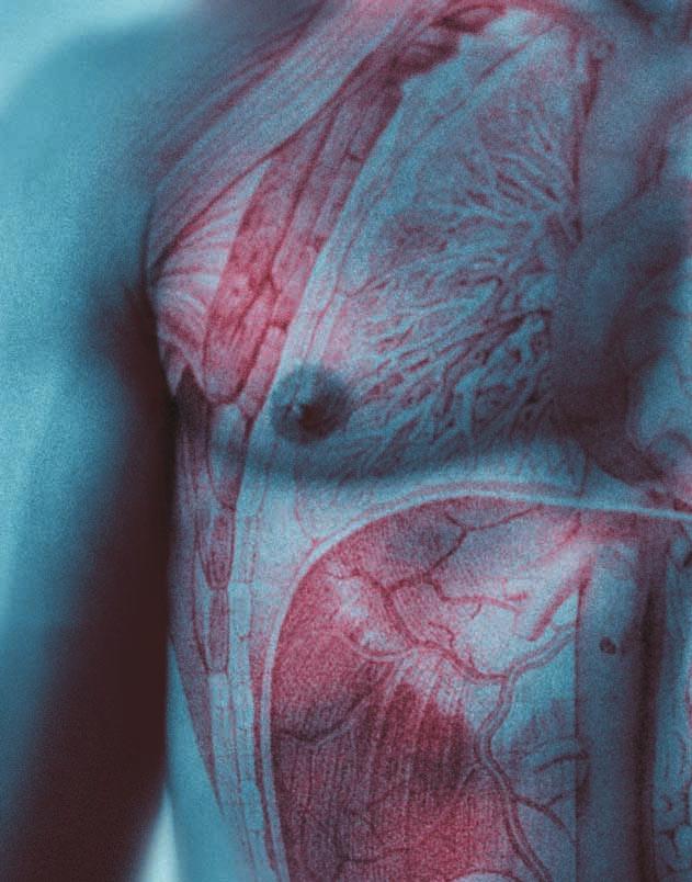
5 FIGURE 2 Muscles of the chest wall platysma muscle sternocleidomastoid muscle subclavius pectoralis major pectoralis minor intercostal muscle serratus anterior deltoid muscle edema in the absence of a pulmonary laceration. It has a very high morbidity and mortality rate following penetrating and blunt trauma. Modern diagnostic techniques, such as the CT scan, have made recognition of this common problem much more frequent. Opacification seen on X- ray within six hours of trauma generally is considered to be a pulmonary contusion. The most common cause of this type of trauma is a compression/decompression injury to the chest, such as may occur during a high-speed automobile crash. Airbags can lessen but do not entirely prevent this type of injury. A compression/decompression injury occurs when a highpressure wave within the thoracic cavity is caused by rapid compression. As the wave moves through the chest wall, the pressure wave reaches the lungs, causing an instant increase in intrathoracic pressure followed by an instant drop (decompression). 2 Pulmonary contusion has two stages: the first is related to the initial injury; the second to resuscitative measures and massive fluid overload. Administration of fluids to patients with unilateral pulmonary contusion can cause extravasation of fluid to the contralateral (uninjured) lung. A reduction in the pulmonary vascular resistance of the uninjured lung causes the right side of the heart to increase hydrostatic pressure within the capillaries to force both fluid and blood out of the capillaries and into the interstitium and alveoli. During this process, the congestion and contusion continue to the next uninjured segment of the lung. Changes within the capillaries leads to extravasation of red blood cells and the accumulation of edema fluid within the interstitium. These processes do not occur simultaneously, but rather in a sequence. The main problem is intra-alveolar hemorrhage and edema followed by a course of fluid resuscitation. The hemorrhage and edema, together with an accumulation of debris and mucous secretions, cause pulmonary atelectasis to occur, leading to large areas of unventilated but still perfused lung. Intrapulmonary shunting is increased, as is the resistance to airflow, causing an increase in respiratory effort to move oxygen into and carbon dioxide out of the lungs. Pulmonary ventilation is decreased, and the patient becomes hypoxic, hypercarbic, and acidotic. As a result, the heart will try to adapt to an increase in cardiac output, but because of the hypoxia, cardiopulmonary decompensation will occur quickly. Treatment of pulmonary contusion generally involves maintenance of adequate ventilation of the lungs. Chest physiotherapy, intercostal nerve blocks, epidural analgesia, and nasotracheal suction are necessary to ensure the patient is adequately oxygenated. Hemothorax Hemothorax is most frequently caused by bleeding from lung injuries. Only 5 to 15% of patients with hemothorax require thoracotomy, due to the fact that the bleeding in the chest, along with a high concentration of thromboplastin in the lungs and low pulmonary arterial pressure, reduces the chance of surgery. 2 A volume of more than ml of blood in the chest cavity must be removed completely as soon as possible. In the chest, large clots act as local anticoagulant by releasing fibrinolysins and fibrogenolysins from their surface. A large amount of blood in the chest can also restrict ventilation and venous return. Bleeding from multiple small intrathoracic vessels will often 24 The Surgical Technologist FEBRUARY 2001
 3.")
 stop fairly rapidly after the volume of blood is completely removed.")
6 Table 2: Types of thoracic trauma 1. Trauma to the outer region of the chest A. subcutaneous emphysema B. paradoxical respiration(flail chest) C. open pneumothorax 2. Trauma to the inner region of the chest A. closed pneumothorax B. hemothorax C. secretional obstruction of the lower airways (as in pulmonary contusion, wet lung, and aspiration pneumonitis) 3. Trauma to the innermost region of the chest A. mediastinal emphysema B. cardiac tamponade C. compression atelectasis (trauma to the diaphragm) stop fairly rapidly after the volume of blood is completely removed. Hemothorax should be suspected following trauma if: breath sounds are reduced; the chest is dull to percussion on the involved side; or fluid collections greater than 300 ml are seen on upright or decubitus roentgenograms of the chest. Treatment depends on the size of the hemothorax. Large hemothoraxes should be treated by insertion of a chest tube (tube thoracostomy). Blood that collects within the pleural cavity causes a reduction in the vital capacity of the lung and an increase in intrathoracic pressure, thereby decreasing minute ventilation and venous return to the heart. During inspiration, the negative intrapleural pressure increases the tendency for blood and air to leak into the pleural cavity through the wound in the lung or chest wall. The patient with an upper airway obstruction has additional air forced into the pleural space during expiration, increasing the possibility of a tension pneumothorax because the intrapleural pressure exceeds atmospheric pressure. FIGURE 3 Flail chest detail rib 3 two fractures rib 4 two fractures rib 5 two fractures rib 6 two fractures FIGURE 4 Pneumothorax pleaural edge mediastinum at midline hemidiaphragms at equal heights Pneumothorax Pneumothorax is the presence of air in the pleural space (Figure 4). Chest X-ray upon admission to the emergency department is the ideal diagnostic tool. However, a clinical diagnosis can be made based on the following criteria: FEBRUARY 2001 The Surgical Technologist 25


7 FIGURE 5 Emergency pericardiocentesis pericardium heart blood sternum Severe respiratory distress Distended neck veins Decreased breath sounds and hyperresonance unilaterally Deviation of the trachea away from the affected side. 2 When pneumothorax is suspected, but not seen clearly on the first chest X-ray, an expiratory film may be helpful. The patient may not show severe symptoms of pneumothorax unless: it occurs in patients with shock or preexisting cardiopulmonary disease; it is a tension pneumothorax; or it occupies more than 40% of the hemothorax. 2 The patient with a small stab wound may experience a delayed pneumothorax that can occur 12 or more hours following the trauma. Serial chest X-rays every six hours for 12 to 24 hours are generally indicated in these patients. Following observation and repeated chest X- rays, the patient may be discharged if no other problems are noted. When pneumothorax is assumed, treatment should be started without waiting for a chest X- ray. Treatment of pneumothorax consists of placement of a large needle into the involved pleural space through a midclavicular or second intercostal space puncture and aspirating any air. This procedure can also confirm the diagnosis of the pneumothorax and may provide temporary relief until a tube thoracostomy can be performed. If a pneumothorax persists in spite of one or more well-placed chest tubes, an emergency bronchoscopy should be performed to clear the bronchi and identify any damage to the tracheobronchial tree that may need repair. Continued air leakage and failure of the lung to expand adequately is an indication for early thoracotomy. 2 Trauma to the diaphragm The most common cause of a diaphragmatic injury is penetrating trauma, particularly gunshot wounds to the lower chest or upper abdomen. Diaphragmatic injury due to blunt trauma is less frequent, only occurring in 4 to 5% of patients with diaphragmatic injury. If a rib fracture is present, the incidence of diaphragmatic injury due to blunt trauma increases to 8 to 10%. 2 Rupture of the diaphragm can cause serious ventilatory problems, but the initial signs and symptoms are generally masked by other injuries. If the diaphragmatic injury is large, the intestinal viscera enters the chest cavity immediately. On the other hand, if the diaphragmatic defect is small, the intestinal viscera may take time to move into the chest cavity, eventually becoming obstructed or strangulated, leading to the formation of a tension pneumothorax. Generally, patients suffering penetrating diaphragmatic injury are diagnosed according to 26 The Surgical Technologist FEBRUARY 2001
8 the location of an entrance wound or intraoperatively. Fifty percent of diaphragmatic injuries are diagnosed surgically (via thoracotomy or laparotomy). Patients suffering blunt trauma may show an abnormality of the lower lung fields or diaphragm on chest X-ray. Additionally, diaphragmatic injuries may be diagnosed using the following methods. 1. Peritoneal lavage with a chest tube in place 2. Upper GI series 3. Pneumoperitoneum with carbon dioxide 4. CT scan with contrast 5. Intraperitoneal technetium sulfur colloid. 2 Optimal repair of a diaphragmatic injury occurs during laparotomy. Cardiac tamponade Cardiac tamponade may be caused by penetrating or blunt trauma to the chest, due to an accumulation of blood within the pericardial sac. The most common penetrating cause of cardiac tamponade is a stab wound to the mid-chest; while blunt compressive forces to the anterior heart cause rupture of the right atrium. Due to the injury, blood fills the pericardial sac causing pressure within the sac to rise rapidly. During this time, the right ventricle may be able to maintain enough blood volume to sustain life for a short interval. The patient will show the same symptoms as seen with a tension pneumothorax. Both cardiac tamponade and tension pneumothorax will cause obstruction of venous return to the heart. Patients will have hypoperfusion, and distended neck veins. As the tamponade progresses, the heart sounds will also be muffled and become more distant as the pericardial pressure increases. 2 The initial treatment is emergency pericardiocentesis (Figure 5), followed by surgical intervention, if necessary, to control bleeding. The patient should be given an intravenous bolus of fluid to fill the right atrium and increase cardiac output. Aspiration of 5 to 10 ml of blood can substantially improve cardiac performance and outcome. Conclusion Rapid diagnosis and immediate treatment are necessary when chest trauma is suspected. Modern radiographic techniques, blood laboratory studies, and pulse oximetry give the patient care team valuable diagnostic treatment shortly after admission to the emergency department allowing treatment to be implemented within minutes, lowering the mortality rate. About the Authors Richard E Wills, MD, MBA, ACSM is the medical director and program director of Steven s Henager Physician Assistant Program and professor of clinical medicine. Michael Norton, DC, is the owner of Chiropractic Health and Fitness and adjunct professor of medicine at Steven s Henager College in Salt Lake City, Utah. Kathryn DeLaney is an EMT and medical assistant in Salt Lake City, Utah. References 1. Tintinalli JE. Emergency Medicine. New York: McGraw-Hill; Schwartz SI. Principles of Surgery, 4th ed. New York: McGraw-Hill; Active First Aid Online Chest Injuries Accessed Medic Boss Chest and Abdominal Injuries ominal%20injuries Accessed Trauma.org Thoracic Trauma /thoracic/index.html Accessed Virtual Hospital Chest Trauma Providers/TeachingFiles/M3M4TeachingModules/Mullan /ChestTrauma.html Accessed Figures 3 and 4 used with permission of Virtual Hospital, Figure 5 from Surgical Technology for the Surgical Technologist: A Positive Care Approach, 1st edition, by Wintermantel C2001. Reprinted with permission of Delmar, a division of Thompson Learning. Fax FEBRUARY 2001 The Surgical Technologist 27
CHEST TRAUMA. Dr Naeem Zia FCPS,FACS,FRCS
 CHEST TRAUMA Dr Naeem Zia FCPS,FACS,FRCS Learning objectives Anatomy of chest wall and thoracic viscera Physiology of respiration and nerve pathways for pain Enumerate different thoracic conditions requiring
In ESH we usually see blunt chest trauma but penetrating injuries also treated here (usually as single injuries, like stab wound)
") Chest Trauma Dr Csaba Dioszeghy MD PhD FRCEM FFICM FERC East Surrey Hospital Emergency Department Scope Thoracic injuries are common and can be life threatening In ESH we usually see blunt chest trauma
Chest Trauma Dr Csaba Dioszeghy MD PhD FRCEM FFICM FERC East Surrey Hospital Emergency Department Scope Thoracic injuries are common and can be life threatening In ESH we usually see blunt chest trauma
CHEST INJURIES. Jacek Piątkowski M.D., Ph. D.
 CHEST INJURIES Jacek Piątkowski M.D., Ph. D. CHEST INJURIES 3-4% of all injuries 8% of patients hospitalized due to injuries 65% of patients who died at the accident place CLASSIFICATION OF THE CHEST INJURIES
Imaging of Thoracic Trauma: Tips and Traps. Arun C. Nachiappan, MD Associate Professor of Clinical Radiology University of Pennsylvania
 Imaging of Thoracic Trauma: Tips and Traps Arun C. Nachiappan, MD Associate Professor of Clinical Radiology University of Pennsylvania None Disclosures Objectives Describe blunt and penetrating traumatic
Chapter 29 - Chest Injuries
 1 2 3 4 5 6 7 8 9 National EMS Education Standard Competencies (1 of 5) Trauma Applies fundamental knowledge to provide basic emergency care and transportation based on assessment findings for an acutely
Medical NREMT-PTE. NREMT Paramedic Trauma Exam.
 Medical NREMT-PTE NREMT Paramedic Trauma Exam https://killexams.com/pass4sure/exam-detail/nremt-pte Question: 41 Which of the following most accurately describes the finding of jugular venous distension
9/10/2012. Chapter 49. Learning Objectives. Learning Objectives (Cont d) Thoracic Trauma
 Thoracic Trauma") Chapter 49 Thoracic Trauma 1 Learning Objectives Explain relevance of thoracic injuries as part of the overall mortality rate from major trauma List thoracic injuries that may result in early death if
Chapter 49 Thoracic Trauma 1 Learning Objectives Explain relevance of thoracic injuries as part of the overall mortality rate from major trauma List thoracic injuries that may result in early death if
3. D Objective: Chapter 4, Objective 4 Page: 79 Rationale: A carbon dioxide level below 35 mmhg indicates hyperventilation.
 1. A Objective: Chapter 1, Objective 3 Page: 14 Rationale: The sudden increase in acceleration produces posterior displacement of the occupants and possible hyperextension of the cervical spine if the
The Management of Chest Trauma. Tom Scaletta, MD FAAEM Immediate Past President, AAEM
 The Management of Chest Trauma Tom Scaletta, MD FAAEM Immediate Past President, AAEM Trichotomizing Rib Fractures Upper 1-3 vascular injuries Middle 4-9 Lower 10-12 12 liver/spleen injuries Management
The ABC s of Chest Trauma
 The ABC s of Chest Trauma J Bradley Pickhardt MD, FACS Providence St Patrick Hospital What s the Problem? 2/3 of trauma patients have chest trauma Responsible for 25% of all trauma deaths Most injuries
Chest Trauma.
 Chest Trauma www.fisiokinesiterapia.biz Objectives Anatomy of Thorax Main Causes of Chest Injuries S/S of Chest Injuries Different Types of Chest Injuries Treatments of Chest Injuries Anatomy of the chest
Children are not small adults Children are Not Small Adults Anatomic considerations Pliable bony & cartilagenous structures - Significant thoracic inj
 PEDIATRIC CHEST TRAUMA Children are not small adults Role of imaging Spectrum of injury Children are not small adults Children are Not Small Adults Anatomic considerations Pliable bony & cartilagenous
Chapter 29 - Chest_and_Abdominal_Trauma
 Introduction to Emergency Medical Care 1 OBJECTIVES 29.1 Define key terms introduced in this chapter. Slides 11, 15, 18, 27 29.2 Describe mechanisms of injury commonly associated with chest injuries. Slides
PEMSS PROTOCOLS INVASIVE PROCEDURES
 PEMSS PROTOCOLS INVASIVE PROCEDURES Panhandle Emergency Medical Services System SURGICAL AND NEEDLE CRICOTHYROTOMY Inability to intubate is the primary indication for creating an artificial airway. Care
PRE-HOSPITAL EMERGENCY CARE COURSE.
 PRE-HOSPITAL EMERGENCY CARE COURSE www.basics.org.uk Chest Assessment & Management BASICS Education March 2016 Objectives To understand the importance of oxygenation and ventilation To be able to describe
CHEST INJURY PULMONARY CONTUSION
 CHEST INJURY PULMONARY CONTUSION Introduction Pulmonary contusion refers to blunt traumatic lung parenchymal injury which results in oedema and haemorrhaging into alveolar spaces. It may also result in
Pneumothorax. Defined as air in the pleural space which can occur through a number of mechanisms
 Pneumothorax Defined as air in the pleural space which can occur through a number of mechanisms Traumatic pneumothorax Penetrating chest trauma Common secondary to bullet or knife penetration Chest tube
RCH Trauma Guideline. Management of Traumatic Pneumothorax & Haemothorax. Trauma Service, Division of Surgery
 RCH Trauma Guideline Management of Traumatic Pneumothorax & Haemothorax Trauma Service, Division of Surgery Aim To describe safe and competent management of traumatic pneumothorax and haemothorax at RCH.
Lecture 2: Clinical anatomy of thoracic cage and cavity II
 Lecture 2: Clinical anatomy of thoracic cage and cavity II Dr. Rehan Asad At the end of this session, the student should be able to: Identify and discuss clinical anatomy of mediastinum such as its deflection,
Advances in MDCT of Thoracic Trauma
 Baltic Congress of Radiology, Riga 2010 Advances in MDCT of Thoracic Trauma Robert A. Novelline, MD Professor of Radiology, Harvard Medical School Director of Emergency Radiology, Massachusetts General
ITLS Advanced Pre-Test Annotated Key 8 th Edition
 1. A Objective: Chapter 1, Objective 3 Page: 14 Rationale: The sudden increase in acceleration produces posterior displacement of the occupants and possible hyperextension of the cervical spine if the
Handling Common Problems & Pitfalls During. Oxygen desaturation in patients receiving mechanical ventilation ACUTE SEVERE RESPIRATORY FAILURE
 Handling Common Problems & Pitfalls During ACUTE SEVERE RESPIRATORY FAILURE Pravit Jetanachai, MD QSNICH Oxygen desaturation in patients receiving mechanical ventilation Causes of oxygen desaturation 1.
Thoracic Trauma The Spectrum
 Thoracic Trauma The Spectrum Joseph Mathew Consultant, s & Emergency dept. 2 Thoracic Trauma Responsible for 20-25% of all deaths attributed to trauma. Contributing cause of death in an additional 25%
1. In a rear-impact motor vehicle crash, which area of the spine is most susceptible to injury? A. Cervical B. Thoracic C. Lumbar D.
 1. In a rear-impact motor vehicle crash, which area of the spine is most susceptible to injury? A. Cervical B. Thoracic C. Lumbar D. Sacral-coccygeal 2. A 36-year-old male sustains blunt force thoracic
Right lung. -fissures:
 -Right lung is shorter and wider because it is compressed by the right copula of the diaphragm by the live.. 2 fissure, 3 lobes.. hilum : 2 bronchi ( ep-arterial, hyp-arterial ), one artery mediastinal
Dana Alrafaiah. - Moayyad Al-Shafei. -Mohammad H. Al-Mohtaseb. 1 P a g e
 - 6 - Dana Alrafaiah - Moayyad Al-Shafei -Mohammad H. Al-Mohtaseb 1 P a g e Quick recap: Both lungs have an apex, base, mediastinal and costal surfaces, anterior and posterior borders. The right lung,
H. Mitchell Shulman MDCM FRCPC CSPQ Assistant Professor, Dept. of Surgery, McGill Medical School Attending Physician, Royal Victoria Hospital,
 H. Mitchell Shulman MDCM FRCPC CSPQ Assistant Professor, Dept. of Surgery, McGill Medical School Attending Physician, Royal Victoria Hospital, Montreal General Hospital, McGill University Health Center
The Respiratory System
 C h a p t e r 24 The Respiratory System PowerPoint Lecture Slides prepared by Jason LaPres North Harris College Houston, Texas Copyright 2009 Pearson Education, Inc., publishing as Pearson Benjamin Cummings
INJURIES CHEST, ABDOMEN, LIMBS. FN Brno November 2011
 INJURIES CHEST, ABDOMEN, LIMBS FN Brno November 2011 Injury Chest Abdomen Limbs Injury to the rib cage Fractured one or more ribs Sharp pain at the site of fracture Pain on taking a deep breath Shallow
Procedure: Chest Tube Placement (Tube Thoracostomy)
") Procedure: Chest Tube Placement (Tube Thoracostomy) Basic Information: The insertion and placement of a chest tube into the pleural cavity for the purpose of removing air, blood, purulent drainage, or
Procedure: Chest Tube Placement (Tube Thoracostomy) Basic Information: The insertion and placement of a chest tube into the pleural cavity for the purpose of removing air, blood, purulent drainage, or
The Primary Survey. C. Clay Cothren, MD FACS. Attending Surgeon, Denver Health Medical Center Assistant Professor of Surgery, University of Colorado
 The Primary Survey C. Clay Cothren, MD FACS Attending Surgeon, Denver Health Medical Center Assistant Professor of Surgery, University of Colorado Outlining the ABCs Why do we need such an approach? The
Trauma. Neck trauma zones. Neck Injuries 1/3/2018. Basic principles A ; Airway B ; Breathing C ; Circulation D ; Disability E ; Exposure
 Trauma 45 minutes highest points Ahmed Mahmoud, MD Basic principles A ; Airway B ; Breathing C ; Circulation D ; Disability E ; Exposure Neck trauma zones Airway ;Rapid sequence intubation Breathing ;Needle
ITLS Pediatric Provider Course Advanced Pre-Test
 ITLS Pediatric Provider Course Advanced Pre-Test 1. You arrive at the scene of a motor vehicle crash and are directed to evaluate a child who was in one of the vehicles. The patient appears to be a child
Chest X-ray Interpretation
 Chest X-ray Interpretation Introduction Routinely obtained Pulmonary specialist consultation Inherent physical exam limitations Chest x-ray limitations Physical exam and chest x-ray provide compliment
Critical Care Monitoring. Indications. Pleural Space. Chest Drainage. Chest Drainage. Potential space. Contains fluid lubricant
 Critical Care Monitoring Indications 1-2- 2 Pleural Space Potential space Contains fluid lubricant Can fill with air, blood, plasma, serum, lymph, pus 3 1 Pleural Space Problems when contain abnormal substances:
PLEURAE and PLEURAL RECESSES
 PLEURAE and PLEURAL RECESSES By Dr Farooq Aman Ullah Khan PMC 26 th April 2018 Introduction When sectioned transversely, it is apparent that the thoracic cavity is kidney shaped: a transversely ovoid space
RESPIRATORY SYSTEM. A. Upper respiratory tract (Fig. 23.1) Use the half-head models.
 Use the half-head models.") RESPIRATORY SYSTEM I. OVERVIEW OF THE RESPIRATORY SYSTEM AND THORAX A. Upper respiratory tract (Fig. 23.1) Use the half-head models. Nasal cavity Pharynx (fare-rinks) B. Lower respiratory tract (Fig. 23.1)
RESPIRATORY SYSTEM I. OVERVIEW OF THE RESPIRATORY SYSTEM AND THORAX A. Upper respiratory tract (Fig. 23.1) Use the half-head models. Nasal cavity Pharynx (fare-rinks) B. Lower respiratory tract (Fig. 23.1)
Respiratory Physiology
 Respiratory Physiology Dr. Aida Korish Associate Prof. Physiology KSU The main goal of respiration is to 1-Provide oxygen to tissues 2- Remove CO2 from the body. Respiratory system consists of: Passages
Esophageal Perforation
 Esophageal Perforation Dr. Carmine Simone Thoracic Surgeon, Division of General Surgery Head, Division of Critical Care May 15, 2006 Overview Case presentation Radiology Pre-operative management Operative
Internal Injury Documentation Guidelines
 Internal Injury Documentation Guidelines General Open Wound of Thorax Injury to Heart Identify episode of care Initial Subsequent Sequela Laterality Sequela of injury Place of occurrence of injury Activity
PEDIATRIC TRAUMA: Implications for Respiratory Care
 PEDIATRIC TRAUMA: Implications for Respiratory Care 17 th Annual Rainbow Respiratory Conference - September 4, 2015 Mike Dingeldein, MD Pediatric Surgeon Pediatric Trauma Medical Director Disclosures none
Aurora Health Care EMS Continuing Education Spring 2011 Packet THORACIC TRAUMA THE PREHOSPITAL APPROACH TO CHEST INJURY MANAGEMENT
 Aurora Health Care EMS Continuing Education Spring 2011 Packet THORACIC TRAUMA Chest injuries are significant contributors to death from major trauma and can be difficult to assess adequately in the pre-hospital
Pulmonary Problems of the Neonate. Jon Palmer, VMD, DACVIM Chief, Neonatal Intensive Care Service New Bolton Center, University of Pennsylvania, USA
 Pulmonary Problems of the Neonate Jon Palmer, VMD, DACVIM Chief, Neonatal Intensive Care Service New Bolton Center, University of Pennsylvania, USA Lower Respiratory Diseases Ventilation/Perfusion Abnormalities
October Paediatric Respiratory Workbook APCP RESPIRATORY COMMITTEE
 October 2017 Paediatric Respiratory Workbook APCP RESPIRATORY COMMITTEE This workbook is designed to introduce to you the difference between paediatric and adult anatomy and physiology. It will also give
Management of Airway Trauma I:
 Management of Airway Trauma I: Tracheobronchial Injuries James P. Kelly, M.D., Watts R. Webb, M.D., Peter V. Moulder, M.D., Charles Everson, M.D., Buford H. Burch, M.D., and Edward S. Lindsey, M.D. ABSTRACT
Activity Three: Where s the Bleeding?
 Activity Three: Where s the Bleeding? There are five main sites of potentially fatal bleeding in trauma, remembered by the phrase on the floor and four more. On the floor refers to losing blood externally
CAE Healthcare istan. Neurological Features Anatomy, Physiology and Clinical signs
 CAE Healthcare istan NYSIM has two wireless istans available for your sessions. We also have them preprogrammed with the NLN Nursing These are available for you to review at the NYSIM Center. They can
You Are the Emergency Medical Responder
 Lesson 32: Injuries to the Chest, Abdomen and Genitalia You Are the Emergency Medical Responder Your police unit responds to a call in a part of town plagued by violence. When you arrive, you find the
Lecturer: Ms DS Pillay ROOM 2P24 25 February 2013
 Lecturer: Ms DS Pillay ROOM 2P24 25 February 2013 Thoracic Wall Consists of thoracic cage Muscle Fascia Thoracic Cavity 3 Compartments of the Thorax (Great Vessels) (Heart) Superior thoracic aperture
Anatomy notes-thorax.
 Anatomy notes-thorax. Thorax: the part extending from the root of the neck to the abdomen. Parts of the thorax: - Thoracic cage (bones). - Thoracic wall. - Thoracic cavity. ** The thoracic cavity is covered
INTRA-THORACIC AND INTRA-ABDO-MINAL PERFORATION OF THE COLON IN TRAUMATIC DIAPHRAGMATIC
 INTRA-THORACIC AND INTRA-ABDO-MINAL PERFORATION OF THE COLON IN TRAUMATIC DIAPHRAGMATIC Pages with reference to book, From 14 To 16 S. Amjad Hussain, Chinda Suriyapa, Karl Grubaugh ( Depts. of Surger and
Shot Through the Heart (And You re to Blame): Penetrating Cardiac Trauma
: Penetrating Cardiac Trauma") Shot Through the Heart (And You re to Blame): Penetrating Cardiac Trauma Yalaunda M. Thomas, MD, FACS The American College of Osteopathic Emergency Physicians Spring Seminar April 5, 2018 Disclosures I
Shot Through the Heart (And You re to Blame): Penetrating Cardiac Trauma Yalaunda M. Thomas, MD, FACS The American College of Osteopathic Emergency Physicians Spring Seminar April 5, 2018 Disclosures I
TRACHEOSTOMY. Tracheostomy means creation an artificial opening in the trachea with tracheostomy tube insertion
 TRACHEOSTOMY Definition Tracheostomy means creation an artificial opening in the trachea with tracheostomy tube insertion Indications for tracheostomy 1-upper airway obstruction with stridor, air hunger,
(SKILLS/HANDS-ON) Chest Tubes. Rebecca Carman, MSN, ACNP-BC. Amanda Shumway, PA-C. Thomas W. White, MD, FACS, CNSC
 Chest Tubes. Rebecca Carman, MSN, ACNP-BC. Amanda Shumway, PA-C. Thomas W. White, MD, FACS, CNSC") (SKILLS/HANDS-ON) Chest Tubes Rebecca Carman, MSN, ACNP-BC Nurse Practitioner, Trauma Services, Intermountain Medical Center, Intermountain Healthcare Amanda Shumway, PA-C APC Trauma and Critical Care
(SKILLS/HANDS-ON) Chest Tubes Rebecca Carman, MSN, ACNP-BC Nurse Practitioner, Trauma Services, Intermountain Medical Center, Intermountain Healthcare Amanda Shumway, PA-C APC Trauma and Critical Care
Thorax Lecture 2 Thoracic cavity.
 Thorax Lecture 2 Thoracic cavity. Spring 2016 Dr. Maher Hadidi, University of Jordan 1 Enclosed by the thoracic wall. Extends between (thoracic inlet) & (thoracic outlet). Thoracic inlet At root of the
European Resuscitation Council
 European Resuscitation Council Incidence of Trauma in Childhood Leading cause of death and disability in children older than one year all over the world Structured approach Primary survey and resuscitation
CAE Healthcare Human Patient Simulator (HPS)
") CAE Healthcare Human Patient Simulator (HPS) The Human Patient Simulator, HPS, is a tethered simulator that is capable of patient assessment and treatment including mechanical ventilation and anesthesia.
CAE Healthcare Human Patient Simulator (HPS) The Human Patient Simulator, HPS, is a tethered simulator that is capable of patient assessment and treatment including mechanical ventilation and anesthesia.
Chapter 3: Thorax. Thorax
 Chapter 3: Thorax Thorax Thoracic Cage I. Thoracic Cage Osteology A. Thoracic Vertebrae Basic structure: vertebral body, pedicles, laminae, spinous processes and transverse processes Natural kyphotic shape,
Anatomy Lecture 8. In the previous lecture we talked about the lungs, and their surface anatomy:
 Anatomy Lecture 8 In the previous lecture we talked about the lungs, and their surface anatomy: 1-Apex:it lies 1 inch above the medial third of clavicle. 2-Anterior border: it starts from apex to the midpoint
Definitive Care Phase: Chest Injuries
 Página 1 de 29 Copyright 2001 Lippincott Williams & Wilkins Greenfield, Lazar J., Mulholland, Michael W., Oldham, Keith T., Zelenock, Gerald B., Lillemoe, Keith D. Surgery: Scientific Principles & Practice,
4/16/2017. Learning Objectives. Interpretation of the Chest Radiograph. Components. Production of the Radiograph. Density & Appearance
 Interpretation of the Arthur Jones, EdD, RRT Learning Objectives Identify technical defects in chest radiographs Identify common radiographic abnormalities This Presentation is Approved for 1 CRCE Credit
Session Number 219 A BLUNT REVIEW OF THE PENETRATING ISSUES IN CHEST TRAUMA
 Session Number 219 A BLUNT REVIEW OF THE PENETRATING ISSUES IN CHEST TRAUMA Lisa C. Laphan-Morad, MSN, APN-C Administrative Director of Surgical & Ambulatory Services Nurse Practitioner Virtua Health Marlton,
McHenry Western Lake County EMS System Paramedic, EMT-B and PHRN Optional Continuing Education 2019 #3 Penetrating Neck Trauma
 McHenry Western Lake County EMS System Paramedic, EMT-B and PHRN Optional Continuing Education 2019 #3 Penetrating Neck Trauma Penetrating neck injury (PNI) comprises 5 to 10 percent of traumatic injuries
Pulmonary Pathophysiology
 Pulmonary Pathophysiology 1 Reduction of Pulmonary Function 1. Inadequate blood flow to the lungs hypoperfusion 2. Inadequate air flow to the alveoli - hypoventilation 2 Signs and Symptoms of Pulmonary
The Anatomy and Physiology of the Respiratory System
 CHAPTER 1 The Anatomy and Physiology of the Respiratory System Sagittal Section of Upper Airway Fig. 1-1. Sagittal section of upper airway. Structure of the Nose Fig. 1-2. Structure of the nose. Sagittal
Lung & Pleura. The Topics :
 Lung & Pleura The Topics : The Trachea. The Bronchi. The Brochopulmonary Segments. The Lungs. The Hilum. The Pleura. The Surface Anatomy Of The Lung & Pleura. The Root & Hilum. - first of all, the lung
Northwest Community EMS System September 2017: Head and Chest Trauma Credit Questions
 NWC EMSS Sept 2017 CE: Head & Chest Trauma. Credit Questions - page 1 Northwest Community EMS System September 2017: Head and Chest Trauma Credit Questions Name: EMS Agency EMSC/Educator reviewer: Date
Esophageal injuries. 新光急診張志華醫師 Facebook.com/jack119
 Esophageal injuries 新光急診張志華醫師 Facebook.com/jack119 Pre-test 1 What is the most common cause of esophageal injuries? A. Traffic accidents B. Gunshot wounds C. Iatrogenic Pre-test 2 Which contrast agent
Syllabus: 6 pages (Page 6 lists corresponding figures for Grant's Atlas 11 th & 12 th Eds.)
") PLEURAL CAVITY AND LUNGS Dr. Milton M. Sholley SELF STUDY RESOURCES Essential Clinical Anatomy 3 rd ed. (ECA): pp. 70 81 Syllabus: 6 pages (Page 6 lists corresponding figures for Grant's Atlas 11 th &
PLEURAL CAVITY AND LUNGS Dr. Milton M. Sholley SELF STUDY RESOURCES Essential Clinical Anatomy 3 rd ed. (ECA): pp. 70 81 Syllabus: 6 pages (Page 6 lists corresponding figures for Grant's Atlas 11 th &
Circulatory System. and. Respiratory System. Ari Min, Yerim Lee and Min Ji Song THE HEART LUNGS. Monday, May 23, 2011
 Human Anatomy Circulatory System and THE HEART Respiratory System LUNGS Ari Min, Yerim Lee and Min Ji Song Purpose of the Circulatory System Function of circulatory system: exchange gases with cardiovascular
Introduction to Chest Radiography
 Introduction to Chest Radiography RSTH 366: DIAGNOSTIC TECHNIQUES Alan Alipoon BS, RCP, RRT Instructor Department of Cardiopulmonary Sciences 1 Introduction Discovered in 1895 by Wilhelm Roentgen Terminology
Chapter 10. The Respiratory System Exchange of Gases. Copyright 2009 Pearson Education, Inc.
 Chapter 10 The Respiratory System Exchange of Gases http://www.encognitive.com/images/respiratory-system.jpg Human Respiratory System UPPER RESPIRATORY TRACT LOWER RESPIRATORY TRACT Nose Passageway for
This is not a required assignment but it is recommended.
 SU 12 Name: This is not a required assignment but it is recommended. BIO 116 - Anatomy & Physiology II Practice Assignment 2 - The Respiratory and Cardiovascular Systems 1. The exchange of oxygen and carbon
Chapter 16. Respiratory System
 Chapter 16 Respiratory System Introduction Respiration = the entire process of exchanging gases between the atmosphere and body cells 1. Ventilation 2. Gas exchange 3. Gas transport : 4. Cellular respiration
Initial Assessment and Management of the Trauma Patient
 Initial Assessment and Management of the Trauma Patient 1 Epidemiology Road Traffic Accidents are major cause of long term morbidity and mortality in developing nations WHO predicts that by 2020, Road
Purpose This Operating Procedure provides guidance on the management of blunt thoracic traumatic injury
 Blunt Thoracic Trauma HELI.CLI.09 Purpose This Operating Procedure provides guidance on the management of blunt thoracic traumatic injury Procedure Management of Blunt Thoracic Traumatic Injury For Review
Respiratory System. Chapter 9
 Respiratory System Chapter 9 Air Intake Air in the atmosphere is mostly Nitrogen (78%) Only ~21% oxygen Carbon dioxide is less than 0.04% Air Intake Oxygen is required for Aerobic Cellular Respiration
Tracheal Trauma: Management and Treatment. Kosmas Iliadis, MD, PhD, FECTS
 Tracheal Trauma: Management and Treatment Kosmas Iliadis, MD, PhD, FECTS Thoracic Surgeon Director of Thoracic Surgery Department Hygeia Hospital, Athens INTRODUCTION Heterogeneous group of injuries mechanism
Tracheostomy and Ventilator Education Program Module 2: Respiratory Anatomy
 Tracheostomy and Ventilator Education Program Module 2: Respiratory Anatomy Disclaimer This material is intended for use by trained family members and caregivers of children with tracheostomies who are
Evaluation & Management of Penetrating Wounds to the NECK
 Evaluation & Management of Penetrating Wounds to the NECK Goal Effectively identify patients with a high probability of injury requiring surgical intervention Define the role of diagnostic tests in assessing
Introduction (1 of 3)
") Chapter 10 Shock Introduction (1 of 3) Shock (hypoperfusion) means a state of collapse and failure of the cardiovascular system. In the early stages, the body attempts to maintain homeostasis. As shock
Chapter 10 Shock Introduction (1 of 3) Shock (hypoperfusion) means a state of collapse and failure of the cardiovascular system. In the early stages, the body attempts to maintain homeostasis. As shock
Tuesday, December 13, 16. Respiratory System
 Respiratory System Trivia Time... What is the fastest sneeze speed? What is the surface area of the lungs? (hint... think of how large the small intestine was) How many breaths does the average person
Anatomy of the Lungs. Dr. Gondo Gozali Department of anatomy
 Anatomy of the Lungs Dr. Gondo Gozali Department of anatomy 1 Pulmonary Function Ventilation and Respiration Ventilation is the movement of air in and out of the lungs Respiration is the process of gas
The Respiratory System. Dr. Ali Ebneshahidi
 The Respiratory System Dr. Ali Ebneshahidi Functions of The Respiratory System To allow gases from the environment to enter the bronchial tree through inspiration by expanding the thoracic volume. To allow
Chapter 16. Thoracic Injuries
 Thoracic Injuries Chapter 16 Thoracic Injuries Introduction About 15% of war injuries involve the chest. Of those, 10% are superficial (soft tissue only) requiring only basic wound treatment. The remaining
The Primary Survey. Clay Cothren Burlew, MD FACS
 The Primary Survey Clay Cothren Burlew, MD FACS Director, Surgical Intensive Care Unit Attending Surgeon, Denver Health Medical Center Associate Professor of Surgery, University of Colorado Outlining the
Shedding Light on Neonatal X-rays. Objectives. Indications for X-Rays 5/14/2018
 Shedding Light on Neonatal X-rays Barbara C. Mordue, MSN, NNP-BC Neonatal Nurse Practitioner LLUH Children s Hospital, NICU Objectives Utilize a systematic approach to neonatal x-ray interpretation Identify
2. List seven functions performed by the respiratory system?
 The Respiratory System C23 Study Guide Tortora and Derrickson 1. In physiology we recognize that the word respiration has three meanings. What are the three different meanings of the word respiration as
Esophageal injuries. Pre-test /11/10. 新光急診張志華醫師 Facebook.com/jack119. O What is the most common cause of esophageal injuries?
 Esophageal injuries 新光急診張志華醫師 Facebook.com/jack119 Pre-test 1 O What is the most common cause of esophageal injuries? A. Traffic accidents B. Gunshot wounds C. Iatrogenic 1 Pre-test 2 O Which contrast
ANATOMY OF THE PLEURA. Dr Oluwadiya KS
 ANATOMY OF THE PLEURA Dr Oluwadiya KS www.oluwadiya.sitesled.com Introduction The thoracic cavity is divided mainly into: Right pleural cavity Mediastinum Left Pleural cavity Pleural cavity The pleural
Respiratory Diseases and Disorders
 Chapter 9 Respiratory Diseases and Disorders Anatomy and Physiology Chest, lungs, and conducting airways Two parts: Upper respiratory system consists of nose, mouth, sinuses, pharynx, and larynx Lower
CHAPTER 7.1 STRUCTURES OF THE RESPIRATORY SYSTEM
 CHAPTER 7.1 STRUCTURES OF THE RESPIRATORY SYSTEM Pages 244-247 DO NOW What structures, do you think, are active participating in the breathing process? 2 WHAT ARE WE DOING IN TODAY S CLASS Finishing Digestion
Thoracic Trauma: 5/19/2010. Keihan Golshani, MD, Assistant Professor of emergency Medicine, Isfahan Medical University,
 Thoracic Trauma: 1 Thoracic trauma: A significant cause of mortality. Many patients with thoracic trauma: die after reaching the hospital Many of them can be prevented by: Prompt diagnosis and treatment.
Proceedings of the World Small Animal Veterinary Association Sydney, Australia 2007
 Proceedings of the World Small Animal Sydney, Australia 2007 Hosted by: Next WSAVA Congress THE LAST GASP II: LUNGS AND THORAX David Holt, BVSc, Diplomate ACVS University of Pennsylvania School of Veterinary
Thoracic Injuries. Chapter 16
 Thoracic Injuries Chapter 16 Thoracic Injuries Introduction About 15% of war injuries involve the torso. Those injuries involving the vasculature of the mediastinum (heart, great vessels, and pulmonary
ADVANCED ASSESSMENT Respiratory System
 ONTARIO BASE HOSPITAL GROUP QUIT ADVANCED ASSESSMENT Respiratory System 2007 Ontario Base Hospital Group ADVANCED ASSESSMENT Respiratory System AUTHOR(S) Mike Muir AEMCA, ACP, BHSc Paramedic Program Manager
Respiratory System. Clinical notes. Published on Second Faculty of Medicine, Charles University ( https://www.lf2.cuni.cz)
") Published on Second Faculty of Medicine, Charles University ( https://www.lf2.cuni.cz) Respiratory System The test of the respiratory system follows the general rules for written tests (see Continuous
Published on Second Faculty of Medicine, Charles University ( https://www.lf2.cuni.cz) Respiratory System The test of the respiratory system follows the general rules for written tests (see Continuous
Chest X rays and Case Studies. No disclosures. Outline 5/31/2018. Carlo Manalo, M.D. Department of Radiology Loma Linda University Children s Hospital
 Chest X rays and Case Studies Carlo Manalo, M.D. Department of Radiology Loma Linda University Children s Hospital No disclosures. Outline Importance of history Densities delineated on radiography An approach
Trauma Life Support Pre-Hospital (TLS-P) Preparatory Materials
 Preparatory Materials") Trauma Life Support Pre-Hospital (TLS-P) Preparatory Materials 1 1. A high-risk bodily fluid for spreading infection is blood. 2. Items that can reduce the spread of infection include masks, gloves, and
Trauma Life Support Pre-Hospital (TLS-P) Preparatory Materials 1 1. A high-risk bodily fluid for spreading infection is blood. 2. Items that can reduce the spread of infection include masks, gloves, and
Chapter 13. The Respiratory System.
 Chapter 13 The Respiratory System https://www.youtube.com/watch?v=hc1ytxc_84a https://www.youtube.com/watch?v=9fxm85fy4sq http://ed.ted.com/lessons/what-do-the-lungs-do-emma-bryce Primary Function of Breathing