From the Vulnerable Atherosclerotic Plaque to CAD Management
|
|
|
- Tamsyn Dean
- 6 years ago
- Views:
Transcription
1 33 rd Panhellenic Congress of Cardiology Athens, November 1-3, 2012 From the Vulnerable Atherosclerotic Plaque to CAD Management Filippos Triposkiadis, MD, FESC, FACC Professor of Cardiology Director, Department of Cardiology Larissa University Hospital Larissa, Greece
2 Introduction
3 Natural History of CAD Obstructive Non-obstructive Positive Remodelling Stenotic Lesions Stress ischemia (+) Stress angina (-/+) Calcium Score (+) Angiography (+) IVUS (+) Normal Non-Stenotic Lesions Stress ischemia (--/+) Stress angina (--/+) Calcium score (--/+) Angiography (--/+) IVUS (+) Triposkiadis F, et al. Curr Cardiol Rev 2007;2: Coronary thrombosis Death Unstable angina Myocardial infarction

4 Presentation of Coronary Artery Disease (%) Series SCD ACS Stable Angina Only 18% of coronary attacks are preceded by longstanding angina pectoris Myerburg RJ, Junttila J. Circulation 2012; 125: Marzocchi A, et al. Int J Cardiol 2012; 157: Hemingway H, et al. 2006; 295:
5 Pathology

6 A Histological Classification of Atherosclerosis Stary HC, et al. Circulation. 1995; 92:
7 Definition of the Vulnerable Plaque Plaque that is at increased risk of thrombosis (or re-thrombosis) and rapid stenosis progression. Schaar, et al. Eur Heart J 2004; 25: Non-obstructive, silent coronary lesion that suddenly becomes obstructive and symptomatic. Moreno, et al. Cardiol Clin 2010; 28:1-30 Plaque susceptible to disruption with superimposed thrombosis, which is responsible for the majority of acute coronary events. Fishbein MC. Cardiovasc Pathol 2010; 19:6-11
 Large necrotic core Increased")


8 Causes of Coronary Thrombosis Plaque rupture (55-60%) Thin fibrous cap (95% 64 μm) Large necrotic core Increased plaque inflammation Positive vascular remodeling Vasa vasorum neovascularization Intraplaque hemorrhage Plaque erosion (30-35%) No specific features suitable for detection Thick, SMC rich fibrous cap Premenopausal women Cigarette smoking Calcified nodule (2-7%) Heavily calcified plaque with or without necrotic core Luminal region of plaque shows disrupted surface fibrous cap and overlying thrombus. More common in older male than women. Virmani R, et al. JACC 2006; 47: Suppl C: C13-8

9 The Thin-Cap Fibroatheroma Inflammation Macrophages infiltration 14±10% in the fibrous cap Thin fibrous cap < 65 μm Spotty calcifications Large necrotic core occupying 23±17% of the plaque area Expansive outward remodeling Remodeling index >1.1 Neoangiogenesis Adventitial vasa vasorum proliferation Intraplaque hemorrhage Virmani R, et al. J Am Coll Cardiol 2006;47:C13 8 Burke AP, et al. N Engl J Med 1997;336: Virmani R, et al.arterioscler Thromb Vasc Biol 2000;20:

10 Arterial Remodelling Schoenhagen P, et al. Circulation 2000;101:
11 Frequency and Distribution of TCFA and Ruptured Plaques Section of coronary arteries from 50 whole hearts taken from pts (mean age 74 years, 64% men) dying of CV (n=33) non-cv (n=13) and unknown (n=4) causes). The majority of TFCA and ruptured plaque localized in the proximal third of the major coronary arteries. Cheruvu, et al. JACC 2007;50:940 9
12 Inflammation in Both Vulnerable and Stable Plaques in Pts Dying of AMI 544 coronary artery segments from 16 patients dying of AMI, 109 segments from 5 patients dying of SA, and 304 coronary segments from 9 patients dying of non-cardiac causes (CTRL) were examined 6.8 ±0.5 vulnerable segments/patient in AMI (in addition to culprit lesions) vs. 0.8 ± 0.3 and 1.4 ± 0.3 in SA and CTRL. In AMI more inflammatory infiltrates vs. SA and CTRL (121.6 ± 12.4 cell X mm2 vs ± 11.9 cell X mm2 vs ± 6.8 cell X mm2, p= ). In AMI, active inflammation evident within culprit lesion, vulnerable plaques, and stable plaques. In AMI vulnerable and stable plaques showed 3- to 4-fold higher inflammation vs. SA and CTRL. Mauriello, et al. J Am Coll Cardiol 2005;45:
 with acute coronary syndromes underwent carotid ultrasonography within 2 days of cardiac catheterization. Triposkiadis F, et al.")
13 Carotid Plaque Composition in Stable and Unstable CAD Twenty-five patients (16 men/9 women, age 63 ± 10 years) with stable coronary artery disease and 61 (41 men/20 women, age 66 ± 16 years) with acute coronary syndromes underwent carotid ultrasonography within 2 days of cardiac catheterization. Triposkiadis F, et al. Am Heart J 2005; 150: 782-9
14 Detection
 targeted to markers of activity (MMP, Ox-LDL, LOX) Different Activity")
15 Morphology vs. Activity Imaging Thermography, Spectroscopy, Molecular Imaging, (radionuclear, MRI, CT ) targeted to markers of activity (MMP, Ox-LDL, LOX) Different Activity Active and inflamed plaque Similar Morphology Inactive and non-inflamed plaque IVUS OCT MRI w/o CM Naghavi M, et al. Circulation 2003; 108:


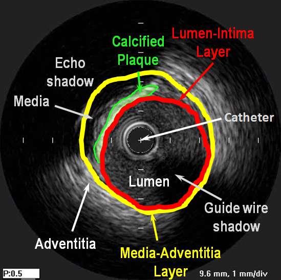
16 Coronary Intravascular Ultrasound Coronary Remodeling Conceals Extensive Disease Stable CAD Remodeling and Clinical Presentation Plaque Rupture Unstable CAD Schoenhagen, et al. Circulation 2000; 101: Nissen S. JACC 2003:41:103S-112S

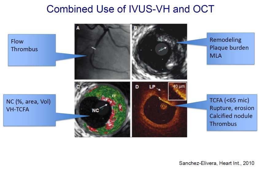
17 IVUS Gray-Scale and Corresponding Virtual Histology Frames MLA: Minimum luminal area; PB: Plaque burden; VCSA: Vessel cross-sectional area. Virtual histology color code: green is fibrous, greenish is fibrofatty, red is necrotic core, and white is dense calcium. Garcia-Garcia HM, et al. Expert Rev Cardiovasc Ther 2008; 6:


18 OCT Coronary Imaging Prati F, et al. Eur Heart J 2010; 31:401-15

19 IVUS vs. OCT OCT IVUS Garcia-Garcia HM, et al. Heart 2009;95: Prati F, et al. Eur Heart J 2010; 31:401-15

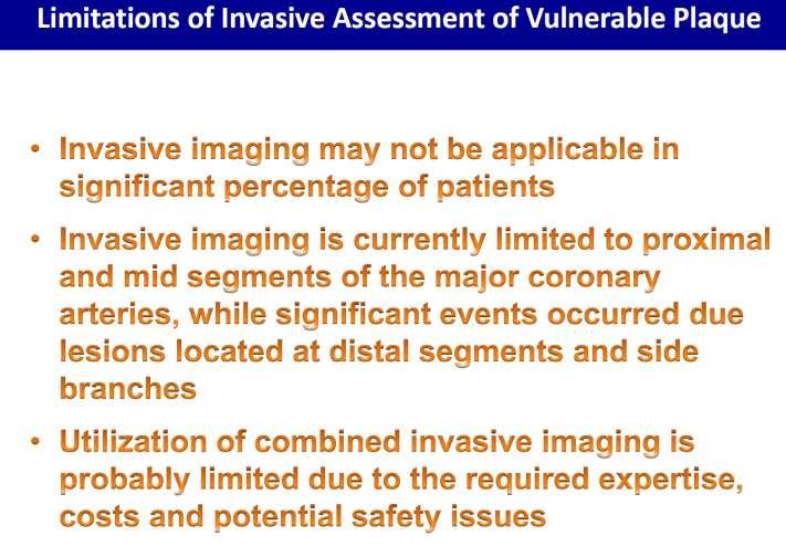
20 Potential Risk of Invasive Assessment of Vulnerable Plaque Stone GW, et al. N Engl J Med 2011;364:226-35

21 Noninvasive Techniques for Vulnerable Plaque Imaging Vancraeynest D, et al. J Am Coll Cardiol 2011;57:


22 Williams MC, et al. Heart 2011;97: Voros S, et al. JACC Img 2011; 4: CT Coronary Imaging
 months. 136 individuals died.")
23 The CONFIRM Registry: ccta and Outcome in Subjects Without Chest Pain Syndrome A registry of 27, 125 pts undergoing ccta and CACS was queried subjects without CPS or CAD history were included. All-cause mortality and the composite of all-cause mortality and nonfatal MI were measured. Median follow-up was 24 (18 35) months. 136 individuals died. The net reclassification improvement resulting from the addition of ccta to a model based on standard risk factors and CACS was negligible! Cho I, et al. Circulation 2012;126:
24 Natural History of Vulnerable Plaque
 in 99 patients. Kubo T, et al.")
25 Dynamic Nature of Coronary Plaque Morphology Baseline and 12-month follow-up VH-IVUS studies of 216 non-culprit lesions (plaque burden 40%) in 99 patients. Kubo T, et al. J Am Coll Cardiol 2010;55:1590 7


26 Changes in Plaque Characteristics Assessed by VH-IVUS VH-TCFA ThCFA VH-TCFA Fibrous plaque VH-TCFA VH-TCFA PIT VH-TCFA ThCFA VH-TCFA Kubo T, et al. J Am Coll Cardiol 2010;55:1590 7
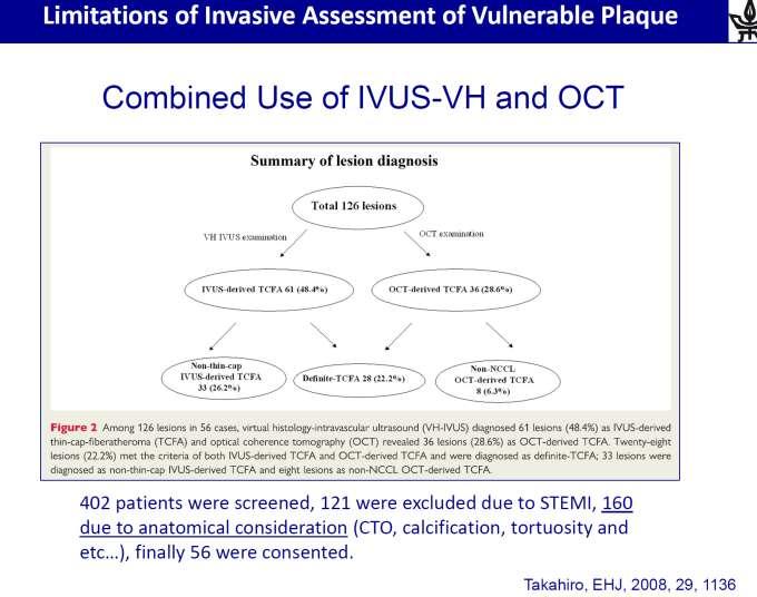
27 The PROSPECT: A Prospective Natural-History Study of Coronary Atherosclerosis 700 pts with ACS who underwent successful 1V or 2V PCI. Pts underwent 3V imaging of culprit artery and non-culprit arteries, with angiography, IVUS, and virtual histology, as well as palpography (n 350 pts). All pts were treated with optimal medical therapy, including aspirin, clopidogrel for one year, and statin therapy. Stone GW, et al. N Engl J Med 2011;364:226-35

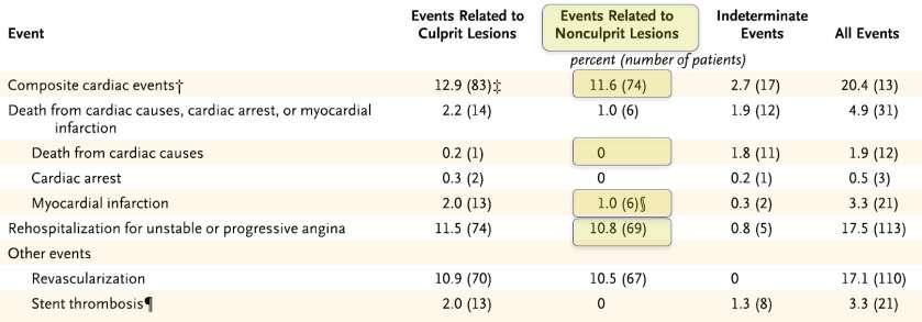
28 The PROSPECT: Event Rates vs. Lesions Stone GW, et al. N Engl J Med 2011;364:226-35

29 The PROSPECT: Plaque Characteristics Associated with Future Events Thin Cap Fibroatheroma Plaque burden 70% Lumen area 4 mm 2 Moreno P, et al. Cardiol Clin 2010; 28:1-30 Stone GW, et al. N Engl J Med 2011;364:226-35

30 The PROSPECT: Characteristics and Significance of Mild Lesions in ACS Incidence of High-Risk Characteristics Detected by IVUS Cumulative Incidence of MACE Brener SJ, et al. JACC Img 2012;5: S86-94


31 The PROSPECT: CV Events and Angiographic Progression Time-to-Event Curves for NC Lesion-Related MACEs Sanidas EA, et al. JACC Img 2012;5: S95-105
32 Residual Plaque Burden in Pts with ACS after Successful PCI MACE During Follow-Up MACE /Lesion During Follow-Up McPherson JA, et al. JACC Img 2012;5: S76-85

33 Predictive Performance of Plaque Characteristics Predictive Performance of Patient Characteristics Kaul S, Diamond GA. JACC Img 2012;5: S
34 Management
35 The Vulnerable Plaque Hypothesis Promise, but Little Progress Is there is a direct 1-to-1 relationship between a TCFA and a high-risk plaque? Is plaque instability a focal, a multifocal, or a disseminated phenomenon? If a plaque is high risk today, will it be high risk in 1 month, 3 months, or a year from now? If we classify a plaque as stable, can we be certain that it not undergo acute conversion to a vulnerable lesion a few weeks later? Nissen S. JACC Cardiovascular Img 2009;2: 483 5
36 The PROSPECT: Is There a Role for PCI in Vulnerable Plaque Management? While the combination of 3 characteristic NCL features (plaque burden 70%, MLA 4.0 mm 2 and presence of a TCFA) conferred an HR of 11, almost 90% of patients with similar plaques did not have a MACE during the 3-year follow-up. Overall, 20.4% of patients had major CV events, of which 11.6% were related to non-culprit lesions; however, only 1% were MIs, while the remaining 10.6% were unstable or progressive angina. Eleven patients (1.6%) had complications that were directly attributable to the 3-vessel imaging procedure. These complications resulted in 3 nonfatal MI (in 0.4% of patients). As the resolution of IVUS is 150 to 200 μm, it is incapable of identifying the fibrous cap thickness component of TCFA (< 65 μm). Another problem with IVUS VH histology is its failure to distinguish lipid pool from necrotic core, which is critical to calling a lesion fibroatheroma. Invasive imaging to locate and then prophylactically treat presumed high-risk lesions with PCI is not warranted Falk E, Willensky RL. JACC Imaging 2012; 5: S38-41 Finn AV, et al. JACC Imaging 2012; 5: 334-6

37 Serial CTA Verified Changes in Plaque Characteristics as an End Point CTA was performed in 32 pts. Of these, 24 received fluvastatin after the baseline study; 8 subjects who refused statin treatment were followed as the control subjects. Serial imaging was performed after a median interval of 12 months. Inoue K, et al. JACC Im 2010;3: 691-8
38 Impact of Statin Therapy on Plaque Composition OCT, grayscale and IB-IVUS of non-target lesions was performed in 42 stable angina pts undergoing elective PCI. 26 received 4 mg pitavastatin, whereas 16 were followed with dietary modification alone. Follow-up imaging was performed after a median interval of 9 months. Hattori K, et al. JACC Im 2012;5:169-77

39 Impact of Therapy on Plaque Composition: Diet Hattori K, et al. JACC Im 2012;5:169-77

40 Impact of Therapy on Plaque Composition: Statin Hattori K, et al. JACC Im 2012;5:169-77
41 Future Perspectives

42 Multimodality Imaging in Atherosclerosis Leuschner F, Nahrendorf M. Circ Res 2011;108:

43 Molecular Imaging Targets in Atherosclerosis Leuschner F, Nahrendorf M. Circ Res 2011;108:

44 Molecular Imaging Targets in Myocardial Infarction Leuschner F, Nahrendorf M. Circ Res 2011;108:

45 Principles of MicroRNA Therapeutics Broderick JA, Zamore PD. Gene Therapy 2011; 18:

46 Therapeutic Modification of Atherosclerosis by MicroRNAs Martin K, et al. Curr Opin Cardiol 2011;26:569-75
47 Conclusions Atherothrombosis continues to evolve in diagnosis and imaging. A single-plaque approach is narrow minded and difficult to prove in clinical practice. The PROSPECT trial provides evidence for increased risk for large, stenotic plaques with VH-TCFA morphology on IVUS. Modern CTA technology is capable of noninvasively detecting morphological markers of lesion vulnerability (location, attenuation, remodeling). Invasive imaging to locate and then prophylactically treat presumed high-risk lesions with PCI is currently not warranted. Statins are currently the treatment of choice for vulnerable plaque stabilization mirnas may provide new therapeutic targets in the not so far future.
48
49
50
51
 and LVEF <50%.")
52 The CONFIRM Registry Risk Stratification by Cardiac CTA CCTA pts with LVEF data were screened. Pts with MI history, coronary Rx, or cardiac Tx were excluded. The NCEP- ATP III risk was calculated for each pt, and CCTA was evaluated for CAD severity (normal, non-obstructive, non high-risk, or high-risk CAD) and LVEF <50%. End point was all-cause mortality; 14,064 pts met the analysis criteria. Mean follow-up was 22.5 (95% CI, ) months. Chow BJW, et al. Circ Cardiovasc Imaging 2011;4:

53 The VIVA (VH-IVUS in Vulnerable Atherosclerosis) Study 170 pts with stable angina or troponin (+) ACS referred for PCI underwent 3V VH-IVUS pre- PCI and also post-pci in culprit vessel. 30,372 mm of VH-IVUS were analyzed. 18 MACE occurred in 16 pts over a median follow-up of 625 days. MACE: 11 PCI, 3 CABG, 2MIs, 2 deaths Calvert PA, et al. JACC Imaging 2011; 4:


54 CTA Characteristics of Plaques Resulting in ACS In 1,059 patients who underwent CTA, atherosclerotic lesions were analyzed for PR and LAP. The remodeling index, and plaque and LAP areas and volumes were calculated. The plaque characteristics of lesions resulting in ACS during the follow-up of 27±10 months were evaluated. 22.2% 3.7% 0.5% 0 Motoyama, et al. J Am Coll Cardiol 2009;54:49 57

55 Causes of Coronary Thrombosis Rupture Erosion Calcified nodule Virmani R, et al. ATVB 2000; 20: Virmani R, et al. JACC 2006; 47: Suppl C: C13-8

56 Characteristics of Thrombosed Plaques Plaque rupture Thin fibrous cap (95% 64 μm) Large necrotic core Increased plaque inflammation Positive vascular remodeling Vasa vasorum neovascularization Intraplaque hemorrhage Plaque erosion No specific features suitable for detection Thick, SMC rich fibrous cap Premenopausal women Cigarette smoking Fishbein MC. Cardiovasc Pathol 2010; 19:6-11
57 Prognosis of Chronic Obstructive CAD Swedish low-dose aspirin trial (SAPAT; 2035 patients; median follow-up 4.2 years). Cardiac death rate of 0.9% per year. Lancet 1992;12;340: Total Ischaemic Burden European Trial (TIBET; 682 patients; 2 years) Cardiac death rate of 1% per year among patients with a positive exercise test. Eur Heart J 1996;17: Angina Prognosis Study In Stockholm (APSIS;809 patients; 3.4 years). Cardiac death rate of 1.2% per year. Eur Heart J 1996;17:76-89 ACTION trial. Cardiovascular mortality rate of 0.9% per year. Lancet 2004;364: Jabbour, et al (693 patients; 4.6 years). Cardiac death rate 0.8 per year. Am J Cardiol 2004; 93: Rates of non-fatal myocardial infarction ranged from 1.0% (APSIS) to 2.6% (TIBET) per year.
 Plaque erosion (25%) Farb A, et al.")
:C13 8. Moreno P, et al.")
58 Causes of Atherothrombosis and Acute Coronary Syndromes Plaque rupture (75%) Plaque erosion (25%) Farb A, et al. Circulation 1996;93(7): Virmani R, et al. J Am Coll Cardiol 2006;47(8 Suppl):C13 8. Moreno P, et al. Cardiol Clin 2010; 28:1-30

59 Calcified Nodule Virmani R, et al. J Am Coll Cardiol 2006;47(8 Suppl):C13 8.
60 Stenosis at the Site of Occlusive Thrombosis: Pathologic Studies a a : Cross-sectional narrowing by plaque Fishbein MC. Cardiovasc Pathol 2010; 19:6 11

61 Stenosis at the Site of Occlusive Thrombosis: Angiographic Studies a a : Cross-sectional narrowing by plaque Fishbein MC. Cardiovasc Pathol 2010; 19:6 11


62 Evolution of Angiographic Non-Stenotic Lesions Little, et al. Circulation 1988;78: Ambrose, et al.j Am Coll Cardiol 1988;12:56 62


63 Analysis of the Thrombolytic Process Thrombolytic recanalization of the obstructed coronary lumen was studied in 32 patients receiving intracoronary streptokinase for 60 to 90 min during acute myocardial infarction. Original stenosis" measured 1.25± 0.32 mm in minimum diameter and 56 ± 14% stenosis when first visualized and was unchanged. In 5 patients catheterized 10 ± 12 weeks before MI, the original stenosis was 1.15 ± 0.22 mm, as compared with 1.17 ± 0.23 mm in its faintly defined form during thrombolytic therapy (p = NS). In 10 cases, this original lesion was less than a 50% stenosis, and in 21 cases less than 60%. Brown, et al. Circulation 1986;73:653-61

64 Assessment of Coronary Stenosis: Angiography vs. Pathology Diameter Stenosis Area Stenosis % cross-sectional involvement by atheroma Actual luminal area decrease Fishbein and Siegel. Circulation 1996;94:2662-6
65 Drawbacks of Coronary Angiography Depicts rather poor representation of cross-sectional coronary anatomy from simple planar silhouette or luminogram of the contrast-filled lumen. Confounded by observer variability, with differences in the estimation of stenosis approaching 50%. Functional testing often reveals discordance between the severity of angiographic lesions and physiologic effects. Necropsy studies and IVUS demonstrate that coronary lesions, particularly after plaque rupture, are complex, with distorted luminal shapes that are difficult to assess using a planar angiographic silhouette. Nissen S. JACC 2003; 41(4 Suppl S): 103S-112S


66 Coronary Imaging With Cardiovascular Magnetic Resonance Chiribiri A, et al. Prog Cardiovasc Dis 2011; 54:240-52
67 Natural History of Vulnerable Plaque

68 The Fate of the Vulnerable Plaque

69 The Fate of the Vulnerable Plaque: Additional Factors (1)

70 The Fate of the Vulnerable Plaque: Additional Factors (2)

71 The Fate of the Vulnerable Plaque: Additional Factors (3)
72
73
74
75 Prediction of Hard Events within 5 Years in the Heinz Nixdorf Recall Study Erbel R, Budoff M. Eur Heart J 2012; 33:
76
77
78
79
80
81
82

83 Fusion between MRA and DE-MRI Chiribiri A, et al. Prog Cardiovasc Dis 2011;54:240-52

84 Co-Registered 18 F-FDG PET/CT Imaging of Inflammation in Human Coronary Arteries Rogers IS, et al. J Am Coll Cardiol Img 2010;3:388 97

85 The Thin-Cap Fibroatheroma Kolodgie FD, et al. Heart 2004; 90:


86 Coronary Plaque Vulnerability Assessed with CTA and IVUS Unstable Angina Unstable Angina Unstable Angina Stable Angina Pugliese, et al. Journal of Cardiovascular Medicine 2009;10:
87 Outcomes From Second-Generation Coronary Stent Trials 1228 patients for were observed 5 years after implantation of second-generation coronary stents. Death, MI, repeat revascularization, and repeat hospitalization for ACS or CHF were attributed to the index stented lesion or other sites in target or other coronary vessels and further classified as procedural,restenosis, or nonrestenosis. Cutlip, et al. Circulation 2004;110:
 pts underwent target lesion PCI for restenosis and 216 (5.")
88 Progression of Asymptomatic Lesions Discovered During PCI Retrospective study to determine features of clinical plaque progression using the NHLBI Dynamic Registry of patients undergoing PCI in 1997 to 1998 and Of 3747 PCI patients, 276 (7.4%) pts underwent target lesion PCI for restenosis and 216 (5.8%) required additional non-target lesion PCI for clinical plaque progression at 1 year (59% presented with new UA, and 9.3% with nonfatal MI. A B C The majority (86.9%) of lesions requiring subsequent PCI were 60% in severity during original PCI, with the mean lesion stenosis 41.8±20.8% at the time of the initial PCI and 83.9 ± 13.9% during the recurrent event. Glaser, et al. Circulation 2005;111:143-9

89 Vasa Vasorum Imaging, by Using Contrast-Enhanced IVUS Vavuranakis, et al. Int J Cardiol 2008; 130:23-9

90 IVUS Morphology of Vulnerable Coronary Plaque 114 coronary sites without significant stenosis by angiography (<50% diameter stenosis) in 106 patients undergoing ICA and IVUS were examined. All sites exhibited atherosclerotic lesions by IVUS. These lesions consisted of 22 concentric and 92 eccentric plaques with a % plaque area averaging 59±12%. Follow-up period of 21.8 ± months (range 1 to 24). 12 pts had an ACS 4.0 ± 3.4 months after initial IVUS study. All plaques related to ACS had an eccentric pattern and their mean percent plaque area was greater than in pts without ACS (67 ± 9% vs. 57 ± 12%, p < 0.05). No difference in lumen area between two patient groups (6.7 ± 3.0 vs. 7.5 ± 3.7 mm 2 ). Among 12 coronary sites with an acute occlusion, 10 sites contained echolucent zones, 8 of these shallow and 2 deep, likely representing a lipid-rich core. In 90 sites without ACS, an echolucent zone in the shallow portion was seen at only 4 sites (p <0.05). Yamagishi, et al. J Am Coll Cardiol 2000; 35:106 11
91 Summary of Invasive Technologies for Vulnerable Plaque Imaging Moreno P, et al. Cardiol Clin 2010; 28:1-30


92 Sudden Coronary Death Caused by PIT without Plaque Formation Tavora F, et al. Cardiovasc Pathol 2011; 20:51-7

93 The Non-Stenotic Lesion as Culprit Nissen S. JACC 2003:41:103S-112S

94 Noninvasive Coronary Imaging Vancraeynest D, et al. J Am Coll Cardiol 2011;57:

95 OCT-Characterization of Coronary Atherosclerosis Jang IK, et al. Circulation 2005;111(12):1551 5

96 OCT-Based Plaque Characteristics Uemura S, et al. Eur Heart J 2012; 33: 78-85

97 Angioscopy--Characterization of Coronary Atherosclerosis Ohtani T, et al. J Am Coll Cardiol 2006;47:

98 CT evaluation of Vulnerable Plaque Opolski MP, et al. Int J Cardiovasc Imaging April 21, 2011
99


100 11 th Cardiology Congress of Northern Greece Thessaloniki May 24-26,2012 The Theory of the Vulnerable Plaque: Impact on Diagnosis and Management of CAD Filippos Triposkiadis, MD, FESC, FACC Professor of Cardiology, Director, Department of Cardiology, Larissa University Hospital, Larissa, Greece
101 Manifestations of Coronary Artery Disease Sudden cardiac death is the initial CAD manifestation in 25% of pts. Sudden cardiac death accounts for 50% of all CAD related deaths. ACS is the initial CAD manifestation in 30% of patients. Only 18% of coronary attacks are preceded by longstanding angina pectoris. Stable angina is the initial CAD manifestation in 50% of patients. In approximately half of the cases the initial CAD manifestation may be detrimental. Approximately half of CAD patients die suddenly. Davies SW. Br Med Bull. 2001;59:17-27 Fox CS, et al. Circulation 2004; 110:522-7 Myerburg RJ, Junttila J. Circulation 2012; 125:
102

103 If human life were long enough to find the ultimate theory, everything would have been solved by previous generations. Nothing would be left to be discovered.
Vulnerable Plaque Pathophysiology, Detection, and Intervention. VP: A Local Problem or Systemic Disease. Erling Falk, Denmark
 Vulnerable Plaque Pathophysiology, Detection, and Intervention VP: A Local Problem or Systemic Disease Erling Falk, Denmark Vulnerable Plaque Pathophysiology, Detection, and Intervention VP: A Local Problem
Added Value of Invasive Coronary Imaging for Plaque Rupture and Erosion
 Assessment of Coronary Plaque Rupture and Erosion Added Value of Invasive Coronary Imaging for Plaque Rupture and Erosion Yukio Ozaki, MD, PhD, FACC, FESC Cardiology Dept., Fujita Health Univ. Toyoake,
Imaging Overview for Vulnerable Plaque: Data from IVUS Trial and An Introduction to VH-IVUS Imgaging
 Imaging Overview for Vulnerable Plaque: Data from IVUS Trial and An Introduction to VH-IVUS Imgaging Gary S. Mintz,, MD Cardiovascular Research Foundation New York, NY Today, in reality, almost everything
Imaging Atheroma The quest for the Vulnerable Plaque
 Imaging Atheroma The quest for the Vulnerable Plaque P.J. de Feijter 1. Department of Cardiology 2. Department of Radiology Coronary Heart Disease Remains the Leading Cause of Death in the U.S, Causing
The PROSPECT Trial. A Natural History Study of Atherosclerosis Using Multimodality Intracoronary Imaging to Prospectively Identify Vulnerable Plaque
 The PROSPECT Trial Providing Regional Observations to Study Predictors of Events in the Coronary Tree A Natural History Study of Atherosclerosis Using Multimodality Intracoronary Imaging to Prospectively
Plaque Characteristics in Coronary Artery Disease. Chourmouzios Arampatzis MD, PhD, FESC
 Plaque Characteristics in Coronary Artery Disease Chourmouzios Arampatzis MD, PhD, FESC Disclosure Statement of Financial Interest Regarding this Presentation NONE Atherosclerosis Model proposed by Stary
Management of Multivessel CAD: Stenting or CABG?
 Management of Multivessel CAD: Stenting or CABG? Filippos Triposkiadis, MD, FESC, FACC Department of Cardiology, University of Thessaly Long-term Outcome of Patients With 3VD Undergoing CABG A Report from
State of the Art. Advances in Cardiovascular Imaging. ESC Congres Stockholm September 1, 2010 Frank E. Rademakers, MD, PhD, FESC
 State of the Art Advances in Cardiovascular Imaging ESC Congres Stockholm September 1, 2010 Frank E. Rademakers, MD, PhD, FESC Coronary Artery Disease Content Patho Physiology Imaging requirements Economical
The PROSPECT Trial. A Natural History Study of Atherosclerosis Using Multimodality Intracoronary Imaging to Prospectively Identify Vulnerable Plaque
 The PROSPECT Trial Providing Regional Observations to Study Predictors of Events in the Coronary Tree A Natural History Study of Atherosclerosis Using Multimodality Intracoronary Imaging to Prospectively
Assessment of Vulnerable Plaque by IVUS and VH-IVUS
 Assessment of Vulnerable Plaque by IVUS and VH-IVUS Akiko Maehara, MD Director of Intravascular Imaging & Physiology Core Laboratories Associate Director of MRI/MDCT Core Laboratory Cardiovascular Research
High-risk vulnerable plaques. Kostis Raisakis G.Gennimatas General Hospital of Athens
 High-risk vulnerable plaques. Kostis Raisakis G.Gennimatas General Hospital of Athens Overview: 1 Definition-Pathology 2 3 Diagnostic Strategies Invasive Non Invasive Prognostic Value of Detection 4 Treatment
Can IVUS Define Plaque Features that Impact Patient Care?
 Can IVUS Define Plaque Features that Impact Patient Care? A Pichard L Satler, K Kent, R Waksman, W Suddath, N Bernardo, N Weissman, M Angelo, D Harrington, J Lindsay, J Panza. Washington Hospital Center
Invasive Coronary Imaging Modalities for Vulnerable Plaque Detection
 Invasive Coronary Imaging Modalities for Vulnerable Plaque Detection Gary S. Mintz, MD Cardiovascular Research Foundation New York, NY Greyscale IVUS studies have shown Plaque ruptures do not occur randomly
Failure of positive. Recanalization and CTO formation. TCFA rupture with (fatal) thrombotic occlusion. TCFA Lipid pool
 thrombotic occlusion. TCFA Lipid pool") Vulnerable Plaque features on coronary CT Jin Ho Choi, MD, PhD Department of Internal Medicine, Emergency Medicine Samsung Medical Center, Sungkyunkwan University School of Medicine, Seoul, Korea IPS /
Vulnerable Plaque features on coronary CT Jin Ho Choi, MD, PhD Department of Internal Medicine, Emergency Medicine Samsung Medical Center, Sungkyunkwan University School of Medicine, Seoul, Korea IPS /
Cardiovascular Research Foundation and Columbia University Medical Center, New York.
 Virtual Histology Intravascular Ultrasound Analysis of Non-culprit Attenuated Plaques Detected by Grayscale Intravascular Ultrasound in Patients with Acute Coronary Syndromes Xiaofan Wu, Akiko Maehara,
Chapter 43 Noninvasive Coronary Plaque Imaging
 hapter 43 Noninvasive oronary Plaque Imaging NIRUDH KOHLI The goal of coronary imaging is to define the extent of luminal narrowing as well as composition of an atherosclerotic plaque to facilitate appropriate
Coronary Artery Thermography
 Coronary Artery Thermography The 10th Anniversary, Interventional Vascular Therapeutics Angioplasty Summit 2005 TCT Asia Pacific Christodoulos Stefanadis Professor of Cardiology Athens Medical School In
Gary S. Mintz,, MD. IVUS Observations in Acute (vs Chronic) Coronary Artery Disease: Structure vs Function
 Coronary Artery Disease: Structure vs Function") Gary S. Mintz,, MD IVUS Observations in Acute (vs Chronic) Coronary Artery Disease: Structure vs Function Important IVUS Observations: Remodeling Originally used (first by Glagov) ) to explain atherosclerosis
Gary S. Mintz,, MD IVUS Observations in Acute (vs Chronic) Coronary Artery Disease: Structure vs Function Important IVUS Observations: Remodeling Originally used (first by Glagov) ) to explain atherosclerosis
OCT; Comparative Imaging Results with IVUS, VH and Angioscopy
 OCT; Comparative Imaging Results with IVUS, VH and Angioscopy Takashi Akasaka, M.D. Department of Cardiovascular Medicine Wakayama, Japan Comparison among coronary imaging techniques OCT IVUS MRI CAG Angioscopy
Pathology of Coronary Artery Disease
 Pathology of Coronary Artery Disease Seth J. Kligerman, MD Pathology of Coronary Artery Disease Seth Kligerman, MD Assistant Professor Medical Director of MRI University of Maryland Department of Radiology
Cottrell Memorial Lecture. Has Reversing Atherosclerosis Become the New Gold Standard in the Treatment of Cardiovascular Disease?
 Cottrell Memorial Lecture Has Reversing Atherosclerosis Become the New Gold Standard in the Treatment of Cardiovascular Disease? Stephen Nicholls MBBS PhD @SAHMRI_Heart Disclosures Research support: AstraZeneca,
CT Imaging of Atherosclerotic Plaque. William Stanford MD Professor-Emeritus Radiology University of Iowa College of Medicine Iowa City, IA
 CT Imaging of Atherosclerotic Plaque William Stanford MD Professor-Emeritus Radiology University of Iowa College of Medicine Iowa City, IA PREVALENCE OF CARDIOVASCULAR DISEASE In 2006 there were 80 million
Can We Identify Vulnerable Patients & Vulnerable Plaque?
 Can We Identify Vulnerable Patients & Vulnerable Plaque? We Know Enough to Treat High-Risk Lesions? Takashi Akasaka, MD, PhD Department of Cardiovascular Medicine, Japan Disclosure Statement of Financial
Pathology of Vulnerable Plaque Angioplasty Summit 2005 TCT Asia Pacific, Seoul, April 28-30, 2005
 Pathology of Vulnerable Plaque Angioplasty Summit 25 TCT Asia Pacific, Seoul, April 28-3, 25 Renu Virmani, MD CVPath, A Research Service of the International Registry of Pathology Gaithersburg, MD Plaque
2yrs 2-6yrs >6yrs BMS 0% 22% 42% DES 29% 41% Nakazawa et al. J Am Coll Cardiol 2011;57:
 Pathology of In-stent Neoatherosclerosis in BMS and DES 197 BMS, 103 SES, and 106 PES with implant duration >30 days The incidence of neoatherosclerosis was significantly greater in DES (31%) than BMS
Κλινική Χρήση IVUS και OCT PERIKLIS A. DAVLOUROS ASSOCIATE PROFESSOR OF CARDIOLOGY INVASIVE CARDIOLOGY & CONGENITAL HEART DISEASE
 Κλινική Χρήση IVUS και OCT PERIKLIS A. DAVLOUROS ASSOCIATE PROFESSOR OF CARDIOLOGY INVASIVE CARDIOLOGY & CONGENITAL HEART DISEASE Conflict of interest None to declare While IVUS is the most used intravascular
Culprit Lesion Remodeling and Long-term (> 5years) Prognosis in Patients with Acute Coronary Syndrome
 Prognosis in Patients with Acute Coronary Syndrome") Culprit Lesion Remodeling and Long-term (> 5years) Prognosis in Patients with Acute Coronary Syndrome Hiroyuki Okura*, MD; Nobuya Matsushita**,MD Kenji Shimeno**, MD; Hiroyuki Yamaghishi**, MD Iku Toda**,
Culprit Lesion Remodeling and Long-term (> 5years) Prognosis in Patients with Acute Coronary Syndrome Hiroyuki Okura*, MD; Nobuya Matsushita**,MD Kenji Shimeno**, MD; Hiroyuki Yamaghishi**, MD Iku Toda**,
OCT Findings: Lesson from Stable vs Unstable Plaques
 ANGIOPLASTY SUMMIT TCTAP 2010 Imaging Workshop OCT Findings: Lesson from Stable vs Unstable Plaques Giulio Guagliumi MD Ospedali Riuniti di Bergamo, Italy DISCLOSURE OF FINANCIAL INTERESTS Consultant Boston
Optical Coherence Tomography for Intracoronary Imaging
 Optical Coherence Tomography for Intracoronary Imaging Lorenz Räber Stephan Windecker Department of Cardiology Swiss Cardiovascular Center and Clinical Trials Unit Bern Bern University Hospital, Switzerland
Left main coronary artery (LMCA): The proximal segment
: The proximal segment") Anatomy and Pathology of Left main coronary artery G Nakazawa Tokai Univ. Kanagawa, Japan 1 Anatomy Difinition Left main coronary artery (LMCA): The proximal segment RCA AV LAD LM LCX of the left coronary
Anatomy and Pathology of Left main coronary artery G Nakazawa Tokai Univ. Kanagawa, Japan 1 Anatomy Difinition Left main coronary artery (LMCA): The proximal segment RCA AV LAD LM LCX of the left coronary
1st Department of Cardiology, University of Athens, Hippokration Hospital, Athens, Greece
 Konstantinos Toutouzas, Maria Riga, Antonios Karanasos, Eleftherios Tsiamis, Andreas Synetos, Maria Drakopoulou, Chrysoula Patsa, Georgia Triantafyllou, Aris Androulakis, Christodoulos Stefanadis 1st Department
OCT. molecular imaging J Jpn Coll Angiol, 2008, 48: molecular imaging MRI positron-emission tomography PET IMT
 48 6 CT MRI PET OCT molecular imaging J Jpn Coll Angiol, 2008, 48: 456 461 atherosclerosis, imaging gold standard computed tomography CT magnetic resonance imaging MRI CT B intima media thickness IMT B
IVUS Virtual Histology. Listening through Walls D. Geoffrey Vince, PhD The Cleveland Clinic Foundation
 IVUS Virtual Histology Listening through Walls D. Geoffrey Vince, PhD Disclosure VH is licenced to Volcano Therapeutics Grant funding from Pfizer, Inc. Grant funding from Boston-Scientific Most Myocardial
Medical sciences 1 (2017) 1 9
 1 9") Medical sciences 1 (2017) 1 9 TISSUE CHARACTERISTICS OF CULPRIT CORONARY LESIONS IN ACUTE CORONARY SYNDROME AND TARGET CORONARY LESIONS IN STABLE ANGINA PECTORIS: VIRTUAL HISTOLOGY AND INTRAVASCULAR ULTRASOUND
Medical sciences 1 (2017) 1 9 TISSUE CHARACTERISTICS OF CULPRIT CORONARY LESIONS IN ACUTE CORONARY SYNDROME AND TARGET CORONARY LESIONS IN STABLE ANGINA PECTORIS: VIRTUAL HISTOLOGY AND INTRAVASCULAR ULTRASOUND
CLINICAL APPLICATIONS OF OPTICAL COHERENCE TOMOGRAPHY. Konstantina P. Bouki, FESC 2 nd Department of Cardiology General Hospital Of Nikea, Pireaus
 CLINICAL APPLICATIONS OF OPTICAL COHERENCE TOMOGRAPHY Konstantina P. Bouki, FESC 2 nd Department of Cardiology General Hospital Of Nikea, Pireaus OPTICAL COHERENCE TOMOGRAPHY (OCT) IVUS and OCT IVUS OCT
Assessment of plaque morphology by OCT in patients with ACS
 Assessment of plaque morphology by OCT in patients with ACS Takashi Akasaka, M.D. Department of Cardiovascular Medicine Wakayama, Japan Unstable plaque Intima Lipid core Plaque rupture and coronary events
as a Mechanism of Stent Failure
 In-Stent t Neoatherosclerosis e osc e os s as a Mechanism of Stent Failure Soo-Jin Kang MD., PhD. University of Ulsan College of Medicine, Heart Institute Asan Medical Center, Seoul, Korea Disclosure I
Subclinical atherosclerosis in CVD: Risk stratification & management Raul Santos, MD
 Subclinical atherosclerosis in CVD: Risk stratification & management Raul Santos, MD Sao Paulo Medical School Sao Paolo, Brazil Subclinical atherosclerosis in CVD risk: Stratification & management Prof.
Invasive Evaluation of High Risk or Vulnerable Plaque A Powerful Tool to Address Potential Pharmacological Agents? Jagat Narula, MD PhD MACC
 Invasive Evaluation of High Risk or Vulnerable Plaque A Powerful Tool to Address Potential Pharmacological Agents? Jagat Narula, MD PhD MACC No Conflicts of Interest to Declare Histological Signatures
Optical Coherence Tomography (OCT): A New Imaging Tool During Carotid Artery Stenting
: A New Imaging Tool During Carotid Artery Stenting") Chapter 6 Optical Coherence Tomography (OCT): A New Imaging Tool During Carotid Artery Stenting Shinichi Yoshimura, Masanori Kawasaki, Kiyofumi Yamada, Arihiro Hattori, Kazuhiko Nishigaki, Shinya Minatoguchi
Chapter 6 Optical Coherence Tomography (OCT): A New Imaging Tool During Carotid Artery Stenting Shinichi Yoshimura, Masanori Kawasaki, Kiyofumi Yamada, Arihiro Hattori, Kazuhiko Nishigaki, Shinya Minatoguchi
Pathology of the Vulnerable Plaque
 Journal of the American College of Cardiology Vol. 47, No. 8 Suppl C 2006 by the American College of Cardiology Foundation ISSN 0735-1097/06/$32.00 Published by Elsevier Inc. doi:10.1016/j.jacc.2005.10.065
EAE Teaching Course. Magnetic Resonance Imaging. Competitive or Complementary? Sofia, Bulgaria, 5-7 April F.E. Rademakers
 EAE Teaching Course Magnetic Resonance Imaging Competitive or Complementary? Sofia, Bulgaria, 5-7 April 2012 F.E. Rademakers Complementary? Of Course N Engl J Med 2012;366:54-63 Clinical relevance Treatment
Patient referral for elective coronary angiography: challenging the current strategy
 Patient referral for elective coronary angiography: challenging the current strategy M. Santos, A. Ferreira, A. P. Sousa, J. Brito, R. Calé, L. Raposo, P. Gonçalves, R. Teles, M. Almeida, M. Mendes Cardiology
Vascular disease. Structural evaluation of vascular disease. Goo-Yeong Cho, MD, PhD Seoul National University Bundang Hospital
 Vascular disease. Structural evaluation of vascular disease Goo-Yeong Cho, MD, PhD Seoul National University Bundang Hospital resistance vessels : arteries
Malaysian Healthy Ageing Society
 Organised by: Co-Sponsored: Malaysian Healthy Ageing Society CAD INVESTIGATIONS PHYSIOLOGICAL / FUNCTIONAL VS ANATOMICAL / STRUCTURAL Disclosure iheal medical centre is a one stop cardiac centre with
Intervention: How and to which extent is technology helping us?
 Cardiological Society of India Congress 12th February 2016 Chennai, India Intervention: How and to which extent is technology helping us? SIMONE BISCAGLIA MD CARDIOVASCULAR INSTITUTE, FERRARA, ITALY Introduction
Usefulness of OCT during coronary intervention
 Usefulness of OCT during coronary intervention Takashi Akasaka, M.D. Department of Cardiovascular Medicine Wakayama, Japan Predictors at 12 Months of Stent Thrombosis and Target Lesion Revascularization
Noninvasive Coronary Imaging: Plaque Imaging by MDCT
 Coronary Physiology & Imaging Summit 2007 Noninvasive Coronary Imaging: Plaque Imaging by MDCT Byoung Wook Choi Department of Radiology Yonsei University, Seoul, Korea Stary, H. C. et al. Circulation
Coronary plaque erosion: a clinical case. Dr. Giampaolo Niccoli, MD, PhD, FESC Institute of Cardiology Catholic University, Rome, Italy
 Coronary plaque erosion: a clinical case, MD, PhD, FESC Institute of Cardiology Catholic University, Rome, Italy Coronary plaque erosion: a clinical case B.M. Age: 59 years Sex: female. Cardiological risk
actually rupture! Challenges to the vulnerable plaque concept
 An Update on the Pathogenesis of the Acute Coronary Syndromes Peter Libby Brigham & Women s Hospital Harvard Medical School ADVANCES IN HEART DISEASE University of California San Francisco December 20,
Cardiac CT Angiography
 Cardiac CT Angiography Dr James Chafey, Radiologist Why do we need a better test for C.A.D? 1. CAD is the leading cause of death in the US CAD 31% Cancer 23% Stroke 7% 2. The prevalence of atherosclerosis
04RC2. The biology of vulnerable plaques. Jozef L. Van Herck 1, Christiaan J. Vrints 1, Arnold G. Herman 2
 04RC2 The biology of vulnerable plaques Jozef L. Van Herck 1, Christiaan J. Vrints 1, Arnold G. Herman 2 1 Department of Cardiology, Antwerp University Hospital, Edegem, Belgium 2 Department of Pharmacology,
COURAGE to Leave Diseased Arteries Alone
 COURAGE to Leave Diseased Arteries Alone Spencer King MD MACC, FSCAI St. Joseph s s Heart and Vascular Institute Professor of Medicine Emeritus Emory Univ. Atlanta, USA Conflict: I am an Interventionalist
2/20/2013. Why use imaging in CV prevention? Update on coronary CTA in 2013 Coronary CTA for 1 0 prevention: pros and cons Are we there yet?
 Evolving Role of Coronary CTA in Primary Cardiovascular Disease Prevention: Are We There Yet? Ron Blankstein, M.D., F.A.C.C. Co-Director, Cardiovascular Imaging Training Program Associate Physician, Preventive
Current and Future Imaging Trends in Risk Stratification for CAD
 Current and Future Imaging Trends in Risk Stratification for CAD Brian P. Griffin, MD FACC Department of Cardiovascular Medicine, Heart and Vascular Institute, Cleveland Clinic Disclosures: None Introduction
Defining Plaque Composition by CTA: The Latest Tool to Monitor Therapy?
 Defining Plaque Composition by CTA: The Latest Tool to Monitor Therapy? John McB. Hodgson, M.D., FSCAI Chairman, Department of Cardiology Geisinger Health System Wilkes Barre,, Pa Disclosure Information
Assessment of vulnerable plaque by OCT
 Assessment of vulnerable plaque by OCT Comparison with histology and possible clinical applications Takashi Akasaka, M.D. Department of Cardiovascular Medicine Wakayama, Japan Identification of vulnerable
Review Article Optical Coherence Tomography Imaging in Acute Coronary Syndromes
 SAGE-Hindawi Access to Research Cardiology Research and Practice Volume 2011, Article ID 312978, 7 pages doi:10.4061/2011/312978 Review Article Optical Coherence Tomography Imaging in Acute Coronary Syndromes
Imaging the Vulnerable Plaque. David A. Dowe, MD Atlantic Medical Imaging
 Imaging the Vulnerable Plaque David A. Dowe, MD Atlantic Medical Imaging Why is this so important? The Acute Situation Coronary disease-important Diagnosis of cardiovascular disease cost $148 billion in
TVA_C02.qxd 8/8/06 10:27 AM Page 19 PART 2. Pathology
 TVA_C2.qxd 8/8/6 :27 AM Page 19 2 PART 2 Pathology TVA_C2.qxd 8/8/6 :27 AM Page TVA_C2.qxd 8/8/6 :27 AM Page 21 2 CHAPTER 2 The pathology of vulnerable plaque Renu Virmani, Allen P Burke, James T Willerson,
CARDIAC IMAGING FOR SUBCLINICAL CAD
 CARDIAC IMAGING FOR SUBCLINICAL CAD WHY DON'T YOU ADOPT MORE SMART TECHNIQUE? Whal Lee, M.D. Seoul National University Hospital Department of Radiology We are talking about Coronary artery Calcium scoring,
The Dynamic Nature of Coronary Artery Lesion Morphology Assessed by Serial Virtual Histology Intravascular Ultrasound Tissue Characterization
 Journal of the American College of Cardiology Vol. 55, No. 15, 2010 2010 by the American College of Cardiology Foundation ISSN 0735-1097/10/$36.00 Published by Elsevier Inc. doi:10.1016/j.jacc.2009.07.078
MR Imaging of Atherosclerotic Plaques
 MR Imaging of Atherosclerotic Plaques Yeon Hyeon Choe, MD Department of Radiology, Samsung Medical Center, Sungkyunkwan University, Seoul MRI for Carotid Atheroma Excellent tissue contrast (fat, fibrous
대한심장학회춘계학술대회 Satellite Symposium
 대한심장학회춘계학술대회 Satellite Symposium Coronary Plaque Regression and Compositional Changes by Lipid-Lowering Therapy: IVUS Substudy in Livalo (Pitavastatin) in Acute Myocardial Infarction Study (LAMIS) Livalo
Title for Paragraph Format Slide
 Title for Paragraph Format Slide Presentation Title: Month Date, Year Atherosclerosis A Spectrum of Disease: February 12, 2015 Richard Cameron Padgett, MD Executive Medical Director, OHVI Pt RB Age 38
Catch-up Phenomenon: Insights from Pathology
 Catch-up Phenomenon: Insights from Pathology Michael Joner, MD CVPath Institute Inc. Gaithersburg, MD USA Path Lessons learned from the BMS and DES (1 st Gen) era Neointimal Thickness [mm] In Stent Re
Imaging ischemic heart disease: role of SPECT and PET. Focus on Patients with Known CAD
 Imaging ischemic heart disease: role of SPECT and PET. Focus on Patients with Known CAD Hein J. Verberne Academic Medical Center, University of Amsterdam, Amsterdam, Netherlands International Conference
FFR and intravascular imaging, which of which?
 FFR and intravascular imaging, which of which? Ayman Khairy MD, PhD, FESC Associate professor of Cardiovascular Medicine Vice Director of Assiut University Hospitals Assiut, Egypt Diagnostic assessment
Microvascular Disease: How to Diagnose and What s its Treatment
 Microvascular Disease: How to Diagnose and What s its Treatment Laxmi S. Mehta, MD, FACC The Ohio State University Medical Center Assistant Professor of Clinical Internal Medicine Clinical Director of
Assessment of Coronary Plaque Vulnerability with Optical Coherence Tomography
 Review Article Acta Cardiol Sin 2014;30:1 9 Assessment of Coronary Plaque Vulnerability with Optical Coherence Tomography Shiro Uemura, Tsunenari Soeda, Yu Sugawara, Tomoya Ueda, Makoto Watanabe and Yoshihiko
Coronary Artery Imaging. Suvipaporn Siripornpitak, MD Inter-hospital Conference : Rajavithi Hospital
 Coronary Artery Imaging Suvipaporn Siripornpitak, MD Inter-hospital Conference : Rajavithi Hospital Larger array : cover scan area Detector size : spatial resolution Rotation speed : scan time Retrospective
Εξελίξεις και νέες προοπτικές στην καρδιαγγειακή απεικόνιση CT. Σταμάτης Κυρζόπουλος Ωνάσειο Καρδιοχειρουργικό Κέντρο
 Εξελίξεις και νέες προοπτικές στην καρδιαγγειακή απεικόνιση CT Σταμάτης Κυρζόπουλος Ωνάσειο Καρδιοχειρουργικό Κέντρο No conflict of interest to disclose Noninvasive Cardiac Imaging Unresolved Issues-Future
THE COMPLEX RELATION BETWEEN VULNERABILITY AND ISCHEMIA a paradigm shift
 THE COMPLEX RELATION BETWEEN VULNERABILITY AND ISCHEMIA a paradigm shift Angioplasty Summit TCT ASIA Seoul, Korea, april 23rd, 2008 Nico H. J. Pijls, MD, PhD Catharina Hospital, Eindhoven, The Netherlands
Chest pain and troponins on the acute take. J N Townend Queen Elizabeth Hospital Birmingham
 Chest pain and troponins on the acute take J N Townend Queen Elizabeth Hospital Birmingham 3 rd Universal Definition of Myocardial Infarction Type 1: Spontaneous MI related to atherosclerotic plaque rupture
FESC, FACC, MAHA, MSCAI, MEAPSI, ESH
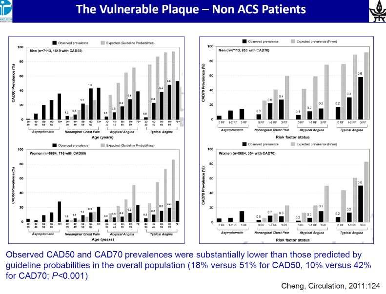
 «Απεικόνιση και φυσιολογία στο αιμοδυναμικό εργαστήριο». Eυάλωτη αθηρωματική πλάκα. Πού βρισκόμαστε? Ηλίας Α. Σανίδας MD, PhD, FESC, FACC, MAHA, MSCAI, MEAPSI, ESH Specialist Επεμβατικός Kαρδιολόγος Επιμελητής,
Combining Coronary Artery Calcium Scanning with SPECT/PET Myocardial Perfusion Imaging
 Combining Coronary Artery Calcium Scanning with SPECT/PET Myocardial Perfusion Imaging Daniel S. Berman, MD Director, Cardiac Imaging Cedars-Sinai Heart Institute Professor of Medicine and Imaging Cedars-Sinai
A Novel Low Pressure Self Expanding Nitinol Coronary Stent (vprotect): Device Design and FIH Experience
: Device Design and FIH Experience") A Novel Low Pressure Self Expanding Nitinol Coronary Stent (vprotect): Device Design and FIH Experience Juan F. Granada, MD Medical Director, Skirball Center for Cardiovascular Research The Cardiovascular
A Novel Low Pressure Self Expanding Nitinol Coronary Stent (vprotect): Device Design and FIH Experience Juan F. Granada, MD Medical Director, Skirball Center for Cardiovascular Research The Cardiovascular
Le espressioni della placca che preoccupano il clinico: progressione o vulnerabilità? CardioLUCCA Marzo 2017
 Le espressioni della placca che preoccupano il clinico: progressione o vulnerabilità? CardioLUCCA Marzo 2017 F Prati San Giovanni H. and CLI Foundation, Rome Euro Image Research Che cosa preoccupa il cardiologo
Who Cares About the Past?
 Risk Factors, the New Calcium Score, Rheology and Atherosclerosis Progression Arthur Agatston 2/21/15 The Vulnerable Plaque vs. Plaque Burden CT Angiogram Is There a Role for Coronary Artery Calcium Scoring
Financial Disclosures. Coronary Artery Calcification. Objectives. Coronary Artery Calcium 6/6/2018. Heart Disease Statistics At-a-Glace 2017
 Coronary Artery Calcification Dharmendra A. Patel, MD MPH Director, Echocardiography Laboratory Associate Program Director Cardiovascular Disease Fellowship Program Erlanger Heart and Lung Institute UT
Tissue Characterization of Coronary Plaques Using Intravascular Ultrasound/Virtual Histology
 REVIEW Korean Circulation J 2006;36:553-558 ISSN 1738-5520 c 2006, The Korean Society of Circulation Tissue Characterization of Coronary Plaques Using Intravascular Ultrasound/Virtual Histology Jang-Ho
Quinn Capers, IV, MD
 Heart Attacks Mended Hearts Presentation, January, 2017 Quinn Capers, IV, MD Associate Professor of Medicine (Cardiovascular Medicine) Director, Transradial Coronary Interventions Division of Cardiovascular
Effect of Intravascular Ultrasound- Guided vs. Angiography-Guided Everolimus-Eluting Stent Implantation: the IVUS-XPL Randomized Clinical Trial
 Effect of Intravascular Ultrasound- Guided vs. Angiography-Guided Everolimus-Eluting Stent Implantation: the IVUS-XPL Randomized Clinical Trial Myeong-Ki Hong, MD. PhD on behalf of the IVUS-XPL trial investigators
Innate Immunity in Atherosclerosis
 Innate Immunity in Atherosclerosis Peter Libby Brigham & Women s Hospital Harvard Medical School IAS Amsterdam May 26, 2015 ACS Stable demand angina Characteristics of Atherosclerotic Plaques Associated
Impact of coronary atherosclerotic burden on clinical presentation and prognosis of patients with coronary artery disease
 Impact of coronary atherosclerotic burden on clinical presentation and prognosis of patients with coronary artery disease Gjin Ndrepepa, Tomohisa Tada, Massimiliano Fusaro, Lamin King, Martin Hadamitzky,
Coronary Artery Disease in the 21 st Century: An Integrated Approach Based on Science and Art
 Coronary Artery Disease in the 21 st Century: An Integrated Approach Based on Science and Art Harisios Boudoulas, MD, Dr, Dr. Hon. Professor, Honorary Professor, Academician Development of Coronary Artery
Side Branch Occlusion
 Side Branch Occlusion Mechanism, Outcome, and How to avoid it From COBIS II Registry Hyeon-Cheol Gwon Cardiac&Vascular Center, Samsung Medical Center Sungkyunkwan University School of Medicine SB occlusion
Multimodality Imaging Atlas of Coronary Atherosclerosis
 JCC: CRDIOVSCUR IMGING VO. 3, NO. 8, 2010 2010 BY THE MERICN COEGE OF CRDIOOGY FOUNDTION ISSN 0735-1097/$36.00 PUBISHED BY ESEVIER INC. DOI:10.1016/j.jcmg.2010.06.006 IMGING VIGNETTE Multimodality Imaging
Diagnostic and Prognostic Value of Coronary Ca Score
 Diagnostic and Prognostic Value of Coronary Ca Score Dr. Ghormallah Alzahrani Cardiac imaging division, Adult Cardiology department Prince Sultan Cardiac Center ( PSCC) Madina, June 2 Coronary Calcium
Assessment of Culprit Lesion Morphology in Acute Myocardial Infarction
 Journal of the American College of Cardiology Vol. 50, No. 10, 2007 2007 by the American College of Cardiology Foundation ISSN 0735-1097/07/$32.00 Published by Elsevier Inc. doi:10.1016/j.jacc.2007.04.082
Coronary Artery Calcification
 Coronary Artery Calcification Julianna M. Czum, MD OBJECTIVES CORONARY ARTERY CALCIFICATION Julianna M. Czum, MD Dartmouth-Hitchcock Medical Center 1. To review the clinical significance of coronary heart
CHAPTER (2) THE VULNERABLE PLAQUE
 THE VULNERABLE PLAQUE") CHAPTER (2) THE VULNERABLE PLAQUE UNSTABLE OR HIGH RISK ATHEROSCLEROTIC PLAQUE - Definition and Composition - Plaque Destabilization and Disruption - Fate of Disrupted Plaque - Clinical Presentation -
CHAPTER (2) THE VULNERABLE PLAQUE UNSTABLE OR HIGH RISK ATHEROSCLEROTIC PLAQUE - Definition and Composition - Plaque Destabilization and Disruption - Fate of Disrupted Plaque - Clinical Presentation -
Pathology of percutaneous interventions (PCI) in coronary arteries. Allard van der Wal, MD.PhD; Pathologie AMC, Amsterdam, NL
 in coronary arteries. Allard van der Wal, MD.PhD; Pathologie AMC, Amsterdam, NL") Pathology of percutaneous interventions (PCI) in coronary arteries Allard van der Wal, MD.PhD; Pathologie AMC, Amsterdam, NL Percutaneous Coronary Intervention (PCI) Definition: transcatheter opening of
Pathology of percutaneous interventions (PCI) in coronary arteries Allard van der Wal, MD.PhD; Pathologie AMC, Amsterdam, NL Percutaneous Coronary Intervention (PCI) Definition: transcatheter opening of
Clinical Value of OCT. Guidance for Coronary Stenting. Giulio Guagliumi, MD
 Clinical Value of OCT Guidance for Coronary Stenting Giulio Guagliumi, MD 100 % Endovascular Imaging Indications of use 87.5 % 75 % 57.5 % 50 % 45 % 25 % 15 % 0 Lesion morphology Stent optimization Lesion
Ischemic heart disease
 Ischemic heart disease Introduction In > 90% of cases: the cause is: reduced coronary blood flow secondary to: obstructive atherosclerotic vascular disease so most of the time it is called: coronary artery
Spontaneous Coronary Artery Dissection
 Spontaneous Coronary Artery Dissection Malissa J. Wood, MD FACC FAHA Co-Director MGH Heart Center Corrigan Women s Heart Health Program Massachusetts General Hospital 40 y/o female transferred from OSH
Inflammation, plaque progression and vulnerability: evidence from intravascular ultrasound imaging
 Review Article Inflammation, plaque progression and vulnerability: evidence from intravascular ultrasound imaging Yu Kataoka, Rishi Puri, Stephen J. Nicholls South Australian Health & Medical Research
FFR-CT Not Ready for Primetime
 FFR-CT Not Ready for Primetime Leslee J. Shaw, PhD, MASNC, FACC, FAHA, FSCCT R. Bruce Logue Professor of Medicine Co-Director, Emory Clinical CV Research Institute Emory University School of Medicine Atlanta,
Appearance of Lipid-Laden Intima and Neovascularization After Implantation of Bare-Metal Stents
 Journal of the American College of Cardiology Vol. 55, No. 1, 2010 2010 by the American College of Cardiology Foundation ISSN 0735-1097/10/$36.00 Published by Elsevier Inc. doi:10.1016/j.jacc.2009.08.032
PERIOPERATIVE MYOCARDIAL INFARCTION THE ANAESTHESIOLOGIST'S VIEW
 PERIOPERATIVE MYOCARDIAL INFARCTION THE ANAESTHESIOLOGIST'S VIEW Bruce Biccard Perioperative Research Group, Department of Anaesthetics 18 June 2015 Disclosure Research funding received Medical Research