CHAP 11 Contrast Enhanced MRI - Perfusion. CHAP 11 Contrast Enhanced MRI - Perfusion
|
|
|
- Paula Stewart
- 6 years ago
- Views:
Transcription
1 CHAP 11 Contrast Enhanced MRI - Perfusion Contrast agents Paramagnetic / superparamagnetic Relaxivity Water exchange Susceptibility effect Brain perfusion pathologies Perfusion measurement T2* (DSC-MRI) T1 (DCE-MRI) CHAP 11 Contrast Enhanced MRI - Perfusion Contrast agents Paramagnetic / superparamagnetic Relaxivity Water exchange Susceptibility effect Brain perfusion pathologies Perfusion measurement T2* (DSC-MRI) T1 (DCE-MRI)
 Use of exogenous contrast to modify relaxation properties of selected")
2 Principles of Contrast Agent Endogenous contrast properties (T1, T2, T2* etc.) Use of exogenous contrast to modify relaxation properties of selected tissues
3
4 Contrast agents - basic principles Paramagnetic ions
5 Superparamagnetic Iron Oxide Particles Gd 3 +


6 Paramagnetic CA
 T1")
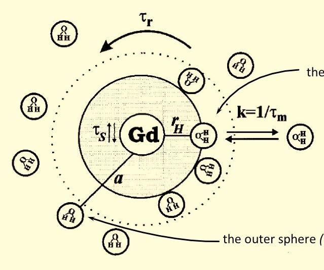
7 Metal chelate use of biocompatible ligand O H H O O Gd O N N H CH 3 O N N O O H 3 C N H O O CHAP 11 Contrast Enhanced MRI - Perfusion Contrast agents Paramagnetic / superparamagnetic Relaxivity Water exchange Susceptibility effect Brain perfusion pathologies Perfusion measurement T2* (DSC-MRI) T1 (DCE-MRI)
8 Contrast Agent Relaxivity in vitro 1/T 1,2 [CA] (mm) Contrast Agent Relaxivity in vitro


9 In vivo CA effects Paramagnetic chelates enhance contrast in T1-w images

 T1 (DCE-MRI) Water exchange effects")
10 CHAP 11 Contrast Enhanced MRI - Perfusion Contrast agents Paramagnetic / superparamagnetic Relaxivity Water exchange Susceptibility effect Brain perfusion pathologies Perfusion measurement T2* (DSC-MRI) T1 (DCE-MRI) Water exchange effects =k 1 + k 2


11

12 Slow/fast exchange
13 Susceptibility induced relaxation contrast agent always unevenly distributed in vivo (multiple intra- extracellular compartments) local susceptibility differences both in blood and interstitium enhanced T2/T2* relaxation CHAP 11 Contrast Enhanced MRI - Perfusion Contrast agents Paramagnetic / superparamagnetic Relaxivity Water exchange Susceptibility effect Brain perfusion pathologies Perfusion measurement T2* (DSC-MRI) T1 (DCE-MRI)

14 Perfusion imaging imaging alteration of blood supply Perfusion imaging: Stroke Ischemic area (e.g. Heart Myocardium) Cancer and Angiogenesis (Tumors) Etc. All areas where we have Microvascular Alterations Perfusion: Blood Supply


15 Perfusion: Blood Supply Perfusion: Blood Supply


16 Cancer & Angiogenesis Angiogenesis in tumors: high vascular density and abnormal vascular morphology Folkman J, Sci Am 1996

 T1 (DCE-MRI) Quantitative Perfusion")
17 CHAP 11 Contrast Enhanced MRI - Perfusion Contrast agents Paramagnetic / superparamagnetic Relaxivity Water exchange Susceptibility effect Brain perfusion pathologies Perfusion measurement T2* (DSC-MRI) T1 (DCE-MRI) Quantitative Perfusion Measurements
18 Perfusion Model and Absolute Value Theoretical considerations General expression of the tissue perfusion (tracer diffusion)


19 Quantitative Perfusion Measurement Diffusible Tracer Theory


 Dynamic Susceptibility")
20 CHAP 11 Contrast Enhanced MRI - Perfusion Contrast agents Paramagnetic / superparamagnetic Relaxivity Water exchange Susceptibility effect Brain perfusion pathologies Perfusion measurement T2* (DSC-MRI) T1 (DCE-MRI) Dynamic Susceptibility Enhanced Perfusion measurement


21 Bolus Perfusion Imaging Concentration-Time curve Conc. Data Fit T0 MTT Perfusion MR


22 Parametric perfusion maps Glioblastoma (high grade) T2-weighted T1-weighted w/ Gd CBV map


23 Oligoastrocytom (low grade) T2-weighted MRI CBV map Glioma grading from ncbv
")


.")
24 Comprehensive Stroke Examination FLAIR GraSE DWI-I ADC NI (rcbv) TTP MTT MRA FLAIR GraSE DWI-I ADC ~ 24 hours Perfusion: Kidneys Parametric maps of a T2W TSE Kidney Perfusion acquisition in a pig using 2 mg Fe/kg b.w ClariScan (Nycomed- Amersham). Top: MTT Bottom: rrbv

25 Breast MRI: Post-contrast Imaging 3D T1W FFE SENSE Body Coil SENSE Factor mm*0.64mm 68 slices of 2.2mm slices Proset 1331 Scantime: 1:53 minutes CHAP 11 Contrast Enhanced MRI - Perfusion Contrast agents Paramagnetic / superparamagnetic Relaxivity Water exchange Susceptibility effect Brain perfusion pathologies Perfusion measurement T2* (DSC-MRI) T1 (DCE-MRI)

26 Dynamic Contrast Enhanced Perfusion measurement
Perfusion MRI. Youngkyoo Jung, PhD Associate Professor Radiology, Biomedical Engineering, and Clinical & Translational Science Institute
 Perfusion MRI Youngkyoo Jung, PhD Associate Professor Radiology, Biomedical Engineering, and Clinical & Translational Science Institute Perfusion The delivery of blood to a capillary bed in tissue Perfusion
Imaging Acute Stroke and Cerebral Ischemia
 Department of Radiology University of California San Diego Imaging Acute Stroke and Cerebral Ischemia John R. Hesselink, M.D. Causes of Stroke Arterial stenosis Thrombosis Embolism Dissection Hypotension
PERFUSION MRI CONTRAST BASED TECHNIQUES
 PERFUSION MRI CONTRAST BASED TECHNIQUES by Kenny K Israni Mar 28, 2006 PERFUSION - MRI Dynamic Susceptibility contrast Dynamic Relaxivity contrast STEADY-STATE STATE TECHNIQUES Steady-state Susceptibility
Functional aspects of anatomical imaging techniques
 Functional aspects of anatomical imaging techniques Nilendu Purandare Associate Professor & Consultant Radiologist Tata Memorial Centre Functional/metabolic/molecular imaging (radioisotope scanning) PET
Outline. Why Image Animals?
 Small Animal Magnetic Resonance Imaging: Current Trends, Challenges and Perspectives for Pathological Imaging C. Chad Quarles Vanderbilt University Institute of Imaging Science Outline Why image animals?
Speed, Comfort and Quality with NeuroDrive
 Speed, Comfort and Quality with NeuroDrive Echelon Oval provides a broad range of capabilities supporting fast, accurate diagnosis of brain conditions and injuries. From anatomical depiction to vascular
PHYSICS OF MRI ACQUISITION. Alternatives to BOLD for fmri
 PHYSICS OF MRI ACQUISITION Quick Review for fmri HST-583, Fall 2002 HST.583: Functional Magnetic Resonance Imaging: Data Acquisition and Analysis Harvard-MIT Division of Health Sciences and Technology
Perfusion Physics. ICMRI2018 March 29-31, 2018 Grand Hilton Hotel, Seoul, Korea. Asian Forum Ⅱ: Perfusion MRI SY24-1.
 SY24-1 Perfusion Physics Hiroyuki Kabasawa MR Collaborations and Development, GE Healthcare, Tokyo, Japan Perfusion is referred as the blood supply to micro capillary in tissue. Perfusion parameter such
Place for Interventional Radiology in Acute Stroke
 Place for Interventional Radiology in Acute Stroke Dr Lakmalie Paranahewa MBBS, MD(Radiology), FRCR Consultant Interventional Radiologist Asiri Group of Hospitals Objectives Imaging in Stroke Neurovascular
MRI/MRS Biomarkers. Robert E. Lenkinski, Ph.D.
 MRI/MRS Biomarkers Robert E. Lenkinski, Ph.D. Disclosure GE Healthcare-Research Grant Aspect MR-Scientific Advisor Aposense-Scientific Advisor Brainwatch-Scientific Advisor I will be discussing off-label
Diagnostic improvement from average image in acute ischemic stroke
 Diagnostic improvement from average image in acute ischemic stroke N. Magne (1), E.Tollard (1), O. Ozkul- Wermester (2), V. Macaigne (1), J.-N. Dacher (1), E. Gerardin (1) (1) Department of Radiology,
Hemodynamic patterns of status epilepticus detected by susceptibility weighted imaging (SWI)
") Hemodynamic patterns of status epilepticus detected by susceptibility weighted imaging (SWI) Poster No.: C-1086 Congress: ECR 014 Type: Scientific Exhibit Authors: J. AELLEN, E. Abela, R. Kottke, E. Springer,
Hemodynamic patterns of status epilepticus detected by susceptibility weighted imaging (SWI) Poster No.: C-1086 Congress: ECR 014 Type: Scientific Exhibit Authors: J. AELLEN, E. Abela, R. Kottke, E. Springer,
Abdominal applications of DWI
 Postgraduate course, SPR San Antonio (Texas), May 14-15, 2013 Abdominal applications of DWI Rutger A.J. Nievelstein Wilhelmina Children s s Hospital, Utrecht (NL) Outline What is DWI? How to perform? Challenges
IMAGING OF INTRACRANIAL INFECTIONS
 IMAGING OF INTRACRANIAL INFECTIONS Dr Carolina Kachramanoglou LYSHOLM DEPARTMENT OF NEURORADIOLOGY NATIONAL HOSPITAL FOR NEUROLOGY AND NEUROSURGERY Plan Introduce MR sequences that are useful in the diagnosis
Multiparametric imaging in oncology
 Multiparametric imaging in oncology p1 T p2 p2 T T p3 p1 p3 T Marco Ravanelli Roberto Maroldi The goal of traditional imaging is high spatial and contrast resolution diagnosis, tumor extent treatment planning,
Assessing Tissue Oxygenation and Predicting Tissue Outcome in Acute Stroke
 The Danish National Research Foundation s Center of Functionally Integrative Neuroscience Aarhus University / Aarhus University Hospital - DENMARK Assessing Tissue Oxygenation and Predicting Tissue Outcome
Quantitative Imaging: Techniques, Applications and Challenges -- MR
 Quantitative Imaging: Techniques, Applications and Challenges -- MR Yue Cao, Ph.D. Departments of Radiation Oncology, Radiology and Biomedical Engineering University of Michigan Acknowledgments Radiation
To analyse whether ADC values have a correlation with survival or EGFR amplification status in glioblastoma
 To analyse whether ADC values have a correlation with survival or EGFR amplification status in glioblastoma R. Zalazar, M. Páramo, M. Hernández, P. Domínguez, J.Etxano, P.García Barquín, H.Quiceno Arias,
Clinical Trials for Adult Brain Tumors - the Imaging Perspective
 Clinical Trials for Adult Brain Tumors - the Imaging Perspective Whitney B. Pope, M.D., Ph.D. Department of Radiology David Geffen School of Medicine at UCLA August 22, 2015 1 Disclosure of Financial Relationships
Perfusion, Viability, Edema and Hemorrhage: How it Can (and Should) Change Clinical Practice. Rohan Dharmakumar, Ph.D.
 Change Clinical Practice. Rohan Dharmakumar, Ph.D.") Perfusion, Viability, Edema and Hemorrhage: How it Can (and Should) Change Clinical Practice Rohan Dharmakumar, Ph.D. Director, Translational Cardiac Imaging Research Associate Director, Biomedical Imaging
Perfusion, Viability, Edema and Hemorrhage: How it Can (and Should) Change Clinical Practice Rohan Dharmakumar, Ph.D. Director, Translational Cardiac Imaging Research Associate Director, Biomedical Imaging
Role of functional MRI in evaluating intraaxial brain tumors Advances and pitfalls.
 Role of functional MRI in evaluating intraaxial brain tumors Advances and pitfalls. Poster No.: C-1685 Congress: ECR 2014 Type: Educational Exhibit Authors: A. R. Udare, A. Mahajan, S. Juvekar, P. Shetty,
Whole Body MRI. Dr. Nina Tunariu. Prostate Cancer recurrence, progression and restaging
 Whole Body MRI Prostate Cancer recurrence, progression and restaging Dr. Nina Tunariu Consultant Radiology Drug Development Unit and Prostate Targeted Therapies Group 12-13 Janeiro 2018 Evolving Treatment
ASFNR Recommendations for Clinical Performance of MR Dynamic Susceptibility Contrast Perfusion Imaging of the Brain
 WHITE PAPER ASFNR Recommendations for Clinical Performance of MR Dynamic Susceptibility Contrast Perfusion Imaging of the Brain K. Welker, J. Boxerman, X A. Kalnin, T. Kaufmann, M. Shiroishi, and M. Wintermark;
PETER PAZMANY CATHOLIC UNIVERSITY Consortium members SEMMELWEIS UNIVERSITY, DIALOG CAMPUS PUBLISHER
 PETER PAZMANY CATHOLIC UNIVERSITY SEMMELWEIS UNIVERSITY Development of Complex Curricula for Molecular Bionics and Infobionics Programs within a consortial* framework** Consortium leader PETER PAZMANY
Invited. Methodology of Brain Perfusion Imaging. Review. Emmanuel L. Barbier, PhD, 1,2 Laurent Lamalle, PhD, 1 and Michel Décorps, PhD 1 *
 JOURNAL OF MAGNETIC RESONANCE IMAGING 13:496 520 (2001) Review Invited Methodology of Brain Perfusion Imaging Emmanuel L. Barbier, PhD, 1,2 Laurent Lamalle, PhD, 1 and Michel Décorps, PhD 1 * Numerous
Effect of intravenous contrast medium administration on prostate diffusion-weighted imaging
 Effect of intravenous contrast medium administration on prostate diffusion-weighted imaging Poster No.: C-1766 Congress: ECR 2015 Type: Authors: Keywords: DOI: Scientific Exhibit J. Bae, C. K. Kim, S.
Renal artery disease: MR Techniques and Interpretation
 Renal artery disease: MR Techniques and Interpretation Prof. Dr. Stefan O. Schoenberg Professor and Chairman of Radiology Department of Clinical Radiology and Nuclear Medicine University Medicine Mannheim
Dynamic contrast enhanced magnetic resonance imaging
 1995 AND 1999 PHOTODISC. INC. Dynamic Magnetic Resonance Imaging of Tumor Perfusion Approaches and Biomedical Challenges MOLECULAR IMAGING II BY DAVID J. COLLINS AND ANWAR R. PADHANI Dynamic contrast enhanced
MRS and Perfusion of Brain Tumors
 Department of Radiology University of California San Diego MRS and Perfusion of Brain Tumors John R. Hesselink, M.D. MRS & Perfusion of Brain Tumors Tumor histology Degree of malignancy Delineate tumor
MR Imaging of Atherosclerotic Plaques
 MR Imaging of Atherosclerotic Plaques Yeon Hyeon Choe, MD Department of Radiology, Samsung Medical Center, Sungkyunkwan University, Seoul MRI for Carotid Atheroma Excellent tissue contrast (fat, fibrous
Prediction of Hemorrhage in Acute Ischemic Stroke Using Permeability MR Imaging
 AJNR Am J Neuroradiol 26:2213 2217, October 2005 Technical Note Prediction of Hemorrhage in Acute Ischemic Stroke Using Permeability MR Imaging Andrea Kassner, Timothy Roberts, Keri Taylor, Frank Silver,
Prognostic value of ADC in glioblastoma multiforme and its correlation with survival and MGMT promoter methylation status.
 Prognostic value of ADC in glioblastoma multiforme and its correlation with survival and MGMT promoter methylation status. R. Zalazar, M.D. Hernández, M. Páramo, P. Slon, M. Millor Muruzabal, J. Solorzano
Amide Proton Transfer Imaging: A Novel MR Method for High-grade Brain Tumors.
 Amide Proton Transfer Imaging: A Novel MR Method for High-grade Brain Tumors. Poster No.: C-1732 Congress: ECR 2013 Type: Scientific Exhibit Authors: M. Ida, M. Ishizuka, T. Suzuki, Y. Kubo, K. Hino, S.
Disclosures. Diffusion and Perfusion Imaging in the Head and Neck. Learning objectives ???
 Disclosures No relevant financial disclosures Diffusion and Perfusion Imaging in the Head and Neck Ashok Srinivasan, MD Associate Professor Director of Neuroradiology University of Michigan Health System
UVM brain MRI protocols upgraded with latest methods
 UVM brain MRI protocols upgraded with latest methods FieldStrength MRI magazine User experiences - March 2017 UVM appreciates latest neuro MR methods for diagnosing and workflow The MRI staff at University
Complete Recovery of Perfusion Abnormalities in a Cardiac Arrest Patient Treated with Hypothermia: Results of Cerebral Perfusion MR Imaging
 pissn 2384-1095 eissn 2384-1109 imri 2018;22:56-60 https://doi.org/10.13104/imri.2018.22.1.56 Complete Recovery of Perfusion Abnormalities in a Cardiac Arrest Patient Treated with Hypothermia: Results
Challenges on Assessment of Treatment Response for Physiologically Adaptive Radiation Therapy
 Challenges on Assessment of Treatment Response for Physiologically Adaptive Radiation Therapy Yue Cao, Ph.D. Departments of Radiation Oncology, Radiology and Biomedical Engineering University of Michigan
NEURORADIOLOGY Part I
 NEURORADIOLOGY Part I Vörös Erika University of Szeged Department of Radiology SZEGED BRAIN IMAGING METHODS Plain film radiography Ultrasonography (US) Computer tomography (CT) Magnetic resonance imaging
Arterial Spin Labeling in Body MR
 Arterial Spin Labeling in Body MR Neil M. Rofsky, MD FACR, FISMRM, FSCBTMR Department of Radiology and Advanced Imaging Research Center None Disclosures Acknowledgements Ananth J. Madhuranthakam, Ph.D.
On Call Guide to CT Perfusion. Updated: March 2011
 On Call Guide to CT Perfusion Updated: March 2011 CT Stroke Protocol 1. Non contrast CT brain 2. CT perfusion: contrast 40cc bolus dynamic imaging at 8 slice levels ~ 60 sec creates perfusion color maps
Non-Invasive MR-based Evaluation of Kidney Function without Exogenous Contrast Agent. Xiang He, PhD Department of Radiology University of Pittsburgh
 Non-Invasive MR-based Evaluation of Kidney Function without Exogenous Contrast Agent Xiang He, PhD Department of Radiology University of Pittsburgh Contents MR-based non-invasive estimation of single kidney
Prevalence of cerebrovascular reserve impairment in patients with severe intracranial stenosis
 Prevalence of cerebrovascular reserve impairment in patients with severe intracranial stenosis Olivier Heck, Naila Boudiaf, Florence Tahon, Arnaud Attye, Kamel Boubagra, Johan Pietras, Olivier Detante,
MRI perfusion of brain tumors: any differences between supratentorial and infratentorial?
 MRI perfusion of brain tumors: any differences between supratentorial and infratentorial? Poster No.: C-2034 Congress: ECR 2012 Type: Scientific Paper Authors: M. Martucci, S. Gaudino, C. Schiarelli, R.
T2, T2*, ute. Yeo Ju Kim. Radiology, Inha University Hospital, Incheon, Korea
 SY28-1 T2, T2*, ute Yeo Ju Kim Radiology, Inha University Hospital, Incheon, Korea T2 relaxation times relate to the rate of transverse magnetization decay, caused by the loss of phase coherence induced
Initial Clinical Experience of TOSHIBA 3T MRI
 The 21st Conference of the Japanese Society of Cardiovascular Imaging & Dynamics Sponsored Seminar The Leading Edge of CT/MRI Diagnosis for the Cardiovascular System Initial Clinical Experience of TOSHIBA
The Egyptian Journal of Hospital Medicine (July 2018) Vol. 72 (10), Page
 Vol. 72 (10), Page") The Egyptian Journal of Hospital Medicine (July 2018) Vol. 72 (10), Page 5398-5402 The Role of Susceptibility Weighted Imaging (SWI) in Evaluation of Acute Stroke Maha Abdelhamed El Nouby*, Eman Ahmed
The Egyptian Journal of Hospital Medicine (July 2018) Vol. 72 (10), Page 5398-5402 The Role of Susceptibility Weighted Imaging (SWI) in Evaluation of Acute Stroke Maha Abdelhamed El Nouby*, Eman Ahmed
Spiral Coronary Angiography Using a Blood Pool Agent
 JOURNAL OF MAGNETIC RESONANCE IMAGING 22:213 218 (2005) Original Research Spiral Coronary Angiography Using a Blood Pool Agent Steffen Ringgaard, PhD, 1 * Michael Pedersen, PhD, 1 Jonas Rickers, MD, 1,2
非對比劑與對比劑增強 MRA. 血管攝影與對比劑 A Course of MRI. 本週課程內容 -MR Angiography (MRA) Unenhanced MRA
 Unenhanced MRA") 本週課程內容 -MR Angiography (MRA) 血管攝影與對比劑 A Course of MRI 盧家鋒助理教授國立陽明大學物理治療暨輔助科技學系 alvin4016@ym.edu.tw 非對比劑增強 MRA(Unenhanced MRA) Time-of-flight (TOF) angiography Phase-contrast (PC) angiography 對比劑增強 MRA(Contrast-enhanced
本週課程內容 -MR Angiography (MRA) 血管攝影與對比劑 A Course of MRI 盧家鋒助理教授國立陽明大學物理治療暨輔助科技學系 alvin4016@ym.edu.tw 非對比劑增強 MRA(Unenhanced MRA) Time-of-flight (TOF) angiography Phase-contrast (PC) angiography 對比劑增強 MRA(Contrast-enhanced
Handzettel 1. Multiparametric Functional Imaging in Radiation Therapy. Functional and Quantitative Imaging with MR
 Multiparametric Functional Imaging in Radiation Therapy Himanshu Bhat, Ph.D. Siemens Healthcare MR in RT Adding valuable information on tissue properties CT provides: Geometric accuracy Delineation of
Remission of diffusion lesions in acute stroke magnetic resonance imaging
 ORIGINAL RESEARCH Remission of diffusion lesions in acute stroke magnetic resonance imaging F. A. Fellner 1, M. R. Vosko 2, C. M. Fellner 1, D. Flöry 1 1. AKH Linz, Institute of Radiology, Austria. 2.
Comparison of Different Post-Processing Algorithms for Dynamic Susceptibility Contrast Perfusion Imaging of Cerebral Gliomas
 Magn Reson Med Sci 2017; 16; 129 136 doi:10.2463/mrms.mp.2016-0036 Published Online: September 20, 2016 MAJOR PAPER Comparison of Different Post-Processing Algorithms for Dynamic Susceptibility Contrast
Related Symposia in AAPM 2007
 Related Symposia in AAPM 7 Functional and Physiological MR Imaging for Therapy Assessment Yue Cao,, Ph.D. Departments of Radiation Oncology and Radiology, University of Michigan President s s symposium:
1 Uniform hyperintense signal intensity (normal). 2 Linear (arrow), wedge-shaped, or diffuse mild hypointensity, usually indistinct margin.
. 2 Linear (arrow), wedge-shaped, or diffuse mild hypointensity, usually indistinct margin.") Figure 3 PI-RADS assessment for peripheral zone on T2-weighted imaging. 1 Uniform hyperintense signal intensity (normal). 2 Linear (arrow), wedge-shaped, or diffuse mild hypointensity, usually indistinct
Figure 3 PI-RADS assessment for peripheral zone on T2-weighted imaging. 1 Uniform hyperintense signal intensity (normal). 2 Linear (arrow), wedge-shaped, or diffuse mild hypointensity, usually indistinct
Acute Ischemic Stroke Imaging. Ronald L. Wolf, MD, PhD Associate Professor of Radiology
 Acute Ischemic Stroke Imaging Ronald L. Wolf, MD, PhD Associate Professor of Radiology Title of First Slide of Substance An Illustrative Case 2 Disclosures No financial disclosures Off-label uses of some
The Paul Evans Memorial Lecture Functional radiotherapy targeting using focused dose escalation. Roberto Alonzi Mount Vernon Cancer Centre
 The Paul Evans Memorial Lecture Functional radiotherapy targeting using focused dose escalation Roberto Alonzi Mount Vernon Cancer Centre Overview Introduction and rationale for focused dose escalation
MRI OF BRAIN METASTASIS. Dr P. AGUETTAZ Hôpital Privé Clairval Marseille
 MRI OF BRAIN METASTASIS Dr P. AGUETTAZ Hôpital Privé Clairval Marseille Purpose To avoid a few common pitfalls when imaging patient suspected of brain metastasis 1. Does hyperintensity on post contrast
MR Functional Imaging to Guide Radiotherapy: Challenges and Opportunities
 Abstract No. 1234 MR Functional Imaging to Guide Radiotherapy: Challenges and Opportunities Michael Milosevic, MD Department of Radiation Oncology, University of Toronto Radiation Medicine Program, Princess
Functional Chest MRI in Children Hyun Woo Goo
 Functional Chest MRI in Children Hyun Woo Goo Department of Radiology and Research Institute of Radiology Asan Medical Center, University of Ulsan College of Medicine, Seoul, Korea No ionizing radiation
MRI to fit your planning. Philips Panorama HFO Oncology Configuration
 MRI to fit your planning Philips Panorama HFO Oncology Configuration MR Imaging that fits Philips Panorama HFO Oncology Configuration allows radiation oncologists to take full advantage of MRI s excellent
STATE OF THE ART IMAGING OF ACUTE STROKE
 STATE OF THE ART IMAGING OF ACUTE STROKE Marin Penkov UH St Ivan Rilski Sofia RadiologyTogether 2-3 June 2017 GOALS The concept and significance of penumbra CT and MRI Basic Principles Clinical application
CT/MRI 第 42 回日本脳卒中学会講演シンポジウム 総説 はじめに. MRI magnetic resonance imaging DWI diffusion-weighted. DWI/CBF malignant profile
 第 42 回日本脳卒中学会講演シンポジウム 総説 CT/MRI 1 要旨 rt-pa MRI magnetic resonance imaging DWI diffusion-weighted image Tmax time-to-maximum DWI/PWI perfusion image mismatch CT computed tomography CBF cerebral blood flow
A New Approach to Treating Acute Ischemic Stroke in Human Brain: Pulsed ElectroMagnetic Fields
 A New Approach to Treating Acute Ischemic Stroke in Human Brain: Pulsed ElectroMagnetic Fields Stefania Setti 1*, Vincenzo Di Lazzaro 2, Fiore Capone 2, Alessia Ongaro 3 Ruggero Cadossi 1 1 IGEA Clinical
The role of neuroimaging in patients with brain tumors is no
 REVIEW ARTICLE Update on Brain Tumor Imaging: From Anatomy to Physiology S. Cha The role of neuroimaging in patients with brain tumors is no longer simply to evaluate structural abnormality and identify
Multi-parametric MRI for Radiotherapy Response Prediction in Rectal Cancer
 Multi-parametric MRI for Radiotherapy Response Prediction in Rectal Cancer Dr Trang Pham Radiation Oncologist PhD Supervisors: Prof Barton, A/Prof Liney, Dr K Wong Current Status in Locally Advanced Rectal
11/6/2013. Vanderbilt University Institute of Imaging Science (VUIIS) 7 Tesla Vascular Imaging. Evaluating stroke risk
 7 Tesla Vascular Imaging. Evaluating stroke risk") Vanderbilt University Institute of Imaging Science (VUIIS) New MI Techniques for Imaging Cerebrovascular Disease Manus J. Donahue Depts. of adiology, Physics, Neurology and Physics Vanderbilt University
Vanderbilt University Institute of Imaging Science (VUIIS) New MI Techniques for Imaging Cerebrovascular Disease Manus J. Donahue Depts. of adiology, Physics, Neurology and Physics Vanderbilt University
Block Copolymer Assemblies for Delivering Drugs and Bioimaging Agents
 Block Copolymer Assemblies for Delivering Drugs and Bioimaging Agents J. S. Riffle, N. Pothayee, R. Zhang, N. Hu, S. Roy- Chaudrury & R. M. Davis Macromolecules and Interfaces Laboratory Virginia Tech
Assessment of Post-Treatment Imaging Changes Following Radiotherapy using Magnetic Susceptibility Techniques
 Western University Scholarship@Western Electronic Thesis and Dissertation Repository August 2016 Assessment of Post-Treatment Imaging Changes Following Radiotherapy using Magnetic Susceptibility Techniques
We are IntechOpen, the world s leading publisher of Open Access books Built by scientists, for scientists. International authors and editors
 We are IntechOpen, the world s leading publisher of Open Access books Built by scientists, for scientists 4,000 116,000 120M Open access books available International authors and editors Downloads Our
Prostate Cancer DFP Case of the Week
 Prostate Cancer DFP Case of the Week Antonio C. Westphalen, MD PhD Clinical Prostate MR Imaging Program, Director Associate Professor of Radiology and Urology University of California, San Francisco Case
JKSMRM 18(1) : 25-33, 2014 Ji Hyun Koo, Young Cheol Yoon, Jae Hoon Kim INTRODUCTION
 : 25-33, 2014 Ji Hyun Koo, Young Cheol Yoon, Jae Hoon Kim INTRODUCTION") www.ksmrm.org JKSMRM 18(1) : 25-33, 2014 pissn 1226-9751 / eissn 2288-3800 Original Article Diffusion-weighted and Dynamic Contrast-enhanced MRI of Metastatic Bone Tumors: Correlation of the Apparent Diffusion
www.ksmrm.org JKSMRM 18(1) : 25-33, 2014 pissn 1226-9751 / eissn 2288-3800 Original Article Diffusion-weighted and Dynamic Contrast-enhanced MRI of Metastatic Bone Tumors: Correlation of the Apparent Diffusion
MR Perfusion Imaging in Moyamoya Syndrome. Potential Implications for Clinical Evaluation of Occlusive Cerebrovascular Disease
 MR Perfusion Imaging in Moyamoya Syndrome Potential Implications for Clinical Evaluation of Occlusive Cerebrovascular Disease F. Calamante, PhD; V. Ganesan, MD; F.J. Kirkham, MB, BChir; W. Jan, MBBS; W.K.
MEDICAL POLICY EFFECTIVE DATE: 12/18/08 REVISED DATE: 12/17/09, 03/17/11, 05/19/11, 05/24/12, 05/23/13, 05/22/14
 MEDICAL POLICY SUBJECT: CT (COMPUTED TOMOGRAPHY) PAGE: 1 OF: 5 If the member's subscriber contract excludes coverage for a specific service it is not covered under that contract. In such cases, medical
Annex III. Amendments to relevant sections of the product information
 Annex III Amendments to relevant sections of the product information Note: These amendments to the relevant sections of the product information are the outcome of the referral procedure. The product information
/13/$ IEEE
 Multivariate Discriminant Analysis of Multiparametric Brain MRI to Differentiate High Grade and Low Grade Gliomas - A Computer- Aided Diagnosis Development Study *, Zeynep Firat, Ilhami Kovanlikaya, Ugur
Biomarkers and the Future of. John R. Votaw CBIS 5 th Year Anniversary Celebration/Look to the future February 8, 2013
 Biomarkers and the Future of Radiology John R. Votaw CBIS 5 th Year Anniversary Celebration/Look to the future February 8, 2013 Statistics/Radiology Collaboration The utility of Radiologic procedures
The Diagnosis of Hypovascular Hepatic Lesions Showing Hypo-intensity in the Hepatobiliary Phase of Gd-EOB- DTPA-enhanced MR Imaging in High-risk
 2013 67 4 239 244 The Diagnosis of Hypovascular Hepatic Lesions Showing Hypo-intensity in the Hepatobiliary Phase of Gd-EOB- DTPA-enhanced MR Imaging in High-risk Patients for Hepatocellular Carcinoma
Role of Perfusion CT Parameter Measurements in Differentiating Treatment Induced Necrosis from Recurrent Tumor
 Journal of US-China Medical Science 12 (2015) 151-157 doi: 10.17265/1548-6648/2015.04.002 D DAVID PUBLISHING Role of Perfusion CT Parameter Measurements in Differentiating Treatment Induced Necrosis from
Diffusion Restriction Precedes Contrast Enhancement in Glioblastoma Multiforme
 Diffusion Restriction Precedes Contrast Enhancement in Glioblastoma Multiforme Adil Bata 1, Jai Shankar 2 1 Faculty of Medicine, Class of 2017 2 Department of Diagnostic Radiology, Division of Neuroradiology,
Philips Gemini TF camera (Philips, Cleveland OH), with an 18 cm coronal and a 57 cm axial
, with an 18 cm coronal and a 57 cm axial") Supplement Material 18 F-DCFBC PET/CT Imaging Protocol 18 F-DCFBC PET/CT imaging was performed on a 3D time of flight (TOF) mode Philips Gemini TF camera (Philips, Cleveland OH), with an 18 cm coronal
Supplement Material 18 F-DCFBC PET/CT Imaging Protocol 18 F-DCFBC PET/CT imaging was performed on a 3D time of flight (TOF) mode Philips Gemini TF camera (Philips, Cleveland OH), with an 18 cm coronal
Stroke imaging. Why image stroke patients? Stroke. Treatment of infarct. Methods for infarct diagnosis. Treatment of infarct.
 Stroke imaging Stroke Infarct: -Arterial thrombosis/embolus -Hypoxic/ischemic -Venous thrombosis Non-traumatic hemorrhage: -Intracerebral -Subarachnoid Johan Wikström MD PhD Associate Professor of Radiology
Clinically focused workflow with unique ability to integrate fmri, DTI, fiber tracks and perfusion in a single, multi-layered 3D rendering
 Clinically focused workflow with unique ability to integrate fmri, DTI, fiber tracks and perfusion in a single, multi-layered 3D rendering Neurosurgeons are demanding more from neuroradiologists and increasingly
Introduction to Modern Imaging Physics and Techniques used in Clinical Neurology
 Introduction to Modern Imaging Physics and Techniques used in Clinical Neurology Benjamin M. Ellingson, Ph.D., M.S. Associate Professor of Radiology, Biomedical Physics, Bioengineering, and Psychiatry
Computed tomography. Department of Radiology, University Medical School, Szeged
 Computed tomography Department of Radiology, University Medical School, Szeged voxel +1-4 +2 +5 +3 +1 0-2 pixel -2 0 +1-4 -6 +5 +2 +1 Department of Radiology, University Medical School, Szeged
OASIS 1.2T: MULTIPARAMETRIC MRI OF PROSTATE CANCER
 OASIS 1.2T: MULTIPARAMETRIC MRI OF PROSTATE CANCER By Dr. John Feller, MD, Radiologist Desert Medical Imaging, Palm Springs, CA MRI is clinically accepted as the best imaging modality for displaying anatomical
Prostate MRI: Not So Difficult. Neil M. Rofsky, MD, FACR, FSCBTMR, FISMRM Dallas, TX
 Prostate MRI: Not So Difficult Neil M. Rofsky, MD, FACR, FSCBTMR, FISMRM Dallas, TX What is the biggest barrier to your practice incorporating prostate MRI? 1) I don t know how to read the cases 2) I don
Innovative Multimodal Imaging Techniques in Brain Tumor Clinical Trials
 Innovative Multimodal Imaging Techniques in Brain Tumor Clinical Trials Benjamin M. Ellingson, Ph.D. Assistant Professor of Radiology, Biomedical Physics, and Bioengineering Brain Tumor Imaging Laboratory
Dynamic contrast-enhanced magnetic resonance imaging: fundamentals and application to the evaluation of the peripheral perfusion
 Review Article Dynamic contrast-enhanced magnetic resonance imaging: fundamentals and application to the evaluation of the peripheral perfusion Yaron Gordon 1, Sasan Partovi 2, Matthias Müller-Eschner
Brain Pain Infections of the CNS
 FRIDAY, OCTOBER 28, 2016 Brain Pain Infections of the CNS Suyash Mohan MD, PDCC Assistant Professor of Radiology & Neurosurgery Division of Neuroradiology, Department of Radiology Perelman School of Medicine
Recent studies have demonstrated that multiparametric
 ORIGINAL RESEARCH D. Kaya A. Dinçer M.E. Yıldız M.O. Çizmeli C. Erzen Acute Ischemic Infarction Defined by a Region of Multiple Hypointense Vessels on Gradient-Echo T2* MR Imaging at 3T BACKGROUND AND
The role of apparent diffusion coefficient (ADC) and relative ADC in the evaluation of breast masses
 and relative ADC in the evaluation of breast masses") The role of apparent diffusion coefficient (ADC) and relative ADC in the evaluation of breast masses Poster No.: C-1749 Congress: ECR 2014 Type: Scientific Exhibit Authors: U. Aksoy Ozcan 1, A. Öz 2, S.
The role of apparent diffusion coefficient (ADC) and relative ADC in the evaluation of breast masses Poster No.: C-1749 Congress: ECR 2014 Type: Scientific Exhibit Authors: U. Aksoy Ozcan 1, A. Öz 2, S.
Innovations in HCC Imaging: MDCT/MRI
 Innovations in HCC Imaging: MDCT/MRI Anthony E. Cheng, M.D. Cardinal MRI Center Cardinal Santos Medical Center, Wilson Street, San Juan Innovations in HCC Imaging: Goals/Objectives MDCT/MRI Learn the diagnostic
The application of SWI and SWIM in imaging stroke, traumatic brain injury and tumors
 The application of SWI and SWIM in imaging stroke, traumatic brain injury and tumors E. Mark Haacke, PhD, nmrimaging@aol.com Director, MR Research Facility, Wayne State University Detroit, Michigan Disclosures
Differentiating Benign and Malignant Breast Lesions with T2*-Weighted First Pass Perfusion Imaging
 Acta Radiologica ISSN: 0284-1851 (Print) 1600-0455 (Online) Journal homepage: https://www.tandfonline.com/loi/iard20 Differentiating Benign and Malignant Breast Lesions with T2*-Weighted First Pass Perfusion
Anatomical and Functional MRI of the Pancreas
 Anatomical and Functional MRI of the Pancreas MA Bali, MD, T Metens, PhD Erasme Hospital Free University of Brussels Belgium mbali@ulb.ac.be Introduction The use of MRI to investigate the pancreas has
Abstract. Introduction. Material and Methods. Results. Conclusion
 Dynamic susceptibility contrast MRI calibrated using T1-based steadystate CBV and vascular space occupancy (VASO): Comparison with model-free arterial spin labelling Emelie Lindgren Supervisors: Linda
Monitoring bony metastases response with diffusion MRI
 Monitoring bony metastases response with diffusion MRI Anwar Padhani MD Mount Vernon Hospital Cancer Centre London, UK Objectives To illustrate the potential of whole body DWI in the therapy response assessment
Whole brain CT perfusion maps with paradoxical low mean transit time to predict infarct core
 Whole brain CT perfusion maps with paradoxical low mean transit time to predict infarct core Poster No.: B-292 Congress: ECR 2011 Type: Scientific Paper Topic: Neuro Authors: S. Chakraborty, M. E. Ahmad,
1) Diffusion weighted imaging DWI is a term used to describe moving molecules due to random thermal motion. This motion is restricted by boundaries
 Diffusion weighted imaging DWI is a term used to describe moving molecules due to random thermal motion. This motion is restricted by boundaries") 1) Diffusion weighted imaging DWI is a term used to describe moving molecules due to random thermal motion. This motion is restricted by boundaries such as ligaments, membranes and macro molecules. Diffusion
1) Diffusion weighted imaging DWI is a term used to describe moving molecules due to random thermal motion. This motion is restricted by boundaries such as ligaments, membranes and macro molecules. Diffusion
Alliance A Symptomatic brain radionecrosis after receiving radiosurgery for
 RANDOMIZED PHASE II STUDY: CORTICOSTEROIDS + BEVACIZUMAB VS. CORTICOSTEROIDS + PLACEBO (BEST) FOR RADIONECROSIS AFTER RADIOSURGERY FOR BRAIN METASTASES Pre-registration Eligibility Criteria Required Initial