Renal artery disease: MR Techniques and Interpretation
|
|
|
- Eustace Booker
- 5 years ago
- Views:
Transcription
1 Renal artery disease: MR Techniques and Interpretation Prof. Dr. Stefan O. Schoenberg Professor and Chairman of Radiology Department of Clinical Radiology and Nuclear Medicine University Medicine Mannheim Annual RSNA meeting, Chicago, 21 Learning objectives 1. To understand the basic technical principles of MR-Angiography with and without IV contrast agents, renal flow, perfusion and filtration measurements using magnetic resonance imaging (MRI) 2. To compare the various functional MRI techniques in terms of diagnostic value, complexity and clinical practicability 3. To demonstrate the added value of a comprehensive use of these techniques for assessment of different types of renovascular and renoparenchymal disease


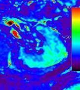
2 Background N Engl J Med 21; 344: From macro- to microcirculation: technical possibilities of MRI MRA Flow velocity [cm/s] time [ms] Michaely HJ, et al. Abdominal Imaging 26


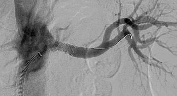
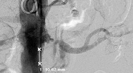
3 Potential of MRI Perfusion Oxygenation Inflammation Tissue structure Problem: eccentric stenoses MR A DSA Voxel size: mm 3 Parallel imaging, PAT 2 26 s acquisition time,.9x.9x.9 mm Schoenberg SO et al. Radiology 25; 235:


4 Accuracy of high resolution MRA vs. IVUS 1 Degree of area stenosis on MRA [%] y =.828x R 2 =.987 y =.883x.6697 R 2 = Degree of area stenosis on IVUS [%] Schoenberg SO et al. Radiology 25; 235: PAT and 3 Tesla: a perfect marriage 1.5T 25s, no PAT >1.5mm³ 1.5T 19s, PAT 2 1mm³ 3T 16s, PAT 3.65mm³ Michaely HJ et al. Mag Res Clin N Am, 27


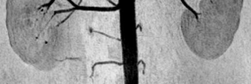

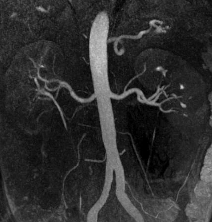
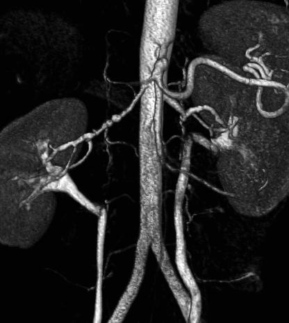
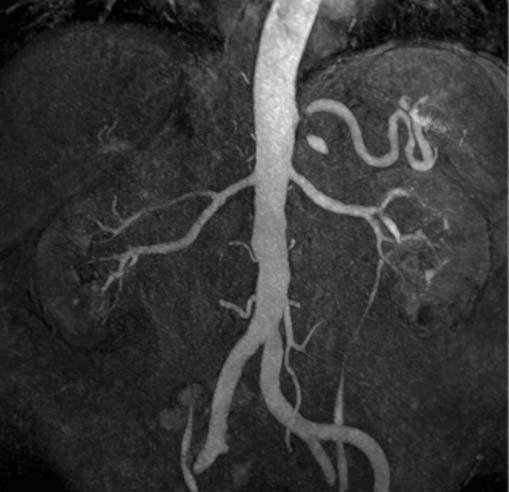
5 Run-off MRA: renal artery findings 3T CTM MRA 1.2 mm isotropic resolution.1mmol/kg BW Gd-DOTA SLE: Microaneurysms & systemic imaging Inflammatory LNs Thick pericardium Pleural effusion


![[ms] 4/1997](/docs-images/93/111252540/images/6-5.jpg "Re-stenosis 8/1997 35")


![6 8 1 12 time [ms]](/docs-images/93/111252540/images/6-8.jpg "Schoenberg SO, et al.")
6 Cine phase-contrast flow measurements velocity [cm/s] V V G t) ( tdt time [ms] 4/1997 Re-stenosis 8/ Right renal artery Apr 97 Aug velocity [cm/s] time [ms] Schoenberg SO, et al. Abdom Imaging 26; 31: 2

![3 2 4 6 8 1 time [ms] 35 3 grade 4 velocity [cm/s] 25 2 15 1 velocity [cm/s]](/docs-images/93/111252540/images/7-1.jpg "25 2 15 1 5 5 2 4 6 8 1 2 4 6 8 1 time [ms] time [ms] Schoenberg SO, et al.")
![JASN 22, 13: 158-169 Improved grading: MRA + PC Flow 6 Velocity [cm/s] 5 4 3](/docs-images/93/111252540/images/7-2.jpg "2 Left renal artery Right renal artery 1 15 125 235 345 455 565 675 time [ms]")
7 Grading scheme of renal artery stenosis 35 3 grade grade 2 velocity [cm/s] velocity [cm/s] time [ms] grade time [ms] 35 3 grade 4 velocity [cm/s] velocity [cm/s] time [ms] time [ms] Schoenberg SO, et al. JASN 22, 13: Improved grading: MRA + PC Flow 6 Velocity [cm/s] Left renal artery Right renal artery time [ms] DSA


![15 1 5 2 4 6 8 1 time [ms] New](/docs-images/93/111252540/images/8-3.jpg "functional methods without contrast")


8 Post-interventional control Schoenberg SO, et al. Radiologe 1997; 37: left velocity [cm/s] right velocity [cm/s] 45 preoperative postoperative time [ms] New functional methods without contrast media Δ p = 8.4mmHg Visual assessment of PC VIPR magnitude images vs. DSA Visualization of flow physiology utilizing time-resolved phase information Courtesy PD Dr. T. Bley, University of Wisconsin
")

![81 11 121 141-2 Time [s] Time](/docs-images/93/111252540/images/9-2.jpg "to Max Michaely HJ, Schoenberg")


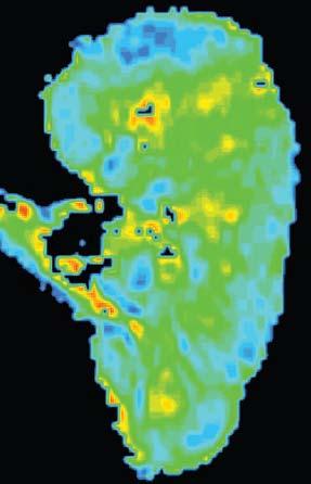
9 Dynamic renal perfusion with Gd chelates 16 ds/dt (maximum) MR-Renography A.U MTT Time [s] Time to Max Michaely HJ, Schoenberg SO, et al. Radiology 26; 238: Post-processing t - s t - 14s t - 7s t - 7s t - 11s t - 12s 7 Signal [a.u.] time in s Signal [a.u.] + First Pass Perfusion 5 1 Signal [a.u.] time in s Filtration time in s


10 Advantages of 3 Tesla for renal perfusion 1.5 Tesla 3 Tesla 1.5 Tesla 3. Tesla P-value Baseline SNR kidney 9.6 ± ± 5.3 p =.5 Peak SNR kidney 6.1 ± ± 38.7 p =.9 Michaely HJ,, Schoenberg SO. Invest Radiology 27; 42: A B C D

![] Before PTA After PTA 8 y o](/docs-images/93/111252540/images/11-2.jpg "female with arterial HTN before")


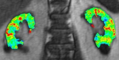
11 RAS: therapy monitoring with MRI Signal intensity [a.u.] Before PTA After PTA 8 y o female with arterial HTN before and after PTA before after PTA Michaely HJ et al, Abdom Imag 26 Severe HTN with low-grade RAS Patient 2 Normal y/o female with severe HTN RAS not significant Biopsy: 2 nephrosclerosis
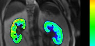
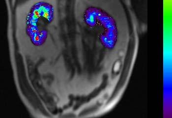
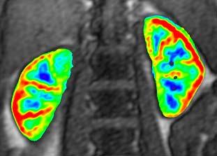
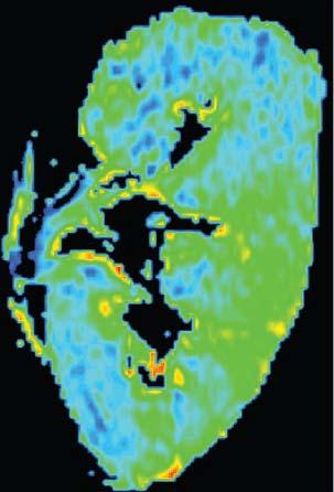
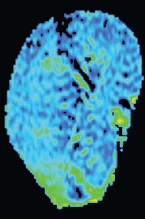
12 Patient with glomerulonephritis Plasma flow (8ml/1ml/min) Tubular flow (8ml/1ml/min) Normal MRA, abnormal perfusion study Comprehensive assessment of renal disease ml/1ml/min ml/1ml/min 47 ml/1ml/min 55 ml/1ml/min Fibromuscular Dysplasia Chronic renal failure

 with new MRI techniques (e.g. DTI)")


13 Algorithm for differentiation of renal disease with functional MRI Strategy MRA + MRP + MRA ± MRP AUC Exclusion of fixed renal damage (e.g. fibrosis) with new MRI techniques (e.g. DTI) Attenberger UI,, Michaely HJ, JMRI 29 From macro- to microcirculation 6 y/o male S/P RTx 5 years ago with rise of serum creatinine No RAS Biopsy revealed chronic ischemia at the upper pole Michaely HJ, Schoenberg SO. Radiology 26; 238:

14 New EMEA-guidelines (Nov 29) High-Risk Medium-Risk Low-Risk GFR < 3ml/min GFR 3-6ml/min Children, LTx Determination SCr Omniscan, Optimark, Magnevist and Generics Forbidden Dose minimization Forbidden Mandatory Multihance, Primovist, Vasovist Dotarem, Prohance, Gadovist Dose minimization* Recommended *single dose, no repetition within 7 days The GRIP Study Gadovist in Renally Impaired Patients prospective international study with > 3 patients included enrollment planned for 1 patients with CRF IV or V


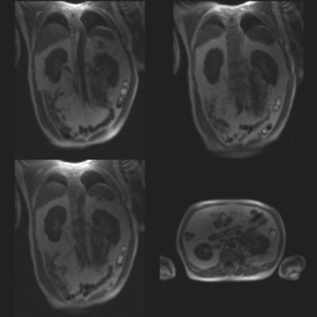
15 Comprehensive protocols in the decade of NSF normal patient Native-MRA (TrueFISP), CE-MRA, 1x1x1mm³, 1.1x1x1.2mm 2-4min 3 Native-MRA (TrueFISP), CE-MRA, 1x1x1mm³, 1.1x1x1.2mm 2-4min 3 Non-contrast MRA: excellent in normal flow states inconsistent results with low-flow Perfusion measurements with ASL (FAIR) Global Inversion and Readout Selective Inversion and Readout after TI=1.2s Subtraction of Images


16 ASL Perfusion Improvement after therapy 56y/o female patient with right sided RAS (>7%) Affected left kidney shows reduced signal intensity After intervention the blood flow to the kidney is restored with signal intensity equal to the opposite side Michaely HJ, Schoenberg SO, et al. Invest Radiol 24; 39: ASL-SNR Healthy Significant RAS Intrarenal oxygenation by BOLD MRI Oxyhemoglobin is diamagnetic Deoxyhemoglobin is paramagnetic R 2 * (1=1/T 2 *) is directly proportional to the tissue content of deoxyhemoglobin A decrease in the slope implies an increase in the Po 2 of blood Surrogate for medullary perfusion Disadvantage: Confounded by blood flow and hemoglobin concentration Prasad PV, et al. Circulation 1996; 94:
17 Post Post-water load Effect of Water Diuresis On Relative Medullary Oxygenation Anatomical R 2 * Map * Map Pre-water load Prasad PV et al. JMRI 1997; 7: Impact of hydration 9 8 Increased blood flow oxygen availability Filtration oxygen consumption Rebound Medulla T2*(ms) Medulla R2* (s-1) Cortex T2* Cortex R2* prä_18:28: post 18:42: 18:47 18:52 18:57 19:2 19:7 time


18 Changes in medullary po2 post-waterload: Effect of endogenous prostaglandins Prasad PV, et al. Kidney Int 1999; 55: y old male with HTN and 5% stenosis R RA Perfusion: mean transit time R Perfusion: plasma flow R Mean flow 34ml/min loss of early systolic peak Mean flow 42ml/min normal flow profile T2* (BOLD): oxygenation R

19 Quantification of therapeutic response after renal stent placement Pre-Therapy Post-Therapy Pre-Therapy Post-Therapy PF ml/1ml/min BOLD R2* loss reflecting renal transplant rejection a b c normal ATN Rejection Sadowski E et al, Radiology 25


20 Threshold value for medullary R2* - 18/sec Medulla Cortex Sadowski E et al, Radiology 25 Sodium MRI with 23 Na at 3 T Maril N et al. (26) Sodium MRI of the Human Kidney at 3 Tesla 3D gradient-echo sequence using an inhousebuilt quadrature surface coil with TR/TE 3/1.8 ms, FOV 38 x 38 x 24 cm 3, and matrix 128 x 128 x 16. Magnetic Resonance in Medicine 56: Water deprivation (12 hr) induced a significant increase of 25% (P<.5) in this gradient.
 Before water")


21 Sodium MRI with 23 Na at 3 T Healthy volunteer (5 x 5 x 5 mm 3 ) Before water load After water load Duration = 16 min; TE = 5,5ms; TR = 12ms; projections = 8; flip angle = 85 Courtesy of Lothar Schad, PhD, CKM and Stefan Haneder MD, IKRN, Mannheim Imaging of inflammatory renal disease ultra small particles of iron oxide (USPIO) Signal changes : -C: -65 % -OM: -62 % -IM: -6 % T2*w MRI pre-uspio T2*w MRI 3 days post-uspio 53 y o male with purpura, acute renal insufficiency and proteinuria Proliferative glomerulopathy with positive CD68 inflammatory cells (55/mm2)

 T2* imaging before and 72h after USPIO")


22 USPIO Imaging of the kidneys Phase II study in 12 patients with renal biopsy as standard of reference USPIO ultra small particles of iron oxide mg Fe/kg KG (Sinerem ) T2* imaging before and 72h after USPIO administration For >5 macrophages/mm² mean signal decrease in renal cortex by 33±18% Detection of acute transplant rejection and ATN Acute tubular necrosis (ATN) day 8 after renal TPx T2* before USPIO T2* 72h after USPIO Hauger O, et al. Eur Radiol 27; 17: USPIO (P94) parenchymal changes Normal Ischemia-Reperfusion CyA-Toxicity In the rat model
 Free diffusion Low cell density, free")


23 Diffusion-Weighted Imaging (DWI) Diffusion-Tensor Imaging (DTI) Free diffusion Low cell density, free water motion in the interstitial room high ADC / isotropy Restricted diffusion High cell density, decreased water motion in the interstitial room low ADC / anisotropy Koh et al. AJR 27 DWI as a contrast media substitute? ADC values dependent on stage of CKD, not age Study with 72 volunteers and 43 patients, b=, b=5, 1.5T Xu X et al., Eur Radiol 21


,")


24 DWI for evaluation of renal fibrosis 7T animal scanner DWI b-values 35, 6, 8, 1, 12 sec/mm² α-sma (smoot muscle actin), cell density Togamu O et al., Radiology 21 RAS: measurement of fractional anisotropy FA map ADC map ADC the left cortex << right FA left cortex > right FA left medulla >> right Notohamiprodjo M et al. Invest Radiol 28; 43:
25 Conclusion 1. MRA: spatial resolution <1mm3 for accurate stenosis quantification 3 Tesla new standard 2. Flow measurements for detection of hemodynamic significance 3. MRP for detection of renal parenchymal disease 4. Combined approach of MRA and MRP detection of clinically significant renal disease 5. NCE-MRA + ASL + BOLD allow (future) protocols without CM 6. USPIO for detection of inflammatory disease on a cellular level Patient selection for intervention based on functional information Acknowledgment Department of Clinical Radiology and Nuclear Medicine, University Medicine Mannheim: Henrik J. Michaely MD, Ulrike I. Attenberger MD, Stefan Haneder MD, Steffen J. Diehl MD Department of computer-assisted clinical medicine (CKM): Lothar R. Schad PhD, Frank Zoellner PhD Department of Radiology, Evanston, University of Chicago: Pottumarthi V. Prasad PhD Department of Radiology, University of Bordeaux, France: Nicolas Grenier MD
Intra-renal Oxygenation. in Human Subjects
 MRI-based Mapping of Intra-renal Oxygenation BOLD in Human Subjects OEF Xiang He, PhD Department of Radiology Background Cortex Brain CBF ~ 1.0 ml/min/g Brain PO 2 ~ 25-35 mm Hg Medullary hypoxia is an
Diffusion weighted MRI in evaluation of transplanted kidney: Preliminary clinical experience
 African Journal of Nephrology (2009) 13: 26-30 Original Article AJN Diffusion weighted MRI in evaluation of transplanted kidney: Preliminary clinical experience Mohamed Abou El-Ghar; M.D, Huda Refaie;
Non-Invasive MR-based Evaluation of Kidney Function without Exogenous Contrast Agent. Xiang He, PhD Department of Radiology University of Pittsburgh
 Non-Invasive MR-based Evaluation of Kidney Function without Exogenous Contrast Agent Xiang He, PhD Department of Radiology University of Pittsburgh Contents MR-based non-invasive estimation of single kidney
MRI qbold Based Evaluation. Renal Oxidative Metabolism. Department of Radiology and Hernando Gomez, MD Critical Care Medicine
 MRI qbold Based Evaluation of Renal Oxidative Metabolism Xiang He, PhD Department of Radiology and Hernando Gomez, MD Critical Care Medicine Background High oxygen-demand and lower medullary blood flow
Magnetic resonance (MR) diffusion-weighted imaging (DWI) is
 diffusion-weighted imaging (DWI) is") Diagn Interv Radiol DOI 10.4261/1305-3825.DIR.3892-10.1 Turkish Society of Radiology 2010 ABDOMINAL IMAGING ORIGINAL ARTICLE Diffusion tensor imaging of the kidney at 3 Tesla: normative values and repeatability
Diagn Interv Radiol DOI 10.4261/1305-3825.DIR.3892-10.1 Turkish Society of Radiology 2010 ABDOMINAL IMAGING ORIGINAL ARTICLE Diffusion tensor imaging of the kidney at 3 Tesla: normative values and repeatability
Protocol for iv. iodine and gadolinium contrast studies
 Protocol for iv. iodine and gadolinium contrast studies Royal College of Radiologists Standard The individual administering the contrast agent must ensure that the patient understands that it is to be
1Pulse sequences for non CE MRA
 MRI: Principles and Applications, Friday, 8.30 9.20 am Pulse sequences for non CE MRA S. I. Gonçalves, PhD Radiology Department University Hospital Coimbra Autumn Semester, 2011 1 Magnetic resonance angiography
Contrast-enhanced MRI: how do changing EU regulations impact daily practice?
 Safety considerations in contrast enhanced procedures Carlo Catalano Sapienza University of Rome Contrast-enhanced MRI: how do changing EU regulations impact daily practice? CE MRI: EU regulations - Initial
Speed, Comfort and Quality with NeuroDrive
 Speed, Comfort and Quality with NeuroDrive Echelon Oval provides a broad range of capabilities supporting fast, accurate diagnosis of brain conditions and injuries. From anatomical depiction to vascular
Val M. Runge, MD Editor-in-Chief Investigative Radiology
 Val M. Runge, MD Editor-in-Chief Investigative Radiology Patients First RSNA 2012 The classical model of medical care which portrays the authoritative physician evaluating and treating an obedient, non-inquisitive
Perfusion MRI. Youngkyoo Jung, PhD Associate Professor Radiology, Biomedical Engineering, and Clinical & Translational Science Institute
 Perfusion MRI Youngkyoo Jung, PhD Associate Professor Radiology, Biomedical Engineering, and Clinical & Translational Science Institute Perfusion The delivery of blood to a capillary bed in tissue Perfusion
Non Contrast MRA. Mayil Krishnam. Director, Cardiovascular and Thoracic Imaging University of California, Irvine
 Non Contrast MRA Mayil Krishnam Director, Cardiovascular and Thoracic Imaging University of California, Irvine No disclosures Non contrast MRA-Why? Limitations of CTA Radiation exposure Iodinated contrast
MR Advance Techniques. Vascular Imaging. Class II
 MR Advance Techniques Vascular Imaging Class II 1 Vascular Imaging There are several methods that can be used to evaluate the cardiovascular systems with the use of MRI. MRI will aloud to evaluate morphology
Kidney Transplant: Functional Assessment with Diffusion-Tensor MR Imaging at 3T 1
 Note: This copy is for your personal non-commercial use only. To order presentation-ready copies for distribution to your colleagues or clients, contact us at www.rsna.org/rsnarights. Original Research
Non-Contrast MRA. How and When 1996! Why Non-Contrast MRA? Angiography: What are our goals? Inflow Techniques Differences in excitation hx
 A major teaching hospital of Harvard Medical School Angiography: What are our goals? Non-Contrast MRA: How and When Neil M. Rofsky, M.D. Professor of Radiology, Harvard Medical School Director of MRI &
Magnetization Preparation Sequences
 Magnetization Preparation Sequences Acquisition method may not give desired contrast Prep block adds contrast (and/or encoding) MP-RAGE = Magnetization prepared rapid acquisition with gradient echo (Mugler,
In vivo diffusion tensor imaging (DTI) of articular cartilage as a biomarker for osteoarthritis
 of articular cartilage as a biomarker for osteoarthritis") In vivo diffusion tensor imaging (DTI) of articular cartilage as a biomarker for osteoarthritis Jose G. Raya 1, Annie Horng 2, Olaf Dietrich 2, Svetlana Krasnokutsky 3, Luis S. Beltran 1, Maximilian F.
In vivo diffusion tensor imaging (DTI) of articular cartilage as a biomarker for osteoarthritis Jose G. Raya 1, Annie Horng 2, Olaf Dietrich 2, Svetlana Krasnokutsky 3, Luis S. Beltran 1, Maximilian F.
A Safety Update on the Gadolinium Chelates
 Control # 1029 A Safety Update on the Gadolinium Chelates Val M. Runge, MD Universitätsinstitut für Diagnostische, Interventionelle und Pädiatrische Radiologie University Hospital Bern Allergic Reactions
Magnetic Resonance Angiography
 Magnetic Resonance Angiography 1 Magnetic Resonance Angiography exploits flow enhancement of GR sequences saturation of venous flow allows arterial visualization saturation of arterial flow allows venous
Renal Functional MRI: Are We Ready for Clinical Application?
 Genitourinary Imaging Perspective Chandarana and Lee Renal Functional MRI Genitourinary Imaging Perspective FOCUS ON: Hersh Chandarana 1 Vivian S. Lee Chandarana H, Lee VS Keywords: blood oxygen level
Flow Quantification from 2D Phase Contrast MRI in Renal Arteries using Clustering
 Flow Quantification from 2D Phase Contrast MRI in Renal Arteries using Clustering Frank G. Zöllner 1,2, Jan Ankar Monnsen 1, Arvid Lundervold 2, Jarle Rørvik 1 1 Department for Radiology, University of
Liver Fat Quantification
 Liver Fat Quantification Jie Deng, PhD, DABMP Department of Medical Imaging May 18 th, 2017 Disclosure Research agreement with Siemens Medical Solutions 2 Background Non-alcoholic fatty liver diseases
Experimental Assessment of Infarct Lesion Growth in Mice using Time-Resolved T2* MR Image Sequences
 Experimental Assessment of Infarct Lesion Growth in Mice using Time-Resolved T2* MR Image Sequences Nils Daniel Forkert 1, Dennis Säring 1, Andrea Eisenbeis 2, Frank Leypoldt 3, Jens Fiehler 2, Heinz Handels
Perfusion, Viability, Edema and Hemorrhage: How it Can (and Should) Change Clinical Practice. Rohan Dharmakumar, Ph.D.
 Change Clinical Practice. Rohan Dharmakumar, Ph.D.") Perfusion, Viability, Edema and Hemorrhage: How it Can (and Should) Change Clinical Practice Rohan Dharmakumar, Ph.D. Director, Translational Cardiac Imaging Research Associate Director, Biomedical Imaging
Perfusion, Viability, Edema and Hemorrhage: How it Can (and Should) Change Clinical Practice Rohan Dharmakumar, Ph.D. Director, Translational Cardiac Imaging Research Associate Director, Biomedical Imaging
Imaging the Kidney using Magnetic Resonance Techniques: Structure to. Function
 Imaging the Kidney using Magnetic Resonance Techniques: Structure to Function Huda Mahmoud 1,2, Charlotte Buchanan 3, Susan T Francis 3 and Nicholas M Selby 1,2 1 Centre for Kidney Research and Innovation
CT Versus MR for the Runoff
 CT Versus MR for the Runoff Robert R. Edelman, M.D. Dept. of Radiology NorthShore University HealthSystem Feinberg School of Medicine, Northwestern University Magnetic Resonance Computed Tomography Radio
Gadolinium-Based Contrast Agents for Magnetic Resonance Imaging (marketed as Magnevist, MultiHance, Omniscan, OptiMARK, ProHance)
") Information for Healthcare Professionals Page 1 of 5 FDA ALERT [6/2006, updated 12/2006 and 5/23/2007: This updated Alert highlights FDA s request for addition of a boxed warning and new warnings about
Information for Healthcare Professionals Page 1 of 5 FDA ALERT [6/2006, updated 12/2006 and 5/23/2007: This updated Alert highlights FDA s request for addition of a boxed warning and new warnings about
PHYSICS OF MRI ACQUISITION. Alternatives to BOLD for fmri
 PHYSICS OF MRI ACQUISITION Quick Review for fmri HST-583, Fall 2002 HST.583: Functional Magnetic Resonance Imaging: Data Acquisition and Analysis Harvard-MIT Division of Health Sciences and Technology
CHAP 11 Contrast Enhanced MRI - Perfusion. CHAP 11 Contrast Enhanced MRI - Perfusion
 CHAP 11 Contrast Enhanced MRI - Perfusion Contrast agents Paramagnetic / superparamagnetic Relaxivity Water exchange Susceptibility effect Brain perfusion pathologies Perfusion measurement T2* (DSC-MRI)
Objectives and Outline
 Development and Clinical Applications of Time- Resolved Magnetic Resonance Angiography Thomas M. Grist, MD, FACR ICRU Gray Symposium AAPM 2017 Denver, CO Objectives and Outline Objectives: Share some key
Index. mri.theclinics.com. Note: Page numbers of article titles are in boldface type.
 Index Note: Page numbers of article titles are in boldface type. A Angiogenesis, and cancer of prostate, 689 690 Angiography, MR. See MR angiography. Apoptosis, MR imaging of, 637 Apparent diffusion coefficient,
High Field MR of the Spine
 Department of Radiology University of California San Diego 3T for MR Applications Advantages High Field MR of the Spine Increased signal-to-noise Better fat suppression Increased enhancement with gadolinium
MR Contrast Agents. Why Use Contrast Agents in MRI? Why Use Contrast Agents in MRI? US Agents. Understanding and Embracing Change
 Why Use Contrast Agents in MRI? Improve disease detection and characterization Increase sensitivity to extent of disease MR Contrast Agents Understanding and Embracing Change Kristan Harrington, MBA, RT
Fully-Automatic Determination of the Arterial Input Function for Dynamic Contrast-Enhanced Pulmonary MR Imaging (DCE-pMRI)
") Fully-Automatic Determination of the Arterial Input Function for Dynamic Contrast-Enhanced Pulmonary MR Imaging (DCE-pMRI) Kohlmann P. 1, Laue H. 1, Anjorin A. 2, Wolf U. 3, Terekhov M. 3, Krass S. 1,
Fully-Automatic Determination of the Arterial Input Function for Dynamic Contrast-Enhanced Pulmonary MR Imaging (DCE-pMRI) Kohlmann P. 1, Laue H. 1, Anjorin A. 2, Wolf U. 3, Terekhov M. 3, Krass S. 1,
Functional aspects of anatomical imaging techniques
 Functional aspects of anatomical imaging techniques Nilendu Purandare Associate Professor & Consultant Radiologist Tata Memorial Centre Functional/metabolic/molecular imaging (radioisotope scanning) PET
Tips and Tricks of State of the art MRA
 Tips and Tricks of State of the art MRA Mayil Krishnam, MD,MBA, MRCP,FRCR(UK) Professor of Radiology Director, Cardiovascular and Thoracic Imaging University of California, Irvine Objectives Technical
P2 Visual - Perception
 P2 Visual - Perception 2014 SOSE Neuroimaging of high-level visual functions gyula.kovacs@uni-jena.de 11/09/06 Functional magnetic resonance imaging (fmri) The very basics What is fmri? What is MRI? The
Diagnosis of Renal Artery Stenosis (RAS)
") May 2001 Diagnosis of Renal Artery Stenosis (RAS) Kurt Fink, Harvard Medical School, Year III Epidemiology Hypertension -Affects 60 million Americans Essential HTN >95% of cases Secondary HTN 1-5% of cases
May 2001 Diagnosis of Renal Artery Stenosis (RAS) Kurt Fink, Harvard Medical School, Year III Epidemiology Hypertension -Affects 60 million Americans Essential HTN >95% of cases Secondary HTN 1-5% of cases
Evaluation of intra-renal oxygenation during water diuresis: A time-resolved study using BOLD MRI
 http://www.kidney-international.org & 2006 International Society of Nephrology original article see commentary on page Evaluation of intra-renal oxygenation during water diuresis: A time-resolved study
New magnetic resonance imaging methods in nephrology
 http://www.kidney-international.org & 2013 International Society of Nephrology New magnetic resonance imaging methods in nephrology Jeff L. Zhang 1, Glen Morrell 1, Henry Rusinek 2, Eric E. Sigmund 2,
Duplex Ultrasound of the Renal Arteries. Duplex Ultrasound. In the Beginning
 Duplex Ultrasound of the Renal Arteries DIMENSIONS IN HEART AND VASCULAR CARE 2013 PENN STATE HEART AND VASCULAR INSTITUTE ROBERT G. ATNIP MD PROFESSOR OF SURGERY AND RADIOLOGY Duplex Ultrasound Developed
Renal. Prof John Buscombe
 Renal Prof John Buscombe Renal nuclear Medicine Only consistent test of kidney func7on Many good tests for renal anatomy Ultrasound good looking at cysts and renal pelvis CT can look at perfusion, size
Contrast-enhanced Breast MRI RSSA 2013
 Contrast-enhanced Breast MRI RSSA 2013 Prof. dr. Maurice van den Bosch University Medical Center Utrecht, the Netherlands Index 1) Breast cancer 2) Why MRI of the breast 3) Technique 4) Interpretation
Diffusion Weighted Imaging in Prostate Cancer
 Diffusion Weighted Imaging in Prostate Cancer Disclosure Information Vikas Kundra, M.D, Ph.D. No financial relationships to disclose. Education Goals and Objectives To describe the utility of diffusion-weighted
Abdominal applications of DWI
 Postgraduate course, SPR San Antonio (Texas), May 14-15, 2013 Abdominal applications of DWI Rutger A.J. Nievelstein Wilhelmina Children s s Hospital, Utrecht (NL) Outline What is DWI? How to perform? Challenges
Supplementary Online Content
 Supplementary Online Content Gregg NM, Kim AE, Gurol ME, et al. Incidental cerebral microbleeds and cerebral blood flow in elderly individuals. JAMA Neurol. Published online July 13, 2015. doi:10.1001/jamaneurol.2015.1359.
Melissa M Ong 1*, Katharina Hausotter 1, Lothar R Pilz 2, Stefan O Schoenberg 1 and Henrik J Michaely 1
 Ong et al. Journal of Cardiovascular Magnetic Resonance 2013, 15:97 RESEARCH Open Access Steady state vascular imaging with extracellular gadobutrol: evaluation of the additional diagnostic benefit in
ACR MRI Accreditation: Medical Physicist Role in the Application Process
 ACR MRI Accreditation: Medical Physicist Role in the Application Process Donna M. Reeve, MS, DABR, DABMP Department of Imaging Physics University of Texas M.D. Anderson Cancer Center Educational Objectives
NSF Coming and Going
 NSF Coming and Going Martin R. Prince, MD, PhD Cornell and Columbia Universities Patent agreements: GE, Philips, Siemens, Hitachi, Medrad, Epix, Lantheus, Bayer, Bracco, Nemoto, Mallinckrodt and Topspins
Raja Muthupillai, PhD. Department of Diagnostic and Interventional Radiology St. Luke s Episcopal Hospital. Research Support: Philips Healthcare
 3D Cardiac Imaging Raja Muthupillai, PhD Department of Diagnostic and Interventional Radiology St. Luke s Episcopal Hospital Houston, TX Disclosures Research Support: Philips Healthcare This presentation
The role of apparent diffusion coefficient (ADC) and relative ADC in the evaluation of breast masses
 and relative ADC in the evaluation of breast masses") The role of apparent diffusion coefficient (ADC) and relative ADC in the evaluation of breast masses Poster No.: C-1749 Congress: ECR 2014 Type: Scientific Exhibit Authors: U. Aksoy Ozcan 1, A. Öz 2, S.
The role of apparent diffusion coefficient (ADC) and relative ADC in the evaluation of breast masses Poster No.: C-1749 Congress: ECR 2014 Type: Scientific Exhibit Authors: U. Aksoy Ozcan 1, A. Öz 2, S.
Daniel Bulte. Centre for Functional Magnetic Resonance Imaging of the Brain. University of Oxford
 Daniel Bulte Centre for Functional Magnetic Resonance Imaging of the Brain University of Oxford Overview Signal Sources BOLD Contrast Mechanism of MR signal change FMRI Modelling Scan design details Factors
Visualization strategies for major white matter tracts identified by diffusion tensor imaging for intraoperative use
 International Congress Series 1281 (2005) 793 797 www.ics-elsevier.com Visualization strategies for major white matter tracts identified by diffusion tensor imaging for intraoperative use Ch. Nimsky a,b,
Department of Radiology University of California San Diego. MR Angiography. Techniques & Applications. John R. Hesselink, M.D.
 Department of Radiology University of California San Diego MR Angiography Techniques & Applications John R. Hesselink, M.D. Vascular Imaging Arterial flow void Flow enhancement Gadolinium enhancement Vascular
Disclosures. Diffusion and Perfusion Imaging in the Head and Neck. Learning objectives ???
 Disclosures No relevant financial disclosures Diffusion and Perfusion Imaging in the Head and Neck Ashok Srinivasan, MD Associate Professor Director of Neuroradiology University of Michigan Health System
To compare the relative amount of of selected gene expression between sham and
 Supplementary Materials and Methods Gene Expression Analysis To compare the relative amount of of selected gene expression between sham and mice given renal ischemia-reperfusion injury (IRI), ncounter
Pediatric* MR Urography
 Abdominal Imaging Clinical Pediatric* MR Urography Richard A. Jones; Stephen Little; J. Damien Grattan-Smith Children s Healthcare of Atlanta, Department of Radiology, Atlanta, GA, USA MR urography represents
ROLE OF CONTRAST ENHANCED MR ANGIOGRAPHY IN AORTIC COARCTATION
 ROLE OF CONTRAST ENHANCED MR ANGIOGRAPHY IN AORTIC COARCTATION By Adel El Badrawy, Ahmed Abdel Razek, Nermin Soliman, Hala El Marsafawy *, Sameh Amer** From Radiodiagnosis, Pediatric Cardiology* & Cardiothoracic
How I do it: Non Contrast-Enhanced MR Angiography (syngo NATIVE)
") Clinical How-I-do-it Cardiovascular How I do it: Non Contrast-Enhanced MR Angiography (syngo NATIVE) Manuela Rick, Nina Kaarmann, Peter Weale, Peter Schmitt Siemens Healthcare, Erlangen, Germany Introduction
Clinical How-I-do-it Cardiovascular How I do it: Non Contrast-Enhanced MR Angiography (syngo NATIVE) Manuela Rick, Nina Kaarmann, Peter Weale, Peter Schmitt Siemens Healthcare, Erlangen, Germany Introduction
MRI/MRS Biomarkers. Robert E. Lenkinski, Ph.D.
 MRI/MRS Biomarkers Robert E. Lenkinski, Ph.D. Disclosure GE Healthcare-Research Grant Aspect MR-Scientific Advisor Aposense-Scientific Advisor Brainwatch-Scientific Advisor I will be discussing off-label
MR Tumor Staging for Treatment Decision in Case of Wilms Tumor
 MR Tumor Staging for Treatment Decision in Case of Wilms Tumor G. Schneider, M.D., Ph.D.; P. Fries, M.D. Dept. of Diagnostic and Interventional Radiology, Saarland University Hospital, Homburg/Saar, Germany
Related Symposia in AAPM 2007
 Related Symposia in AAPM 7 Functional and Physiological MR Imaging for Therapy Assessment Yue Cao,, Ph.D. Departments of Radiation Oncology and Radiology, University of Michigan President s s symposium:
Introduction to Clinical Diagnosis Nephrology
 Introduction to Clinical Diagnosis Nephrology I. David Weiner, M.D. C. Craig and Audrae Tisher Chair in Nephrology Professor of Medicine and Physiology and Functional Genomics University of Florida College
6/23/2009. Inversion Recovery (IR) Techniques and Applications. Variations of IR Technique. STIR, FLAIR, TI and TI Null. Applications of IR
 Techniques and Applications. Variations of IR Technique. STIR, FLAIR, TI and TI Null. Applications of IR") The Anatomy of Basic R Pulse Sequences Inversion Recovery () Techniques and Applications Chen Lin, PhD Indiana University School of edicine & Clarian Health Partners agnetization Preparation Section Chemical
The Anatomy of Basic R Pulse Sequences Inversion Recovery () Techniques and Applications Chen Lin, PhD Indiana University School of edicine & Clarian Health Partners agnetization Preparation Section Chemical
Current Role of Renal Artery Stenting in Patients with Renal Artery Stenosis
 Current Role of Renal Artery Stenting in Patients with Renal Artery Stenosis Young-Guk Ko, M.D. Severance Cardiovascular Hospital, Yonsei University College of Medicine, Seoul, Korea Etiology Fibromuscular
MR Advance Techniques. Cardiac Imaging. Class III
 MR Advance Techniques Cardiac Imaging Class III Black Blood Imaging & IR Blue= O2 poor blood Red=O2 rich blood Inversion pulses can produce black blood imaging in GRE pulse sequences. Specially on the
The Use of Magnetic Resonance to Evaluate Tissue Oxygenation in Renal Artery Stenosis
 The Use of Magnetic Resonance to Evaluate Tissue Oxygenation in Renal Artery Stenosis Stephen C. Textor, James F. Glockner, Lilach O. Lerman, Sanjay Misra, Michael A. McKusick, Stephen J. Riederer, Joseph
DTI fiber tracking at 3T MR using b-1000 value in the depiction of periprostatic nerve before and after nervesparing prostatectomy
 DTI fiber tracking at 3T MR using b-1000 value in the depiction of periprostatic nerve before and after nervesparing prostatectomy Poster No.: C-2328 Congress: ECR 2012 Type: Scientific Paper Authors:
Imaging Acute Stroke and Cerebral Ischemia
 Department of Radiology University of California San Diego Imaging Acute Stroke and Cerebral Ischemia John R. Hesselink, M.D. Causes of Stroke Arterial stenosis Thrombosis Embolism Dissection Hypotension
Diffusion Tensor Imaging in brain tumours
 Diffusion Tensor Imaging in brain tumours @MarionSmits, MD PhD Associate Professor of Neuroradiology Dept. of Radiology, Erasmus MC, Rotterdam (NL) Honorary Consultant and Reader UCLH National Hospital
ASL BASICS II. Learning Objectives. Outline. Acquisition. M. A. Fernández-Seara, Ph. D. Arterial spin labeled perfusion MRI: basic theory
 Acquisition ASL BASICS II M. A. Fernández-Seara, Ph. D. Neuroimaging Laboratory Center for Applied Medical Research University of Navarra Pamplona, Spain Outline Arterial spin labeled perfusion MRI: basic
Evaluation of Transplanted Kidneys Using Blood Oxygenation Level Dependent MRI at 3 T: A Preliminary Study
 Genitourinary Imaging Original Research Park et al. OLD MRI of Renal llografts Genitourinary Imaging Original Research Sung Yoon Park 1 Chan Kyo Kim 1 yung Kwan Park 1 Wooseong Huh 2 Sung Ju Kim 3 ohyun
The Latest on CT Fractional Flow Reserve. Dimitris Mitsouras, Ph.D.
 The Latest on CT Fractional Flow Reserve Dimitris Mitsouras, Ph.D. Assistant Professor of Radiology Harvard Medical School Director, Applied Imaging Science Lab Brigham and Women s Hospital Disclosures
Oak foundation for donating the 3T Siemens Verio scanner. Board of directors BBH and Frh Hospitals for supporting the
 Knee pain and inflammation in the infrapatellar fat pad estimated by conventional and dynamic contrast-enhanced magnetic resonance imaging in obese patients with osteoarthritis: a crosssectional study
Supplementary information Detailed Materials and Methods
 Supplementary information Detailed Materials and Methods Subjects The experiment included twelve subjects: ten sighted subjects and two blind. Five of the ten sighted subjects were expert users of a visual-to-auditory
General Cardiovascular Magnetic Resonance Imaging
 2 General Cardiovascular Magnetic Resonance Imaging 19 Peter G. Danias, Cardiovascular MRI: 150 Multiple-Choice Questions and Answers Humana Press 2008 20 Cardiovascular MRI: 150 Multiple-Choice Questions
Index. Note: Page numbers of article titles are in boldface type.
 Magn Reson Imaging Clin N Am 12 (2004) 587 591 Index Note: Page numbers of article titles are in boldface type. A Adenoma(s), adrenal, gadolinium-enhanced MR imaging in, 533 534 hyperfunctioning versus
ACR MRI Accreditation Program. ACR MRI Accreditation Program Update. Educational Objectives. ACR accreditation. History. New Modular Program
 ACR MRI Accreditation Program Update Donna M. Reeve, MS, DABR, DABMP Department of Imaging Physics University of Texas M.D. Anderson Cancer Center Educational Objectives Present requirements of the new
Hepatobiliary Contrast Agents for Liver MRI
 Hepatobiliary Contrast Agents for Liver MRI Scott B. Reeder, MD, PhD International Society for Magnetic Resonance in Medicine Sociedad Mexicana de Radiologia e Imagen (SMRI) Mexico City June 4, 2014 Department
ASL Perfusion Imaging: Concepts and Applications
 ASL Perfusion Imaging: Concepts and Applications David C. Alsop, Ph.D. Beth Israel Deaconess Medical Center and Harvard Medical School, Boston USA INTRODUCTION Arterial Spin Labeling (ASL) perfusion imaging
Effect of intravenous contrast medium administration on prostate diffusion-weighted imaging
 Effect of intravenous contrast medium administration on prostate diffusion-weighted imaging Poster No.: C-1766 Congress: ECR 2015 Type: Authors: Keywords: DOI: Scientific Exhibit J. Bae, C. K. Kim, S.
Case 8038 Renal allograft complicated with renal artery stenosis
 Case 8038 Renal allograft complicated with renal artery stenosis Santiago I, Canelas A, Pinto AP Section: Cardiovascular Published: 2009, Nov. 30 Patient: 61 year(s), male Clinical History A 61-year-old
40 th Annual Meeting of the SCBT/MR
 Liver Fat and Iron Quantification 40 th Annual Meeting of the SCBT/MR Nashville, TN September 11, 2017 Scott B. Reeder, MD, PhD 100% 0% Department of Radiology University of Wisconsin Madison, WI Disclosures
Imaging for Peripheral Vascular Disease
 Imaging for Peripheral Vascular Disease James G. Jollis, MD Director, Rex Hospital Cardiovascular Imaging Imaging for Peripheral Vascular Disease 54 year old male with exertional calf pain in his right
Sung A Chang Department of Internal Medicine, Division of Cardiology, Sungkyunkwan University School of Medicine, Samsung Medical Center
 CMR Perfusion and Viability A STICH Out of Time? Sung A Chang Department of Internal Medicine, Division of Cardiology, Sungkyunkwan University School of Medicine, Samsung Medical Center Can Imaging Improve
Cardiac Imaging. Kimberly Delcour, DO, FACC. Mahi Ashwath, MD, FACC, FASE. Director, Cardiac CT. Director, Cardiac MRI
 Cardiac Imaging Kimberly Delcour, DO, FACC Director, Cardiac CT Mahi Ashwath, MD, FACC, FASE Director, Cardiac MRI Cardiac Imaging Discuss the clinical applications of and indications for: Cardiac CT Nuclear
Overview. Fundamentals of functional MRI. Task related versus resting state functional imaging for sensorimotor mapping
 Functional MRI and the Sensorimotor System in MS Nancy Sicotte, MD, FAAN Professor and Vice Chair Director, Multiple Sclerosis Program Director, Neurology Residency Program Cedars-Sinai Medical Center
Renal artery stenosis (RAS) evaluation with Nonenhanced MR Angiography.
 evaluation with Nonenhanced MR Angiography.") Renal artery stenosis (RAS) evaluation with Nonenhanced MR Angiography. Poster No.: C-1329 Congress: ECR 2012 Type: Scientific Exhibit Authors: B. Corcioni, C. Gaudiano, F. Busato, M. G. Orrei, D. Valerio,
Renal artery stenosis (RAS) evaluation with Nonenhanced MR Angiography. Poster No.: C-1329 Congress: ECR 2012 Type: Scientific Exhibit Authors: B. Corcioni, C. Gaudiano, F. Busato, M. G. Orrei, D. Valerio,
Table 1. Summary of PET and fmri Methods. What is imaged PET fmri BOLD (T2*) Regional brain activation. Blood flow ( 15 O) Arterial spin tagging (AST)
 Regional brain activation. Blood flow ( 15 O) Arterial spin tagging (AST)") Table 1 Summary of PET and fmri Methods What is imaged PET fmri Brain structure Regional brain activation Anatomical connectivity Receptor binding and regional chemical distribution Blood flow ( 15 O)
Table 1 Summary of PET and fmri Methods What is imaged PET fmri Brain structure Regional brain activation Anatomical connectivity Receptor binding and regional chemical distribution Blood flow ( 15 O)
Successful Breast MRI Program : The ingredients
 Successful Breast MRI Program : The ingredients Dr. Smriti Hari Associate Professor Deptt. Of Radiology All India Institute of Medical Sciences New Delhi How to perform Breast MRI Breast MRI descriptors
Essentials of Clinical MR, 2 nd edition. 99. MRA Principles and Carotid MRA
 99. MRA Principles and Carotid MRA As described in Chapter 12, time of flight (TOF) magnetic resonance angiography (MRA) is commonly utilized in the evaluation of the circle of Willis. TOF MRA allows depiction
Why Talk About Technique? MRI of the Knee:
 Why Talk About Technique? MRI of the Knee: Part 1 - Imaging Techniques Mark Anderson, M.D. University of Virginia Health Sciences Center Charlottesville, Virginia Always had an interest teach our fellows
In-vivo Cerebral Aneurysm Wall Imaging
 In-vivo Cerebral Aneurysm Wall Imaging David M. Hasan, MD Associate Professor Chief of Vascular Neurosurgery University of Iowa Hospitals and Clinics Disclosures NIH funding Founder and CEO of Advanced
University of Virginia Institutional Review Board - Health Sciences Research Guidelines for Researchers Using Gadolinium-Enhanced MRI in Research
 University of Virginia Institutional Review Board - Health Sciences Research Guidelines for Researchers Using Gadolinium-Enhanced MRI in Research Page 1 of 16 Table of Contents New Information:... 3 IRB-HSR
FUNCTIONAL MRI OF THE KIDNEYS
 FUNCTIONAL MRI OF THE KIDNEYS INVESTIGATING MINIMALLY INVASIVE STRESSORS FOR FUNCTIONAL MRI OF THE KIDNEYS By MARLA A. SHAVER, B.Eng. A Thesis Submitted to the School of Graduate Studies in Partial Fulfilment
PERFUSION MRI CONTRAST BASED TECHNIQUES
 PERFUSION MRI CONTRAST BASED TECHNIQUES by Kenny K Israni Mar 28, 2006 PERFUSION - MRI Dynamic Susceptibility contrast Dynamic Relaxivity contrast STEADY-STATE STATE TECHNIQUES Steady-state Susceptibility
Anatomical and Functional MRI of the Pancreas
 Anatomical and Functional MRI of the Pancreas MA Bali, MD, T Metens, PhD Erasme Hospital Free University of Brussels Belgium mbali@ulb.ac.be Introduction The use of MRI to investigate the pancreas has
Functional Chest MRI in Children Hyun Woo Goo
 Functional Chest MRI in Children Hyun Woo Goo Department of Radiology and Research Institute of Radiology Asan Medical Center, University of Ulsan College of Medicine, Seoul, Korea No ionizing radiation
Methods of MR Fat Quantification and their Pros and Cons
 Methods of MR Fat Quantification and their Pros and Cons Michael Middleton, MD PhD UCSD Department of Radiology International Workshop on NASH Biomarkers Washington, DC April 29-30, 2016 1 Disclosures
Functional Magnetic Resonance Imaging with Arterial Spin Labeling: Techniques and Potential Clinical and Research Applications
 pissn 2384-1095 eissn 2384-1109 imri 2017;21:91-96 https://doi.org/10.13104/imri.2017.21.2.91 Functional Magnetic Resonance Imaging with Arterial Spin Labeling: Techniques and Potential Clinical and Research