22 nd Annual Conference of the Saudi Heart Association Riyadh, Saudi Arabia
|
|
|
- Janice Norris
- 5 years ago
- Views:
Transcription


1 22 nd Annual Conference of the Saudi Heart Association Riyadh, Saudi Arabia New Echocardiographic Modalities to Evaluate Ventricular Function in Congenital Heart Disease: Tissue Doppler & Strain Rate Imaging Benjamin W Eidem, MD, FACC, FASE Professor of Pediatrics & Medicine Divisions of Pediatric Cardiology& Cardiovascular Diseases Mayo Clinic No Disclosures
2 Overview
3 Evaluation of Ventricular Function Intrinsic myocyte function Ventricular pump function Wall properties Elasticity Fiber structure Global geometry Active force development (pressure generation) Deformation (volume ejection) Peripheral circulation Loading conditions


4 Evaluation of LV Systolic Function Volumetric parameters LVSF % and LVEF % Readily performed Load dependent Do not evaluate Regional function Longitudinal function Echocardiography
5 Evaluation of LV Diastolic Function
6 Evaluation of RV Function Echocardiography h Unique challenges with RV Geometry Anatomic location Different hemodynamics compared to LV Right - left heart interaction Effects of co-existing congenital heart disease Loading conditions
7 Evaluation of Ventricular Function Congenital Heart Disease Challenges in congenital heart disease Complex anatomy Variable loading conditions Regional myocardial function RV function as relevant as LV function


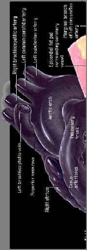
8 Evaluation of Congenital Heart Disease Segmental Approach Situs & Position Systemic & Pulmonary Venous Connections Atrial & AV Connection Ventricular morphology VA Connection Great Arteries Hypoplastic Left Heart
9 Evaluation of Ventricular Function Echocardiographic Segmental Approach LV systolic function M-mode parameters Stress-velocity index LVSF % and LVEF % LV diastolic function Mitral inflow Doppler Pulmonary venous Doppler Color flow propagation LA volume LV global function Myocardial performance index LV longitudinal function Tissue Doppler LV regional function Strain and strain rate LV torsion


10 Evaluation of Ventricular Function 3-Dimensional Echocardiography Courtesy Phillips & G Shirali
11 Evaluation of Ventricular Function New Echo Modalities Tissue Doppler Strain & strain rate imaging
12 Tissue Doppler & Strain Rate Imaging Potential ti Advantages Quantitative evaluation of myocardial motion & deformation Less geometric limitations Less impacted by loading conditions Better suited to serial evaluation Enable early identification of ventricular dysfunction Facilitate earlier and better treatment modalities
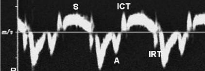
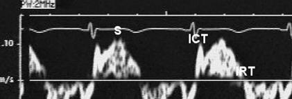
13 Tissue Doppler Imaging Systole 20 cm/s IVC Peak systole 20 cm/s IVRT E A Diastole


14 Tissue Doppler Imaging Background Longitudinal and radial shortening and lengthening Load independent Myocardial Systolic & diastolic components Heterogeneous distribution of TDI velocities Cardiac translation & tethering Orientation of myocardial fibers Systolic and diastolic velocity gradients CP


15 Pulsed Wave Tissue Doppler Longitudinal Velocities Lateral mitral annulus Septal annulus Tricuspid annulus
 Loading")

16 Tissue Doppler Imaging Assessment of LV Diastolic Function Magnitude of Ea velocity blunted with diastolic dysfunction (cath-derived tau) Loading conditions Ea less influenced by loading conditions Diastolic dysfunction Pseudonormal mitral inflow Elevated LV filling pressures Constriction vs restriction
17 Tissue Doppler in Normal Children TDI velocities impacted by cardiac growth LV end-diastolic diastolic dimension LV mass TDI velocities also impacted by Age Heart rate TDI not influenced by Gender Mitral inflow Doppler LV SF, LV EF, LV & RV MPI Eidem et al: JASE, 2004 Mit ral annula ar E-wave velocity (c cm/sec) tral annu lar E-wav ve velocity (cm/sec) Mi LVEDD (cm) Age (yr)
18 Clinical Outcome in DCM Variable OR 5-95% CI P Multivariate analysis Tricuspid Ea velocity LVEF Univariate analysis Tricuspid Ea velocity <0.001 RV Tei index LVEF < LVEF in DCM Tricuspid Ea in DCM fraction (% %) Ejection Cntl DCM P- DCM P+ Tri icuspid late eral annulus Ea velocit ty (cm/s) N NH H/I D/T PEP+ McMahon CJ: Br Heart J, 2004
 Myocardial")

19 Tissue Doppler in HCM Myocardial velocities in HCM VO 2 max (ml/min/kg) Myocardial velocities &VO 2 max y=54-2x r=-0.74 P< Septal E/Ea
20 Tissue Doppler Preclinical Diagnosis of HCM 21 Peak Systolic Septal Velocities Septal Sa (cm/s) 18 Reduced peak systolic 15 velocities in M+ with normal wall thickness Normal M + LVH - M + LVH + n=30 n=13 n=30 Nagueh SF: Circulation, 2001
21 Assessment of Ventricular Function Tissue Doppler - IVA Tissue Doppler Velocities Affected by loading Isovolumic Acceleration Begins at initial onset of LV pressure rise Earliest of systolic events Vogel M et al, Circ 2003
22 Assessment of Ventricular Function Tissue Doppler - IVA LV & RV Contractility Independent of physiologic s in preload & afterload Correlates with invasive E es and dp/dt max Correlates with s s in contractile state Force - frequency Atrial pacing Esmolol Vogel M et al, Circ 2002 & 2003


23 Tissue Doppler Detection of Allograft Rejection Baseline Sa, Ea vs controls IVA similar with rejection FS% M-mode score Sa, Ea IVA Pauliks et al: JHLT 2005 Baseline tissue Doppler Tissue Doppler with rejection


24 Tissue Doppler Limitations Angle dependency Influenced by global heart motion Cardiac translation & torsion V Myocardial motion 2 influenced by adjacent V segments (tethering) V 1 Load dependency
25 Strain Rate Imaging Elastic Deformation Properties Strain = myocardial deformation caused by fiber contraction ti I Strain = I I (L L 0 ) / L 0 0 V 1 Strain rate = rate of regional d I myocardial shortening & 0 V2 V 2 V 1 -V 2 lengthening Strain rate = d Myocardial Doppler velocities at 2 different locations spatial L/L SR = 0 v(i 0 ) - v ( L + L 0 ) = velocity gradient t L 0 Less load dependent Less variation in different (t) = t L (t + dt) - L(t) myocardial wall segments t0 L(t)
26 Strain Rate Imaging Velocities Natural Strain Rate Natural Strain r v 1 v.. v 2 Calculate spatial gradient Integrate temporally Strain (rate) estimation = velocity estimation + post processing
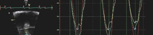
27 Strain Rate Imaging Radial Function Peak systolic strain rate Thickening Thinning SYS AVC MVO DIAST Strain rate (sec -1 ) Peak systolic strain Strain (%) Courtesy L Mertens
28 Strain Rate Imaging Longitudinal Function Shortening Lengthening Peak systolic strain rate SYS DIAST Strain rate (sec -1 ) Peak systolic strain -20 Strain (%) Courtesy L Mertens
29 Strain Imaging in Children Normal Longitudinal Deformation Systolic Strain LV Sept LV Lat LV Inf RV Free b m a b m a b m a b m a -20% -20% -40% -40% -60% -60% Weidemann: JASE, 2002
30 Case Presentation 4 month old presents to local ER Poor feeding & weight gain Tachypnea, pallor Physical exam Tachycardic, hypotensive III/VIHSMapex-- --> axilla Poor distal pulses & perfusion CXR cardiomegaly & pulmonary edema ECG Q-waves in leads I and AVL Echocardiogram performed in ER
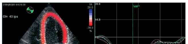
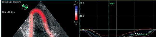
31 Echocardiogram


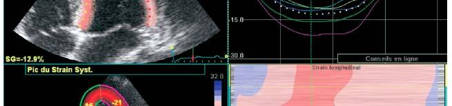
32 ALCAPA Strain Rate Imaging Clinical Outcome in CHD Causes global LV ischemia LV histological changes of hibernating myocardium Re-implantation of coronary artery results in normalization of LV function Courtesy BCH


33 ALCAPA Radial LV Function % 80 Preop 1 week postop 5 mo postop Strain 40 Mertens L, Cardiol Young 2001
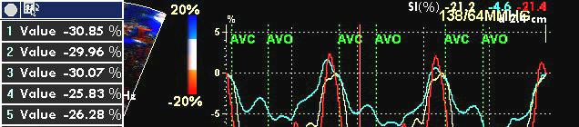
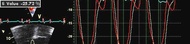
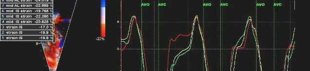
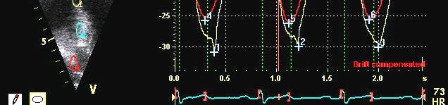
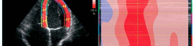
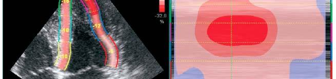
34 ALCAPA Longitudinal Strain I Normal ALCAPA II III DiSalvo G, AJC 2004 AVC Strain curve [%] AVC
35 ALCAPA Longitudinal Strain Rate Normal 2 2 I -2-2 ALCAPA II -2 2 III 2-2 AVC -2 AVC DiSalvo G: AJC, 2004
36 Strain Rate Imaging Clinical Outcome in Cardiomyopathy Duchene Muscular Dystrophy Mutation in dystrophin gene Genetic model for dilated cardiomyopathy Young patients often have normal LV function > 90% develop DCM later in life
37 Routine Echocardiographic Results DMD <12 yr Normals Age 7.4± ±2.8 Heart rate 102.2± ±13.8* LVEDD/BSA 4.3± ±0.6 LVESD/BSA 2.9± ±0.4 LV FS (%) 33.7± ±3.7 LV EF (%) 64.9± ±5.6 Mitral E 1.0± ±0.1 Mitral A 0.5± ±0.1 E/A ratio 2.1± ±0.6 Deceleration time 126.6± ±22.8 Mertens L: AEPC, 2004 *P<
38 Correlation of Strain & Age in DMD DMD 3-6 yr %) Strain ( 70 r= P<0.05 train (% %) DMD 7-11 yr Controls S Age Age Mertens L, AEPC 2004


39 Hypertrophic p Cardiomyopathy y Echocardiography
 apex mid Ao Ac Mc base 0-10 -20-30 Mid")
 Yang H,")
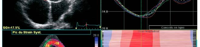
40 Strain Rate Imaging g in HCM Septal Deformation Strain (%) apex mid Ao Ac Mc base Mid septal lengthening was found in 50 % of the HCM s patients t time (ms) Yang H, JASE 2003


41 Strain in Pediatric HCM Strain vs Z score for IVSD thickness
42 LV Deformation in Pediatric HCM 0.0 SR S basal SR mid S mid SR basal S basal septum septum septum septum lateral lateral * HCM Controls * * P<0.001 vs controls; *P<0.01 vs controls Ganame: JASE, 2005
43 Hypertrophic Cardiomyopathy Strain & Strain Rate Imaging Strain in HCM Strain Family history of HCM
44 Strain & Strain Rate Imaging Limitations 1-dimensional technique Load dependent Angle dependent - a r l Frame rate >150 fps Aliasing + a Reverberation artifacts Data processing l r


45 Strain & Strain Rate Imaging Myocardial Twist & Torsion

46 Strain & Strain Rate Imaging Speckle Tracking


47 Strain & Strain Rate Imaging Myocardial Twist & Torsion


48 Strain & Strain Rate Imaging g g Myocardial Twist & Torsion

49 Strain & Strain Rate Imaging Myocardial Twist & Torsion Myocardial twist mechanics Subendocardium Right handed helix Subepicardium Left handed helix Clockwise twist of base vs CCW at cardiac apex Systole - global LV twist related to ejection + stored energy Diastole -2 phases Early rapid recoil Gradual later untwisting
50 Myocardial Deformation Imaging Age, Loading Conditions & LV Twist LV twist from childhood adulthood Apical CCW rotation ti constant t Base CW rotation changes with age Infancy CCW rotation Childhood d neutral Adolescence CW rotation Sengupta, JACC 2008
51 Hypertrophic p Cardiomyopathy y Deformation Imaging Richland et al, AJC 2007
52 Evaluation of Systemic RV Function Pressure - Volume Relationship RV Pressure - Volume Loop LV Pressure - Volume Loop Redington A, Cardiol Clin 2002
53 Evaluation of Systemic RV Function Strain & Strain Rate Systemic RV Circumferential > longitudinal RV strain Similar to normal LV strain pattern Circumferential > longitudinal SR compared to normal llvsr Petterson E, JACC 2007
54 TDI & Strain Rate Imaging in CHD Conclusions Sensitive techniques for detecting regional abnormalities New insights into pathophysiology of CHD Early identification and treatment of ventricular dysfunction Ongoing clinical validation studies
Strain/Untwisting/Diastolic Suction
 What Is Diastole and How to Assess It? Strain/Untwisting/Diastolic Suction James D. Thomas, M.D., F.A.C.C. Cardiovascular Imaging Center Department of Cardiology Cleveland Clinic Foundation Cleveland,
Velocity, strain and strain rate: Doppler and Non-Doppler methods. Thoraxcentre, Erasmus MC,Rotterdam
 Velocity, strain and strain rate: Doppler and Non-Doppler methods J Roelandt J. Roelandt Thoraxcentre, Erasmus MC,Rotterdam Basics of tissue Doppler imaging Instantaneous annular velocity profiles IVCT
Evalua&on)of)Le-)Ventricular)Diastolic) Dysfunc&on)by)Echocardiography:) Role)of)Ejec&on)Frac&on)
of)Le-)Ventricular)Diastolic) Dysfunc&on)by)Echocardiography:) Role)of)Ejec&on)Frac&on)") Evalua&on)of)Le-)Ventricular)Diastolic) Dysfunc&on)by)Echocardiography:) Role)of)Ejec&on)Frac&on) N.Koutsogiannis) Department)of)Cardiology) University)Hospital)of)Patras)! I have no conflicts of interest
Evalua&on)of)Le-)Ventricular)Diastolic) Dysfunc&on)by)Echocardiography:) Role)of)Ejec&on)Frac&on) N.Koutsogiannis) Department)of)Cardiology) University)Hospital)of)Patras)! I have no conflicts of interest
Diastology Disclosures: None. Dias2011:1
 Diastology 2011 James D. Thomas, M.D., F.A.C.C. Cardiovascular Imaging Center Department of Cardiology Cleveland Clinic Foundation Cleveland, Ohio, USA Disclosures: None Dias2011:1 Is EVERYBODY a member!?!
Strain imaging in children: from Tissue Doppler to 3 D
 Strain imaging in children: from Tissue Doppler to 3 D Mark kk. Friedberg Fi Outline Deformation in the fetus and neonate Deformation in pediatric cardiomyopathy y (briefly!) Deformation in Congenital
Strain and Strain Rate Imaging How, Why and When?
 Strain and Strain Rate Imaging How, Why and When? João L. Cavalcante, MD Advanced Cardiac Imaging Fellow Cleveland Clinic Foundation Disclosures: No conflicts of interest Movement vs Deformation Movement
LV FUNCTION ASSESSMENT: WHAT IS BEYOND EJECTION FRACTION
 LV FUNCTION ASSESSMENT: WHAT IS BEYOND EJECTION FRACTION Jamilah S AlRahimi Assistant Professor, KSU-HS Consultant Noninvasive Cardiology KFCC, MNGHA-WR Introduction LV function assessment in Heart Failure:
Tissue Doppler Imaging in Congenital Heart Disease
 Tissue Doppler Imaging in Congenital Heart Disease L. Youngmin Eun, M.D. Department of Pediatrics, Division of Pediatric Cardiology, Kwandong University College of Medicine The potential advantage of ultrasound
Nancy Goldman Cutler, MD Beaumont Children s Hospital Royal Oak, Mi
 Nancy Goldman Cutler, MD Beaumont Children s Hospital Royal Oak, Mi Identify increased LV wall thickness (WT) Understand increased WT in athletes Understand hypertrophic cardiomyopathy (HCM) Enhance understanding
How does the heart pump? From sarcomere to ejection volume
 How does the heart pump? From sarcomere to ejection volume Piet Claus Cardiovascular Imaging and Dynamics Department of Cardiovascular Diseases University Leuven, Leuven, Belgium Course on deformation
DISCLOSURE. Myocardial Mechanics. Relevant Financial Relationship(s) Off Label Usage
 Off Label Usage") 7th Annual Team Echocardiography: The Heart of Cardiovascular Medicine Tissue Doppler, Strain, Speckle: What? How? Christopher J Kramer RDCS Aurora Medical Group Advanced Cardiovascular Services, Aurora
7th Annual Team Echocardiography: The Heart of Cardiovascular Medicine Tissue Doppler, Strain, Speckle: What? How? Christopher J Kramer RDCS Aurora Medical Group Advanced Cardiovascular Services, Aurora
Quantitation of right ventricular dimensions and function
 SCCS Basics of cardiac assessment Quantitation of right ventricular dimensions and function Tomasz Kukulski, MD PhD Dept of Cardiology, Congenital Heart Disease and Electrotherapy Silesian Medical University
Diastole is Not a Single Entity Four Components of Diastolic Dysfunction
 Physiology of Diastolic Function Made Easy James D. Thomas, MD, FACC, FASE Director, Center for Heart Valve Disease Bluhm Cardiovascular Institute Professor of Medicine, Feinberg School of Medicine, Northwestern
10/7/2013. Systolic Function How to Measure, How Accurate is Echo, Role of Contrast. Thanks to our Course Director: Neil J.
 Systolic Function How to Measure, How Accurate is Echo, Role of Contrast Neil J. Weissman, MD MedStar Health Research Institute & Professor of Medicine Georgetown University Washington, D.C. No Disclosures
Tissue Doppler and Strain Imaging. Steven J. Lester MD, FRCP(C), FACC, FASE
, FACC, FASE") Tissue Doppler and Strain Imaging Steven J. Lester MD, FRCP(C), FACC, FASE Relevant Financial Relationship(s) None Off Label Usage None a. Turn the wall filters on and turn down the receiver gain. b. Turn
Tissue Doppler and Strain Imaging Steven J. Lester MD, FRCP(C), FACC, FASE Relevant Financial Relationship(s) None Off Label Usage None a. Turn the wall filters on and turn down the receiver gain. b. Turn
An Integrated Approach to Study LV Diastolic Function
 An Integrated Approach to Study LV Diastolic Function Assoc. Prof. Adriana Ilieşiu, FESC University of Medicine Carol Davila Bucharest, Romania LV Diastolic Dysfunction impaired relaxation (early diastole)
Imaging in Heart Failure: A Multimodality Approach. Thomas Ryan, MD
 Imaging in Heart Failure: A Multimodality Approach Thomas Ryan, MD Heart Failure HFrEF HFpEF EF50% Lifetime risk 20% Prevalence 6M Americans Societal costs - $30B 50% 5-year survival 1 Systolic
2/2/2011. Strain and Strain Rate Imaging How, Why and When? Movement vs Deformation. Doppler Myocardial Velocities. Movement. Deformation.
 Strain and Strain Rate Imaging How, Why and When? João L. Cavalcante, MD Advanced Cardiac Imaging Fellow Cleveland Clinic Foundation Disclosures: No conflicts of interest Movement vs Deformation Movement
Altered left ventricular geometry and torsional mechanics in high altitude-induced pulmonary hypertension:
 Altered left ventricular geometry and torsional mechanics in high altitude-induced pulmonary hypertension: a 3-D echocardiographic study B.W. De Boeck,* S. Kiencke, C. Dehnert, K. Auinger, # M. Maggiorini,
Basic Approach to the Echocardiographic Evaluation of Ventricular Diastolic Function
 Basic Approach to the Echocardiographic Evaluation of Ventricular Diastolic Function J A F E R A L I, M D U N I V E R S I T Y H O S P I T A L S C A S E M E D I C A L C E N T E R S T A F F C A R D I O T
Highlights from EuroEcho 2009 Echo in cardiomyopathies
 Highlights from EuroEcho 2009 Echo in cardiomyopathies Bogdan A. Popescu University of Medicine and Pharmacy, Bucharest, Romania ESC Congress 2010 Hypertrophic cardiomyopathy To determine the differences
RIGHT VENTRICULAR SIZE AND FUNCTION
 RIGHT VENTRICULAR SIZE AND FUNCTION Edwin S. Tucay, MD, FPCC, FPCC, FPSE Philippine Society of Echocardiography Quezon City, Philippines Echo Mission, BRTTH, Legaspi City, July 1-2, 2016 NO DISCLOSURE
Restrictive Cardiomyopathy
 ESC Congress 2011, Paris Imaging Unusual Causes of Cardiomyopathy Restrictive Cardiomyopathy Kazuaki Tanabe, MD, PhD Professor of Medicine Chair, Division of Cardiology Izumo, Japan I Have No Disclosures
LV geometric and functional changes in VHD: How to assess? Mi-Seung Shin M.D., Ph.D. Gachon University Gil Hospital
 LV geometric and functional changes in VHD: How to assess? Mi-Seung Shin M.D., Ph.D. Gachon University Gil Hospital LV inflow across MV LV LV outflow across AV LV LV geometric changes Pressure overload
Alicia Armour, MA, BS, RDCS
 Alicia Armour, MA, BS, RDCS No disclosures Review 2D Speckle Strain (briefly) Discuss some various patient populations & disease pathways where Strain can be helpful Discuss how to acquire images for Strain
The importance of left atrium in LV diastolic function
 II Baltic Heart Failure Meeting and Congress of Latvian Society of Cardiology The importance of left atrium in LV diastolic function Dr. Artem Kalinin Eastern Clinical University Hospital Riga 30.09.2010.
Evaluation of Left Ventricular Diastolic Dysfunction by Doppler and 2D Speckle-tracking Imaging in Patients with Primary Pulmonary Hypertension
 ESC Congress 2011.No 85975 Evaluation of Left Ventricular Diastolic Dysfunction by Doppler and 2D Speckle-tracking Imaging in Patients with Primary Pulmonary Hypertension Second Department of Internal
Echo assessment of the failing heart
 Echo assessment of the failing heart Mark K. Friedberg, MD The Labatt Family Heart Center The Hospital for Sick Children Toronto, Ontario, Canada Cardiac function- definitions Cardiovascular function:
Tissue Doppler and Strain Imaging
 Tissue Doppler and Strain Imaging Steven J. Lester MD, FRCP(C), FACC, FASE Relevant Financial Relationship(s) None Off Label Usage None 1 Objective way with which to quantify the minor amplitude and temporal
Novel echocardiographic modalities: 3D echo, speckle tracking and strain rate imaging. Potential roles in sports cardiology. Stefano Caselli, MD, PhD
 Novel echocardiographic modalities: 3D echo, speckle tracking and strain rate imaging. Potential roles in sports cardiology. Stefano Caselli, MD, PhD Ospedale San Pietro Fatebenefratelli Rome, Italy Differential
Myocardial Strain Imaging in Cardiac Diseases and Cardiomyopathies.
 Myocardial Strain Imaging in Cardiac Diseases and Cardiomyopathies. Session: Cardiomyopathy Tarun Pandey MD, FRCR. Associate Professor University of Arkansas for Medical Sciences Disclosures No relevant
Diastolic Function Assessment Practical Ways to Incorporate into Every Echo
 Diastolic Function Assessment Practical Ways to Incorporate into Every Echo Jae K. Oh, MD Echo Hawaii 2018 2018 MFMER 3712003-1 Learning Objectives My presentation will help you to Appreciate the importance
The Patient with Atrial Fibrilation
 Assessment of Diastolic Function The Patient with Atrial Fibrilation Assoc. Prof. Adriana Ilieşiu, FESC University of Medicine Carol Davila Bucharest, Romania Associated Conditions with Atrial Fibrillation
Heart Failure in Women: Dr Goh Ping Ping Cardiologist Asian Heart & Vascular Centre
 Heart Failure in Women: More than EF? Dr Goh Ping Ping Cardiologist Asian Heart & Vascular Centre Overview Review pathophysiology as it relates to diagnosis and management Rational approach to workup:
Left ventricular diastolic function and filling pressure in patients with dilated cardiomyopathy
 Left ventricular diastolic function and filling pressure in patients with dilated cardiomyopathy Bogdan A. Popescu University of Medicine and Pharmacy Bucharest, Romania My conflicts of interest: I have
Mechanisms of heart failure with normal EF Arterial stiffness and ventricular-arterial coupling. What is the pathophysiology at presentation?
 Mechanisms of heart failure with normal EF Arterial stiffness and ventricular-arterial coupling What is the pathophysiology at presentation? Ventricular-arterial coupling elastance Central arterial pressure
Imaging to Measure Cardiac Contractility: Current and Future. Safety Pharmacology Society 2012 Jon Heyen on behalf of Bob Coatney
 Imaging to Measure Cardiac Contractility: Current and Future Safety Pharmacology Society 2012 Jon Heyen on behalf of Bob Coatney Background / Context / Scope How do we bridge? Contractility = Force generated
VECTORS OF CONTRACTION
 1/3/216 Strain, Strain Rate, and Torsion: Myocardial Mechanics Simplified and Applied VECTORS OF CONTRACTION John Gorcsan, MD University of Pittsburgh, Pittsburgh, PA Shortening Thickening Twisting No
Tissue Doppler and Strain Imaging
 Tissue Doppler and Strain Imaging Steven J. Lester MD, FRCP(C), FACC, FASE Relevant Financial Relationship(s) None Off Label Usage None 1 Objective way with which to quantify the minor amplitude and temporal
Little is known about the degree and time course of
 Differential Changes in Regional Right Ventricular Function Before and After a Bilateral Lung Transplantation: An Ultrasonic Strain and Strain Rate Study Virginija Dambrauskaite, MD, Lieven Herbots, MD,
Review Article Transthoracic Echocardiography in Children and Young Adults with Congenital Heart Disease
 International Scholarly Research Network ISRN Pediatrics Volume 2012, Article ID 753481, 15 pages doi:10.5402/2012/753481 Review Article Transthoracic Echocardiography in Children and Young Adults with
Index. K Knobology, TTE artifact, image resolution, ultrasound, 14
 A Acute aortic regurgitation (AR), 124 128 Acute aortic syndrome (AAS) classic aortic dissection diagnosis, 251 263 evolutive patterns, 253 255 pathology, 250 251 classifications, 247 248 incomplete aortic
Diastolic Function: What the Sonographer Needs to Know. Echocardiographic Assessment of Diastolic Function: Basic Concepts 2/8/2012
 Diastolic Function: What the Sonographer Needs to Know Pat Bailey, RDCS, FASE Technical Director Beaumont Health System Echocardiographic Assessment of Diastolic Function: Basic Concepts Practical Hints
How To Perform Strain Imaging; Step By Step Approach. Maryam Bo Khamseen Echotechnoligist II EACVI, ARDMS, RCS King Abdulaziz Cardiac Center- Riyadh
 How To Perform Strain Imaging; Step By Step Approach Maryam Bo Khamseen Echotechnoligist II EACVI, ARDMS, RCS King Abdulaziz Cardiac Center- Riyadh Outlines: Introduction Describe the basic of myocardium
Incorporating the New Echo Guidelines Into Everyday Practice
 Incorporating the New Echo Guidelines Into Everyday Practice Clinical Case RIGHT VENTRICULAR FAILURE Gustavo Restrepo MD President Elect Interamerican Society of Cardiology Director Fellowship Training
Appendix II: ECHOCARDIOGRAPHY ANALYSIS
 Appendix II: ECHOCARDIOGRAPHY ANALYSIS Two-Dimensional (2D) imaging was performed using the Vivid 7 Advantage cardiovascular ultrasound system (GE Medical Systems, Milwaukee) with a frame rate of 400 frames
Echocardiographic assessment of the right ventricle in paediatric pulmonary hypertension.
 Echocardiographic assessment of the right ventricle in paediatric pulmonary hypertension. Mark K. Friedberg, MD No disclosures Outline RV response to increased afterload Echo assessment of RV function
Hypertensive heart disease and failure
 Hypertensive heart disease and failure Prof. Dr. Alan Fraser Cardiff University The heart in hypertension Pathophysiology of LV adaptation Regional development of hypertrophy Stress testing - inducible
좌심실수축기능평가 Cardiac Function
 Basic Echo Review Course 좌심실수축기능평가 Cardiac Function Seonghoon Choi Cardiology Hallym university LV systolic function Systolic function 좌심실수축기능 - 심근의수축으로심실에서혈액을대동맥으로박출하는기능 실제임상에서 LV function 의의미 1Diagnosis
Diastology State of The Art Assessment
 Diastology State of The Art Assessment Dr. Mohammad AlGhamdi Assistant professor, KSAU-HS Consultant Cardiologist King AbdulAziz Cardiac Center Ministry of National Guard Health Affairs Diagnostic Clinical
Aortic valve Stenosis: Insights in the evaluation of LV function. Erwan DONAL Cardiologie CHU Rennes
 Aortic valve Stenosis: Insights in the evaluation of LV function Erwan DONAL Cardiologie CHU Rennes erwan.donal@chu-rennes.fr Preload Afterload Myocardial Fiber Shortening Circumferential Longitudinal
DECLARATION OF CONFLICT OF INTEREST. None
 DECLARATION OF CONFLICT OF INTEREST None Hot Topics in Echocardiography: The position of the EAE EAE / ASE recommendation about Echo Assessment of Cardiac Mechanics Jens-Uwe Voigt Dpt. of Cardiovascular
Echo-Doppler evaluation of left ventricular diastolic function. Michel Slama Amiens France
 Echo-Doppler evaluation of left ventricular diastolic function Michel Slama Amiens France Left ventricular pressure Pressure A wave [ LVEDP LVEDP préa Congestive cardiac failure with preserved systolic
We are IntechOpen, the world s leading publisher of Open Access books Built by scientists, for scientists. International authors and editors
 We are IntechOpen, the world s leading publisher of Open Access books Built by scientists, for scientists 4,100 116,000 120M Open access books available International authors and editors Downloads Our
Three-dimensional Wall Motion Tracking:
 Three-dimensional Wall Motion Tracking: A Novel Echocardiographic Method for the Assessment of Ventricular Volumes, Strain and Dyssynchrony Jeffrey C. Hill, BS, RDCS, FASE Jennifer L. Kane, RCS Gerard
Echocardiographic Evaluation of the Cardiomyopathies. Stephanie Coulter, MD, FACC, FASE April, 2016
 Echocardiographic Evaluation of the Cardiomyopathies Stephanie Coulter, MD, FACC, FASE April, 2016 Cardiomyopathies (CMP) primary disease intrinsic to cardiac muscle Dilated CMP Hypertrophic CMP Infiltrative
L ecocardiografia nello Scompenso Cardiaco Acuto e cronico: vecchi dogmi e nuovi trends.
 V SESSIONE SCOMPENSO CARDIACO 2015 Genova, 13-14 Novembre 2015 L ecocardiografia nello Scompenso Cardiaco Acuto e cronico: vecchi dogmi e nuovi trends. Gian Paolo Bezante, MD, FACC UOC Clinica di Malattie
Acute impairment of basal left ventricular rotation but not twist and untwist are involved in the pathogenesis of acute hypertensive pulmonary oedema
 Acute impairment of basal left ventricular rotation but not twist and untwist are involved in the pathogenesis of acute hypertensive pulmonary oedema A.D. Margulescu 1,2, R.C. Sisu 1,2, M. Florescu 2,
How NOT to miss Hypertrophic Cardiomyopathy? Adaya Weissler-Snir, MD University Health Network, University of Toronto
 How NOT to miss Hypertrophic Cardiomyopathy? Adaya Weissler-Snir, MD University Health Network, University of Toronto Introduction Hypertrophic cardiomyopathy is the most common genetic cardiomyopathy,
Evaluation of the Right Ventricle and Risk Stratification for Sudden Cardiac Death
 Evaluation of the Right Ventricle and Risk Stratification for Sudden Cardiac Death Presenters: Sabrina Phillips, MD FACC FASE Director, Adult Congenital Heart Disease Services The University of Oklahoma
Index of subjects. effect on ventricular tachycardia 30 treatment with 101, 116 boosterpump 80 Brockenbrough phenomenon 55, 125
 145 Index of subjects A accessory pathways 3 amiodarone 4, 5, 6, 23, 30, 97, 102 angina pectoris 4, 24, 1l0, 137, 139, 140 angulation, of cavity 73, 74 aorta aortic flow velocity 2 aortic insufficiency
Tissue Doppler Imaging
 Cronicon OPEN ACCESS Hesham Rashid* Tissue Doppler Imaging CARDIOLOGY Editorial Department of Cardiology, Benha University, Egypt *Corresponding Author: Hesham Rashid, Department of Cardiology, Benha University,
Global left ventricular circumferential strain is a marker for both systolic and diastolic myocardial function
 Global left ventricular circumferential strain is a marker for both systolic and diastolic myocardial function Toshinari Onishi 1, Samir K. Saha 2, Daniel Ludwig 1, Erik B. Schelbert 1, David Schwartzman
DOPPLER HEMODYNAMICS (1) QUANTIFICATION OF PRESSURE GRADIENTS and INTRACARDIAC PRESSURES
 QUANTIFICATION OF PRESSURE GRADIENTS and INTRACARDIAC PRESSURES") THORAXCENTRE DOPPLER HEMODYNAMICS (1) QUANTIFICATION OF PRESSURE GRADIENTS and INTRACARDIAC PRESSURES J. Roelandt DOPPLER HEMODYNAMICS Intracardiac pressures and pressure gradients Volumetric measurement
THORAXCENTRE DOPPLER HEMODYNAMICS (1) QUANTIFICATION OF PRESSURE GRADIENTS and INTRACARDIAC PRESSURES J. Roelandt DOPPLER HEMODYNAMICS Intracardiac pressures and pressure gradients Volumetric measurement
HYPERTROPHY: Behind the curtain. V. Yotova St. Radboud Medical University Center, Nijmegen
 HYPERTROPHY: Behind the curtain V. Yotova St. Radboud Medical University Center, Nijmegen Disclosure of interest: none Relative wall thickness (cm) M 0.22 0.42 0.43 0.47 0.48 0.52 0.53 F 0.24 0.42 0.43
Strain Imaging: Myocardial Mechanics Simplified and Applied
 9/28/217 Strain Imaging: Myocardial Mechanics Simplified and Applied John Gorcsan III, MD Professor of Medicine Director of Clinical Research Division of Cardiology VECTORS OF CONTRACTION Shortening Thickening
Early Regional Myocardial Dysfunction in Young Patients With Duchenne Muscular Dystrophy
 Early Regional Myocardial Dysfunction in Young Patients With Duchenne Muscular Dystrophy Luc Mertens, MD, PhD, Javier Ganame, MD, PhD, Piet Claus, PhD, Nathalie Goemans, MD, Daisy Thijs, MSc, Bénédicte
GENERAL PRINCIPLES FOR ECHO ASSESSMENT OF DIASTOLIC FUNCTION (For full recommendation refer to the Left Ventricular Diastolic Function Guideline)
") 1 THE AMERICAN SOCIETY OF ECHOCARDIOGRAPHY RECOMMENDATIONS FOR THE EVALUATION OF LEFT VENTRICULAR DIASTOLIC FUNCTION BY ECHOCARDIOGRAPHY: A QUICK REFERENCE GUIDE FROM THE ASE WORKFLOW AND LAB MANAGEMENT
1 THE AMERICAN SOCIETY OF ECHOCARDIOGRAPHY RECOMMENDATIONS FOR THE EVALUATION OF LEFT VENTRICULAR DIASTOLIC FUNCTION BY ECHOCARDIOGRAPHY: A QUICK REFERENCE GUIDE FROM THE ASE WORKFLOW AND LAB MANAGEMENT
Diastolic Function Assessment New Guideline Update Practical Approach
 Mayo Clinic Department of Cardiovascular Diseases Mayo Clinic Echocardiography Review Course for Boards and Recertification Diastolic Function Assessment New Guideline Update Practical Approach Jae K.
Adel Hasanin Ahmed 1
 Adel Hasanin Ahmed 1 PERICARDIAL DISEASE The pericardial effusion ends anteriorly to the descending aorta and is best visualised in the PLAX. PSAX is actually very useful sometimes for looking at posterior
Athlete s Heart vs. Cardiomyopathy
 Athlete s Heart vs. Cardiomyopathy Linda D. Gillam, MD, MPH, FASE Chair, Department of Cardiovascular Medicine Medical Director, Cardiovascular Service Line Former Team Cardiologist to the New York Jets
3D-stress echocardiography Bernard Cosyns, MD, PhD
 3D-stress echocardiography Bernard Cosyns, MD, PhD No Disclosure The Pro-Technology bias Sicari et al. Cardiovascular Ultrasound 2006, 4:11 Overview 2D stress echocardiography: main limitations 3D echocardiography:
Mechanisms of False Positive Exercise Electrocardiography: Is False Positive Test Truly False?
 Mechanisms of False Positive Exercise Electrocardiography: Is False Positive Test Truly False? Masaki Izumo a, Kengo Suzuki b, Hidekazu Kikuchi b, Seisyo Kou b, Keisuke Kida b, Yu Eguchi b, Nobuyuki Azuma
Value of echocardiography in chronic dyspnea
 Value of echocardiography in chronic dyspnea Jahrestagung Schweizerische Gesellschaft für /Schweizerische Gesellschaft für Pneumologie B. Kaufmann 16.06.2016 Chronic dyspnea Shortness of breath lasting
Stephen Glen ISCHAEMIC HEART DISEASE AND LEFT VENTRICULAR FUNCTION
 Stephen Glen ISCHAEMIC HEART DISEASE AND LEFT VENTRICULAR FUNCTION Overview Coronary arteries Terminology to describe contractility Measuring ventricular function Systolic dysfunction Practice cases- LV
Chamber Quantitation Guidelines - Update II
 Chamber Quantitation Guidelines - Update II Right Heart Measurements Steven A. Goldstein MD FACC FASE Professor of Medicine Georgetown University Medical Center MedStar Heart Institute Washington Hospital
Rotation: Echocardiography: Transthoracic Echocardiography (TTE)
") Rotation: Echocardiography: Transthoracic Echocardiography (TTE) Rotation Format and Responsibilities: Fellows rotate in the echocardiography laboratory in each clinical year. Rotations during the first
Rotation: Echocardiography: Transthoracic Echocardiography (TTE) Rotation Format and Responsibilities: Fellows rotate in the echocardiography laboratory in each clinical year. Rotations during the first
MYOCARDIAL DEFORMATION IMAGING ON EXERCISE IN CHRONIC PRIMARY MITRAL REGURGITATION
 MYOCARDIAL DEFORMATION IMAGING ON EXERCISE IN CHRONIC PRIMARY MITRAL REGURGITATION A thesis submitted to the University of Manchester for the degree of Doctor of Medicine in the Faculty of Medical and
Diastolic Heart Function: Applying the New Guidelines Case Studies
 Diastolic Heart Function: Applying the New Guidelines Case Studies Mitral Regurgitation The New ASE William Guidelines: A. Zoghbi Role MD, of FASE, 2D/3D MACCand CMR Professor and Chairman, Department
Ιπποκράτειες μέρες καρδιολογίας Θεσσαλονίκη, 9-10 Μαρτίου Φωτεινή Α. Λαζαρίδου Επιμελήτρια Α Γενικό Νοσοκομείο Αγιος Παύλος, Θεσσαλονίκη
 Ιπποκράτειες μέρες καρδιολογίας Θεσσαλονίκη, 9-10 Μαρτίου 2018 Φωτεινή Α. Λαζαρίδου Επιμελήτρια Α Γενικό Νοσοκομείο Αγιος Παύλος, Θεσσαλονίκη RV shape Triangular shape in frontal plane crescent shape in
OPTIMIZING ECHO ACQUISTION FOR STRAIN AND DIASTOLOGY
 OPTIMIZING ECHO ACQUISTION FOR STRAIN AND DIASTOLOGY October 8, 2017 Deborah Agler, ACS, RDCS, FASE Coordinator of Education and Training Cleveland Clinic General Principles Diastology Clinical Data Heart
Advanced Multi-Layer Speckle Strain Permits Transmural Myocardial Function Analysis in Health and Disease:
 Advanced Multi-Layer Speckle Strain Permits Transmural Myocardial Function Analysis in Health and Disease: Clinical Case Examples Jeffrey C. Hill, BS, RDCS Echocardiography Laboratory, University of Massachusetts
Ventricular-Pulmonary Vascular Coupling after the Total Cavopulmonary Anastomosis (Fontan)
") Ventricular-Pulmonary Vascular Coupling after the Total Cavopulmonary Anastomosis (Fontan) 6th International Neonatal & Childhood Pulmonary Vascular Disease Conference Fontan Physiology Single ventricular
Ventricular-Pulmonary Vascular Coupling after the Total Cavopulmonary Anastomosis (Fontan) 6th International Neonatal & Childhood Pulmonary Vascular Disease Conference Fontan Physiology Single ventricular
Assessment of LV systolic function
 Tutorial 5 - Assessment of LV systolic function Assessment of LV systolic function A knowledge of the LV systolic function is crucial in the undertanding of and management of unstable hemodynamics or a
Advanced imaging of the left atrium - strain, CT, 3D, MRI -
 Advanced imaging of the left atrium - strain, CT, 3D, MRI - Monica Rosca, MD Carol Davila University of Medicine and Pharmacy, Bucharest, Romania Declaration of interest: I have nothing to declare Case
Pericardial Disease: Case Examples. Echo Fiesta 2017
 Pericardial Disease: Case Examples Echo Fiesta 2017 2014 2014 MFMER MFMER 3346252-1 slide-1 Objectives Have a systematic approach to evaluation of constriction 2014 MFMER 3346252-2 CASE 1 2013 MFMER 3248567-3
Assessing Function by Echocardiography in VHD Asymptomatic Severe Organic MR. Dr. Julien Magne, PhD Sart Tilman Liège, BELGIUM
 Assessing Function by Echocardiography in VHD Asymptomatic Severe Organic MR Dr. Julien Magne, PhD Sart Tilman Liège, BELGIUM Conflict of Interest Disclosure None Why to assess LV function in asymptomatic
How to Assess Diastolic Dysfunction?
 How to Assess Diastolic Dysfunction? Fausto J Pinto, MD, PhD, FESC, FACC, FASE Lisbon University Dyastolic Dysfunction Impaired relaxation Elevated filling pressures Ischemic heart disease Cardiomyopathies
The road to successful CRT implantation: The role of echo
 The road to successful CRT implantation: The role of echo Tae-Ho Park Dong-A University Hospital, Busan, Korea Terminology Cardiac Resynchronization Therapy (CRT) = Biventricular pacing (BiV) = Left ventricular
Ref 1. Ref 2. Ref 3. Ref 4. See graph
 Ref 1 Ref 2 Ref 3 1. Ages 6-23 y/o 2. Significant LVM differences by gender 3. For males 95 th percentiles: a. LVM/BSA = 103 b. LVM/height = 100 4. For females 95 th percentiles: a. LVM/BSA = 84 b. LVM/height
E/Ea is NOT an essential estimator of LV filling pressures
 Euroecho Kopenhagen Echo in Resynchronization in 2010 E/Ea is NOT an essential estimator of LV filling pressures Wilfried Mullens, MD, PhD December 10, 2010 Ziekenhuis Oost Limburg Genk University Hasselt
Cardiac Chamber Quantification by Echocardiography
 Cardiac Chamber Quantification by Echocardiography Maryam Bokhamseen, RCS, RCDS, EACVI Echotechnologist ǁ, Non invasive Cardiac Laboratory King Abdulaziz Cardiac Center. Outline: Introduction. Background
Echocardiographic and Doppler Assessment of Cardiac Functions in Patients of Non-Insulin Dependent Diabetes Mellitus
 ORIGINAL ARTICLE JIACM 2002; 3(2): 164-8 Echocardiographic and Doppler Assessment of Cardiac Functions in Patients of Non-Insulin Dependent Diabetes Mellitus Rajesh Rajput*, Jagdish**, SB Siwach***, A
Evaluation of Left Ventricular Function and Hypertrophy Gerard P. Aurigemma MD
 Evaluation of Left Ventricular Function and Hypertrophy Gerard P. Aurigemma MD Board Review Course 2017 43 year old health assistant Severe resistant HTN LT BSA 2 Height 64 1 Here is the M mode echocardiogram
Dr. Dermot Phelan MB BCh BAO PhD European Society of Cardiology 2012
 Relative Apical Sparing of Longitudinal Strain Using 2- Dimensional Speckle-Tracking Echocardiography is Both Sensitive and Specific for the Diagnosis of Cardiac Amyloidosis. Dr. Dermot Phelan MB BCh BAO
Effect of loading and geometry on functional parameters
 Effect of loading and geometry on functional parameters Piet Claus Cardiovascular Imaging and Dynamics Department of Cardiovascular Diseases Leuven University, Leuven, Belgium 5 th European Echocardiography
The Athlete s Heart. Critical Role of Echo. Neil J. Weissman, MD MedStar Health Research Institute & Professor of Medicine Georgetown University
 The Athlete s Heart Critical Role of Echo Neil J. Weissman, MD MedStar Health Research Institute & Professor of Medicine Georgetown University Washington, D.C. Disclosures Grant support (to institution)
ASCeXAM / ReASCE. Practice Board Exam Questions Monday Morning
 ASCeXAM / ReASCE Practice Board Exam Questions Monday Morning Ultrasound Physics Artifacts Doppler Physics Imaging, Knobology, and Artifacts Echocardiographic Evaluation of the RV Tricuspid and Pulmonary
Objectives. Diastology: What the Radiologist Needs to Know. LV Diastolic Function: Introduction. LV Diastolic Function: Introduction
 Objectives Diastology: What the Radiologist Needs to Know. Jacobo Kirsch, MD Cardiopulmonary Imaging, Section Head Division of Radiology Cleveland Clinic Florida Weston, FL To review the physiology and
PRESENTER DISCLOSURE INFORMATION. There are no potential conflicts of interest regarding current presentation
 PRESENTER DISCLOSURE INFORMATION There are no potential conflicts of interest regarding current presentation Better synchrony and diastolic function for septal versus apical right ventricular permanent