MSRS 6473 Vascular Noninvasive Imaging Procedures
|
|
|
- Ross Jefferson
- 5 years ago
- Views:
Transcription
1 MSRS 6473 Vascular Noninvasive Imaging Procedures Rex T. Christensen MHA RT (R) (MR) (CT) (ARRT) CIIP Basic Physics Equipment Cardiac Positioning Perfusion Pathology MRI 1

2 Animal Magnetism MRI Basic Physics 2


3 Hydrogen Atom Has the simplest atomic structure. Contains one proton and one electron. The nucleus of a hydrogen atom contains only a proton. Therefore, hydrogen atoms are often referred as protons Abundant in the body. Water Molecule H 2 O Hydrogen in the body is found predominately in three forms: 1. Free water in body fluids (CSF) 2. Water bound to large molecules (proteins) 3. Hydrogen atoms within fat. 3
 If two of these characteristics are present it induces the third magnetism Called a magnetic moment Precession")
4 MR Active Nuclei (Hydrogen) Angular momentum or spin. Posses an electrical charge (+) If two of these characteristics are present it induces the third magnetism Called a magnetic moment Precession Precession is the wobble of a top Precessional Frequency is the speed it wobbles. (Mhz) 4

5 Larmor Frequency: The Larmor frequency is also called the Precessional frequency. Precession is a wobble of the hydrogen atom. Hydrogen Atoms before being placed in the MRI: 5


6 Hydrogen Atoms after being placed in the MRI: Hydrogen and Field Strength As field strength increases the number of Hydrogen atoms becomes more visible. 6


7 Resonance: When an RF pulse is turned on and its frequency matches that of the frequency of the hydrogen atom energy is transferred. This tips the hydrogen atoms down into the transverse plane (90 degrees). Resonance Precessional frequency of hydrogen is MHz/T Precessional frequency of hydrogen: 1.0T = MHz 1.5T = MHz 3.0T = MHz 7
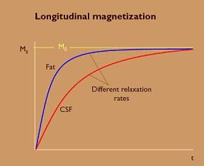
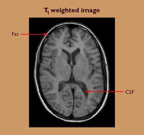
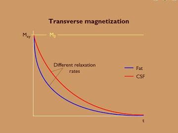
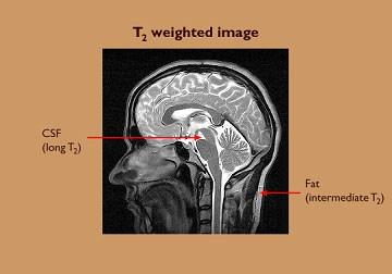
8 T1 Relaxation: T2 Relaxation: 8
")

9 Longitudinal (T1) and Transverse (T2) Magnetization (relaxation) Relaxation times for different tissues: 9

10 MRI Equipment 10
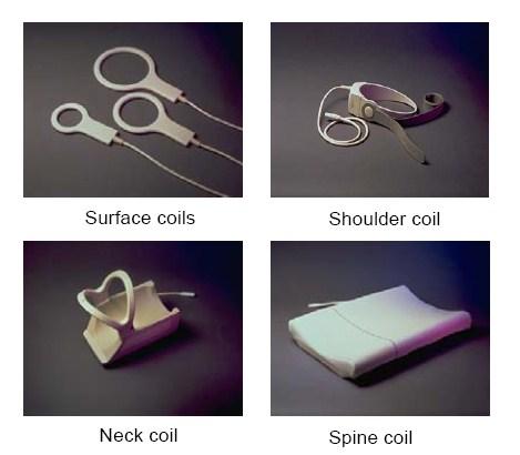
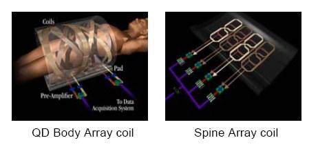
11 Coils 11


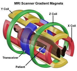
12 Slice selecting gradients. Spatial Localization A combination of these gradients form an oblique slice Axial Sagittal Coronal Imaging Planes 12


13 Magnet Safety As you approach the magnet, the fringe magnetic field gets STRONGER 25 Room Safety MR Safe MR Conditional MR Unsafe 13


14 MRI Safety Bad Day! MRI Safety Ouch! EKG patch burn Pulse Oximeter wire burn 14
 and mitral valve (MV) Also called vertical long axis view LA MV LAA")
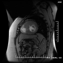
15 MRI Cardiac Positioning Two-Chamber View Center parallel to long axis of left ventricle Always center from breathhold (BH) transverse The resultant image will demonstrate the left ventricle, left atrium, left atrial appendage (LAA) and mitral valve (MV) Also called vertical long axis view LA MV LAA LV 15

16 Two-Chamber View Aliased Two-Chamber View The field of view (FOV) that was selected was too small for body habitus; therefore fold over or wrap has degraded this image Wrap 16
 image The resultant image will")

17 Four-Chamber View Center parallel to the twochamber view Center in between the papillary muscles on short axis (SA) image The resultant image will demonstrate the right and left atrium, the right and left ventricle, and MV and tricuspid valve Also known as horizontal long axis RA LA TV MV RV LV Four Chamber View 17
18 Malpositioned Four-Chamber View This image was positioned too superior on the twochamber view The resultant image has the aortic valve (AV) in the view making it a fivechamber image Aortic root Malpositioned Four-Chamber View 18

 RV LV Short Axis with")

19 Short Axis Center perpendicular to the septum on the four-chamber view The resultant image demonstrates the left and right ventricle in a nice round shape (donuts) RV LV Short Axis with Motion Artifact Impaired image quality because patient had difficulty holding their breath Motion artifact 19


20 Left Ventricular Outflow Tract of Aorta Center perpendicular to the aorta The resultant image demonstrates the left ventricle, AV and outflow tract Also known as coronal view RA LVOT & AV RV LV PA Left Ventricular Outflow Tract of Aorta 20

 image Flow artifact Flow")
21 Flow Artifact Turbulent flow in the aorta causing a flow artifact on left ventricular outflow tract (LVOT) image Flow artifact Flow Artifact 21


22 Para-Axial Aorta Center parallel to the AV using the LVOT image The resultant image demonstrates the cusps of the AV Para-Axial Aorta 22


23 Three-Chamber View Center parallel to the long axis of the left ventricle and aorta This image RV demonstrates the AV, AV MV, and right and left ventricle LA MV LV Parasagittal Aorta Center parallel to the ascending and descending aorta The resultant image shows the aorta in a candy cane view Descending aorta RPA Ascending aorta Descending aorta Ascending aorta 23
24 Parasagittal Aorta MRI Challenges Claustrophobia Motion Cardiac Triggering (arrhythmias) Coil selection Pacemakers Gradient strength (faster imaging) Coronary Arteries/Stents 24

25 MRI Perfusion Heart 25
26 Why MRI Perfusion? Resolution < 2mm No radionuclide Visualize subendocardial defects Morphology and of the heart Comparisons Nuclear(including PET) MRI Spatial Resolution cm mm Voxel resolution 3 5 cm 8 10 mm SNR 6 20 CNR 8 >100 Difference in resolution PET, position emission tomography CNR, Contrast to noise ratio SNR, signal to noise ratio 26
 (2D)")


.")

27 Magnetic Resonance Imaging Hyperenhancement Images Post Percutaneous Transluminal Coronary Angiography (PTCA) (2D) Post (6 weeks) 41 YO M with 1-mm ST Elevation Myocardinal Infarction 1 Week Ago and Negative Nuclear Scan Yesterday A very thin lateral wall subendocardial infarction below the limits for detection by standard nuclear imaging well visualized by the high spatial resolution afforded by cardiovascular magnetic resonance (CMR). Note, the high CMR resolution to demonstrate the dual supply of the posterior lateral papillary muscle suggested by variable scar (middle arrow). 27


28 MRI Pathology Hypertrophic Cardiomyopathy 28


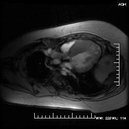
29 Right Ventricle Perforation: 85 YO Un Life threatening rupture on CMR CT 29

 = Distance the table travels per rotation")
30 Multi Detector CT (MDCT) Pitch Pitch combines the table distance traveled per 360- degree rotation with the slice thickness. Pitch (P) = Distance the table travels per rotation (d)/total collimation (W) 30
 lowers heart rate Cardiac Gating Contrast Contraindications")
31 Protocols Coronary Angiography Factors to Consider Timing of the contrast bolus Number of beats per slice IV Metropolol (beta blocker) lowers heart rate Cardiac Gating Contrast Contraindications 31

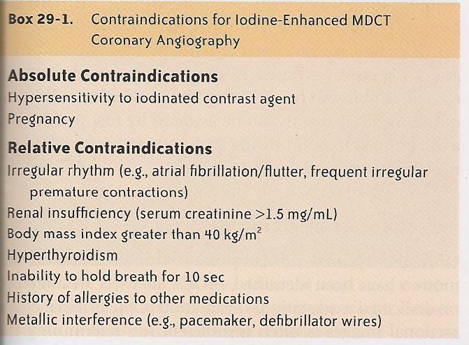
32 Cardiac Gating CT Contraindications for Iodine 32
33 Contrast Protocols Iodine Concentration (300 to 400 mg iodine/ml) Volume and Rate of contrast injection (80 ml 140 ml) (3 6 ml per second) Timing of the delivery (bolus tracking HU) Coronary Artery Nomenclature LMCA Left Main Coronary Artery (10 15mm) Bifurcates LAD and CX Trifurcates LAD, CX, and IR LAD Left Anterior Descending. Anterior 2/3 of septum, lateral edge of lt. ventricular wall CX Circumflex Artery. Lateral and Posterior walls of the lt. ventricle IR Intermediate Ramus 33

34 Coronary Artery Nomenclature RCA Right Coronary Artery. Lateral wall of rt. Ventricle AM Acute Marginal Branch PD Posterior Descending Artery PV Posterior Left ventricular Artery DG Diagonal Branch. A branch off one of the main arteries. Normal Coronary Anatomy Aortic or Valsalva Sinus. Located just above the Aortic Valve. RCS Right Coronary Sinus LCS Left Coronary Sinus NCS Non-Coronary Sinus PA Pulmonary Artery 34


35 Aortic Valve Right Coronary Artery Anomalies 35


36 Right Coronary Artery Anomaly RCA originates off the LMCA Left Main Coronary Artery Anomalies 36

37 Left Main Coronary Anomaly Absence of Left Main Coronary Artery LCMA absent in 0.41% of patients Left Main Coronary Anomaly LMCA originates off the Right Coronary Sinus 37


38 LAD Coronary Artery Anomalies Left Circumflex Coronary Artery Anomalies 38

39 Left CX Anomaly CX originating off the RCA Normal LAD 39

40 Normal RCA Myocardial Perfusion Normal Myocardium enhances homogeneously in both rest and stress scans 40
41 Myocardial Perfusion Myocardial Perfusion Defect Ischemia myocardial defect presents at stress, disappears at rest. Necrosis myocardial defect presents at stress and at rest. Myocardial Perfusion Intramural perfusion defect is placed in the inner layers of myocardium Transmural perfusion defect is located in the entire myocardial wall Subendocardial perfusion defect is placed in the subendocardial myocardium. Usually this is not well identified by SPECT studies 41


42 Myocardial Perfusion Myocardial Viability Negative viability the contrast material accumulates in nonviable tissue, showing a typical hyperenhancement Positive viability The density on the hypoattenuated myocardial segment is homogeneous in comparison with normal segments CT Perfusion Normal Stress Rest and Stress 42

43 Cardiac Scoring Cardiac Scoring 43
44 Agatston Score Based on Hounsfield Units (HU) 1 = HU 2 = HU 3 = HU 4 = 400 or greater HU Agatston Score Summaries all the HU s from all of the slices 1 10 = Minimal = Mild = Moderate 400 or more = Severe 44


45 CCTA and IVUS method IVUS miniaturized transducer is introduced during coronary catheterization 45
46 Attributes Cross-platform for Myocardial Infarction Imaging SPECT, single photon emission computed tomography; ECHO, echocardiography; MRI, magnetic resonance imaging Characteristic SPECT ECHO MRI Spatial resolution Sensitivity Specificity Quantitation Speed Cost Cost-effectiveness Platform availability Claustrophobia Proven in MI User-independent Reproducibility Subendocardial imaging Variability Viability References: Westbrook, Catherine and Kaut, Carolyn (1998). MRI In Practice, 3rd ed., Blackwell Science, Inc. Malden, Ma. Biederman, Robert et al. (2008). Cardiovascular MRI Tutorial: Lectures and Learning. Lippincott Williams & Wilkins. Philadelphia, Pa. Haaga, John et al. (2009). CT and MRI of the Whole Body. Mosby Elsevier. Philadelphia, Pa. 46
Index. radiologic.theclinics.com. Note: Page numbers of article titles are in boldface type.
 Index Note: Page numbers of article titles are in boldface type. A ALCAPA. See Anomalous left coronary artery from the pulmonary artery. Angiosarcoma computed tomographic assessment of, 809 811 Anomalous
MR Advance Techniques. Cardiac Imaging. Class IV
 MR Advance Techniques Cardiac Imaging Class IV Heart The heart is a muscular organ responsible for pumping blood through the blood vessels by repeated, rhythmic contractions. Layers of the heart Endocardium
cardiac imaging planes planning basic cardiac & aortic views for MR
 cardiac imaging planes planning basic cardiac & aortic views for MR Dianna M. E. Bardo, M. D. Assistant Professor of Radiology & Cardiovascular Medicine Director of Cardiac Imaging cardiac imaging planes
CARDIAC AND CORONARY ARTERY ANATOMY NO DISCLOSURES. Axial Anatomy of Heart. Axial Anatomy of Heart. Axial Anatomy of Heart
 CARDIAC AND CORONARY ARTERY ANATOMY NO DISCLOSURES NASCI MEETING, ORLANDO FLORIDA 2009 KOSTAKI G. BIS, MD, FACR DEPARTMENT OF RADIOLOGY WILLIAM BEAUMONT HOSPITAL Royal Oak, Michigan OBJECTIVES CARDIAC
Radiologic Assessment of Myocardial Viability
 November 2001 Radiologic Assessment of Myocardial Viability Joshua Moss, Harvard Medical School Year III Patient EF 66yo female with a 3-year history of intermittent chest pain previously relieved by sublingual
Case 47 Clinical Presentation
 93 Case 47 C Clinical Presentation 45-year-old man presents with chest pain and new onset of a murmur. Echocardiography shows severe aortic insufficiency. 94 RadCases Cardiac Imaging Imaging Findings C
2019 Qualified Clinical Data Registry (QCDR) Performance Measures
 Performance Measures") 2019 Qualified Clinical Data Registry (QCDR) Performance Measures Description: This document contains the 18 performance measures approved by CMS for inclusion in the 2019 Qualified Clinical Data Registry
2019 Qualified Clinical Data Registry (QCDR) Performance Measures Description: This document contains the 18 performance measures approved by CMS for inclusion in the 2019 Qualified Clinical Data Registry
CARDIAC MRI. Cardiovascular Disease. Cardiovascular Disease. Cardiovascular Disease. Overview
 CARDIAC MRI Dr Yang Faridah A. Aziz Department of Biomedical Imaging University of Malaya Medical Centre Cardiovascular Disease Diseases of the circulatory system, also called cardiovascular disease (CVD),
MR Advance Techniques. Vascular Imaging. Class II
 MR Advance Techniques Vascular Imaging Class II 1 Vascular Imaging There are several methods that can be used to evaluate the cardiovascular systems with the use of MRI. MRI will aloud to evaluate morphology
General Cardiovascular Magnetic Resonance Imaging
 2 General Cardiovascular Magnetic Resonance Imaging 19 Peter G. Danias, Cardiovascular MRI: 150 Multiple-Choice Questions and Answers Humana Press 2008 20 Cardiovascular MRI: 150 Multiple-Choice Questions
Imaging congestive heart failure: role of coronary computed tomography angiography (CCTA)
") Imaging congestive heart failure: role of coronary computed tomography angiography (CCTA) Gianluca Pontone, MD, PhD, FESC, FSCCT Director of MR Unit Deputy Director of Cardiovascul CT Unit Clinical Cardiology
Imaging congestive heart failure: role of coronary computed tomography angiography (CCTA) Gianluca Pontone, MD, PhD, FESC, FSCCT Director of MR Unit Deputy Director of Cardiovascul CT Unit Clinical Cardiology
Can SCMR CMR protocol recommendations
 Can SCMR CMR protocol recommendations V1.3 - April 2009 CanSCMR CMR Protocol and SOP Recommendation 2009 (15 minutes) 2 Planning of LV fct. real time multiple axes Realtime 3 cine long axis 6 long axes
Cardiac MR -Complimentary -Competitor -Conqueror?
 Cardiac MR -Complimentary -Competitor -Conqueror? Dr Girish Dwivedi MRCP (UK), PhD (UK), FASE Staff Cardiologist, Assistant Professor in Medicine University of Ottawa Heart Institute University of Ottawa,
Dr Felix Keng. Imaging of the heart is technically difficult because: Role of Cardiac MSCT. Current: Cardiac Motion Respiratory Motion
 Siemens Philips Dr Felix Keng GE Toshiba Role of Cardiac MSCT Current: Structural / congenital heart imaging Extra-cardiac / Great vessel imaging Volumes and ejection fractions (cine + gating) Calcium
Blood supply of the Heart & Conduction System. Dr. Nabil Khouri
 Blood supply of the Heart & Conduction System Dr. Nabil Khouri Arterial supply of Heart Right coronary artery Left coronary artery 3 Introduction: Coronary arteries - VASAVASORUM arising from aortic sinuses
Cardiac Imaging Tests
 Cardiac Imaging Tests http://www.medpagetoday.com/upload/2010/11/15/23347.jpg Standard imaging tests include echocardiography, chest x-ray, CT, MRI, and various radionuclide techniques. Standard CT and
The Value of Stress MRI in Evaluation of Myocardial Ischemia
 The Value of Stress MRI in Evaluation of Myocardial Ischemia Dr. Saeed Al Sayari, MBBS, EBCR, MBA Department of Radiology and Nuclear Medicine Mafraq Hospital, Abu Dhabi United Arab Emirates Introduction
Why Cardiac MRI? Presented by:
 Why Cardiac MRI? Presented by: Lisa G. Carkner, MD, FACC 1 Disclosures I have no financial disclosures Objectives Review basic principles of Cardiac MRI. What patient characteristics do I need to consider
Introduction. Cardiac Imaging Modalities MRI. Overview. MRI (Continued) MRI (Continued) Arnaud Bistoquet 12/19/03
 MRI (Continued) Arnaud Bistoquet 12/19/03") Introduction Cardiac Imaging Modalities Arnaud Bistoquet 12/19/03 Coronary heart disease: the vessels that supply oxygen-carrying blood to the heart, become narrowed and unable to carry a normal amount
Introduction Cardiac Imaging Modalities Arnaud Bistoquet 12/19/03 Coronary heart disease: the vessels that supply oxygen-carrying blood to the heart, become narrowed and unable to carry a normal amount
Cardiac Computed Tomography
 Cardiac Computed Tomography Authored and approved by Koen Nieman Stephan Achenbach Francesca Pugliese Bernard Cosyns Patrizio Lancellotti Anastasia Kitsiou Contents CARDIAC COMPUTED TOMOGRAPHY Page 1.
Use of Nuclear Cardiology in Myocardial Viability Assessment and Introduction to PET and PET/CT for Advanced Users
 Use of Nuclear Cardiology in Myocardial Viability Assessment and Introduction to PET and PET/CT for Advanced Users February 1 5, 2011 University of Santo Tomas Hospital Angelo King A-V Auditorium Manila,
Cardiac MRI: Cardiomyopathy
 Cardiac MRI: Cardiomyopathy Laura E. Heyneman, MD I do not have any relevant financial relationships with any commercial interests Cardiac MRI: Cardiomyopathy Laura E. Heyneman, MD Duke University Medical
Objectives 8/17/2011. Challenges in Cardiac Imaging. Challenges in Cardiac Imaging. Basic Cardiac MRI Sequences
 8/17/2011 Traditional Protocol Model for Tomographic Imaging Cardiac MRI Sequences and Protocols Frandics Chan, M.D., Ph.D. Stanford University Medical Center Interpretation Lucile Packard Children s Hospital
12 Lead ECG Interpretation
 12 Lead ECG Interpretation Julie Zimmerman, MSN, RN, CNS, CCRN Significant increase in mortality for every 15 minutes of delay! N Engl J Med 2007;357:1631-1638 Who should get a 12-lead ECG? Also include
Syllabus References. Resources. Video: MRI Introduction
 MRI Lesson Outline Syllabus References 9.6.4.2.5 Define precessing and relate the frequency of the precessing to the composition of the nuclei and the strength of the applied external magnetic field 9.6.4.2.6
Magnetic Resonance Angiography
 Magnetic Resonance Angiography 1 Magnetic Resonance Angiography exploits flow enhancement of GR sequences saturation of venous flow allows arterial visualization saturation of arterial flow allows venous
THE ROLE OF HIGH END MULTI DETECTOR CT IN CORONARY IMAGING ESSAY
 THE ROLE OF HIGH END MULTI DETECTOR CT IN CORONARY IMAGING ESSAY Submitted for partial fulfillment of Master degree in Radiodiagnosis By Ahmed Yehia Ahmed (M.B.B.Ch., Cairo University) Supervisors Prof.
I have no financial disclosures
 Manpreet Singh MD I have no financial disclosures Exercise Treadmill Bicycle Functional capacity assessment Well validated prognostic value Ischemic assessment ECG changes ST segments Arrhythmias Hemodynamic
Multiple Gated Acquisition (MUGA) Scanning
 Scanning") Multiple Gated Acquisition (MUGA) Scanning Dmitry Beyder MPA, CNMT Nuclear Medicine, Radiology Barnes-Jewish Hospital / Washington University St. Louis, MO Disclaimers/Relationships Standard of care research
Multiple Gated Acquisition (MUGA) Scanning Dmitry Beyder MPA, CNMT Nuclear Medicine, Radiology Barnes-Jewish Hospital / Washington University St. Louis, MO Disclaimers/Relationships Standard of care research
AP2 Lab 3 Coronary Vessels, Valves, Sounds, and Dissection
 AP2 Lab 3 Coronary Vessels, Valves, Sounds, and Dissection Project 1 - BLOOD Supply to the Myocardium (Figs. 18.5 &18.10) The myocardium is not nourished by the blood while it is being pumped through the
Noncoronary Cardiac MDCT
 Noncoronary Cardiac MDCT David A. Bluemke, M.D., Ph.D. Professor, of Radiology and Medicine Johns Hopkins University School of Medicine Baltimore, Maryland Toshiba Disclosures Grant support Noncoronary
Mapping and Ablation of Challenging Outflow Tract VTs: Pulmonary Artery, LVOT, Epicardial
 Mapping and Ablation of Challenging Outflow Tract VTs: Pulmonary Artery, LVOT, Epicardial Samuel J. Asirvatham, MD Mayo Clinic Rochester California Heart Rhythm Symposium San Francisco, CA September 8,
Cardiac ultrasound protocols
 Cardiac ultrasound protocols IDEXX Telemedicine Consultants Two-dimensional and M-mode imaging planes Right parasternal long axis four chamber Obtained from the right side Displays the relative proportions
PRACTICAL GUIDE TO FETAL ECHOCARDIOGRAPHY IC Huggon and LD Allan
 PRACTICAL GUIDE TO FETAL ECHOCARDIOGRAPHY IC Huggon and LD Allan Fetal Cardiology Unit, Harris Birthright Research Centre for Fetal Medicine, King's College Hospital, London, UK IMPORTANCE OF PRENATAL
Cardiac Imaging in abnormal rhythm Role of MDCT
 Cardiac Imaging in abnormal rhythm Role of MDCT Cardiac Imaging in abnormal rhythm Role of MDCT Scope of the problem CT in Atrial Fibrillation CT and pacing Ventricular arrhythmia Other applications 1
Cardiac MRI: Clinical Application to Disease
 Cardiac MRI: Clinical Application to Disease Jessi Smith, MD Cardiothoracic imaging, Indiana University Slides courtesy of Stacy Rissing, MD Outline Imaging planes Disease findings Pulse sequences used
Multiplane Magnetic Resonance Imaging of the Heart and Major Vessels:
 661 Charles B. Higgins1 David Stark Michael McNamara Peter Lanzer Lawrence E. Crooks Leon Kaufman Received October 25, 1983; accepted after revision January 5, 1984. This work was supported in part by
Cardiac MRI in ACHD What We. ACHD Patients
 Cardiac MRI in ACHD What We Have Learned to Apply to ACHD Patients Faris Al Mousily, MBChB, FAAC, FACC Consultant, Pediatric Cardiology, KFSH&RC/Jeddah Adjunct Faculty, Division of Pediatric Cardiology
New Cardiovascular Devices and Interventions: Non-Contrast MRI for TAVR Abhishek Chaturvedi Assistant Professor. Cardiothoracic Radiology
 New Cardiovascular Devices and Interventions: Non-Contrast MRI for TAVR Abhishek Chaturvedi Assistant Professor Cardiothoracic Radiology Disclosure I have no disclosure pertinent to this presentation.
ULTRASOUND OF THE FETAL HEART
 ULTRASOUND OF THE FETAL HEART Cameron A. Manbeian, MD Disclosure Statement Today s faculty: Cameron Manbeian, MD does not have any relevant financial relationships with commercial interests or affiliations
Lab Activity 23. Cardiac Anatomy. Portland Community College BI 232
 Lab Activity 23 Cardiac Anatomy Portland Community College BI 232 Cardiac Muscle Histology Branching cells Intercalated disc: contains many gap junctions connecting the adjacent cell cytoplasm, creates
PROSTHETIC VALVE BOARD REVIEW
 PROSTHETIC VALVE BOARD REVIEW The correct answer D This two chamber view shows a porcine mitral prosthesis with the typical appearance of the struts although the leaflets are not well seen. The valve
Coronary Artery Anomalies from Birth to Adulthood; the Role of CT Coronary Angiography in Sudden Cardiac Death Screening
 Coronary Artery Anomalies from Birth to Adulthood; the Role of CT Coronary Angiography in Sudden Cardiac Death Screening E O Dwyer 1, C O Brien 1, B Loo 1, A Snow Hogan 1, O Buckley1 2, B 1. Department
Normal TTE/TEE Examinations
 Normal TTE/TEE Examinations Geoffrey A. Rose, MD FACC FASE Sanger Heart & Vascular Institute Before you begin imaging... Obtain the patient s Height Weight BP PLAX View PLAX View Is apex @ 9-10 o clock?
Non Contrast MRA. Mayil Krishnam. Director, Cardiovascular and Thoracic Imaging University of California, Irvine
 Non Contrast MRA Mayil Krishnam Director, Cardiovascular and Thoracic Imaging University of California, Irvine No disclosures Non contrast MRA-Why? Limitations of CTA Radiation exposure Iodinated contrast
Detection and Assessment of MI: Use of Imaging Methods. Robert O. Bonow, M.D.
 Detection and Assessment of MI: Use of Imaging Methods Robert O. Bonow, M.D. Detection and Assessment of MI: Use of Imaging Methods Robert O. Bonow, M.D. No Relationships to Disclose Expert Consensus Document
ADVANCED CARDIOVASCULAR IMAGING. Medical Knowledge. Goals and Objectives PF EF MF LF Aspirational
 Medical Knowledge Goals and Objectives PF EF MF LF Aspirational Know the basic principles of magnetic resonance imaging (MRI) including the role of the magnetic fields and gradient coil systems, generation
Your heart is a muscular pump about the size of your fist, located
 How Your Heart Works Your heart is a muscular pump about the size of your fist, located slightly to the left and behind your breastbone. Its function is to pump blood throughout your body. As your heart
the Cardiovascular System I
 the Cardiovascular System I By: Dr. Nabil A Khouri MD, MsC, Ph.D MEDIASTINUM 1. Superior Mediastinum 2. inferior Mediastinum Anterior mediastinum. Middle mediastinum. Posterior mediastinum Anatomy of
Breakout Session: Transesophageal Echocardiography
 Breakout Session: Transesophageal Echocardiography Doris Ockert, MD Andrew Schroeder, MD University of Wisconsin School of Medicine and Public Health Jutta Novalija, MD, PhD Medical College of Wisconsin
Giovanni Di Salvo MD, PhD, FESC Second University of Naples Monaldi Hospital
 Giovanni Di Salvo MD, PhD, FESC Second University of Naples Monaldi Hospital VSD is one of the most common congenital cardiac abnormalities in the newborn. It can occur as an isolated finding or in combination
Gated blood pool ventriculography: Is there still a role in myocardial viability?
 Gated blood pool ventriculography: Is there still a role in myocardial viability? Oliver C. Alix, MD Adult Clinical and Nuclear Cardiology St. Luke s Medical Centre - Global City Case Presentation A 62-year-old
Cardiac MRI: Clinical Application to Disease
 Cardiac MRI: Clinical Application to Disease Stacy Rissing, MD! Cardiothoracic imaging, Indiana University! Outline Imaging planes Disease findings Pulse sequences used for each indication Pathophysiology
Cardiac Imaging. Kimberly Delcour, DO, FACC. Mahi Ashwath, MD, FACC, FASE. Director, Cardiac CT. Director, Cardiac MRI
 Cardiac Imaging Kimberly Delcour, DO, FACC Director, Cardiac CT Mahi Ashwath, MD, FACC, FASE Director, Cardiac MRI Cardiac Imaging Discuss the clinical applications of and indications for: Cardiac CT Nuclear
, David Stultz, MD. Cardiac CT. David Stultz, MD Cardiology Fellow, PGY 6 March 28, 2006
 Cardiac CT David Stultz, MD Cardiology Fellow, PGY 6 March 28, 2006 Courtesy Tom Kracus Courtesy Kettering Tom Medical Kracus Cente Kettering Medical Center 2003-2006, David Stultz, MD Courtesy Tom Kracus
Coronary Anomalies & Hemodynamic Identification
 Coronary Anomalies & Hemodynamic Identification David Stultz, MD Cardiology Fellow, PGY 6 May 2, 2006 Anomaly #1 Anomaly #2 Anomaly #3 Figure 18-27 Anomalous origin of the left circumflex artery.
The Normal Echocardiogram
 The Normal Echocardiogram Pravin V. Patil, MD FACC Lewis Katz School of Medicine at Temple University Acknowledgments Dr. Susan Wiegers Dr. Martin Keane Temple Cardiac Sonographers Disclosures No relevant
Adult Echocardiography Examination Content Outline
 Adult Echocardiography Examination Content Outline (Outline Summary) # Domain Subdomain Percentage 1 2 3 4 5 Anatomy and Physiology Pathology Clinical Care and Safety Measurement Techniques, Maneuvers,
The Heart. Happy Friday! #takeoutyournotes #testnotgradedyet
 The Heart Happy Friday! #takeoutyournotes #testnotgradedyet Introduction Cardiovascular system distributes blood Pump (heart) Distribution areas (capillaries) Heart has 4 compartments 2 receive blood (atria)
Fulfilling the Promise
 Fulfilling the Promise of Cardiac MR Non-contrast, free-breathing technique generates comprehensive evaluation of the coronary arteries By Maggie Fung, MR Cardiovascular Clinical Development Manager; Wei
Low-dose prospective ECG-triggering dual-source CT angiography in infants and children with complex congenital heart disease: first experience
 Low-dose prospective ECG-triggering dual-source CT angiography in infants and children with complex congenital heart disease: first experience Ximing Wang, M.D., Zhaoping Cheng, M.D., Dawei Wu, M.D., Lebin
MRI ACS-ben. Tamás Simor MD, PhD, Med Hab. University of Pécs, Heart Institute
 MRI ACS-ben Tamás Simor MD, PhD, Med Hab Time Course of Changes in Infarct Size, Viable Myocardium, and LV Mass After Reperfused and Nonreperfused MI Blue lines denote reperfused myocardial infarction
Advanced Imaging MRI and CTA
 Advanced Imaging MRI and CTA Who and why may benefit. Matthew W. Martinez, M.D. FACC Lehigh Valley Health Network Director, Cardiovascular Imaging Learning Objectives Review basics of CMR and CTA Review
Current Indications for Cardiac MRI: What You See is What You Get?
 Current Indications for Cardiac MRI: What You See is What You Get? Javier Ganame, MD, PhD, FASE No disclosures Cardiology Update, Niagara, Sept 24th, 2016 The Ideal Diagnostic Technique Easy to apply Accurate
Index of subjects. effect on ventricular tachycardia 30 treatment with 101, 116 boosterpump 80 Brockenbrough phenomenon 55, 125
 145 Index of subjects A accessory pathways 3 amiodarone 4, 5, 6, 23, 30, 97, 102 angina pectoris 4, 24, 1l0, 137, 139, 140 angulation, of cavity 73, 74 aorta aortic flow velocity 2 aortic insufficiency
CORONARY ANATOMY WITH MULTIDETECTOR COMPUTED TOMOGRAPHY ANGIOGRAPHY*
 Updating CORONRY NTOMY WITH MULTIDETECTOR COMPUTED TOMOGRPHY NGIOGRPHY* Joalbo Matos ndrade 1 * Study developed at Instituto do Coração, Fundação Zerbini, rasília, DF, razil. 1. MD, Radiologist at Departament
MR Assessment of Myocardial Viability
 MR Assessment of Myocardial Viability Definition of Viability Clinical Metabolism: Presence of glucose uptake Perfusion / Perfusion reserve Morphology: Wall thickness, wall thickening Contractility: Recovery
Sung A Chang Department of Internal Medicine, Division of Cardiology, Sungkyunkwan University School of Medicine, Samsung Medical Center
 CMR Perfusion and Viability A STICH Out of Time? Sung A Chang Department of Internal Medicine, Division of Cardiology, Sungkyunkwan University School of Medicine, Samsung Medical Center Can Imaging Improve
Managing Hypertrophic Cardiomyopathy with Imaging. Gisela C. Mueller University of Michigan Department of Radiology
 Managing Hypertrophic Cardiomyopathy with Imaging Gisela C. Mueller University of Michigan Department of Radiology Disclosures Gadolinium contrast material for cardiac MRI Acronyms Afib CAD Atrial fibrillation
IAEA. Department of Technical Cooperation. And. Nuclear Medicine Section RAS 6/063
 IAEA Department of Technical Cooperation And Nuclear Medicine Section RAS 6/063 Strengthening the Application of Nuclear Medicine in the Management of Cardiovascular Diseases Cardiac Imaging CT and MR
Case 1. Case 2. Case 3
 Case 1 The correct answer is D. Occasionally, the Brugada syndrome can present similar morphologies to A and also change depending on the lead position but in the Brugada pattern the r is wider and ST
Cardiac CT - Coronary Calcium Basics Workshop II (Basic)
") Cardiac CT - Coronary Calcium Basics Workshop II (Basic) J. Jeffrey Carr, MD, MSCE Dept. of Radiology & Public Health Sciences Wake Forest University School of Medicine Winston-Salem, NC USA No significant
Cardiac CT - Coronary Calcium Basics Workshop II (Basic) J. Jeffrey Carr, MD, MSCE Dept. of Radiology & Public Health Sciences Wake Forest University School of Medicine Winston-Salem, NC USA No significant
Imaging Guide Echocardiography
 Imaging Guide Guide to Small Animal Echocardiography using the Vevo Imaging Systems System Compatibility: This guide contains instructions and suggestions for work on the Vevo2100, VevoLAZR, Vevo 3100
Fellows on this rotation are expected to attend nuclear conferences and multimodality imaging conference.
 Rotation: Imaging 1 Imaging 1 provides COCATS Level 1 experience for nuclear cardiology (including SPECT and PET) and cardiac CT. Fellows will administer, process, and read cardiac nuclear studies with
Chapter 20 (1) The Heart
 The Heart") Chapter 20 (1) The Heart Learning Objectives Describe the location and structure of the heart Describe the path of a drop of blood from the superior vena cava or inferior vena cava through the heart out
Chapter 20 (1) The Heart Learning Objectives Describe the location and structure of the heart Describe the path of a drop of blood from the superior vena cava or inferior vena cava through the heart out
Certificate in Clinician Performed Ultrasound (CCPU) Syllabus. Rapid Cardiac Echo (RCE)
 Syllabus. Rapid Cardiac Echo (RCE)") Certificate in Clinician Performed Ultrasound (CCPU) Syllabus Rapid Cardiac Echo (RCE) Purpose: Rapid Cardiac Echocardiography (RCE) This unit is designed to cover the theoretical and practical curriculum
Certificate in Clinician Performed Ultrasound (CCPU) Syllabus Rapid Cardiac Echo (RCE) Purpose: Rapid Cardiac Echocardiography (RCE) This unit is designed to cover the theoretical and practical curriculum
MRI protocol for post-repaired TOF
 2012 NASCI MRI protocol for post-repaired TOF Taylor Chung, M.D. Associate Director, Body and Cardiovascular Imaging Department of Diagnostic Imaging Children s Hospital & Research Center Oakland Oakland,
Diagnostic Imaging
 www.fisiokinesiterapia.biz Diagnostic Imaging Diagnostic Imaging is no longer limited to radiography. Major technological advancements have lead to the use of new and improved imaging technologies. The
Myocardial viability testing. What we knew and what is new
 Myocardial viability testing. What we knew and what is new Dr B K S Sastry, MD, DM. CARE Hospitals, Hyderabad What is Viability Viability Dysfunctional myocardium subtended by diseased coronary arteries
Lab 16. The Cardiovascular System Heart and Blood Vessels. Laboratory Objectives
 Lab 16 The Cardiovascular System Heart and Blood Vessels Laboratory Objectives Describe the anatomical structures of the heart to include the pericardium, chambers, valves, and major vessels. Describe
Imaging of Coronary Artery Disease: II
 Acta Radiológica Portuguesa, Vol.XIX, nº 74, pág. 45-51, Abr.-Jun., 2007 Imaging of Coronary Artery Disease: II Jean Jeudy University of Maryland School of Medicine Department of Diagnostic Radiology Armed
This lab activity is aligned with Visible Body s A&P app. Learn more at visiblebody.com/professors
 1 This lab activity is aligned with Visible Body s A&P app. Learn more at visiblebody.com/professors 2 PRE-LAB EXERCISES: A. Watch the video 29.1 Heart Overview and make the following observations: 1.
Multimodality Imaging of Anomalous Left Coronary Artery from the Pulmonary
 1 IMAGES IN CARDIOVASCULAR ULTRASOUND 2 3 4 Multimodality Imaging of Anomalous Left Coronary Artery from the Pulmonary Artery 5 6 7 Byung Gyu Kim, MD 1, Sung Woo Cho, MD 1, Dae Hyun Hwang, MD 2 and Jong
Achieva 1.5T scanner devoted to congenital heart disease
 I s s u e 3 1 - M a rc h 2 0 0 7 F i e l d Strength Publication for the Philips MRI Community Achieva 1.5T scanner devoted to congenital heart disease Drs. Geva, Powell integrate MRI into Cardiovascular
Anatomy of left ventricular outflow tract'
 Anatomy of left ventricular outflow tract' ROBERT WALMSLEY British Heart Journal, 1979, 41, 263-267 From the Department of Anatomy and Experimental Pathology, The University, St Andrews, Scotland SUMMARY
Case # 1. Page: 8. DUKE: Adams
 Case # 1 Page: 8 1. The cardiac output in this patient is reduced because of: O a) tamponade physiology O b) restrictive physiology O c) coronary artery disease O d) left bundle branch block Page: 8 1.
syngo MR D13 Operator Manual - Cardio syngo MR D
 Siemens Healthcare Sector Cs2 Informatik, syngo Operator Cardio 06/2012 English n.a. 02 01 06 630 MR-05015 2010-2012 MR MRAG, Manual D13 Cape syngo MR D13 Operator Manual - Cardio syngo MR D13 www.siemens.com/healthcare
Abnormal, Autoquant Adenosine Myocardial Perfusion Heart Imaging. ID: GOLD Date: Age: 46 Sex: M John Doe Phone (310)
") Background: Reason: preoperative assessment of CAD, Shortness of Breath Symptom: atypical chest pain Risk factors: hypertension Under influence: a beta blocker Medications: digoxin Height: 66 in. Weight:
Background: Reason: preoperative assessment of CAD, Shortness of Breath Symptom: atypical chest pain Risk factors: hypertension Under influence: a beta blocker Medications: digoxin Height: 66 in. Weight:
Improvement of Image Quality with ß-Blocker Premedication on ECG-Gated 16-MDCT Coronary Angiography
 16-MDCT Coronary Angiography Shim et al. 16-MDCT Coronary Angiography Sung Shine Shim 1 Yookyung Kim Soo Mee Lim Received December 1, 2003; accepted after revision June 1, 2004. 1 All authors: Department
The use of Cardiac CT and MRI in Clinical Practice
 The use of Cardiac CT and MRI in Clinical Practice Matthew W. Martinez, MD Assistant Professor of Medicine LVPG - Lehigh Valley Heart Specialists Lehigh Valley Health Network Oct. 3, 2009 DISCLOSURE Relevant
MRI Sequences: What to use for what
 MRI Sequences: What to use for what MRI basics T 1 and T 2 relaxation Common Imaging Protocols Mechanical function (cine) Tissue characterization LGE Edema imaging (T 2 weighted) T1 Special protocols MRA
Disclosures. GETTING TO THE HEART OF THE MATTER WITH MULTIMODALITY CARDIAC IMAGING Organ Review Meeting 25 September. Overview
 GETTING TO THE HEART OF THE MATTER WITH MULTIMODALITY CARDIAC IMAGING Organ Review Meeting 25 September Disclosures None relevant to this presentation Mini Pakkal Assistant Professor of Radiology University
SPECT-CT: Τι πρέπει να γνωρίζει ο Καρδιολόγος
 SPECT-CT: Τι πρέπει να γνωρίζει ο Καρδιολόγος Δρ Αναστασία Κίτσιου Διευθύντρια, Καρδιολογική Κλινική, Σισμανόγλειο ΓΝΑ Chair, Education Committee, Section on Nuclear Cardiology & Cardiac CT, EACVI, ESC
J. Schwitter, MD, FESC Section of Cardiology
 J. Schwitter, MD, FESC Section of Cardiology CMR Center of the CHUV University Hospital Lausanne - CHUV Switzerland Centre de RM Cardiaque J. Schwitter, MD, FESC Section of Cardiology CMR Center of the
Matters of the Heart: Comprehensive Cardiology SARAH BEANLANDS RN BSCN MSC
 Matters of the Heart: Comprehensive Cardiology SARAH BEANLANDS RN BSCN MSC Who am I? Class Outline Gross anatomy of the heart Trip around the heart Micro anatomy: cellular and tissue level Introduction
ARTIFACTS: THEORY AND ILLUSTRATIVE EXAMPLES
 ARTIFACTS: THEORY AND ILLUSTRATIVE EXAMPLES Robert A. Levine, M.D. Marielle Scherrer-Crosbie, M.D. Eric M. Isselbacher, M.D. No conflicts of interest Philippe Bertrand, Pieter Vendervoort, Hasselt and
9/8/2009 < 1 1,2 3,4 5,6 7,8 9,10 11,12 13,14 15,16 17,18 > 18. Tetralogy of Fallot. Complex Congenital Heart Disease.
 Current Indications for Pediatric CTA S Bruce Greenberg Professor of Radiology Arkansas Children s Hospital University of Arkansas for Medical Sciences greenbergsbruce@uams.edu 45 40 35 30 25 20 15 10
Cardiac CT Angiography
 Cardiac CT Angiography Dr James Chafey, Radiologist Why do we need a better test for C.A.D? 1. CAD is the leading cause of death in the US CAD 31% Cancer 23% Stroke 7% 2. The prevalence of atherosclerosis
Cardiac Radiology In-Training Test Questions for Diagnostic Radiology Residents
 Cardiac Radiology In-Training Test Questions for Diagnostic Radiology Residents March, 2013 Sponsored by: Commission on Education Committee on Residency Training in Diagnostic Radiology 2013 by American
CARDIOVASCULAR SYSTEM
 CARDIOVASCULAR SYSTEM Overview Heart and Vessels 2 Major Divisions Pulmonary Circuit Systemic Circuit Closed and Continuous Loop Location Aorta Superior vena cava Right lung Pulmonary trunk Base of heart