La calcolosi urinaria :patologia di interesse multidisciplinare
|
|
|
- Beatrix Griselda Bradley
- 6 years ago
- Views:
Transcription
1 La calcolosi urinaria :patologia di interesse multidisciplinare Nuovi standard radiologici e di medicina nucleare nello studio della litiasi urinaria CT Dott. PAOLO BRESCIANI U.O.C. RADIOLOGIA Azienda Ospedaliero Universitaria Parma I MARTEDI DELL ORDINE PARMA 1 MARZO 2016


2 Imaging of urinary calculi Diagnostic protocols: KUB + US Hill, AJR 1984 Ervin, Radiology 1984 The new gold standard for imaging urinary stones 1993 KUB + US and IVU in unsolved cases Dalla Palma L, Clin Radiol 1993 Unenhanced CT (UHCT) Smith RC, Radiology


3
4 UHCT Advantages It can be performed rapidly. It doesn t require administration of contrast media. It s highly sensitive for the detection of stones of all sizes. Informations about stone composition. It allows detection of other unsuspected extraurinary and urinary abnormalities.
5 UHCT Technique Tailored to the indications. Moderate bladder distention. Thinner reconstruction sections(1-3 mm.) Multiplanarreconstruction( coronaland sagittal) are veryusefultoimprovedetection ofsmallstonesat renalpolesand facilitate differentiation of phleboliths.


6 Imaging of urinary calculi UHCT Direct sign: identification of the stone Sensitivity: 95%-98% Specificity: 96%-100% Smith RC, AJR 1996 Fielding JR, J Urol 1997 Chen MYM, J Emerg Med 1999 Niall O, J Urol 1999

 90% KATZ et al.")
 84% NIALL et al.")
7 Imaging of urinary calculi UHCT Secondary signs Ureteral dilatation Frequency SMITH et al. (1996) 90% KATZ et al. (1996) 67% YILMAZ et al. (1998) 84% NIALL et al. (1999) 96% SOURTZIS et al. (1999) 64%
 82% KATZ et al. (1996) 65% YILMAZ et al. (1998) 70% NIALL et al. (1999) 71% SOURTZIS et al.")
8 Imaging of urinary calculi UHCT Secondary signs Ureteral dilatation Perinephric and periureteral stranding Frequency SMITH et al. (1996) 82% KATZ et al. (1996) 65% YILMAZ et al. (1998) 70% NIALL et al. (1999) 71% SOURTZIS et al. (1999) 36%

 77% KAWASHIMA et al. (1997) 50% NIALL et al.")
9 Imaging of urinary calculi UHCT Secondary signs Ureteral dilatation Perinephric and periureteral stranding Rim sign Frequency SMITH et al. (1996) 69% HENEGHAN et al. (1997) 77% KAWASHIMA et al. (1997) 50% NIALL et al. (1999) 64% SOURTZIS et al. (1999) 75%
 71% YILMAZ et al. (1998) 53% NIALL et al.")
10 Imaging of urinary calculi UHCT Secondary signs Ureteral dilatation Perinephric and periureteral stranding Rim sign Renal enlargement Frequency SMITH et al. (1996) 71% YILMAZ et al. (1998) 53% NIALL et al. (1999) 36%

11 Imaging of urinary calculi UHCT Secondary signs Ureteral dilatation Perinephric and periureteral stranding Rim sign Renal enlargement Renal sinus fat blurring


 Contralateral (35 HU)")
12 Imaging of urinary calculi UHCT Secondary signs Ureteral dilatation Perinephric and periureteral stranding Rim sign Renal enlargement Renal sinus fat blurring Thickening of lateroconal fascia Reduced attenuation (>5HU) of the of the renal parenchyma GeorgiadesCS, AJR 2001 Goldman SM, AJR 2004 Not specific for acute obstruction; may also be caused by interstitial edema from acute pyelonephritis and by venous congestion from renal vein thrombosis Colic (27 HU) Contralateral (35 HU) Georgiades CS, AJR 2001
13 Imaging of urinary calculi UHCT Pitfalls and limitations A major pitfall in the interpretation of UHCT in the evaluation of patients with suspected ureterolithiasis is the frequent inability to identify accurately the ureter amongst periureteral vessels and to differentiate with certainty ureteral stones from extraurinary calcifications Hartman RP et al., Helical CT in the diagnosis of urolithiasis. In: MorcosSK, Cohan, RH. New Techniques in Uroradiology, Taylor & Francis, NY, 2006

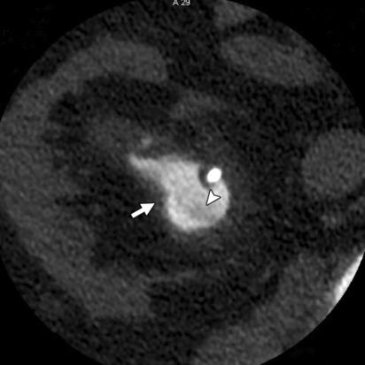
14 Imaging of urinary calculi UHCT Differential diagnosis between urinary stones and extraurinary calcifications Rim sign is specific for urinary stone sensitivity 50-77% specificity % Rim sign

15 Imaging of urinary calculi UHCT Differential diagnosis between urinary stones and extraurinary calcifications Rim sign is specific for urinary stone The comet tail sign is a useful sign in diagnosing phleboliths Bell TV, Radiology 1998 Boriady IC, Radiology 1999 The comet tail sign does not preclude a coexisting ipsilateral calculus Guest AR, AJR 2001 Eccentric tapering of soft tissue extending from one surface of the calcification Comet tail sign

16 Imaging of urinary calculi UHCT Differential diagnosis between urinary stones and extraurinary calcifications Rim sign is specific for urinary stone The comet tail sign is a useful sign in diagnosing phleboliths Most phleboliths are round or oval, most ureteral calculi are slightly angular in shape Traubici J, AJR 1999


17 Imaging of urinary calculi UHCT Differential diagnosis between urinary stones and extraurinary calcifications Rim sign is specific for urinary stone The comet tail sign is a useful sign in diagnosing phleboliths Most phleboliths are round or oval, most ureteral calculi are slightly angular in shape Phleboliths may contain a central lucent area Rarely seen at CT! Traubici J, AJR 1999 Hartman RP et al., In: Morcos SK, Cohan, RH. New Techniques in Uroradiology, Taylor & Francis, NY, 2006

18 Imaging of urinary calculi CE-CT Differentiation of stones from phleboliths Differentiation of parapelvic cysts from hydronephrosis Clinically suspected complicated pyelonephritis Usefulin conditionssuchasureteralstrictures, duplicatedsystem or ureteropelvic junction obstructions. Evaluation of alternative causes of flank pain Diverticulitis, appendicitis Pelvic masses Large renal tumors Bowel obstruction Aortic aneurysm. Most of them are identified at UHCT
19 Stone evaluation Stone burden( size evaluation better with bone windows). Stone fragility( heterogeneousor homogeneous). Stone composition. Treatment planning. Posttreatment evaluation.


20 Stone burden Number Size( at least2 planesbetter withbone-window) Location

21 Stone fragility

22 Stone composition Composition Uric acid Struvite Cystine Density UH UH UH Calcium phosphate UH Calcium oxalate monohydrateand brushite UH Stones of mixed compostion have overlapping attenuation ranges in vivo. CT attenuation measurementshavebeenmostvaluablein differentiation of 100% uric acid stones from other stones. Dual-Energy CT?
23 Treatment planning MultidetectorCT not only assists in the selection of an appropriate calixfor percutaneousaccess, but it also helps ascertain a safe path for puncture by depicting the relationship of the kidney to various surrounding organs such as the spleen, liver, and colon. Evaluation of SSD. Evaluation of infundible-pelvic angle.

24
25 Posttreatment Evaluation Confirm stone-free status. Identify the presence of residual stones. Rule out obstruction in the urinary system. Detection of complications such as perirenal hematoma and urinoma( CECT).


26

27
28 Imaging of urinary calculi UHCT The gold standard for imaging patients with urinary calculi..but radiation dose is a significant problem Radiation exposure of regular-dose CT Ruppert-Kohlmayr et al, Radiation exposure of different modalities Becker et al, 1999 Most stones8.8 have chance to pass msv Cohnen et al, 2003 spontaneously KUB Keske et al, ChestX ray : 0,02 msv. msv Young patients at significant life time risk for recurrent renal colics KUB Aroua et al, IVU Denton et al, IVU Liu et al, IVU Thomson et al,
29 Imaging of urinary calculi UHCT The new gold standard for imaging patients with renal colic..but radiation dose is a significant problem Developement of low dose and ultra low-dose CT protocols
30 is 0.05% (1 in 2,000) for 10 msvof ionizing radiation [5, 17]. This estimate is determined by linear extrapolation from the risk of 5% per each sievert established by the International Commission on Radiological Protection in Rxtorace : 0,02 msv.
31 Strategies for dose reduction Appropriate patients selection. Limiting the scan range. Using automatic tube current modulation suchas care dose. Weight based selection of tube voltage.
32 UHCT in all cases? Nearly55 % ofpatientsundergoingct forevaluationofacute flankpaindidnothavestonedisease. 15 % had other abnormalities. Stones 5 mm oflessin diameterhada spontaneouspassage rate of68 %, stonesgreaterthan5 mm butlessthanor equal to10 mm. hada spontaneouspassagerate of47 %. Young patients, high life timerecurrenceofrenalcolic. NO
33 Imaging of urinary calculi Is UHTC needed in all patients with renal colic? UHCT is the best imaging procedure for evaluation of patients with renal colic But US will provide enough clinically useful information in most cases without radiation High end US equipment, appropriate training to improve operator s skill Identifies hydronephrosis Detection of ureteral stones can improve Identifies clinically significant extraurinary pathologies


34 Suggesting CT reporting points for urolithiasis
35 TAKE HOME POINTS Correct indication. Adeguate technique. Post-processing. Dose reduction.
36
37 La calcolosi urinaria :patologia di interesse multidisciplinare Nuovi standard radiologici e di medicina nucleare nello studio della litiasi urinaria CT Dott. PAOLO BRESCIANI U.O.C. RADIOLOGIA Azienda Ospedaliero Universitaria Parma I MARTEDI DELL ORDINE PARMA 1 MARZO 2016
Acute flank pain in children: Imaging considerations
 Acute flank pain in children: Imaging considerations Carlos J. Sivit MD Rainbow Babies and Children s Hospital Case Western Reserve School of Medicine Flank pain Results from distention of ureter or renal
Acute renal colic Radiological investigation in patients with renal colic
 Acute renal colic Radiological investigation in patients with renal colic Mikael Hellström Professor Department of Radiology Sahlgrenska University Hospital Göteborg University 0.9-1.8/1.000 inhabitants
Unenhanced Spiral CT in Acute Ureteral Colic: A Replacement for Excretory Urography?
 Unenhanced Spiral CT in cute Ureteral Colic: Replacement for Excretory Urography? Jeong-h Ryu, MD 1 ohyun Kim, MD 1 Yong Hwan Jeon, MD 1 Jongmee Lee, MD 1 Jin-Wook Lee, MD 1 Seong Soo Jeon, MD 2 Kwan Hyun
Non-calculus causes of renal colic on CT KUB
 Non-calculus causes of renal colic on CT KUB Poster No.: C-1341 Congress: ECR 2010 Type: Scientific Exhibit Topic: Genitourinary Authors: A. Afaq, E. L. Leen; London/UK Keywords: renal colic, CT KUB, appendicitis
Low-Dose Unenhanced Computed Tomography with Iterative Reconstruction for Diagnosis of Ureter Stones
 Original Article Yonsei Med J 2018 May;59(3):389-396 pissn: 0513-5796 eissn: 1976-2437 Low-Dose Unenhanced Computed Tomography with Iterative Reconstruction for Diagnosis of Ureter Stones Byung Hoon Chi
Residents Section Structured Review Article
 Residents Section Structured Review rticle O Connor et al. CT Urography Residents Section Structured Review rticle Residents inradiology Owen J. O Connor 1 Michael M. Maher O Connor OJ, Maher MM Keywords:
Low-Dose Versus Standard-Dose CT Protocol in Patients with Clinically Suspected Renal Colic
 CT of Renal Colic Genitourinary Imaging Original Research Pierre-Alexandre Poletti 1 Alexandra Platon 1 Olivier T. Rutschmann 2 Franz R. Schmidlin 3 Christophe E. Iselin 3 Christoph D. ecker 1 Poletti
EVALUATION OF SUSPECTED RENAL COLIC PATIENTS WITH UNENHANCED LOW-DOSE MULTI-DETECTOR COMPUTED TOMOGRAPHY
 190 EAST AFRICAN MEDICAL JOURNAL April 2009 East African Medical Journal Vol. 85 No. 4 April 2009 EVALUATION OF SUSPECTED RENAL COLIC PATIENTS WITH UNENHANCED LOW-DOSE MULTI-DETECTOR COMPUTED TOMOGRAPHY
Patients with ureteral stones usually come to the
 Original Article 182 Decreased Renal Parenchymal Density on Unenhanced Helical Computed Tomography for Diagnosis of Ureteral Stone Disease in Emergent Patients with Acute Flank Pain Chen-Chih Huang, MD;
이학종분당서울대학교병원. Ultrasound in Urinary Colic
 이학종분당서울대학교병원 Ultrasound in Urinary Colic U l t r a s o u n d i n U r i n a US: Normal Kidney r y C o l i c Contents 1. 1. Definition and clinical consideration 2. 2. Pathophysiology 3. 3. US in in obstructive
Plain Radiographs in Non-Traumatic Abdominal Pain. Plain Radiographs in Non-Traumatic Abdominal Pain
 Jake Block, MD Associate Professor Associate Vice-Chairman for Clinical Operations Director, Musculoskeletal and Emergency Radiology Department of Radiology and Radiological Sciences Vanderbilt University
kingstonegems.com Precious Stones: Gems of the urogenital system Nordic Forum 2017, Helsinki, Finland Ken F Linnau MD, MS Emergency Radiology
 kingstonegems.com Precious Stones: Gems of the urogenital system Nordic Forum 2017, Helsinki, Finland Ken F Linnau MD, MS Emergency Radiology 59 year old woman Intermittent right flank pain Pain radiates
R adio logical investigations of urinary system
 R adio logical investigations of urinary system There are 4 main radiological Ix: 1 IVU: Intravenous urography. 2- U/S 3-CT scan 4-Radioisotope scan. Others (not frequently used): MRI, arteriography, antegrade
Request Card Task ANSWERS
 Request Card Task ANSWERS Medical Student Workbook Author: Dr Sam Leach, SpR Case 1 What differential diagnoses are most likely? Which investigation is most appropriate? Case 1 The most likely diagnosis
Dual Energy CT: a new tool in evaluation of the urinary tract stones composition in clinical practice - initial study
 Dual Energy CT: a new tool in evaluation of the urinary tract stones composition in clinical practice - initial study Poster No.: C-2279 Congress: ECR 2013 Type: Scientific Exhibit Authors: M. Guzi#ski,
Title: Radiological Imaging for Renal Calculi: Guidelines and a Clinical and Cost Effectiveness Review
 Title: Radiological Imaging for Renal Calculi: Guidelines and a Clinical and Cost Effectiveness Review Date: 29 February 2008 Context and policy issues: About 10% of the population will have an episode
Radiological evaluation of complications of extracorporeal shock wave lithotripsy for urolithiasis
 Radiological evaluation of complications of extracorporeal shock wave lithotripsy for urolithiasis Poster No.: C-0749 Congress: ECR 2013 Type: Educational Exhibit Authors: J. A. Merino Bonilla, H. Guerra
Outline. Introduction to imaging modalities of the urinary system. Case base learning of common diseases in urinary tract
 Outline Introduction to imaging modalities of the urinary system Case base learning of common diseases in urinary tract Outline Introduction to imaging modalities of the urinary system Case base learning
Outline. Introduction to imaging modalities of the urinary system. Case base learning of common diseases in urinary tract
 Outline Introduction to imaging modalities of the urinary system Case base learning of common diseases in urinary tract Diagnostic Investigations in Urinary System PLAIN KUB EXCRETORY UROGRAPHY RETROGRADE
AUA Guidelines for Imaging Known or Suspected Ureteral Calculi. Michael Ferrandino, MD Assoc Professor of Urology Duke University Medical Center
 AUA Guidelines for Imaging Known or Suspected Ureteral Calculi Michael Ferrandino, MD Assoc Professor of Urology Duke University Medical Center Imaging for Urolithiasis Justification for the Guidelines
Renal colic and its mimickers: Pearls and pitfalls on CT to avoid misdiagnosis
 Renal colic and its mimickers: Pearls and pitfalls on CT to avoid misdiagnosis Poster No.: C-2229 Congress: ECR 2013 Type: Educational Exhibit Authors: C. Esteves, C. Maciel, F. Rego Costa, A. F. S. Simões,
CASE REPORT RENAL TUBERCULOSIS CAUSE OF RENAL REPLACEMENT LIPOMATOSIS : A RARE ASSOCIATION
 CASE REPORT RENAL TUBERCULOSIS CAUSE OF RENAL REPLACEMENT LIPOMATOSIS : A RARE ASSOCIATION DR ANAND AARTI 1, DR CHANDAK PRIYA 2,DR SURESH PARVATHY 3 1. PROF AND HOD, DEPARTMENT OF RADIODIAGNOSIS, GOVERNMENT
PROFESSIONAL SKILLS 1 3RD YEAR SEMESTER 6 RADIOGRAPHY. THE URINARY SYSTEM Uz. Fatema shmus aldeen Tel
 PROFESSIONAL SKILLS 1 3RD YEAR SEMESTER 6 RADIOGRAPHY THE URINARY SYSTEM Uz. Fatema shmus aldeen Tel. 0925111552 Professional skills-2 THE URINARY SYSTEM The urinary system (review anatomy and physiology)
Role of imaging in evaluation of genitourinary i trauma Spectrum of GU injuries Relevance of imaging findings in determining management Focus on MDCT
 Genitourinary Tract Injuries 6 th Nordic Course Scott D. Steenburg, MD Assistant Professor University of Maryland Department of Radiology Division of Trauma and Emergency Radiology R Adams Cowley Shock
Imaging Features of Acute Pyelonephritis in Contrast Computed Tomography as Predictors of Need for Intervention
 Imaging Features of Acute Pyelonephritis in Contrast Computed Tomography as Predictors of Need for Intervention Poster No.: C-0088 Congress: ECR 2014 Type: Scientific Exhibit Authors: C. Y. Lee, C. W.
The Diagnostic Value of Color Doppler Ultrasound in Ureteral Calculi
 International Journal of Medical Imaging 2018; 6(2): 12-17 http://www.sciencepublishinggroup.com/j/ijmi doi: 10.11648/j.ijmi.20180602.11 ISSN: 2330-8303 (Print); ISSN: 2330-832X (Online) The Diagnostic
Nongynecological causes of acute and chronicpelvic pain. Amela Sofić UKC Sarajevo Bosnia and Herzegovina
 Nongynecological causes of acute and chronicpelvic pain Amela Sofić UKC Sarajevo Bosnia and Herzegovina One of the most challenging problems in a clinical routine is the pelvic pain It is useful to classify
Appropriate Imaging Tests Lead to Meaningful Results. Dr. Richard Wasley May 2011
 Appropriate Imaging Tests Lead to Meaningful Results Dr. Richard Wasley May 2011 Summarize the advantages and limitations of specific imaging tests and why clinical information is so important to radiologists
Lec-8 جراحة بولية د.نعمان
 4th stage Lec-8 جراحة بولية د.نعمان 11/10/2015 بسم هللا الرحمن الرحيم Ureteric, Vesical, & urethral stones Ureteric Calculus Epidemiology like renal stones Etiology like renal stones Risk factors like
Alternate and Incidental Diagnoses on Noncontrast- Enhanced Spiral Computed Tomography for Acute Flank Pain
 Endourology and Stone Disease Alternate and Incidental Diagnoses on Noncontrast- Enhanced Spiral Computed Tomography for Acute Flank Pain M Hammad Ather, Kulsoom Faizullah, Ilyas Achakzai, Rizwan Siwani,
Excretory urography (EU) or IVP US CT & radionuclide imaging
 or IVP US CT & radionuclide imaging") Excretory urography (EU) or IVP US CT & radionuclide imaging MRI arteriography studies requiring catherization or direct puncture of collecting system EU & to a lesser extent CT provide both functional
Excretory urography (EU) or IVP US CT & radionuclide imaging MRI arteriography studies requiring catherization or direct puncture of collecting system EU & to a lesser extent CT provide both functional
Unenhanced CT in the evaluation of renal/ureteric colic
 Review Unenhanced CT in the evaluation of renal/ureteric colic Unenhanced CT of kidneys, ureters and bladder (CTKUB) is now the recommended imaging modality in the investigation of patients with acute
Hydronephrosis. What is hydronephrosis?
 What is hydronephrosis? Hydronephrosis Hydronephrosis describes the situation where the urine collecting system of the kidney is dilated. This may be a normal variant or it may be due to an underlying
US in non-traumatic acute abdomen. Lalita, M.D. Radiologist Department of radiology Faculty of Medicine ChiangMai university
 US in non-traumatic acute abdomen Lalita, M.D. Radiologist Department of radiology Faculty of Medicine ChiangMai university Sagittal Orientation Transverse (Axial) Orientation Coronal Orientation Intercostal
Case Report Three-Dimensional Dual-Energy Computed Tomography for Enhancing Stone/Stent Contrasting and Stone Visualization in Urolithiasis
 Case Reports in Urology Volume 2013, Article ID 646087, 4 pages http://dx.doi.org/10.1155/2013/646087 Case Report Three-Dimensional Dual-Energy Computed Tomography for Enhancing Stone/Stent Contrasting
Audit of split-bolus CT urography for the investigation of haematuria over a 12 month period at two district general hospitals
 Audit of split-bolus CT urography for the investigation of haematuria over a 12 month period at two district general hospitals Poster No.: C-1349 Congress: ECR 2010 Type: Educational Exhibit Topic: Genitourinary
What the Radiologist Needs to Know About Urolithiasis: Part 2 CT Findings, Reporting, and Treatment
 Integrative Imaging Review Cheng et al. CT of Urolithiasis Integrative Imaging Review CME SM What the Radiologist Needs to Know bout Urolithiasis FOCUS ON: Phillip M. Cheng 1 Paymann Moin 2 Matthew D.
ROLE OF ULTRASOUND IN EVALUATION OF RENAL COLIC AND ASSESSMENT OF RISK FACTOR FOR RENAL CALCULI
 Arif Pervez & Ammar Arif ORIGINAL ARTICLE ROLE OF ULTRASOUND IN EVALUATION OF RENAL COLIC AND ASSESSMENT OF RISK FACTOR FOR RENAL CALCULI Arif Pervez 1 and Ammar Arif 2 1 Bolan Medical College Hospital
Role of imaging in RCC. Ultrasonography. Solid lesion. Cystic RCC. Solid RCC 31/08/60. From Diagnosis to Treatment: the Radiologist Perspective
 Role of imaging in RCC From Diagnosis to Treatment: the Radiologist Perspective Diagnosis Staging Follow up Imaging modalities Limitations and pitfalls Duangkamon Prapruttam, MD Department of Therapeutic
Bedside Ultrasound in the Emergency Department to Detect Hydronephrosis for the Evaluation of Suspected Ureteric Colic
 Bedside Ultrasound in the Emergency Department to Detect Hydronephrosis for the Evaluation of Suspected Ureteric Colic Shrestha R, Shakya RM, Khan A ABSTRACT Background Department of Emergency Medicine
Urolithiasis. Ali Kasraeian, MD, FACS Kasraeian Urology Advanced Laparoscopic, Robotic & Minimally Invasive Urologic Surgery
 Urolithiasis Ali Kasraeian, MD, FACS Kasraeian Urology Advanced Laparoscopic, Robotic & Minimally Invasive Urologic Surgery Urolithiasis: Why should we care? Affects 5% of US men and women Men twice as
Ultra-low dose CT of the acute abdomen: Spectrum of imaging findings
 Ultra-low dose CT of the acute abdomen: Spectrum of imaging findings Poster No.: C-1452 Congress: ECR 2010 Type: Educational Exhibit Topic: GI Tract Authors: P. A. Vlachou, C. Kloeters, S. Kandel, P. Hein,
The 82 nd UWI/BAMP CME Conference November 18, Jeetu Nebhnani MBBS D.M. Urology Consultant Urologist
 The 82 nd UWI/BAMP CME Conference November 18, 2017 Jeetu Nebhnani MBBS D.M. Urology Consultant Urologist Disclosures Outline Index case Introduction Etiology Risk factors Acute stone event Conservative
Diagnostic value of 64 slice spiral computed tomography imaging of the urinary tract during the excretory phase for urinary tract obstruction
 EXPERIMENTAL AND THERAPEUTIC MEDICINE 14: 4761-4766, 2017 Diagnostic value of 64 slice spiral computed tomography imaging of the urinary tract during the excretory phase for urinary tract obstruction DE
Abdominal Ultrasound : Aorta, Kidneys, Bladder
 Abdominal Ultrasound : Aorta, Kidneys, Bladder Nilam J. Soni, MD, MSc Associate Professor of Medicine Divisions of Hospital Medicine and Pulmonary/Critical Care Medicine Department of Medicine University
IMAGING OF UPPER UT TCC
 IMAGING OF UPPER UT TCC IS THERE AN EVIDENCE BASED STRATEGY? S A MOUSSA FRCS Ed, FRCR WESTERN GENERAL HOSPITAL EDINBURGH UPPER TRACT TCC 0.7-4% of patients with primary bladder cancer develops UT-TCC.
Nonenhanced Helical CT and US in the Emergency Evaluation of Patients with Renal Colic: Prospective Comparison 1
 Emergency Radiology Douglas H. Sheafor, MD Barbara S. Hertzberg, MD Kelly S. Freed, MD Barbara A. Carroll, MD Mary T. Keogan, MD Erik K. Paulson, MD David M. DeLong, PhD Rendon C. Nelson, MD Index terms:
Urinary Stone Disease: Comparison of Standard-Dose and Low-Dose with 4D MDCT Tube Current Modulation
 MDCT of Urinary Stone Disease Genitourinary Imaging Original Research Tom H. Mulkens 1 Sofie Daineffe 1 Roel De Wijngaert 1 Patrick Bellinck 1 André Leonard 2 Guido Smet 2 Jean-Luc Termote 1 Mulkens TH,
Predictive Value of Preoperative Unenhanced Computed Tomography During Ureteroscopic Lithotripsy: A Single Institute s Experience
 www.kjurology.org http://dx.doi.org/0.4/kju.03.54..77 Endourology/Urolithiasis Predictive Value of Preoperative Unenhanced Computed Tomography During Ureteroscopic Lithotripsy: A Single Institute s Experience
Treatment of choice for end stage renal disease Imaging to establish baseline and diagnosis of potential complications Review common surgical
 Treatment of choice for end stage renal disease Imaging to establish baseline and diagnosis of potential complications Review common surgical techniques Review normal appearance Discuss US diagnosis of
Renal masses - the role of diagnostic imaging
 Renal masses - the role of diagnostic imaging Poster No.: C-2471 Congress: ECR 2015 Type: Educational Exhibit Authors: V. Rai#; Bjelovar/HR Keywords: Cysts, Cancer, Structured reporting, Ultrasound, MR,
The impact of ureteral Double-J stent insertion following ureterorenoscopy in patients with ureteral stones accompanied by perirenal fat stranding
 ORIGINAL PAPER DOI: 10.4081/aiua.2018.1.15 The impact of ureteral Double-J stent insertion following ureterorenoscopy in patients with ureteral stones accompanied by perirenal fat stranding Ercan Ogreden
CYSTIC DISEASES of THE KIDNEY. Dr. Nisreen Abu Shahin
 CYSTIC DISEASES of THE KIDNEY Dr. Nisreen Abu Shahin 1 Types of cysts 1-Simple Cysts 2-Dialysis-associated acquired cysts 3-Autosomal Dominant (Adult) Polycystic Kidney Disease 4-Autosomal Recessive (Childhood)
Research Article Ureteral Dilatation with No Apparent Cause on Intravenous Urography: Normal or Abnormal? A Pilot Study
 Advances in Urology Volume 2015, Article ID 681836, 6 pages http://dx.doi.org/10.1155/2015/681836 Research Article Ureteral Dilatation with No Apparent Cause on Intravenous Urography: Normal or Abnormal?
Nephrographic and Pyelographic Analysis of CT Urography: Principles, Patterns, and Pathophysiology
 Genitourinary Imaging Review Wolin et al. CT Urography Principles, Patterns, and Genitourinary Imaging Review FOCUS ON: Ely A. Wolin 1 David S. Hartman J. Ryan Olson Wolin EA, Hartman DS, Olson JR Keywords:
Urologic Stone Disease. Urologic Stone Disease. Urologic Stone Disease. Urologic Stone Disease. Urologic Stone Disease 5/7/2010
 Diagnosis and Treatment Stephen E. Strup MD William Farish Professor and Chief of Urology Director of Minimally Invasive Urologic Surgery University of Kentucky I will not cut, even for the stone, but
URINARY SYSTEM. Lecturer Dr.Firdous M.Jaafar Department of anatomy/histology section Lecture 3
 URINARY SYSTEM Lecturer Dr.Firdous M.Jaafar Department of anatomy/histology section Lecture 3 Objectives 1- Describe the structure of the urinary bladder, 2- Describe the structure of the ureters, bladder,
Sex: 女 Age: 51 Occupation: 無 Admission date:92/07/22
 Sex: 女 Age: 51 Occupation: 無 Admission date:92/07/22 Chief complaint Unknown fever for one month Hand tremor and left huge renal tumor was noted Present illness Suffered from fever for one month, hand
Original Article INTRODUCTION. Abstract
 Original Article Print ISSN: 2321-6379 Online ISSN: 2321-595X DOI: 10.17354/ijss/2017/411 Role of Non-contrast Computed Tomography - Kidney, Ureter, and Bladder in Predicting the Stone Fragility and Extracorporeal
Nonurographic evaluation of renal calculous disease 1
 Contributions Nonurographic evaluation of renal calculous disease 1 Gregory P. Borkowski, M.D. Craig R. George, M.D. Peter B. O'Donovan, M.D. While excretory urography has been useful in the evaluation
Obstructive Nephropathy
 Obstructive Nephropathy Liza A. Lucero RN, FNP-C, MSN Renal Medicine Associates Conflicts No conflict of interests Obstructive Nephropathy Objectives Definition of Obstructive Nephropathy Causes Clinical
Detection of Renal Stones on Portal Venous Phase CT: Comparison of Thin Axial and Coronal Maximum- Intensity-Projection Images
 Genitourinary Imaging Original Research Corwin et al. Detection of Renal Stones on Portal Venous Phase CT Genitourinary Imaging Original Research Michael T. Corwin 1 Justin S. Lee 1 Ghaneh Fananapazir
CT Imaging of the Kidney
 September 2001 CT Imaging of the Kidney Images: Netter, FH: Atlas of Human Anatomy, 2 nd ed. Novartis, 1997 Anthony Powell, HMS IV Beth Israel Deaconess Medical Center Images: BIDMC, Dept of Radiology,
Correlation of volume, position of stone, and hydronephrosis with microhematuria in patients with solitary urolithiasis
 e-issn 1643-3750 DOI: 10.12659/MSM.889077 Received: 2012.02.21 Accepted: 2012.04.04 Published: 2013.04.24 Correlation of volume, position of stone, and hydronephrosis with microhematuria in patients with
CT Urography. Ureter. Stuart G. Silverman, M.D.
 CT Urography Stuart G. Silverman, M.D. Professor of Radiology Harvard Medical School Director, Abdominal Imaging and Intervention Brigham and Women s Hospital Ureter Boston, MA CT Urography Stuart G. Silverman,
Question 2. What percentage of abdominal trauma involve the kidney? a) 5 % b) 10% c) 15 % d) 20 %
 5 % b) 10% c) 15 % d) 20 %") Quiz Question 1 After injecting 2ml/kg of contrast for a patient needing a single-shot IVP before kidney exploration, What is the best turnaround time to take the X-ray? a) 3 minutes b) 5 minutes c) 10
Quiz Question 1 After injecting 2ml/kg of contrast for a patient needing a single-shot IVP before kidney exploration, What is the best turnaround time to take the X-ray? a) 3 minutes b) 5 minutes c) 10
Low-dose unenhanced helical computed tomography (CT) is an effective modality for the assessment of urinary calculus disease ( 1 7 ). It can help diag
 is an effective modality for the assessment of urinary calculus disease ( 1 7 ). It can help diag") Note: This copy is for your personal, non-commercial use only. To order presentation-ready copies for distribution to your colleagues or clients, contact us at www.rsna.org/rsnarights. ORIGINAL RESEARCH
Note: This copy is for your personal, non-commercial use only. To order presentation-ready copies for distribution to your colleagues or clients, contact us at www.rsna.org/rsnarights. ORIGINAL RESEARCH
Proceedings of the 34th World Small Animal Veterinary Congress WSAVA 2009
 www.ivis.org Proceedings of the 34th World Small Animal Veterinary Congress WSAVA 2009 São Paulo, Brazil - 2009 Next WSAVA Congress : Reprinted in IVIS with the permission of the Congress Organizers IMAGING
What s Your Diagnosis??? Renée Fahrenholz, Class of 2012
 Renée Fahrenholz, Class of 2012 What s Your Diagnosis??? Signalment Emma, a 9 year old, Female, Spayed, Domestic Short Haired Feline Presenting Complaint Weight loss, vomited the morning of her visit,
Autosomal Dominant Polycystic Kidney Disease
 Case Studies [1] July 01, 2014 By Amar Udare, MBBS [2] Case History: 45-year-old female with vague pain in the abdomen. Case History: A 45-year-old female presented with vague pain in the abdomen. A USG
Pediatric Urology Are Stone Protocol Computed Tomography Scans Mandatory for Children With Suspected Urinary Calculi?
 Pediatric Urology Are Stone Protocol Computed Tomography Scans Mandatory for Children With Suspected Urinary Calculi? Emilie K. Johnson, Gary J. Faerber, William W. Roberts, J. Stuart Wolf, Jr., John M.
Value of color doppler ultrasound, kub and urinalysis in diagnosis of renal colic due to ureteral stones
 ORIGINAL ARTICLE Value of color doppler ultrasound, kub and urinalysis in diagnosis of renal colic due to ureteral stones Mahmoud Abdel-Gawad, Ravi Kadasne, Chandrashekar Anjikar, Emad Elsobky Department
Quantifying Dual-energy computed tomography (DECT) in patients with renal calculi using a Toshiba Aquilion One Scanner.
 in patients with renal calculi using a Toshiba Aquilion One Scanner.") Quantifying Dual-energy computed tomography (DECT) in patients with renal calculi using a Toshiba Aquilion One Scanner. Poster No.: C-2613 Congress: ECR 2015 Type: Authors: Keywords: DOI: Scientific Exhibit
Quantifying Dual-energy computed tomography (DECT) in patients with renal calculi using a Toshiba Aquilion One Scanner. Poster No.: C-2613 Congress: ECR 2015 Type: Authors: Keywords: DOI: Scientific Exhibit
Abdominal ultrasound:
 Abdominal ultrasound: Non-traumatic acute abdomen Wittanee Na-ChiangMai, MD Department of Radiology ChiangMai University 26/04/2017 Contents Technique of examination Normal anatomy Emergency conditions
Acute Pyelonephritis
 Acute Pyelonephritis Variant 1: Acute pyelonephritis. Uncomplicated patient (eg, no history of diabetes or immune compromise or history of stones or obstruction or prior renal surgery or lack of response
Value of Multislice Helical CT Scans and Maximum-Intensity-Projection Images to Improve Detection of Ureteral Stones at Abdominal Radiography
 Bernard E. Van Beers 1 Stéphane Dechambre 1 Pierre Hulcelle 1 Roland Materne 1 Jacques Jamart 2 Received December 11, 2000; accepted after revision May 16, 2001. 1 Department of Radiology, Université Catholique
Received June 30, 2006; Revised September 7, 2006; Accepted September 14, 2006; Published
 Case Study TheScientificWorldJOURNAL (2006) 6, 2495 2504 TSW Urology ISSN 1537-744X; DOI 10.1100/tsw.2006.389 Scintigraphic Demonstration of Urine Extravasation Secondary to Acute Ureteral Obstruction:
My Patient Has Abdominal Pain PoCUS of the Biliary Tract and the Urinary Tract
 My Patient Has Abdominal Pain PoCUS of the Biliary Tract and the Urinary Tract Objectives PoCUS for Biliary Disease PoCUS for Renal Colic PoCUS for Urinary Retention Biliary Disease A patient presents
Kidney stones and imaging: What can your radiologist do for you?
 World J Urol (2015) 33:193 202 DOI 10.1007/s00345-014-1416-0 TOPIC PAPER Kidney stones and imaging: What can your radiologist do for you? Raphaële Renard-Penna Aurélie Martin Pierre Conort Pierre Mozer
Nephrolithiasis Outline Epidemiology
 Nephrolithiasis Brian Duty, M.D. Assistant Professor Department of Urology Oregon Health & Sciences University Outline Epidemiology Pathophysiology Clinical Presentation Diagnosis Management Medical Surgical
Unenhanced CT KUB for urinary colic : It's not just about the stones
 Unenhanced CT KUB for urinary colic : It's not just about the stones Poster No.: C-0762 Congress: ECR 2016 Type: Educational Exhibit Authors: P. Jagmohan, S. Dhanda, B. ang, S. T. Quek; SINGAPORE/SG Keywords:
Primary epiploic appendagitis versus omental infarction : The role of MDCT
 Primary epiploic appendagitis versus omental infarction : The role of MDCT e-poster: EE-125 Congress: ESGAR 2010 Type: Educational Exhibit Topic: Diagnostic / Mesentery and Peritoneum Authors: P. Kraniotis,
Safe surgical treatment of peripelvic renal cyst combined with renal calculi by percutaneous nephroscopy
 CASE REPORT Safe surgical treatment of peripelvic renal cyst combined with renal calculi by percutaneous nephroscopy Bowei Yang 1, Jiongming Li 1, Jianhe Liu 1, Yuepan Bao 2, Prashant Mishra 1, Haixiang
The relationship Between The Divided Shape of Kidney and the Duplication of Ureter
 The relationship Between The Divided Shape of Kidney and the Duplication of Ureter Poster No.: C-1373 Congress: ECR 2014 Type: Authors: Keywords: DOI: Scientific Exhibit J. Lee, B. S. Cho, S. J. Kim, K.
Surgically Discovered Xanthogranulomatous Pyelonephritis Invading Inferior Vena Cava with Coexisting Renal Cell Carcinoma
 Case Study TheScientificWorldJOURNAL (2009) 9, 5 9 TSW Urology ISSN 1537-744X; DOI 10.1100/tsw.2009.6 Surgically Discovered Xanthogranulomatous Pyelonephritis Invading Inferior Vena Cava with Coexisting
How do the Parameters affect Image Quality and Dose for Abdominal CT? Image Review
 How do the Parameters affect Image Quality and Dose for Abdominal CT? Image Review Mannudeep K. Kalra, MD, DNB Massachusetts General Hospital Harvard Medical School Financial Disclosure This presentation
Kristina M. Nowitzki, M.D., Ph.D. and Hao S. Lo, M.D. University of Massachusetts Medical School, Worcester, MA
 Kristina M. Nowitzki, M.D., Ph.D. and Hao S. Lo, M.D. University of Massachusetts Medical School, Worcester, MA Outline I. Introduction highlighting normal renal enhancement physiology including normal
A Giant Hydronephrotic Kidney with Ureteropelvic Junction Obstruction with Blunt Renal Trauma in a Boy
 A Giant Hydronephrotic Kidney with Ureteropelvic Junction Obstruction with Blunt Renal Trauma in a Boy BY JUNYA TSURUKIRI, HIDEFUMI SANO, YOSUKE TANAKA, TAKAO SATO, HIROKAZU TAGUCHI Abstract An 18-year-old
HHS Public Access Author manuscript Abdom Imaging. Author manuscript; available in PMC 2017 June 27.
 Motion Artifacts in Kidney Stone Imaging Using Single-Source and Dual-Source Dual-Energy CT Scanners. A Phantom Study El-Sayed H. Ibrahim 1,2,*, Joseph G. Cernigliaro 1, Robert A. Pooley 1, James C. Williams
PICTORIAL ESSAY. Experiences of using a single post-contrast CT scan of the urinary tract after triphasic contrast injection
 Experiences of using a single post-contrast CT scan of the urinary tract after triphasic contrast injection P C Pretorius, FCRad (Diag) SA Drs Visser, Erasmus, Vawda & Partners, Port Elizabeth Corresponding
Imaging abdominal vascular emergencies. V.Stoynova
 Imaging abdominal vascular emergencies V.Stoynova Abdominal vessels V. Stoynova 2 Acute liver bleeding trauma anticoagulant therapy liver disease : HCC, adenoma, meta, FNH, Hemangioma Diagnosis :CT angiography
Imaging findings in renal infections
 Imaging findings in renal infections Poster No.: C-0221 Congress: ECR 2013 Type: Educational Exhibit Authors: I. lópez blasco, D. Soriano Mena, R. Pastor Toledo, S. Paz Maya, A. M. Julve Parreño, J. Palmero
elical CT plays an important role
 bdominal Imaging Yu et al. Helical CT of cute RLQ Pain Pictorial Essay Jinxing Yu 1 nn S. Fulcher Mary nn Turner Robert. Halvorsen Yu J, Fulcher S, Turner M, Halvorsen R Helical CT Evaluation of cute Right
11/1/2014. Radiologic incidentalomas Ordering pitfalls Newer technology and applications
 Bilal Tahir, MD Gitasree Borthakur, MD Indiana University School of Medicine Department of Radiology & Imaging Sciences October 31, 2014 ACP 2014 Dr. V. Aaron Nuclear (vaaron@iupui.edu) Dr. S. Westphal
ASSESSING THE PLAIN ABDOMINAL RADIOGRAPH M A A M E F O S U A A M P O F O
 ASSESSING THE PLAIN ABDOMINAL RADIOGRAPH M A A M E F O S U A A M P O F O Introduction The abdomen (less formally called the belly, stomach, is that part of the body between the thorax (chest) and pelvis,
Genitourinary. Common Clinical Scenarios Protocoling Module. Patty Ojeda & Mariam Shehata
 The following training module was developed as a quality improvement project to serve as an educational tool for junior radiology residents. The following diagnostic radiology protocoling modules were
Emergency Ultrasound and Urinalysis in the Evaluation of Flank Pain
 1180 Gaspari and Horst d EMERGENCY ULTRASOUND IN FLANK PAIN Emergency Ultrasound and Urinalysis in the Evaluation of Flank Pain Romolo J. Gaspari, MD, MSc, RDMS, Kurt Horst, MD Abstract Objectives: To
Is Structured Reporting More Accurate Than Conventional Reporting in CT Reporting of the Abdomen and Pelvis?
 Is Structured Reporting More Accurate Than Conventional Reporting in CT Reporting of the Abdomen and Pelvis? A M Almuslim, MBBS; J G Ryan, MD; A Murtaza, MD Purpose The purpose of this research is to determine