R adio logical investigations of urinary system
|
|
|
- Veronica Lily Hart
- 5 years ago
- Views:
Transcription
1 R adio logical investigations of urinary system There are 4 main radiological Ix: 1 IVU: Intravenous urography. 2- U/S 3-CT scan 4-Radioisotope scan. Others (not frequently used): MRI, arteriography, antegrade or retrograde pyelogram.
2 Imaging of urinary system: 1-IVU 2-Antegrade pyelogram 3-Reterograde pyelogram 4-micturating cystourethrography
3 Antegrade pyelogram : reguire insertion of a fine neddle in to pelvicalyceal system under US or radiographic control
4 Retrograde pylography: can be performed by inserting catheter in to ureteric orifice at cystoscopy
5 Pylography mainly used to detect the level of obstruction
6 IVU STUDY
7 OBJECTIVES Indication Advantages Disadvantages Procedure Examples
8 INDICATIONS Renal colic to visualize stones or obstructions Heamaturia Recurrent UTI to assess congenital anomalies strictures or urothelial lesions Renal obstruction e.g. renal mass assessment of renal outline pylocalyceal system ureters bladder urothelium Others e.g. Prostatitis Neurogenic bladder Trauma
9 IVU is not indicated in Pylonephritis Renal failure Renal parenchymal disease Disease of hyper tension
10 ADVANTAGE Widely available Excellent to identify uroepithelium Can visualize the whole length of ureters Can quantify the degree urinary tact obstruction
11 DISADVANTAGE Need for patient preparation Risk of radiation Contrast media problems such as allergy May get suboptimal results due to 1. Patient movement 2. Overlying bowel gas 3. Poor concentration media due to poor renal function

12 PATIENT PREPRATION 1. Laxatives is taken 24 hr before the examination 2. Nil by mouth for 4-6 hr before the examination 3. The patient should be ambulant as long as possible to decrease air swallowing 4. Certain measures is taken to certain group of patients e.g. DM and children
13

14 Basic position AP position

15 BASIC PATIENT POSITION The patient must lie supine at mid line of table
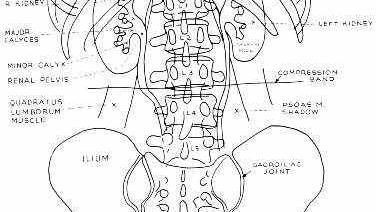
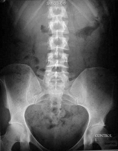
16 Plain film Calcification 1-T.B 2-stone 3-hydatid cyst 4- tumors 5- fibroid in female Abdominal cavity 1- kidney outline 2- shadow of psoase muscle 3- bones
17
 (24 x 30 cm)")
18 Preliminary film (Control) (24 x 30 cm) Inspiration
19 CONTRAST INJECTION The median cubital vein is punctured with a 19 gauge needle and the warmed (40*C) contrast agent is injected rapidly. Films are then taken at intervals to demonstrate the whole of the renal tract. The most common contrast used is non ionized iodine strength
20 End of Injection (24 x 30cm) A.P. of the renal areas to show the nephrogram, i.e. the renal parenchyma opacified by the contrast medium in the renal tubules

21 5 Minute film, (24 x 30cm) A.P. of the renal areas to determine if excretion is symmetrical or if uptake is poor and a further dose of contrast agent is required
22 Compression may be applied in some centers at this point to distend the pelvicalyceal systems to demonstrate any filling defects and a film taken at 10 minutes of the renal areas. Compression should not be used in cases of suspected renal colic, renal trauma or after recent abdominal surgery
 to demonstrate the pelvicalyceal systems and the")
23 15 Minute film (35 x 43cm) release if compression has been applied) to demonstrate the pelvicalyceal systems and the ureters.

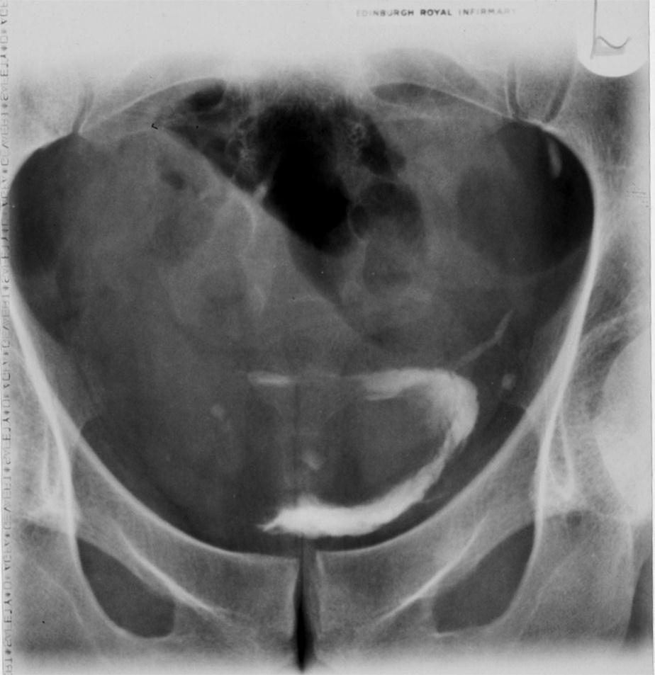
24 25 Minute film 24 x 30cm) 15 caudal angulation centered 5 cm above the upper border of the symphysis pubis to demonstrate the distended bladder.

25 Post Micturition film 24 x 30cm) 15 caudal angulation centered 5 cm above the upper border of the symphysis pubis to demonstrate the bladder emptying success, and the return of the previously distended lower ends of ureters to normal.


26 CONGENITAL ANOMALLY RENAL agenesis and horse shoe kidney

27 Congenital anomaly Duplex kidney


28 RENAL CALCULI Right URETERIC pelviureteric junction AND LOWER BLADDER CALCULUS


29 RENAL CACULUS Left stag horn calculus and right ureteric stone

30 NEOPLASIM CA BLADDER

31 RENAL NEO PLASIM CA KIDNEY
32 ULTRASOUND Non invasive. cheap &easy Indication : 1. Patient with urinary symptoms. 2. Follow-up after lithotripsy 3. Patient with renal transplant to check rejection, stone & hydronephrosis. 4. Renal failure 5. Patient with recurrent UTI (in children) to diagnose reflux.
33 CT SCAN Used for 1. Staging of tumor. 2. Detection of radiolucent stones (xanthine & uric acid stones). 3. Retroperitoneal mass, fibrosis 4. Sagittal reformal (to see kidney in lateral viewer), coronal reformat. 5. Renal trauma (bleeding, hernatoma). in CT scan we 1 take a plain CT without contrast to see if there s calcification.
34 RADIO-IOSOTOPE mainly for function of the kidney or when we can't do IVU. We inject Tc 99 DFPA intraenously & only the functioning part of the kidney will appear.
35 MRI Two main indications 1. Renal vasculature before transplant for both the donor & recipient. 2. Renal artery stenosis (5 mm normally) with post stenotic dilation If we want to do operation, we do angiography also.
PROFESSIONAL SKILLS 1 3RD YEAR SEMESTER 6 RADIOGRAPHY. THE URINARY SYSTEM Uz. Fatema shmus aldeen Tel
 PROFESSIONAL SKILLS 1 3RD YEAR SEMESTER 6 RADIOGRAPHY THE URINARY SYSTEM Uz. Fatema shmus aldeen Tel. 0925111552 Professional skills-2 THE URINARY SYSTEM The urinary system (review anatomy and physiology)
Excretory urography (EU) or IVP US CT & radionuclide imaging
 or IVP US CT & radionuclide imaging") Excretory urography (EU) or IVP US CT & radionuclide imaging MRI arteriography studies requiring catherization or direct puncture of collecting system EU & to a lesser extent CT provide both functional
Excretory urography (EU) or IVP US CT & radionuclide imaging MRI arteriography studies requiring catherization or direct puncture of collecting system EU & to a lesser extent CT provide both functional
Outline. Introduction to imaging modalities of the urinary system. Case base learning of common diseases in urinary tract
 Outline Introduction to imaging modalities of the urinary system Case base learning of common diseases in urinary tract Outline Introduction to imaging modalities of the urinary system Case base learning
Outline. Introduction to imaging modalities of the urinary system. Case base learning of common diseases in urinary tract
 Outline Introduction to imaging modalities of the urinary system Case base learning of common diseases in urinary tract Diagnostic Investigations in Urinary System PLAIN KUB EXCRETORY UROGRAPHY RETROGRADE
Radiographic Procedures III (RAD 228)
") Radiographic Procedures III (RAD 228) Urinary System RADIOGRAPHIC EXAMINATIONS Urinary System Antegrade Exam IVU Functional test Hypertensive evaluation as per protocol Retrograde Exams Retrograde Urography
Radiographic Procedures III (RAD 228) Urinary System RADIOGRAPHIC EXAMINATIONS Urinary System Antegrade Exam IVU Functional test Hypertensive evaluation as per protocol Retrograde Exams Retrograde Urography
Acute renal colic Radiological investigation in patients with renal colic
 Acute renal colic Radiological investigation in patients with renal colic Mikael Hellström Professor Department of Radiology Sahlgrenska University Hospital Göteborg University 0.9-1.8/1.000 inhabitants
Hydronephrosis. What is hydronephrosis?
 What is hydronephrosis? Hydronephrosis Hydronephrosis describes the situation where the urine collecting system of the kidney is dilated. This may be a normal variant or it may be due to an underlying
Contents. Review anatomy of the urinary tract Imaging modalities
 Contents Review anatomy of the urinary tract Imaging modalities The Urinary Tract Kidneys ตาแหน งไต (position) อย ใน retroperitoneum ระด บ T12-L3 โดยไต ขวาจะม ระด บตากว าไตซ ายเล กน อย ร ปร าง (shape)
IVU ((INTRAVENOUSUROGRAM))
)") IVU ((INTRAVENOUSUROGRAM)) Anatomy The urinary system consists of the following : 2 kidneys, 2 ureters,1 bladder, 1 urethra Renal pelvis Minor calyx Major calyx Proximal ureter Pelvi-uretric junction
IVU ((INTRAVENOUSUROGRAM)) Anatomy The urinary system consists of the following : 2 kidneys, 2 ureters,1 bladder, 1 urethra Renal pelvis Minor calyx Major calyx Proximal ureter Pelvi-uretric junction
Acute flank pain in children: Imaging considerations
 Acute flank pain in children: Imaging considerations Carlos J. Sivit MD Rainbow Babies and Children s Hospital Case Western Reserve School of Medicine Flank pain Results from distention of ureter or renal
Lec-8 جراحة بولية د.نعمان
 4th stage Lec-8 جراحة بولية د.نعمان 11/10/2015 بسم هللا الرحمن الرحيم Ureteric, Vesical, & urethral stones Ureteric Calculus Epidemiology like renal stones Etiology like renal stones Risk factors like
Pediatric Ure-Radiology*
 Pediatric Ure-Radiology* HERMAN GROSSMAN, M.D. Professor of Radiology and Pediatrics, Duke University Medical Center, Durham, North Carolina "Routine" radiologic studies do not, often enough, concentrate
Acute Pyelonephritis
 Acute Pyelonephritis Variant 1: Acute pyelonephritis. Uncomplicated patient (eg, no history of diabetes or immune compromise or history of stones or obstruction or prior renal surgery or lack of response
Intravenous Pyelogram (IVP)
") Scan for mobile link. Intravenous Pyelogram (IVP) Intravenous pyelogram (IVP) is an x-ray exam that uses an injection of contrast material to evaluate your kidneys, ureters and bladder and help diagnose
Scan for mobile link. Intravenous Pyelogram (IVP) Intravenous pyelogram (IVP) is an x-ray exam that uses an injection of contrast material to evaluate your kidneys, ureters and bladder and help diagnose
ASSESSING THE PLAIN ABDOMINAL RADIOGRAPH M A A M E F O S U A A M P O F O
 ASSESSING THE PLAIN ABDOMINAL RADIOGRAPH M A A M E F O S U A A M P O F O Introduction The abdomen (less formally called the belly, stomach, is that part of the body between the thorax (chest) and pelvis,
IVU ((INTRAVENOUSUROGRAM))
)") IVU ((INTRAVENOUSUROGRAM)) Anatomy The urinary system consists of : 2 kidneys, 2 ureters,1 bladder, 1 urethra Renal pelvis Minor calyx Major calyx Renal parenchyma Proximal ureter Pelvi-uretric junction
IVU ((INTRAVENOUSUROGRAM)) Anatomy The urinary system consists of : 2 kidneys, 2 ureters,1 bladder, 1 urethra Renal pelvis Minor calyx Major calyx Renal parenchyma Proximal ureter Pelvi-uretric junction
Uroradiology For Medical Students
 Uroradiology For Medical Students Lesson 4: Cystography & Urethrography - Part 2 American Urological Association Review Cystography is useful in evaluating the bladder, the urethra and the competence of
IMAGING OF THE UROGENITAL TRACT
 IMAGING OF THE UROGENITAL TRACT 1 A) URINARY TRACT There are many methods of imaging the urinary tract but plain abdominal X-ray and ultrasound scan are usually done first in most cases, especially in
My Patient Has Abdominal Pain PoCUS of the Biliary Tract and the Urinary Tract
 My Patient Has Abdominal Pain PoCUS of the Biliary Tract and the Urinary Tract Objectives PoCUS for Biliary Disease PoCUS for Renal Colic PoCUS for Urinary Retention Biliary Disease A patient presents
Radiological Assessment of the Kidney in Patients with Hematuria
 March 2005 Radiological Assessment of the Kidney in Patients with Hematuria Jeremy L. McKay, Harvard Medical School Year III Hematuria Signs and Symptoms Microscopic or gross hematuria Abdominal pain Fever
Cystitis cystica is a rare chronic reactive inflammatory
 JOURNAL OF ENDOUROLOGY CASE REPORTS Volume 3.1, 2017 Mary Ann Liebert, Inc. Pp. 34 38 DOI: 10.1089/cren.2017.0010 Case Report Cystitis Cystica as a Large Solitary Bladder Cyst Stephanie Potts, MBChB and
Five Views of Transitional Cell Carcinoma: One Man s Journey
 September 2006 Five Views of Transitional Cell Carcinoma: One Man s Journey Amsalu Dabela, Harvard Medical School III Outline Overview: Renal Anatomy Our Patient s Story Diagnostic Imaging Studies Appearance
Clinics in Diagnostic Imaging (61)
") Singapore Med J 2001 Vol 42(5) : 233-237 M e d i c a l E d u c a t i o n Clinics in Diagnostic Imaging (61) K B J Sheah, S K H Yip, V T Joseph Fig. 1a Control abdominal radiograph. Fig. 1b Coned oblique
Singapore Med J 2001 Vol 42(5) : 233-237 M e d i c a l E d u c a t i o n Clinics in Diagnostic Imaging (61) K B J Sheah, S K H Yip, V T Joseph Fig. 1a Control abdominal radiograph. Fig. 1b Coned oblique
Case Report Crossed Renal Ectopia without Fusion An Unusual Cause of Acute Abdominal Pain: A Case Report
 Case Reports in Urology Volume 2012, Article ID 728531, 4 pages doi:10.1155/2012/728531 Case Report Crossed Renal Ectopia without Fusion An Unusual Cause of Acute Abdominal Pain: A Case Report D. P. Ramaema,
URINARY TRACT IMAGING - BASIC PRINCIPLES
 URINARY TRACT IMAGING - BASIC PRINCIPLES Clinical Radiology Every physician needs a basic understanding of diagnostic imaging to understand how to order the appropriate studies and to understand the resulting
Case MDCT 3D reconstructed features of posterior urethral valve
 Case 12688 MDCT 3D reconstructed features of posterior urethral valve Hidayatullah Hamidi Third year Resident of Radiology French medical institute for children Radiology Department; Kabul, Afghanistan;
Audit of split-bolus CT urography for the investigation of haematuria over a 12 month period at two district general hospitals
 Audit of split-bolus CT urography for the investigation of haematuria over a 12 month period at two district general hospitals Poster No.: C-1349 Congress: ECR 2010 Type: Educational Exhibit Topic: Genitourinary
RENAL SCINTIGRAPHY IN THE 21 st CENTURY
 RENAL SCINTIGRAPHY IN THE 21 st CENTURY 99m Tc- MAG 3 with zero time injection of Furosemide (MAG 3 -F 0 ) : A Fast and Easy Protocol, One for All Indications Clinical Experience Congenital Disorders PROTOCOL
4 th Year Urology Core Objectives Keith Rourke (Revised June 1, 2007)
") 4 th Year Urology Core Objectives Keith Rourke (Revised June 1, 2007) I. Genitourinary Trauma: 1. Goal: The student will be able to demonstrate a basic clinical approach to the management & diagnosis of
4 th Year Urology Core Objectives Keith Rourke (Revised June 1, 2007) I. Genitourinary Trauma: 1. Goal: The student will be able to demonstrate a basic clinical approach to the management & diagnosis of
URINARY SYSTEM I. Kidneys II. Nephron Unit and Urine Formation
 URINARY SYSTEM I. Kidneys A. Location and Structure 1. Retroperitoneal 2. Between T12 and L3 3. Rt. kidney slightly lower 4. Two bean shaped organs 5. Adrenal gland 6. Internal construction a. Renal cortex
Find Medical Solutions to Your Problems HYDRONEPHROSIS. (Distension of Renal Calyces & Pelvis)
") HYDRONEPHROSIS (Distension of Renal Calyces & Pelvis) Hydronephrosis is the distension of the renal calyces and pelvis due to accumulation of the urine as a result of the obstruction to the outflow of
HYDRONEPHROSIS (Distension of Renal Calyces & Pelvis) Hydronephrosis is the distension of the renal calyces and pelvis due to accumulation of the urine as a result of the obstruction to the outflow of
Obstructive Nephropathy
 Obstructive Nephropathy Liza A. Lucero RN, FNP-C, MSN Renal Medicine Associates Conflicts No conflict of interests Obstructive Nephropathy Objectives Definition of Obstructive Nephropathy Causes Clinical
Request Card Task ANSWERS
 Request Card Task ANSWERS Medical Student Workbook Author: Dr Sam Leach, SpR Case 1 What differential diagnoses are most likely? Which investigation is most appropriate? Case 1 The most likely diagnosis
Obstetrics Content Outline Obstetrics - Fetal Abnormalities
 Obstetrics Content Outline Obstetrics - Fetal Abnormalities Effective February 2007 10 16% renal agenesis complete absence of the kidneys occurs when ureteric buds fail to develop Or degenerate before
Urologic investigations
 Urologic investigations د. Laboratory studies EXAMINATION OF URINE Urinalysis: Urinalysis is one of the most important and useful urologic tests available. Reasons for inadequate urinalyses include: (1)
ISUOG Basic Training. Distinguishing between Normal & Abnormal Appearances of the Urinary Tract. Seshadri Suresh, India
 ISUOG Basic Training Distinguishing between Normal & Abnormal Appearances of the Urinary Tract Seshadri Suresh, India Learning objectives 13 & 14 At the end of the lecture you will be able to: describe
Obstructive Uropathy. PATHOPHYSIOLOGIC CHANGES UUO vs BUO. Arry Rodjani Urology Department Ciptomangunkusumo Hospital Jakarta
 Obstructive Uropathy PATHOPHYSIOLOGIC CHANGES UUO vs BUO Arry Rodjani Urology Department Ciptomangunkusumo Hospital Jakarta INTRODUCTION Obstructive uropathy refers to the functional or anatomic obstruction
Intrarenal reflux and the scarred kidney
 Archives of Disease in Childhood, 1974, 49, 531. Intrarenal reflux and the scarred kidney G. L. ROLLESTON, T. M. J. MALING, and C. J. HODSON* From the Department of Radiology, Christchurch Hospital and
The 82 nd UWI/BAMP CME Conference November 18, Jeetu Nebhnani MBBS D.M. Urology Consultant Urologist
 The 82 nd UWI/BAMP CME Conference November 18, 2017 Jeetu Nebhnani MBBS D.M. Urology Consultant Urologist Disclosures Outline Index case Introduction Etiology Risk factors Acute stone event Conservative
Comparison of DMSA Scan 99 m and EC Scan 99 m in Diagnosis of Cortical Defect and Differential Renal Function
 Global Journal of Health Science; Vol. 6, No. 7; 2014 ISSN 1916-9736 E-ISSN 1916-9744 Published by Canadian Center of Science and Education Comparison of DMSA Scan 99 m and EC Scan 99 m in Diagnosis of
Kidney & Urinary Tract Ultrasound. Fatina Fadel Hafez Bazaraa
 Kidney & Urinary Tract Ultrasound Fatina Fadel Hafez Bazaraa Ultrasonography Ultrasound Available Rapid Inexpensive Painless & no sedation needed No adverse effects/ complications Can be repeated Useful
US in non-traumatic acute abdomen. Lalita, M.D. Radiologist Department of radiology Faculty of Medicine ChiangMai university
 US in non-traumatic acute abdomen Lalita, M.D. Radiologist Department of radiology Faculty of Medicine ChiangMai university Sagittal Orientation Transverse (Axial) Orientation Coronal Orientation Intercostal
Radiology of the abdomen Lecture -1-
 Radiology of the abdomen Lecture -1- Objectives To know radiology modalities used in abdomen imaging mainly GI tract. To know advantages and disadvantages of each modality. To know indications and contraindications
Urologic Surgical Complications In Renal Transplantation
 Urologic Surgical Complications In Renal Transplantation Chris Freise, MD Professor of Surgery UCSF Transplant Division Urologic Complications Review of Bladder Anastomosis Complications and Management
Ureters, Urinary Bladder & Urethra
 Ureters, Urinary Bladder & Urethra Please check our Editing File هذا العمل ال يغني عن المصدر األساسي للمذاكرة Lecture 2 } و م ن ي ت و ك ع ل ا لل ه ف ه و ح س ب ه { Objectives o Describe the course of ureter
Urologic Stone Disease. Urologic Stone Disease. Urologic Stone Disease. Urologic Stone Disease. Urologic Stone Disease 5/7/2010
 Diagnosis and Treatment Stephen E. Strup MD William Farish Professor and Chief of Urology Director of Minimally Invasive Urologic Surgery University of Kentucky I will not cut, even for the stone, but
ISSN East Cent. Afr. J. surg. (Online)
") 87 Ureteroscopy in a Resource Limited Setting: The Tikur Anbessa General Specialized Hospital Experience in Addis Ababa, Ethiopia. D. Andualem, L. Be-ede, T. Mulat, L. Samodi Addis Ababa University-School
87 Ureteroscopy in a Resource Limited Setting: The Tikur Anbessa General Specialized Hospital Experience in Addis Ababa, Ethiopia. D. Andualem, L. Be-ede, T. Mulat, L. Samodi Addis Ababa University-School
Guidelines, Policies and Statements D5 Statement on Abdominal Scanning
 Guidelines, Policies and Statements D5 Statement on Abdominal Scanning Disclaimer and Copyright The ASUM Standards of Practice Board have made every effort to ensure that this Guideline/Policy/Statement
Chapter 6: Genitourinary and Gastrointestinal Systems 93
 Chapter 6: Genitourinary and Gastrointestinal Systems 93 Chapter 6 Genitourinary and Gastrointestinal Systems Embryology Three sets of excretory organs or kidneys develop in human embryos: Pronephros:
RADIOLOGY (MEDICAL IMAGING)
") RADIOLOGY (MEDICAL IMAGING) Radiology is the study of the diagnosis of disease by the use of radiant energy (radiation). In the past this meant the use of X-rays to make an image. Today many other forms
RADIOLOGY (MEDICAL IMAGING) Radiology is the study of the diagnosis of disease by the use of radiant energy (radiation). In the past this meant the use of X-rays to make an image. Today many other forms
PICTORIAL ESSAY. Experiences of using a single post-contrast CT scan of the urinary tract after triphasic contrast injection
 Experiences of using a single post-contrast CT scan of the urinary tract after triphasic contrast injection P C Pretorius, FCRad (Diag) SA Drs Visser, Erasmus, Vawda & Partners, Port Elizabeth Corresponding
Plain abdomen The standard films are supine & erect AP views (alternative to erect, lateral decubitus film is used in ill patients).
.") Plain abdomen The standard films are supine & erect AP views (alternative to erect, lateral decubitus film is used in ill patients). The stomach can be readily identified by its location, gastric rugae
Plain abdomen The standard films are supine & erect AP views (alternative to erect, lateral decubitus film is used in ill patients). The stomach can be readily identified by its location, gastric rugae
Abdominal Ultrasound : Aorta, Kidneys, Bladder
 Abdominal Ultrasound : Aorta, Kidneys, Bladder Nilam J. Soni, MD, MSc Associate Professor of Medicine Divisions of Hospital Medicine and Pulmonary/Critical Care Medicine Department of Medicine University
Name of the module: Urology clerkship (selective) Number of module: Course description: Basic clinical urology.
 Number of module: Course description: Basic clinical urology.") Name of the module: Urology clerkship (selective) Number of module: 471-8-5097 Credit points: 2.5 ECTS: Academic year: 5 th & 6 th Semester: 1st, 2 nd Duration: 2 weeks Location: Urology depts. in Soroka
Name of the module: Urology clerkship (selective) Number of module: 471-8-5097 Credit points: 2.5 ECTS: Academic year: 5 th & 6 th Semester: 1st, 2 nd Duration: 2 weeks Location: Urology depts. in Soroka
8/14/2017. Kidney location & visualization. Brief Review with tips & Case Based Illustrations. Size = x L2. Size =
 Dr. Russell Tucker, DACVR Brief Review with tips & Case Based Illustrations Kidney location & visualization K9 Kidneys: Rt @ T13-L1 Lt @ L2-L4 Kidney visualization K9 Kidneys: Rt @ T13-L1 Lt @ L2-L4 Size
AUA Guidelines for Imaging Known or Suspected Ureteral Calculi. Michael Ferrandino, MD Assoc Professor of Urology Duke University Medical Center
 AUA Guidelines for Imaging Known or Suspected Ureteral Calculi Michael Ferrandino, MD Assoc Professor of Urology Duke University Medical Center Imaging for Urolithiasis Justification for the Guidelines
1. Congenital Anomalies of Kidney and Ureter 1
 CONTENTS 1. Congenital Anomalies of Kidney and Ureter 1 1.1 Antenatal Pelviureteric Junction Obstruction 1 1.2 Bilateral Pelviureteric Junction Obstruction 3 1.3 Circumcaval Ureter 6 1.4 Crossed Renal
Urinary system Ultrasound (Renal & Urinary bladder)
") Urinary system Ultrasound (Renal & Urinary bladder) Edited & Presented by ; Hussien A.B ALI DINAR. Msc.Phd ISRRT Associate Member Lecturer (National university) Reporting Sonographer (PHC) Objective By
Urinary system Ultrasound (Renal & Urinary bladder) Edited & Presented by ; Hussien A.B ALI DINAR. Msc.Phd ISRRT Associate Member Lecturer (National university) Reporting Sonographer (PHC) Objective By
Lab Monitor Images Dissection of the Abdominal Vasculature + Lower Digestive System
 Lab Monitor Images Dissection of the Abdominal Vasculature + Lower Digestive System Stomach & Duodenum Frontal (AP) View Nasogastric tube 2 1 3 4 Stomach Pylorus Duodenum 1 Duodenum 2 Duodenum 3 Duodenum
EVALUATION OF SUSPECTED RENAL COLIC PATIENTS WITH UNENHANCED LOW-DOSE MULTI-DETECTOR COMPUTED TOMOGRAPHY
 190 EAST AFRICAN MEDICAL JOURNAL April 2009 East African Medical Journal Vol. 85 No. 4 April 2009 EVALUATION OF SUSPECTED RENAL COLIC PATIENTS WITH UNENHANCED LOW-DOSE MULTI-DETECTOR COMPUTED TOMOGRAPHY
Nongynecological causes of acute and chronicpelvic pain. Amela Sofić UKC Sarajevo Bosnia and Herzegovina
 Nongynecological causes of acute and chronicpelvic pain Amela Sofić UKC Sarajevo Bosnia and Herzegovina One of the most challenging problems in a clinical routine is the pelvic pain It is useful to classify
What s Your Diagnosis??? Renée Fahrenholz, Class of 2012
 Renée Fahrenholz, Class of 2012 What s Your Diagnosis??? Signalment Emma, a 9 year old, Female, Spayed, Domestic Short Haired Feline Presenting Complaint Weight loss, vomited the morning of her visit,
Nonurographic evaluation of renal calculous disease 1
 Contributions Nonurographic evaluation of renal calculous disease 1 Gregory P. Borkowski, M.D. Craig R. George, M.D. Peter B. O'Donovan, M.D. While excretory urography has been useful in the evaluation
Diagnostic value of 64 slice spiral computed tomography imaging of the urinary tract during the excretory phase for urinary tract obstruction
 EXPERIMENTAL AND THERAPEUTIC MEDICINE 14: 4761-4766, 2017 Diagnostic value of 64 slice spiral computed tomography imaging of the urinary tract during the excretory phase for urinary tract obstruction DE
Coding Companion for Urology/Nephrology. A comprehensive illustrated guide to coding and reimbursement
 Coding Companion for Urology/Nephrology A comprehensive illustrated guide to coding and reimbursement 2014 Contents Getting Started with Coding Companion...i Integumentary...1 Arteries and Veins...15 Lymph
URINARY SYSTEM. MEDICAL TERMINOLOGY Chapter Six HIT #141. Anatomy
 URINARY SYSTEM MEDICAL TERMINOLOGY Chapter Six HIT #141 Anatomy Kidneys = bean-shaped organs, located on each side of the spinal column, removal of waste from the blood. Nephron = microscopic located in
Spectrum of Micturating Cystourethrogram Revisited: A Pictorial Assay
 603 International Journal of Collaborative Research on Internal Medicine & Public Health Spectrum of Micturating Cystourethrogram Revisited: A Pictorial Assay Abhinav Jain 1, Vivek Setia 1, Shweta Agnihotri
Imaging Ejaculatory Disorders and Hematospermia
 ATHENS 4-6 October 2018 European Society of Urogenital Radiology Imaging Ejaculatory Disorders and Hematospermia Parvati Ramchandani, MD Professor, Radiology and Surgery University of Pennsylvania Medical
Case Report Congenital Midureteric Stricture: Challenges in Diagnosis and Management
 Case Reports in Urology Volume 2015, Article ID 969246, 5 pages http://dx.doi.org/10.1155/2015/969246 Case Report Congenital Midureteric Stricture: Challenges in Diagnosis and Management Raashid Hamid,
Shadow because the air
 Thyroid Ultrasound Thyroid US examination needs: 1. high frequency transducer 2. extended patient's neck 3. check all the neck area because the swelling could be in areas other than the thyroid such as
Body MRI from the Liver to the Bladder
 Body MRI from the Liver to the Bladder I Want You! Audience Participation Methodist Hospital Continuing Education Seminar Jordan Swensson, MD November 7, 2015 Objectives Observe the uses of MRI for organs
Indications and effectiveness of the open surgery in vesicoureteral reflux
 Indications and effectiveness of the open surgery in vesicoureteral reflux Suzi DEMIRBAG, MD Department of Pediatric Surgery, Gulhane Military Medical Academy, Ankara, TURKEY Vesicoureteral reflux (VUR)
Research Article Ureteral Dilatation with No Apparent Cause on Intravenous Urography: Normal or Abnormal? A Pilot Study
 Advances in Urology Volume 2015, Article ID 681836, 6 pages http://dx.doi.org/10.1155/2015/681836 Research Article Ureteral Dilatation with No Apparent Cause on Intravenous Urography: Normal or Abnormal?
RADIOLOGICAL INVESTIGATION OF THE UROGENITAL SYSTEM
 MINISTRY OF HEALTH OF UKRAINE ZAPORIZHZHIA STATE MEDICAL UNIVERSITY Department of Urology, Radiologic Diagnostics and Therapy E. G. Nordio, N. V. Tumanska, T. M. Kichangina RADIOLOGICAL INVESTIGATION OF
Renal Artery Stenosis With Severe Hypertension: A Case Report
 CASE REPORT Renal Artery Stenosis With Severe Hypertension: A Case Report Suwaid MA ABSTRACT Background: Renal artery stenosis (RAS) is found in 77% of hypertensive patients and is responsible for 1-2%
Proceedings of the 34th World Small Animal Veterinary Congress WSAVA 2009
 www.ivis.org Proceedings of the 34th World Small Animal Veterinary Congress WSAVA 2009 São Paulo, Brazil - 2009 Next WSAVA Congress : Reprinted in IVIS with the permission of the Congress Organizers IMAGING
MICTURATING CYSTOURETHROGRAPHY- A PICTORIAL ESSAY
 PICTORIAL REVIEW MICTURATING CYSTOURETHROGRAPHY- A PICTORIAL ESSAY Palle Lalitha, 1 M. Ch. Balaji Reddy, 1 K. Jagannath Reddy, 1 Vijaya Kumari 2 1 2 Department of Radiology, Focus Diagnostic Center, Punjagutta,
Department of Radiology Teaching Hospital, Kandy
 Department of Radiology Teaching Hospital, Kandy Welcome to the Department of Radiology Department of Radiology is one of the oldest departments to be established in Teaching hospital Kandy and, involved
Imaging spectrum of genitourinary tuberculosis: Our experience at a tertiary care centre of a third world country
 Imaging spectrum of genitourinary tuberculosis: Our experience at a tertiary care centre of a third world country Poster No.: C-361 Congress: ECR 2009 Type: Educational Exhibit Topic: Genitourinary Authors:
Objectives: To analyze various factors predicting success of retrograde ureteric stenting in managing patients with ureteric obstruction.
 ISPUB.COM The Internet Journal of Urology Volume 14 Number 1 Factors Predicting Success Rate Of Retrograde Ureteric Stenting In Managing Patients With Ureteric Obstruction- Our Experiences In A South Indian
Renal Trauma: Management Options
 Renal Trauma: Management Options Immediate surgical repair Nephrectomy Conservative management Alonso RC et al. Kidney in Danger: CT Findings of Blunt and Penetrating Renal Trauma. RadioGraphics 2009;
Effective Utilization of Imaging. John V. Roberts, M.D. Premier Radiology Abdominal Imaging
 Effective Utilization of Imaging John V. Roberts, M.D. Premier Radiology Abdominal Imaging Safety Contrast and Radiation What to order Abdomen/Pelvis Brain/Spine Chest Musculoskeletal Ob/Gyn Head and Neck
HMM 4401 Genito-urinary tract diseases
 HMM 4401 Genito-urinary tract diseases Urine production Core elements: Glomerulus, proximal and distal convoluted tube, loop of Henle, collecting tubules, ureters, bladder, sphincter, uretra, and out
NEUROGENIC BLADDER. Dr Harriet Grubb Dr Alison Seymour Dr Alexander Joseph
 NEUROGENIC BLADDER Dr Harriet Grubb Dr Alison Seymour Dr Alexander Joseph OUTLINE Definition Anatomy and physiology of bladder function Types of neurogenic bladder Assessment and management Complications
Bladder Cancer in Primary Care. Dr Penny Kehagioglou Consultant Clinical Oncologist
 Bladder Cancer in Primary Care Dr Penny Kehagioglou Consultant Clinical Oncologist Objectives Patient presentation in primary care Investigating bladder cancer Management of bladder cancer Differential
Pelvioureteric junction obstruction of the lower collecting system associated with incomplete ureteral duplication: A case report
 Ped Urol Case Rep 2014;1(6):11-15 DOI:10.14534/PUCR.201468061 PUCR Ped Urol Case Rep PEDIATRIC UROLOGY CASE REPORTS ISSN: 2148 2969 Journal homepage: http://www.pediatricurologycasereports.com Pelvioureteric
16.1 Risk of UTI recurrence in children
 16. UTI prognosis 16.1 Risk of UTI recurrence in children Key question: What is the risk of recurrent UTI in children with no known structural or functional abnormalities of the urinary tract with a first
Role of Imaging in the evaluation of Renal Trauma
 Role of Imaging in the evaluation of Renal Trauma M. H. Ather,M.A. Noor ( Department of Surgery, The Aga Khan University, Karachi. ) Trauma is the leading cause of morbidity and mortality among young adults
Paediatric surgical emergencies. Mani Thyagarajan BWCH
 Paediatric surgical emergencies Mani Thyagarajan BWCH General points Always discuss Call consultant for help ASAP CT scan is a bad modality in paediatrics Ultrasound? Intussusception? Renal colic? UTI
Bladder Cancer Early Detection, Diagnosis, and Staging
 Bladder Cancer Early Detection, Diagnosis, and Staging Detection and Diagnosis Catching cancer early often allows for more treatment options. Some early cancers may have signs and symptoms that can be
Clinical aspects in urogenital injuries
 Clinical aspects in urogenital injuries Rolf Wahlqvist Oslo Urological University Clinic Aker University Hospital Nordic Rad.2008 1 Urogenital injuries in trauma patients Renal injury Ureteral injury (infrequent/iatrogenic)
Perineal Sonography in Diagnosis of an Ectopic Ureteric Opening Into the Urethra
 Case Series Perineal Sonography in Diagnosis of an Ectopic Ureteric Opening Into the Urethra S. Boopathy Vijayaraghavan, MD, DMRD Objective. To study the role of perineal sonography in the diagnosis of
Kristina M. Nowitzki, M.D., Ph.D. and Hao S. Lo, M.D. University of Massachusetts Medical School, Worcester, MA
 Kristina M. Nowitzki, M.D., Ph.D. and Hao S. Lo, M.D. University of Massachusetts Medical School, Worcester, MA Outline I. Introduction highlighting normal renal enhancement physiology including normal
Urinary tract obstruction
 Urinary tract obstruction Common causes : stone, blood clot Radiographic findings depend on I. Level of obstruction II. Severity of obstruction : partial or complete III. Timing of obstruction Pathophysiology
DAll that you need to know
 DAll that you need to know ouble - J Stenting D.Dalela UroHealth Education Cell UroHealth Research Centre, Lucknow What is a Double-J Stent? Double J (D.J.) Stent is a fine tube made of silicone coated
Right Ovarian Vein Syndrome. Nasser Algharem, MD, FRCR, EBIR.
 Right Ovarian Vein Syndrome Nasser Algharem, MD, FRCR, EBIR. Disclosure Speaker name: Nasser Algharem... I do not have any potential conflict of interest Safi A 47-year-old multiparous woman who had conceived
Radiological changes of renal papillary necrosis
 Kidney International, Vol. 13 (1978), pp. 93-1 06 Radiological changes of renal papillary necrosis NILs LINDVALL Department of Diagnostic Radiology, Karolinska Sjukhuset, Stockholm, Sweden Necrosis of
Imaging the Urogenital System
 maging the Urogenital System Tony Pease, DVM, MS, DACVR Assistant Professor of Radiology North Carolina State University Reading Thrall Chapters 42-46 Prostate Gland Not visible radiographically in normal
10. Diagnostic imaging for UTI
 10. Diagnostic imaging for UTI Key question: What is the most effective imaging test for the diagnosis of structural abnormalities of the urinary tract and/or kidney damage in children with UTI? Current