Outline. Introduction to imaging modalities of the urinary system. Case base learning of common diseases in urinary tract
|
|
|
- Darlene White
- 5 years ago
- Views:
Transcription
1
2 Outline Introduction to imaging modalities of the urinary system Case base learning of common diseases in urinary tract
3 Outline Introduction to imaging modalities of the urinary system Case base learning of common diseases in urinary tract
4 Diagnostic Investigations in Urinary System PLAIN KUB EXCRETORY UROGRAPHY RETROGRADE PYELOGRAPHY ULTRASOUND COMPUTED TOMOGRAPHY MAGNETIC RESONANCE IMAGING ANGIOGRAM
5 The general used investigations in urinary tract system PLAIN RADIOGRAPHY: PLAIN KUB EXCRETORY UROGRAPHY RETROGRADE PYELOGRAPHY ULTRASOUND COMPUTED TOMOGRAPHY (CT)

6 PLAIN RADIOGRAPHY Density levels 1. Air density 2. Fat density 3. Soft tissue/fluid/water density 4. Bone/calcific density 5. Metallic density

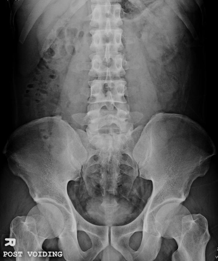
7 PLAIN KUB Renal shadows Psoas shadows Bladder shadow Calcification Bony structure Other soft tissue structures: liver/spleen, abnormal mass

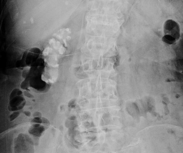
8 STAGHORN STONE 8

9 VESICAL STONE 9
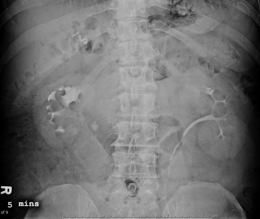
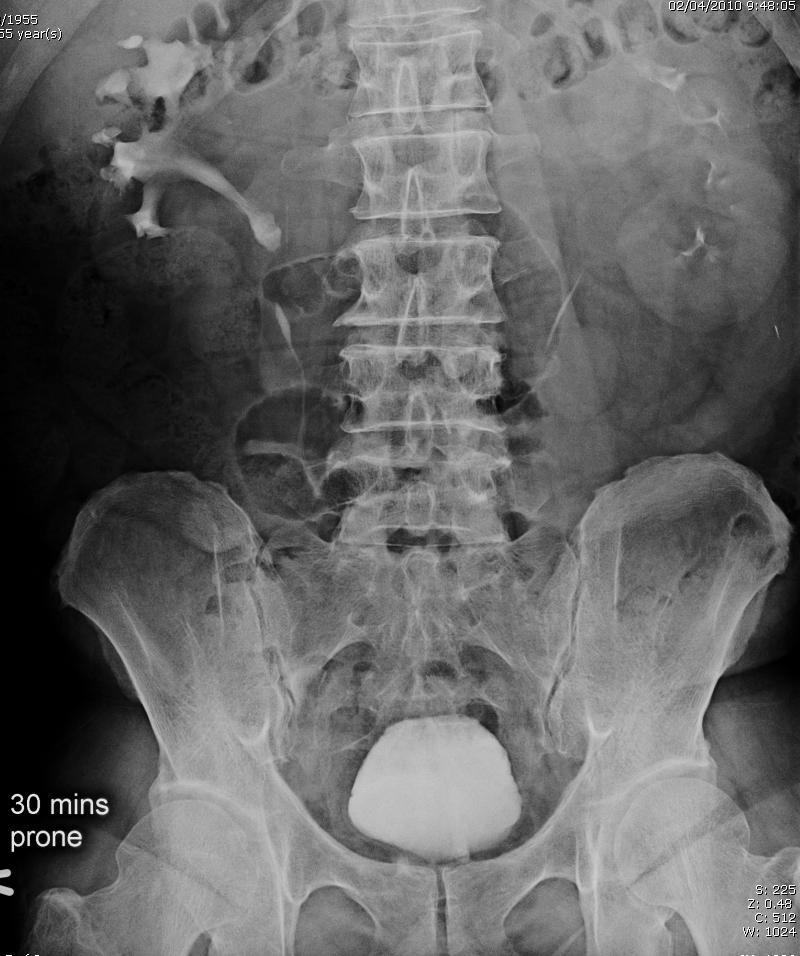
10 Excretory Urography Intravenous pyelography: IVP Intravenous urography: IVU Advantages -Anatomy: kidney and collecting system -Excretory function -Obstructive uropathy 10
11 Excretory Urography Scout image Dynamic study - 1-min film / 5-min of both kidneys min or other delayed images - full bladder image Post-void image 11

12 Normal Pyelogram (IVP 5 min) minor calyx major calyx renal papilla renal papilla renal pelvis ureter


13 NORMAL IVP 13
14 Excretory Urography 1. Anatomy: Renal size, shape, axis and position 2. Excretory function: normal, delay, poor, no excretion 3. Obstructive uropathy: hydronephrosis, hydroureter 4. Distortion or destruction of collecting system 5. Filling defect in pelvocalyceal system, ureter or bladder 14

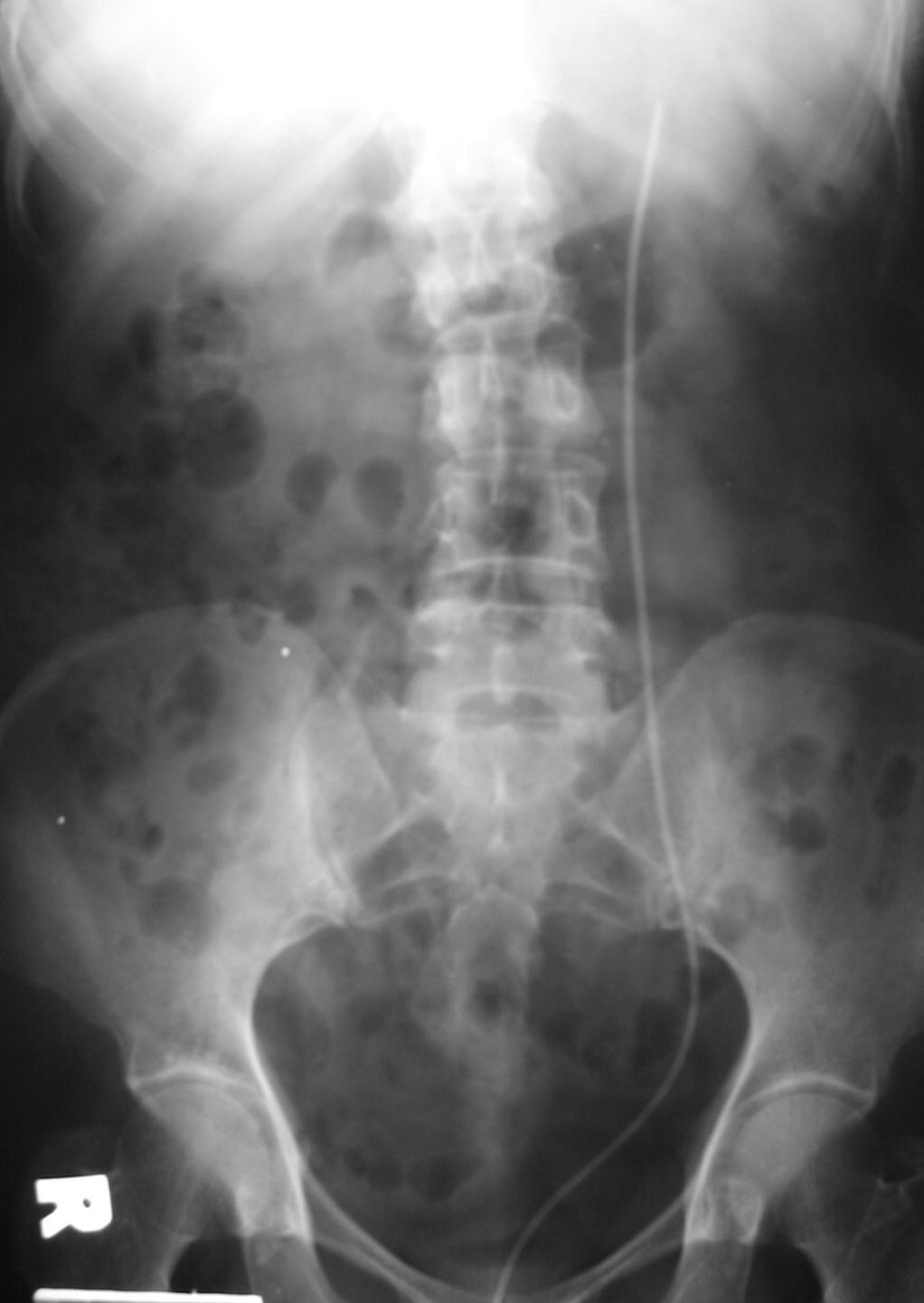
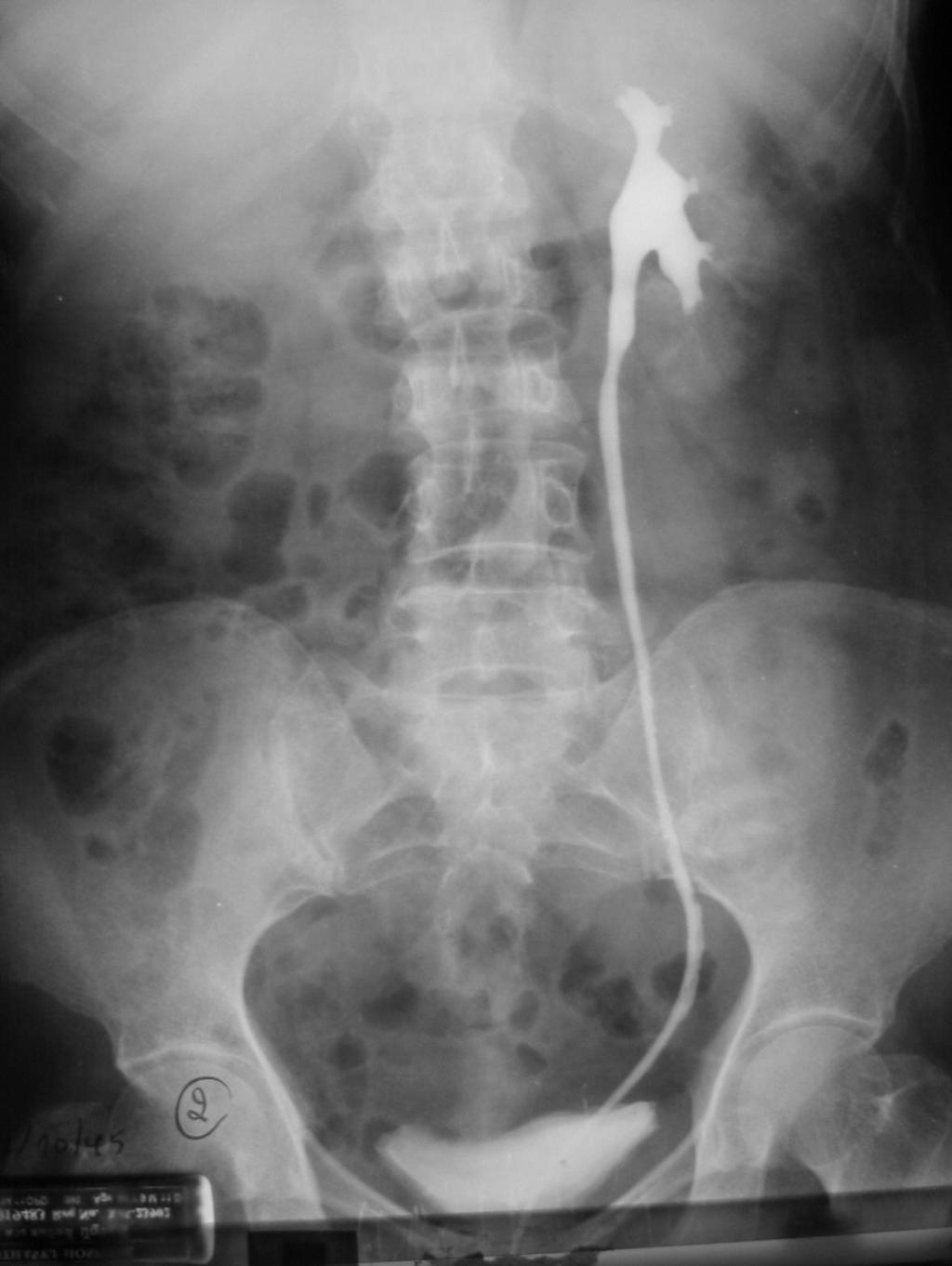
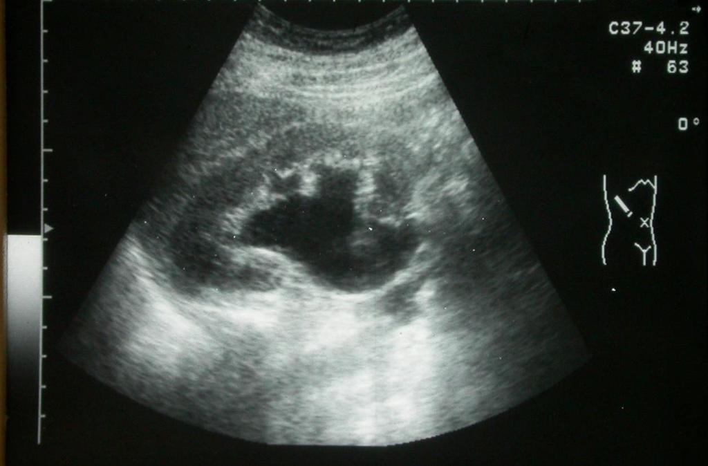
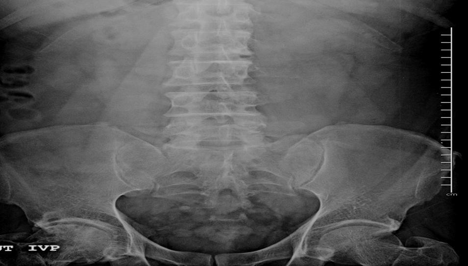
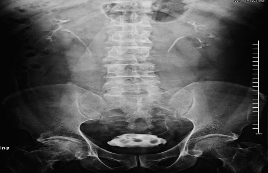
15 A 55-year-old man with hematuria 15

16 16
17 17
18 Retrograde Pyelography (RP) Direct injection of contrast medium into pelvicalyceal system or ureters via the catheter Demonstrates pelvicalyceal system and ureters Limitations - invasive technique: cystoscope - cannot evaluate renal function 18
19 Retrograde Pyelography Indications Evaluate collecting system in patients with hematuria Findings on IVP, US, or CT are not clear Poor renal excretion Contraindication Urinary tract infection 19
20 20
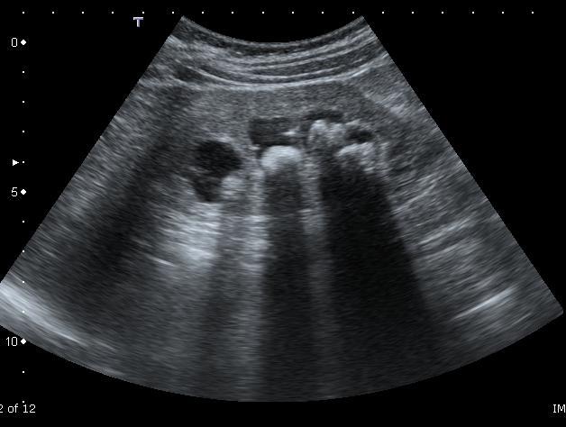
21 ULTRASOUND (US) Safe and cheap Portable No radiation No intravenous contrast medium Advantages for patients with renal failure, children and pregnancy Limitation to evaluate renal function or obese patient
22 KIDNEY Normal sonographic finding Shape: bean shape Size: 9-12 cm. in long axis Echogenicity: - Renal parenchyma: hypoecho and 1 cm in thickness - Central part or renal sinus: hyperecho
23
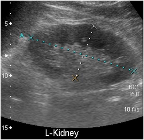
24 HYDRONEPHROSIS

25 CHRONIC RENAL DISEASE 25

26 NORMAL BLADDER 26
27 Color-Doppler Ultrasound Evaluate renal vascular disease - Renal artery - Renal vein Evaluate tumor vascularization

28 Color-Doppler Ultrasound 28
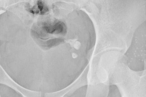
29 COMPUTED TOMOGRAPHY (CT) Indications Detection and characterization of adrenal gland, KUB system and pelvic organ Evaluate retroperitoneal disease Staging tumor and follow up after treatment Limitations Pregnancy: radiation Renal insufficiency/failure : CT need IV contrast medium administration

30 Renal cell carcinoma

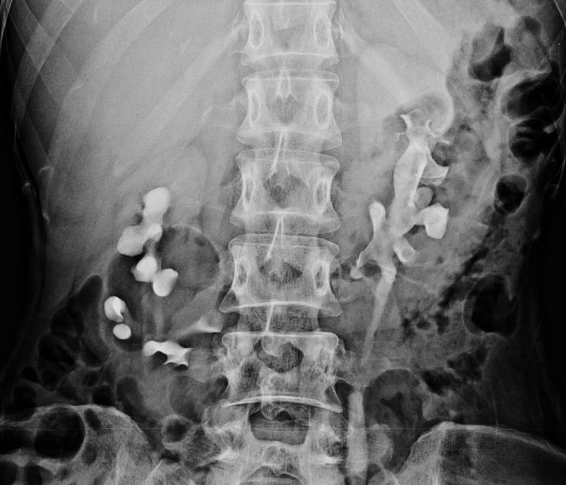
31 CT Urography (CTU)
32 CT ANGIOGRAPHY (CTA)
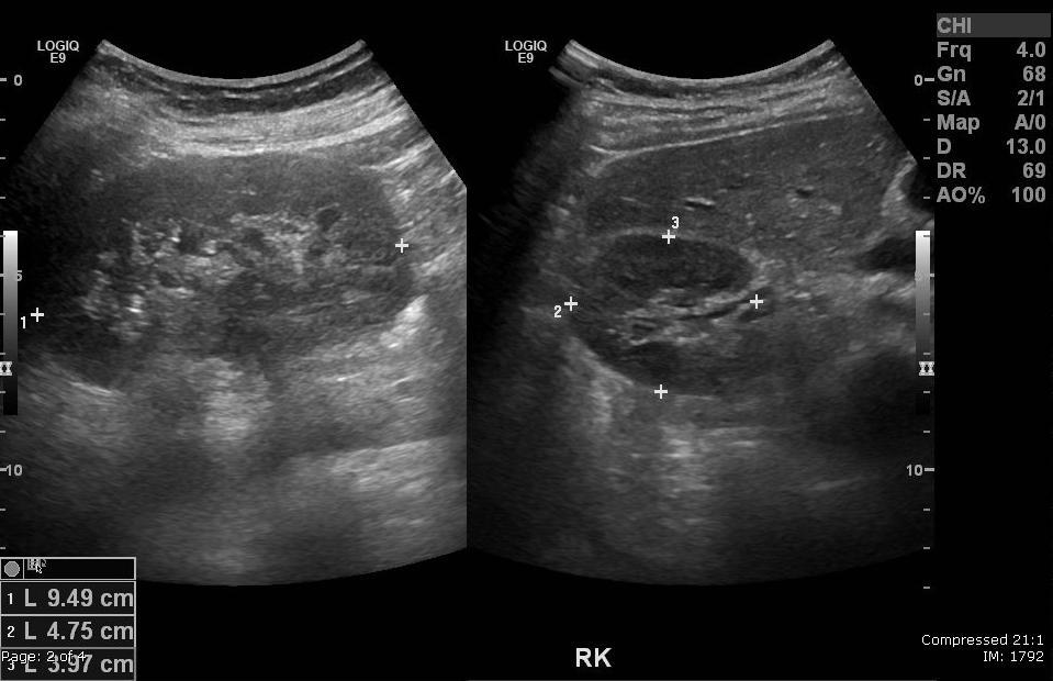
33 Outline Introduction to urinary tract Case base learning of common disease in urinary tract
34 Hematuria Causes of hematuria Stone Tumor Infection


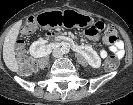
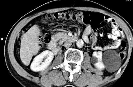
35 Case 1 : A 46-year-old woman with right colicky pain and hematuria
36 Urinary calculi Opaque stones : calcium oxalate, carbonate, phosphate Poor density stones : uric acid, xanthine, cystine
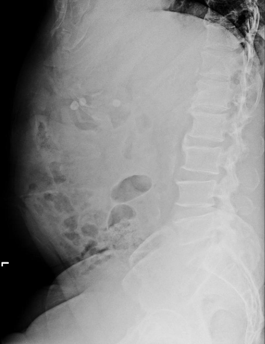
37 Urinary calculi Plain KUB / IVP Location: calyx, renal pelvis, ureter, bladder Shape: staghorn, round/oval (ureteric stone), lamellation (vesical stone) IVP Severity and location of obstruction Renal function
38 Semiopaque vesical stones
39 Differential diagnosis Filling defects in collecting system blood clot tumor stone fungal ball polyp air bubble
40 Ultrasound Urinary calculi Diagnosis of non-opaque stone echogenic material with acoustic shadow Severity and location of obstruction Renal size and cortical thickness

41 Renal stone with hydronephrosis Vesical stone


42 Urinary calculi CT scan Lt.renal stone Lt.renal stone

43 Quiz : Renal or extrarenal calcification?
44 Quiz : Renal or extrarenal calcification?


45 Quiz : Renal or extrarenal calcification?
46 Extra-urinary calcific shadows 1. Calcified costal cartilage 2. Calcified mesenteric LN 3. Calcified iliac and splenic vessels 4. Phleboliths 5. Gallstones, CBD, and cystic duct stones 6. Transverse process of L-spine 7. Intestinal content 8. Calcified appendicolith 9. Pancreatic calcification
47 Case 2 : A 50-year-old woman with underlying DM presented with fever and sepsis


48 Case 2 : A 50-year-old woman with underlying DM and present with fever and sepsis
49 Emphysematous pyelonephritis Rare, life-threatening condition Severe pyelonephritis with gas-producing organism (Gram-neg. bacilli: E.coli) Elderly diabetic patient (DM %) Clinical symptoms & signs : chills, fever, flank pain, lethargy, confusion, septicemic shock
50 Emphysematous pyelonephritis IVP Gas bubbles in renal bed, upper renal collecting system Renal enlargement Delayed or absent renal excretion Obliteration of the renal pelvis CT Investigation of choice Intraparenchymal, intracalyceal, intrapelvic gas and extension into perinephric space

51 Case 3: Spot diagnosis?
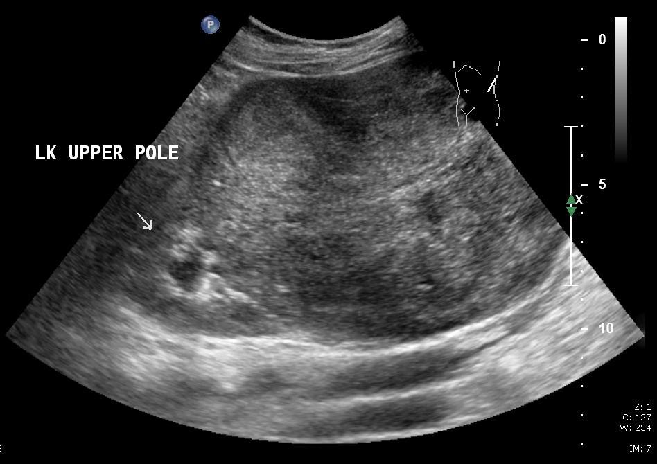
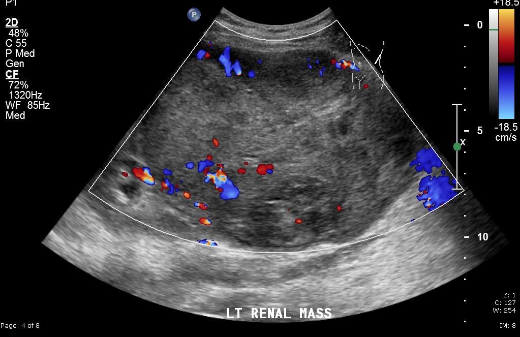
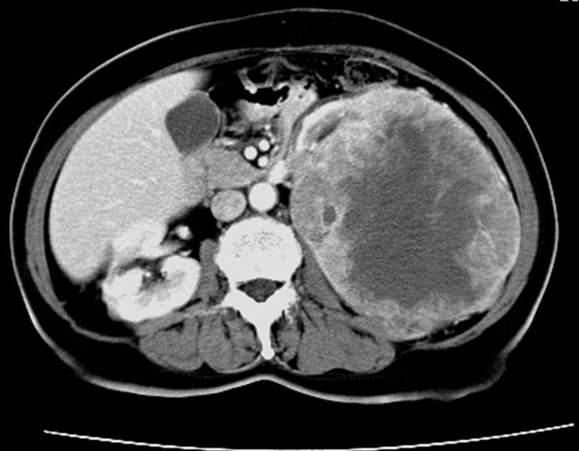
52 Renal Tuberculosis: Typical Findings Early : difficult to detect due to miliary tubercles in renal parenchyma Moderate : minimal irregularity of pyramid, fornix or isthmus : destruction of papilla : isthmus stenosis due to fibrosis : deformity of pelvicalyceal system : cavity (caseous material & necrosis) : reduction in renal size Severe : nephrocalcinosis (putty calcification) : autonephrectomy

53 Case 4: Spot diagnosis?
54 HORSESHOE KIDNEY Most common renal anomaly (1:400 live birth) Male : female = 2:1 Midline connection may be by functioning renal perenchyma or fibrotic band

55 Case 5: Diagnosis?

56 Case 5: Diagnosis?
57 SIMPLE CYST The most common renal masses ~ % of cases Occur in patients > 50 years old Solitary or multiple Bilateral cysts: less common Size: variable No symptom, incidental finding Serous containing
58 SIMPLE CYST US well defined, smooth wall anechoic posterior enhancement CT homogeneous water density thin septation may be identified no enhancement

59 Case 6: A 60-year-old man with hematuria
60
61 Renal cell carcinoma (RCC) Most common primary malignant renal tumor in adults Male : female = 2.5:1 Age : years Increased incidence Tobacco Acquired cystic disease of uremia Hemodialysis
62 Renal cell carcinoma (RCC) Clinical presentation: Classic triad: gross hematuria (60%) flank pain (50%) palpable renal mass (<10%) Distant metastases: lung, bone, liver and brain
63 Renal cell carcinoma (RCC) IVP distortion, enlarged renal portion displaced / obliterated collecting system, filling defect diminished function soft tissue mass US : Hyperechoic mass with inhomogeneity CT : Hypervascular renal mass

64 Case 7: What is it?

65 Case 8: What is type of calcification?
66 Which cases did you learn today? 1.Calculous disease D/D calcification on plain KUB 2.Infection bacterial, TB 3.Renal cystic disease 4.Congenital anomaly - horseshoe kidney 5.Renal tumor 6.Calcified ovarian dermoid/uterine fibroid myoma


67 REFERENCES 1. Armstong P, Rockall A, Wastie M. Diagnostic imaging. 6th ed. Williston: Wiley Blackwell; Dunnick R, Sandler C, Newhouse J. Textbook of Uroradiology. 5th ed. Lippincott Williams & Wilkins;
Outline. Introduction to imaging modalities of the urinary system. Case base learning of common diseases in urinary tract
 Outline Introduction to imaging modalities of the urinary system Case base learning of common diseases in urinary tract Diagnostic Investigations in Urinary System PLAIN KUB EXCRETORY UROGRAPHY RETROGRADE
Contents. Review anatomy of the urinary tract Imaging modalities
 Contents Review anatomy of the urinary tract Imaging modalities The Urinary Tract Kidneys ตาแหน งไต (position) อย ใน retroperitoneum ระด บ T12-L3 โดยไต ขวาจะม ระด บตากว าไตซ ายเล กน อย ร ปร าง (shape)
Excretory urography (EU) or IVP US CT & radionuclide imaging
 or IVP US CT & radionuclide imaging") Excretory urography (EU) or IVP US CT & radionuclide imaging MRI arteriography studies requiring catherization or direct puncture of collecting system EU & to a lesser extent CT provide both functional
Excretory urography (EU) or IVP US CT & radionuclide imaging MRI arteriography studies requiring catherization or direct puncture of collecting system EU & to a lesser extent CT provide both functional
US in non-traumatic acute abdomen. Lalita, M.D. Radiologist Department of radiology Faculty of Medicine ChiangMai university
 US in non-traumatic acute abdomen Lalita, M.D. Radiologist Department of radiology Faculty of Medicine ChiangMai university Sagittal Orientation Transverse (Axial) Orientation Coronal Orientation Intercostal
PROFESSIONAL SKILLS 1 3RD YEAR SEMESTER 6 RADIOGRAPHY. THE URINARY SYSTEM Uz. Fatema shmus aldeen Tel
 PROFESSIONAL SKILLS 1 3RD YEAR SEMESTER 6 RADIOGRAPHY THE URINARY SYSTEM Uz. Fatema shmus aldeen Tel. 0925111552 Professional skills-2 THE URINARY SYSTEM The urinary system (review anatomy and physiology)
Proceedings of the 34th World Small Animal Veterinary Congress WSAVA 2009
 www.ivis.org Proceedings of the 34th World Small Animal Veterinary Congress WSAVA 2009 São Paulo, Brazil - 2009 Next WSAVA Congress : Reprinted in IVIS with the permission of the Congress Organizers IMAGING
Acute flank pain in children: Imaging considerations
 Acute flank pain in children: Imaging considerations Carlos J. Sivit MD Rainbow Babies and Children s Hospital Case Western Reserve School of Medicine Flank pain Results from distention of ureter or renal
Sex: 女 Age: 51 Occupation: 無 Admission date:92/07/22
 Sex: 女 Age: 51 Occupation: 無 Admission date:92/07/22 Chief complaint Unknown fever for one month Hand tremor and left huge renal tumor was noted Present illness Suffered from fever for one month, hand
R adio logical investigations of urinary system
 R adio logical investigations of urinary system There are 4 main radiological Ix: 1 IVU: Intravenous urography. 2- U/S 3-CT scan 4-Radioisotope scan. Others (not frequently used): MRI, arteriography, antegrade
Urinary system Ultrasound (Renal & Urinary bladder)
") Urinary system Ultrasound (Renal & Urinary bladder) Edited & Presented by ; Hussien A.B ALI DINAR. Msc.Phd ISRRT Associate Member Lecturer (National university) Reporting Sonographer (PHC) Objective By
Urinary system Ultrasound (Renal & Urinary bladder) Edited & Presented by ; Hussien A.B ALI DINAR. Msc.Phd ISRRT Associate Member Lecturer (National university) Reporting Sonographer (PHC) Objective By
My Patient Has Abdominal Pain PoCUS of the Biliary Tract and the Urinary Tract
 My Patient Has Abdominal Pain PoCUS of the Biliary Tract and the Urinary Tract Objectives PoCUS for Biliary Disease PoCUS for Renal Colic PoCUS for Urinary Retention Biliary Disease A patient presents
Abdominal ultrasound:
 Abdominal ultrasound: Non-traumatic acute abdomen Wittanee Na-ChiangMai, MD Department of Radiology ChiangMai University 26/04/2017 Contents Technique of examination Normal anatomy Emergency conditions
Lec-8 جراحة بولية د.نعمان
 4th stage Lec-8 جراحة بولية د.نعمان 11/10/2015 بسم هللا الرحمن الرحيم Ureteric, Vesical, & urethral stones Ureteric Calculus Epidemiology like renal stones Etiology like renal stones Risk factors like
Uroradiology For Medical Students
 Uroradiology For Medical Students Lesson 4: Cystography & Urethrography - Part 2 American Urological Association Review Cystography is useful in evaluating the bladder, the urethra and the competence of
Urinary tract obstruction
 Urinary tract obstruction Common causes : stone, blood clot Radiographic findings depend on I. Level of obstruction II. Severity of obstruction : partial or complete III. Timing of obstruction Pathophysiology
Nonurographic evaluation of renal calculous disease 1
 Contributions Nonurographic evaluation of renal calculous disease 1 Gregory P. Borkowski, M.D. Craig R. George, M.D. Peter B. O'Donovan, M.D. While excretory urography has been useful in the evaluation
Autosomal Dominant Polycystic Kidney Disease
 Case Studies [1] July 01, 2014 By Amar Udare, MBBS [2] Case History: 45-year-old female with vague pain in the abdomen. Case History: A 45-year-old female presented with vague pain in the abdomen. A USG
Renal masses - the role of diagnostic imaging
 Renal masses - the role of diagnostic imaging Poster No.: C-2471 Congress: ECR 2015 Type: Educational Exhibit Authors: V. Rai#; Bjelovar/HR Keywords: Cysts, Cancer, Structured reporting, Ultrasound, MR,
CASE REPORT RENAL TUBERCULOSIS CAUSE OF RENAL REPLACEMENT LIPOMATOSIS : A RARE ASSOCIATION
 CASE REPORT RENAL TUBERCULOSIS CAUSE OF RENAL REPLACEMENT LIPOMATOSIS : A RARE ASSOCIATION DR ANAND AARTI 1, DR CHANDAK PRIYA 2,DR SURESH PARVATHY 3 1. PROF AND HOD, DEPARTMENT OF RADIODIAGNOSIS, GOVERNMENT
Hydronephrosis. What is hydronephrosis?
 What is hydronephrosis? Hydronephrosis Hydronephrosis describes the situation where the urine collecting system of the kidney is dilated. This may be a normal variant or it may be due to an underlying
Abdominal Ultrasound : Aorta, Kidneys, Bladder
 Abdominal Ultrasound : Aorta, Kidneys, Bladder Nilam J. Soni, MD, MSc Associate Professor of Medicine Divisions of Hospital Medicine and Pulmonary/Critical Care Medicine Department of Medicine University
Acute renal colic Radiological investigation in patients with renal colic
 Acute renal colic Radiological investigation in patients with renal colic Mikael Hellström Professor Department of Radiology Sahlgrenska University Hospital Göteborg University 0.9-1.8/1.000 inhabitants
Uroradiology Tutorial For Medical Students
 Uroradiology Tutorial For Medical Students Lesson 3: Cystography & Urethrography Part 1 American Urological Association Introduction Conventional radiography of the urinary tract includes several diagnostic
URINARY TRACT IMAGING - BASIC PRINCIPLES
 URINARY TRACT IMAGING - BASIC PRINCIPLES Clinical Radiology Every physician needs a basic understanding of diagnostic imaging to understand how to order the appropriate studies and to understand the resulting
Imaging the Urinary Tract
 Imaging the Urinary Tract Laura Armbrust, DVM, DACVR Gregory F. Grauer, DVM, MS, DACVIM Kansas State University Radiographic and ultrasound imaging in addition to history, physical examination, and clinicopathologic
Role of imaging in RCC. Ultrasonography. Solid lesion. Cystic RCC. Solid RCC 31/08/60. From Diagnosis to Treatment: the Radiologist Perspective
 Role of imaging in RCC From Diagnosis to Treatment: the Radiologist Perspective Diagnosis Staging Follow up Imaging modalities Limitations and pitfalls Duangkamon Prapruttam, MD Department of Therapeutic
Five Views of Transitional Cell Carcinoma: One Man s Journey
 September 2006 Five Views of Transitional Cell Carcinoma: One Man s Journey Amsalu Dabela, Harvard Medical School III Outline Overview: Renal Anatomy Our Patient s Story Diagnostic Imaging Studies Appearance
Radiological Assessment of the Kidney in Patients with Hematuria
 March 2005 Radiological Assessment of the Kidney in Patients with Hematuria Jeremy L. McKay, Harvard Medical School Year III Hematuria Signs and Symptoms Microscopic or gross hematuria Abdominal pain Fever
Imaging of liver and pancreas
 Imaging of liver and pancreas.. Disease of the liver Focal liver disease Diffusion liver disease Focal liver disease Benign Cyst Abscess Hemangioma FNH Hepatic adenoma HCC Malignant Fibrolamellar carcinoma
Obstetrics Content Outline Obstetrics - Fetal Abnormalities
 Obstetrics Content Outline Obstetrics - Fetal Abnormalities Effective February 2007 10 16% renal agenesis complete absence of the kidneys occurs when ureteric buds fail to develop Or degenerate before
Acute Pyelonephritis
 Acute Pyelonephritis Variant 1: Acute pyelonephritis. Uncomplicated patient (eg, no history of diabetes or immune compromise or history of stones or obstruction or prior renal surgery or lack of response
Chapter 6: Genitourinary and Gastrointestinal Systems 93
 Chapter 6: Genitourinary and Gastrointestinal Systems 93 Chapter 6 Genitourinary and Gastrointestinal Systems Embryology Three sets of excretory organs or kidneys develop in human embryos: Pronephros:
Kidney & Urinary Tract Ultrasound. Fatina Fadel Hafez Bazaraa
 Kidney & Urinary Tract Ultrasound Fatina Fadel Hafez Bazaraa Ultrasonography Ultrasound Available Rapid Inexpensive Painless & no sedation needed No adverse effects/ complications Can be repeated Useful
URINARY SYSTEM I. Kidneys II. Nephron Unit and Urine Formation
 URINARY SYSTEM I. Kidneys A. Location and Structure 1. Retroperitoneal 2. Between T12 and L3 3. Rt. kidney slightly lower 4. Two bean shaped organs 5. Adrenal gland 6. Internal construction a. Renal cortex
RADIOLOGY OF THE URINARY TRACT CHAPTER 9 239
 RADIOLOGY OF THE URINARY TRACT CHAPTER 9 239 in length. They lie cephalad to the kidneys, with the right just posterior to the inferior vena cava (IVC) and the left anteromedial to the upper pole of the
이학종분당서울대학교병원. Ultrasound in Urinary Colic
 이학종분당서울대학교병원 Ultrasound in Urinary Colic U l t r a s o u n d i n U r i n a US: Normal Kidney r y C o l i c Contents 1. 1. Definition and clinical consideration 2. 2. Pathophysiology 3. 3. US in in obstructive
Abdomen and Retroperitoneum Ultrasound Protocols
 Abdomen and Retroperitoneum Ultrasound Protocols Reviewed By: Anna Ellermeier, MD Last Reviewed: March 2018 Contact: (866) 761-4200, Option 1 **NOTE for all examinations: 1. If documenting possible flow
IMAGING OF THE UROGENITAL TRACT
 IMAGING OF THE UROGENITAL TRACT 1 A) URINARY TRACT There are many methods of imaging the urinary tract but plain abdominal X-ray and ultrasound scan are usually done first in most cases, especially in
Candida Bezoars with Urinary Tract Obstruction in Two Women without Immunocompromising Conditions
 Case Study TheScientificWorldJOURNAL (2011) 11, 1168 1172 TSW Urology ISSN 1537-744X; DOI 10.1100/tsw.2011.119 Candida Bezoars with Urinary Tract Obstruction in Two Women without Immunocompromising Conditions
Ultrasonographic diagnosis and typing of renal tuberculosis
 International Journal of Urology (2008) 15, 135 139 doi: 10.1111/j.1442-2042.2007.01962.x, Original Article: Clinical Investigation Ultrasonographic diagnosis and typing of renal tuberculosis Xuefang Rui,
Find Medical Solutions to Your Problems HYDRONEPHROSIS. (Distension of Renal Calyces & Pelvis)
") HYDRONEPHROSIS (Distension of Renal Calyces & Pelvis) Hydronephrosis is the distension of the renal calyces and pelvis due to accumulation of the urine as a result of the obstruction to the outflow of
HYDRONEPHROSIS (Distension of Renal Calyces & Pelvis) Hydronephrosis is the distension of the renal calyces and pelvis due to accumulation of the urine as a result of the obstruction to the outflow of
Sonographic Features of Necrosed Renal Papillae Causing Hydronephrosis
 Case Series Sonographic Features of Necrosed Renal Papillae Causing Hydronephrosis S. Boopathy Vijayaraghavan, MD, DMRD, Sangampalayam Vedhanayagam Kandasamy, MS, MCh, Mylsamy Arul, MS, DNB (Uro), Muniappan
In Canada, there was a 25% reduction in incidence of genitourinary TB in the period compared with An interesting speculation
 Renal T B EPIDEMIOLOGY Young to middle age usually affected, rare in children Male : female ratio = 2:1 True prevalence and incidence not known as patients are usually asymptomatic With HIV pandemic, there
Genitourinary Radiology In-Training Test Questions for Diagnostic Radiology Residents
 Genitourinary Radiology In-Training Test Questions for Diagnostic Radiology Residents March, 2013 Sponsored by: Commission on Education Committee on Residency Training in Diagnostic Radiology 2013 by American
Guidelines, Policies and Statements D5 Statement on Abdominal Scanning
 Guidelines, Policies and Statements D5 Statement on Abdominal Scanning Disclaimer and Copyright The ASUM Standards of Practice Board have made every effort to ensure that this Guideline/Policy/Statement
Imaging findings in renal infections
 Imaging findings in renal infections Poster No.: C-0221 Congress: ECR 2013 Type: Educational Exhibit Authors: I. lópez blasco, D. Soriano Mena, R. Pastor Toledo, S. Paz Maya, A. M. Julve Parreño, J. Palmero
Radiology of hepatobiliary diseases
 GI cycle - Lecture 14 436 Teams Radiology of hepatobiliary diseases Objectives 1. To Interpret plan x-ray radiograph of abdomen with common pathologies. 2. To know the common pathologies presentation.
Case MDCT 3D reconstructed features of posterior urethral valve
 Case 12688 MDCT 3D reconstructed features of posterior urethral valve Hidayatullah Hamidi Third year Resident of Radiology French medical institute for children Radiology Department; Kabul, Afghanistan;
ASSESSING THE PLAIN ABDOMINAL RADIOGRAPH M A A M E F O S U A A M P O F O
 ASSESSING THE PLAIN ABDOMINAL RADIOGRAPH M A A M E F O S U A A M P O F O Introduction The abdomen (less formally called the belly, stomach, is that part of the body between the thorax (chest) and pelvis,
Plain abdomen The standard films are supine & erect AP views (alternative to erect, lateral decubitus film is used in ill patients).
.") Plain abdomen The standard films are supine & erect AP views (alternative to erect, lateral decubitus film is used in ill patients). The stomach can be readily identified by its location, gastric rugae
Plain abdomen The standard films are supine & erect AP views (alternative to erect, lateral decubitus film is used in ill patients). The stomach can be readily identified by its location, gastric rugae
Abdominal Ultrasound. Diane Hallinen, MD. Bloodroot
 Abdominal Ultrasound Diane Hallinen, MD Bloodroot Abdominal Ultrasound Vasculature Hepatobiliary Spleen Kidney Bladder Bowel Where to put the probe? Vasculature We are going to talk about Celiac Trunk
Imaging of common diseases of hepatobiliary and GI system
 Imaging of common diseases of hepatobiliary and GI system Natthaporn Tanpowpong, M.D. Diagnostic radiology Faculty of Medicine, Chulalongkorn University Normal plain radiograph A = Common bile duct
 INTRAUTERINE DEVICE = IUD INTRAUTERINE DEVICE = IUD CONGENITAL DISORDERS Pyometra = pyometrea is a uterine infection, it is accumulation of purulent material in the uterine cavity. Ultrasound is usually
Abdomen and Pelvis CT (1) By the end of the lecture students should be able to:
 By the end of the lecture students should be able to:") RAD 451 Abdomen and Pelvis CT (1) By the end of the lecture students should be able to: State the common indications for Abdomen and pelvis CT exams Identify possible contra indications for Abdomen and
RAD 451 Abdomen and Pelvis CT (1) By the end of the lecture students should be able to: State the common indications for Abdomen and pelvis CT exams Identify possible contra indications for Abdomen and
Urinary System Part of the Excretory System
 Urinary System Part of the Excretory System Bellwork **only write the term and underlined definition INCONTINENCE involuntary urination, often seem in older persons, or due to illness and disease ENURESIS
Primary Renal Candidiasis
 Case Series Primary Renal Candidiasis Importance of Imaging and Clinical History in Diagnosis and Management Barry J. Sadegi, MD, Bhargavi K. Patel, MD, Andrew C. Wilbur, MD, Anil Khosla, MD, Ejaz Shamim,
Imaging the Urogenital System
 maging the Urogenital System Tony Pease, DVM, MS, DACVR Assistant Professor of Radiology North Carolina State University Reading Thrall Chapters 42-46 Prostate Gland Not visible radiographically in normal
Separating and Distorted Nephroliths Signs of Renal Squamous Cell Carcinoma
 Chin J Radiol 2003; 28: 203-208 203 Separating and Distorted Nephroliths Signs of Renal Squamous Cell Carcinoma TZE-YU LEE SHEUNG-FAT KO CHUNG-CHENG HUANG YU-FENG CHENG Department of Radiology, Chang Gung
Audit of split-bolus CT urography for the investigation of haematuria over a 12 month period at two district general hospitals
 Audit of split-bolus CT urography for the investigation of haematuria over a 12 month period at two district general hospitals Poster No.: C-1349 Congress: ECR 2010 Type: Educational Exhibit Topic: Genitourinary
The 82 nd UWI/BAMP CME Conference November 18, Jeetu Nebhnani MBBS D.M. Urology Consultant Urologist
 The 82 nd UWI/BAMP CME Conference November 18, 2017 Jeetu Nebhnani MBBS D.M. Urology Consultant Urologist Disclosures Outline Index case Introduction Etiology Risk factors Acute stone event Conservative
A Case of Calcified Ureteritis Cystica: An Indiscernible Condition from Ureterolithiasis
 Prague Medical Report / Vol. 110 (2009) No. 3, p. 245 249 245) A Case of Calcified Ureteritis Cystica: An Indiscernible Condition from Ureterolithiasis Alicioglu B. 1, Kaplan M. 2, Aktoz T. 3, Atakan I.
Rad Lab 4 Unknowns: Genitourinary!
 Rad Lab 4 Unknowns: Genitourinary! Peter Clarke MD! Don Di Salvo, MD! Clerkship Directors for Radiology! Harvard Medical School! Brigham and Women s Hospital! Dana Farber Cancer Institute! Case 1: 69 year
Role of imaging in evaluation of genitourinary i trauma Spectrum of GU injuries Relevance of imaging findings in determining management Focus on MDCT
 Genitourinary Tract Injuries 6 th Nordic Course Scott D. Steenburg, MD Assistant Professor University of Maryland Department of Radiology Division of Trauma and Emergency Radiology R Adams Cowley Shock
Caveat sonologist Mistakes to avoid in Kidney Ultrasound
 Caveat sonologist Mistakes to avoid in Kidney Ultrasound Simon Freeman Derriford Hospital, Plymouth simonfreeman@nhs.net Bear trap 1 Report: There is a 4cm solid mass arising from the left kidney likely
Kidneys and Urinary Tract Content Outline. Anatomy Coverings. Location. (Effective February 2007) (16%-24%)
 (16%-24%)") Kidneys and Urinary Tract Content Outline (Effective February 2007) (16%-24%) Anatomy Coverings true capsule perirenal fat surrounds capsule Gerota s fascia separates perirenal from extraperitoneal fat
Kidneys and Urinary Tract Content Outline (Effective February 2007) (16%-24%) Anatomy Coverings true capsule perirenal fat surrounds capsule Gerota s fascia separates perirenal from extraperitoneal fat
By GEORGE E. NELIGAN, M.C., M.A., B.M,, B.Ch. (Oxon.), F.R.C.S. (Swrgeon with charge of Out-patients and Surgeon in charge of the Genito-Urinary
, F.R.C.S. (Swrgeon with charge of Out-patients and Surgeon in charge of the Genito-Urinary") 426 POST-GRADUATE MEDICAL JOURNAL November, 1935 RENAL TUMOURS. By GEORGE E. NELIGAN, M.C., M.A., B.M,, B.Ch. (Oxon.), F.R.C.S. (Swrgeon with charge of Out-patients and Surgeon in charge of the Genito-Urinary
426 POST-GRADUATE MEDICAL JOURNAL November, 1935 RENAL TUMOURS. By GEORGE E. NELIGAN, M.C., M.A., B.M,, B.Ch. (Oxon.), F.R.C.S. (Swrgeon with charge of Out-patients and Surgeon in charge of the Genito-Urinary
XANTHOGRANULOMATOUS PYELONEPHRITIS: radiologic review.
 XANTHOGRANULOMATOUS PYELONEPHRITIS: radiologic review. Poster No.: C-0557 Congress: ECR 2014 Type: Educational Exhibit Authors: M. Barral, J. M. Sánchez Crespo, J. C. Pérez Herrera, J. L. 1 2 3 1 1 1 Ortega
Genitourinary. Common Clinical Scenarios Protocoling Module. Patty Ojeda & Mariam Shehata
 The following training module was developed as a quality improvement project to serve as an educational tool for junior radiology residents. The following diagnostic radiology protocoling modules were
RENAL SCINTIGRAPHY IN THE 21 st CENTURY
 RENAL SCINTIGRAPHY IN THE 21 st CENTURY 99m Tc- MAG 3 with zero time injection of Furosemide (MAG 3 -F 0 ) : A Fast and Easy Protocol, One for All Indications Clinical Experience Congenital Disorders PROTOCOL
CYSTIC DISEASES of THE KIDNEY. Dr. Nisreen Abu Shahin
 CYSTIC DISEASES of THE KIDNEY Dr. Nisreen Abu Shahin 1 Types of cysts 1-Simple Cysts 2-Dialysis-associated acquired cysts 3-Autosomal Dominant (Adult) Polycystic Kidney Disease 4-Autosomal Recessive (Childhood)
Imaging spectrum of genitourinary tuberculosis: Our experience at a tertiary care centre of a third world country
 Imaging spectrum of genitourinary tuberculosis: Our experience at a tertiary care centre of a third world country Poster No.: C-361 Congress: ECR 2009 Type: Educational Exhibit Topic: Genitourinary Authors:
Hepatobiliary Ultrasound Rimon Bengiamin, MD, RDMS Assistant Clinical Professor Director of Emergency Ultrasound UCSF Fresno. Objectives. Why?
 Hepatobiliary Ultrasound Rimon Bengiamin, MD, RDMS Assistant Clinical Professor Director of Emergency Ultrasound UCSF Fresno Objectives Discuss the goals of point-of-care biliary ultrasound Review the
Radiographic Procedures III (RAD 228)
") Radiographic Procedures III (RAD 228) Urinary System RADIOGRAPHIC EXAMINATIONS Urinary System Antegrade Exam IVU Functional test Hypertensive evaluation as per protocol Retrograde Exams Retrograde Urography
Radiographic Procedures III (RAD 228) Urinary System RADIOGRAPHIC EXAMINATIONS Urinary System Antegrade Exam IVU Functional test Hypertensive evaluation as per protocol Retrograde Exams Retrograde Urography
Fetal Renal Malformations: The Role of Ultrasound in Diagnosis & Management
 Fetal Renal Malformations: The Role of Ultrasound in Diagnosis & Management 12 weeks Alfred Abuhamad, M.D. Eastern Virginia Medical School 13 weeks 2nd trimester Medullary pyramids Renal Sinus Cortex 2nd
Lab Monitor Images Dissection of the Abdominal Vasculature + Lower Digestive System
 Lab Monitor Images Dissection of the Abdominal Vasculature + Lower Digestive System Stomach & Duodenum Frontal (AP) View Nasogastric tube 2 1 3 4 Stomach Pylorus Duodenum 1 Duodenum 2 Duodenum 3 Duodenum
The Diagnostic Value of Color Doppler Ultrasound in Ureteral Calculi
 International Journal of Medical Imaging 2018; 6(2): 12-17 http://www.sciencepublishinggroup.com/j/ijmi doi: 10.11648/j.ijmi.20180602.11 ISSN: 2330-8303 (Print); ISSN: 2330-832X (Online) The Diagnostic
ULTRASONOGRAPHY RADIOLOGY Simple abdominal X-ray Intravenous urography Retrograde/anterograde pieloureterography Renal angiography CT
 ULTRASONOGRAPHY RADIOLOGY Simple abdominal X-ray Intravenous urography Retrograde/anterograde pieloureterography Renal angiography CT NUCLEAR MEDICINE Static studies: static renal scintigraphy Dynamic
ID data. Sex: female Age: 46y/o Birthday: 1955/10/13
 ID data Sex: female Age: 46y/o Birthday: 1955/10/13 Chief Complain Right upper quadrate abdominal tenderness for one month. Present illness (1) This 46 years old female patient was in a healthy condition
CLINICAL PRESENTATION AND RADIOLOGY QUIZ QUESTION
 Donald L. Renfrew, MD Radiology Associates of the Fox Valley, 333 N. Commercial Street, Suite 100, Neenah, WI 54956 1/22/2011 Radiology Quiz of the Week # 4 Page 1 CLINICAL PRESENTATION AND RADIOLOGY QUIZ
Male genital tract tumors. SiCA. Division of Urology, Department of Surgery, Faculty of Medicine Siriraj Hospital.
 Male genital tract tumors Division of Urology, Department of Surgery, Faculty of Medicine Siriraj Hospital. adenocarcinoma Prostate Cancer most common male cancer in western countries more detected in
EVALUATION OF SUSPECTED RENAL COLIC PATIENTS WITH UNENHANCED LOW-DOSE MULTI-DETECTOR COMPUTED TOMOGRAPHY
 190 EAST AFRICAN MEDICAL JOURNAL April 2009 East African Medical Journal Vol. 85 No. 4 April 2009 EVALUATION OF SUSPECTED RENAL COLIC PATIENTS WITH UNENHANCED LOW-DOSE MULTI-DETECTOR COMPUTED TOMOGRAPHY
CLINICAL PRESENTATION AND RADIOLOGY QUIZ QUESTION
 Donald L. Renfrew, MD Radiology Associates of the Fox Valley, 333 N. Commercial Street, Suite 100, Neenah, WI 54956 8/20/2011 Radiology Quiz of the Week # 34 Page 1 CLINICAL PRESENTATION AND RADIOLOGY
Congenital Pediatric Anomalies: A Collection of Abdominal Scintigraphy Findings: An Imaging Atlas
 ISPUB.COM The Internet Journal of Nuclear Medicine Volume 5 Number 1 Congenital Pediatric Anomalies: A Collection of Abdominal Scintigraphy Findings: An Imaging Atlas V Vijayakumar, T Nishino Citation
Obstructive Uropathy. PATHOPHYSIOLOGIC CHANGES UUO vs BUO. Arry Rodjani Urology Department Ciptomangunkusumo Hospital Jakarta
 Obstructive Uropathy PATHOPHYSIOLOGIC CHANGES UUO vs BUO Arry Rodjani Urology Department Ciptomangunkusumo Hospital Jakarta INTRODUCTION Obstructive uropathy refers to the functional or anatomic obstruction
Imaging Ejaculatory Disorders and Hematospermia
 ATHENS 4-6 October 2018 European Society of Urogenital Radiology Imaging Ejaculatory Disorders and Hematospermia Parvati Ramchandani, MD Professor, Radiology and Surgery University of Pennsylvania Medical
Original Research Article
 Original Research Article Role of (Non Gynaecological Causes) Kaleem Ahmad 1, Rishav Kumar Jain 2, Ashok Yadav 3, Shilpa Vahikar 4 1 Associate Professor, Department of Radiodiagnosis, 2 Professor, Department
Kristina M. Nowitzki, M.D., Ph.D. and Hao S. Lo, M.D. University of Massachusetts Medical School, Worcester, MA
 Kristina M. Nowitzki, M.D., Ph.D. and Hao S. Lo, M.D. University of Massachusetts Medical School, Worcester, MA Outline I. Introduction highlighting normal renal enhancement physiology including normal
Pediatric Ure-Radiology*
 Pediatric Ure-Radiology* HERMAN GROSSMAN, M.D. Professor of Radiology and Pediatrics, Duke University Medical Center, Durham, North Carolina "Routine" radiologic studies do not, often enough, concentrate
Nephrographic and Pyelographic Analysis of CT Urography: Principles, Patterns, and Pathophysiology
 Genitourinary Imaging Review Wolin et al. CT Urography Principles, Patterns, and Genitourinary Imaging Review FOCUS ON: Ely A. Wolin 1 David S. Hartman J. Ryan Olson Wolin EA, Hartman DS, Olson JR Keywords:
Pediatric Retroperitoneal Masses Radiologic-Pathologic Correlation
 Acta Radiológica Portuguesa, Vol.XVIII, nº 70, pág. 61-70, Abr.-Jun., 2006 Pediatric Retroperitoneal Masses Radiologic-Pathologic Correlation Marilyn J. Siegel Mallinckrodt Institute of Radiology, Washington
In any operation. Indications. Anaesthesia. Position of the patient. Incision. Steps of the operation. Complications.
 In any operation Indications. Anaesthesia. Position of the patient. Incision. Steps of the operation. Complications. Abdominal operation I position for operation Supine Abdominal operation I position for
GU Ultrasound in First Trimester
 Fetal Renal Malformations: The Role of Ultrasound in Diagnosis & Management Outline 1. Renal Anomalies Urinary Tract Dilation Aberrant Early Development Defects Terminal Maturation Alfred Abuhamad, M.D.
Lecture 56 Kidney and Urinary System
 Lecture 56 Kidney and Urinary System The adrenal glands are located on the superomedial aspect of the kidney The right diagram shows a picture of the kidney with the abdominal walls and organs removed
Chris McGuire Gillian Lieberman, MD. May Schistosomiasis. Chris McGuire, Harvard Medical School Year III Gillian Lieberman, MD
 May 2003 Schistosomiasis Chris McGuire, Harvard Medical School Year III Overview Introduction to schistosomiasis Katayama syndrome Patient A Patient B Ultrasound detection Bladder infection Kidney infection
Appendix 5. EFSUMB Newsletter. Gastroenterological Ultrasound
 EFSUMB Newsletter 87 Examinations should encompass the full range of pathological conditions listed below A log book listing the types of examinations undertaken should be kept Training should usually
HMM 4401 Genito-urinary tract diseases
 HMM 4401 Genito-urinary tract diseases Urine production Core elements: Glomerulus, proximal and distal convoluted tube, loop of Henle, collecting tubules, ureters, bladder, sphincter, uretra, and out
Urinary Tract Infections KIDNEY INFECTIONS. Dr. AMMAR FADIL
 Urinary Tract Infections KIDNEY INFECTIONS Dr. AMMAR FADIL General principles Urinary tract infections (UTIs) is inflammatory response of the urothelium to bacterial invasion. are common affect men and
PICTORIAL ESSAY. Experiences of using a single post-contrast CT scan of the urinary tract after triphasic contrast injection
 Experiences of using a single post-contrast CT scan of the urinary tract after triphasic contrast injection P C Pretorius, FCRad (Diag) SA Drs Visser, Erasmus, Vawda & Partners, Port Elizabeth Corresponding
Kidney Case 1 SURGICAL PATHOLOGY REPORT
 Kidney Case 1 Surgical Pathology Report February 9, 2007 Clinical History: This 45 year old woman was found to have a left renal mass. CT urography with reconstruction revealed a 2 cm medial mass which
Paediatric surgical emergencies. Mani Thyagarajan BWCH
 Paediatric surgical emergencies Mani Thyagarajan BWCH General points Always discuss Call consultant for help ASAP CT scan is a bad modality in paediatrics Ultrasound? Intussusception? Renal colic? UTI