CASE REPORT RENAL TUBERCULOSIS CAUSE OF RENAL REPLACEMENT LIPOMATOSIS : A RARE ASSOCIATION
|
|
|
- Shauna Fowler
- 5 years ago
- Views:
Transcription
1 CASE REPORT RENAL TUBERCULOSIS CAUSE OF RENAL REPLACEMENT LIPOMATOSIS : A RARE ASSOCIATION DR ANAND AARTI 1, DR CHANDAK PRIYA 2,DR SURESH PARVATHY 3 1. PROF AND HOD, DEPARTMENT OF RADIODIAGNOSIS, GOVERNMENT 2. SENIOR RESIDENT, DEPARTMENT OF RADIODIAGNOSIS, GOVERNMENT 3. JUNIOR RESIDENT, DEPARTMENT OF RADIODIAGNOSIS, GOVERNMENT Corresponding author: DR AARTI ANAND, 165 Shivaji Nagar, Nagpur, Maharashtra, INDIA. Pin: Publication history: Received on 20/09/2017, Published online 29/09/2017 ABSTRACT: Renal replacement lipomatosis is an uncommon, chronic debilitating disorder, usually occurring unilaterally. There is marked proliferation of fatty tissue within the renal sinus, hilum and peri-renal space, usually secondary to destruction or atrophy of renal parenchyma due to longstanding inflammation. This condition most commonly follows calculus disease. However, associations with conditions such as aging, renal tuberculosis and post renal transplantation have also been reported. (5). Here we presenting a case of renal replacement lipomatosis with co existing renal tuberculosis.only one case has been reported of such association (1). Keywords- Renal replacement lipomatosis, tuberculosis INTRODUCTION A 70 year old female patient came with complains of recurrent episodes pain in right flank. On examination,vague lump was palpable in the region. Radiograph revealed vague lucency in right renal area. The normal renal shadow was not visible.on further evaluation, Ultrasound revealed hyperechoic mass in renal fossa with hyperechoic focus with distal shadowing in proximal ureter representing ureteric calculi and dilation of upper ureter. Contrast enhanced computed tomography revealed extensive fatty infiltration of right kidney with atrophy of renal parenchyma with paper thin cortex.this fatty infiltration was seen extending along the ureter and along peri-nephric space.(fig-1) Multiple calculi were noted in mid-ureter. Right renal artery & renal vein were narrow in caliber.there was associated small collection adjacent to ureter. (Fig-2.1,2.2)Right kidney was found to be non excretory. 45


2 Patient was operated and right sided nephrectomy was done (Fig 3).On histopathology, there was replacement of renal tissue by fibro-fatty tissue with foci of necrosis and multiple granulomas. There was mixed inflammatory infiltrate with lymphoid follicle formation and with areas of fibrosis.(fig 4). However the specimen AFB was negative which can occur in 45% of cases. fig.1.1 fig.1.2 Figure 1.1 &1.2 :Contrast enhanced CT shows enlargement and extensive fatty infiltration of right kidney and perinephric space with atrophy of renal parenchyma. fig.2.1 fig.2.2 Figure 2.1 &2.2 : multiple calculi noted in ureter with extension of fat along right ureter 46


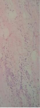
3 Figure 3:Gross operative specimen of nephrectomy shows fatty replacement of kidney. Fig. 4.1 Fig.4.2 Fig.4.3 Figure 4.1Section showing residual renal tubules, focal inflammation and thick walled blood vesselsfigure4.2section showing mature adipose 47
4 DISCUSSION : Fatty proliferation in kidney represents a spectrum of disorders ranging from mild lipomatosis in the renal sinus with underlying normal parenchyma (renal sinus lipomatosis) to a severe variety with lipomatosis involving renal sinus, hilum and perinephric region with underlying atrophic parenchyma (renal replacement lipomatosis). The presence of atrophic renal parenchyma distinguishes this condition from other causes of fibro-fatty proliferation in and around the kidney, as in obesity, Cushing's disease or excessive corticosteroid therapy and idiopathic. (7) The condition is resultant of chronic infective process, commonly as a consequence of renal /ureteric calculi. It is found in association with xanthogranulomatous pyelonephritis, however in this case it was associated with renal tuberculosis. Patient will usually have complaints like fever, pain, flank mass, haematuria etc. The shape of kidney is maintained and different degrees of hydronephrosis might be associated Contrary to other neoplastic fat containing lesions like angiomyolipomaswhich develop in the renal parenchyma, here the fat is proliferating in the normal renal sinus and occupying the adjacent space to atrophied renal parenchyma. Radiography may show renal calculus with surrounding lucent area replacing the normal fluid density of kidney. There may be associated mass effect. Intravenous pyelography is often not useful as the kidney is mostly non-excretory (3). IVP can be useful in assessing excretion of the contralateral kidney for planning of nephrectomy.ultrasound will show hyperechoic mass in renal fossa with hypoechoic rim of preserved renal parenchyma. Associated renal calculus may be seen.computed tomography accurately demonstrates the condition. Fat density mass is seen with atrophy of the renal parenchyma. Associated conditions like collections, calculi can be assessed using computed tomography.milder form of renal lipomatosis can be difficult to distinguish from conditions like lipoma, xanthogranulomatous pyelonephritis and angiomyolipoma. Narrowing of renal vessels and its stretching over the sinus fat can be seen in renal angiography. There may mild increase in vascular blush due to infectious process. In xanthogranulomatous pyelonephritis there is infiltration of renal parenchyma by lipid laden macrophages, hence the attenuation will be between the attenuation of fat and fluid within the renal parenchyma.(3)the normal renal outline is lost, and there is renal enlargment with contracted renal pelvis. The calyces in contrast, are dilated giving a multiloculated appearance. (7)Using combined modalities, it is possible to differentiate RRL from other fat-containing neoplasms in the renal fossa, such as angiomyolipoma, lipoma and liposarcoma. CONCLUSION Renal replacement lipomatosis is associated with chronic infection which in this case was tuberculosis. The diagnosis of cause is important as it will change the treatment. The coexistence of xanthogranulomatous pyelonephritis and renal replacement lipomatosis was been found in numerous literature, however the association of tuberculosis was been reported only once. Source of Support: Nil Conflict of Interest: None declared. 48
5 REFERENCES : 1. Casas, J. Darío, et al. "Replacement lipomatosis related to renal tuberculosis: imaging findings in one case." European radiology 12.4 (2002): Ambos, MARJORIE A., et al. "Replacement lipomatosis of the kidney." American Journal of Roentgenology (1978): Kullendorff, B., U. Nyman, and P. Aspelin. "Computed tomography in renal replacement lipomatosis." ActaRadiologica 28.4 (1987): Xu, Y., et al. "Renal replacement lipomatosis." European surgical research38.4 (2006): Prasad, K. R., H. Satish Chandra, and KR Vijay Kumar. "Renal replacement lipomatosis." Indian journal of urology: IJU: journal of the Urological Society of India 28.1 (2012): Acunas, Bűlent, et al. "Coexistent xanthogranulomatous pyelonephritis and massive replacement lipomatosis of the kidney: CT diagnosis." Urologic radiology 12.1 (1990): SM Goldman1,etal.CT of XanthogranulomatousPyelonephntis: Radiologic-Pathologic Correl.American Journal of Roentgenology. 1984;142: Paper cited as: Danand Aarti, Chandak Priya, Suresh Parvathy. RENAL TUBERCULOSIS CAUSE OF RENAL REPLACEMENT LIPOMATOSIS : A RARE ASSOCIATION. International Journal of Medical and Applied Sciences. 2017;6(2):
Matsunaga, Naofumi; Saito, Yutaka. Citation Acta medica Nagasakiensia. 1991, 36
 NAOSITE: Nagasaki University's Ac Title Author(s) A Large Periureteral Lipoma Associa Hydronephrosis. Hayashi, Tomayoshi; Imamura, Atushi Matsunaga, Naofumi; Saito, Yutaka Citation Acta medica Nagasakiensia.
Sex: 女 Age: 51 Occupation: 無 Admission date:92/07/22
 Sex: 女 Age: 51 Occupation: 無 Admission date:92/07/22 Chief complaint Unknown fever for one month Hand tremor and left huge renal tumor was noted Present illness Suffered from fever for one month, hand
XANTHOGRANULOMATOUS PYELONEPHRITIS: radiologic review.
 XANTHOGRANULOMATOUS PYELONEPHRITIS: radiologic review. Poster No.: C-0557 Congress: ECR 2014 Type: Educational Exhibit Authors: M. Barral, J. M. Sánchez Crespo, J. C. Pérez Herrera, J. L. 1 2 3 1 1 1 Ortega
Abdominal Ultrasound : Aorta, Kidneys, Bladder
 Abdominal Ultrasound : Aorta, Kidneys, Bladder Nilam J. Soni, MD, MSc Associate Professor of Medicine Divisions of Hospital Medicine and Pulmonary/Critical Care Medicine Department of Medicine University
Outline. Introduction to imaging modalities of the urinary system. Case base learning of common diseases in urinary tract
 Outline Introduction to imaging modalities of the urinary system Case base learning of common diseases in urinary tract Outline Introduction to imaging modalities of the urinary system Case base learning
Outline. Introduction to imaging modalities of the urinary system. Case base learning of common diseases in urinary tract
 Outline Introduction to imaging modalities of the urinary system Case base learning of common diseases in urinary tract Diagnostic Investigations in Urinary System PLAIN KUB EXCRETORY UROGRAPHY RETROGRADE
Excretory urography (EU) or IVP US CT & radionuclide imaging
 or IVP US CT & radionuclide imaging") Excretory urography (EU) or IVP US CT & radionuclide imaging MRI arteriography studies requiring catherization or direct puncture of collecting system EU & to a lesser extent CT provide both functional
Excretory urography (EU) or IVP US CT & radionuclide imaging MRI arteriography studies requiring catherization or direct puncture of collecting system EU & to a lesser extent CT provide both functional
Role of imaging in RCC. Ultrasonography. Solid lesion. Cystic RCC. Solid RCC 31/08/60. From Diagnosis to Treatment: the Radiologist Perspective
 Role of imaging in RCC From Diagnosis to Treatment: the Radiologist Perspective Diagnosis Staging Follow up Imaging modalities Limitations and pitfalls Duangkamon Prapruttam, MD Department of Therapeutic
Separating and Distorted Nephroliths Signs of Renal Squamous Cell Carcinoma
 Chin J Radiol 2003; 28: 203-208 203 Separating and Distorted Nephroliths Signs of Renal Squamous Cell Carcinoma TZE-YU LEE SHEUNG-FAT KO CHUNG-CHENG HUANG YU-FENG CHENG Department of Radiology, Chang Gung
Kidney Case 1 SURGICAL PATHOLOGY REPORT
 Kidney Case 1 Surgical Pathology Report February 9, 2007 Clinical History: This 45 year old woman was found to have a left renal mass. CT urography with reconstruction revealed a 2 cm medial mass which
Five Views of Transitional Cell Carcinoma: One Man s Journey
 September 2006 Five Views of Transitional Cell Carcinoma: One Man s Journey Amsalu Dabela, Harvard Medical School III Outline Overview: Renal Anatomy Our Patient s Story Diagnostic Imaging Studies Appearance
Acute renal colic Radiological investigation in patients with renal colic
 Acute renal colic Radiological investigation in patients with renal colic Mikael Hellström Professor Department of Radiology Sahlgrenska University Hospital Göteborg University 0.9-1.8/1.000 inhabitants
Surgically Discovered Xanthogranulomatous Pyelonephritis Invading Inferior Vena Cava with Coexisting Renal Cell Carcinoma
 Case Study TheScientificWorldJOURNAL (2009) 9, 5 9 TSW Urology ISSN 1537-744X; DOI 10.1100/tsw.2009.6 Surgically Discovered Xanthogranulomatous Pyelonephritis Invading Inferior Vena Cava with Coexisting
What s Your Diagnosis??? Renée Fahrenholz, Class of 2012
 Renée Fahrenholz, Class of 2012 What s Your Diagnosis??? Signalment Emma, a 9 year old, Female, Spayed, Domestic Short Haired Feline Presenting Complaint Weight loss, vomited the morning of her visit,
Imaging findings in renal infections
 Imaging findings in renal infections Poster No.: C-0221 Congress: ECR 2013 Type: Educational Exhibit Authors: I. lópez blasco, D. Soriano Mena, R. Pastor Toledo, S. Paz Maya, A. M. Julve Parreño, J. Palmero
Acute flank pain in children: Imaging considerations
 Acute flank pain in children: Imaging considerations Carlos J. Sivit MD Rainbow Babies and Children s Hospital Case Western Reserve School of Medicine Flank pain Results from distention of ureter or renal
Find Medical Solutions to Your Problems HYDRONEPHROSIS. (Distension of Renal Calyces & Pelvis)
") HYDRONEPHROSIS (Distension of Renal Calyces & Pelvis) Hydronephrosis is the distension of the renal calyces and pelvis due to accumulation of the urine as a result of the obstruction to the outflow of
HYDRONEPHROSIS (Distension of Renal Calyces & Pelvis) Hydronephrosis is the distension of the renal calyces and pelvis due to accumulation of the urine as a result of the obstruction to the outflow of
54 year-old Female with Recurrent Bronchopneumonia and Tumor of the Left Kidney
 Original Report TheScientificWorldJOURNAL (2004) 4 (S1), 353 356 ISSN 1537-744X; DOI 10.1100/tsw.2004.89 54 year-old Female with Recurrent Bronchopneumonia and Tumor of the Left Kidney Burkhard Ubrig,
Normal Sonographic Anatomy
 hapter 2:The Liver DUNSTAN ABRAHAM Normal Sonographic Anatomy Homogeneous, echogenic texture (Figure 2-1) Measures approximately 15 cm in length and 10 12.5 cm anterior to posterior; measurement taken
Personal data. Age : 63 Gender : male
 Personal data Age : 63 Gender : male Chief complain No specific symptom or discomfort A hepatic mass, found by abdominal sonography of routine health exam on 88-12-08 Past history 1984-3-3 Old CVA with
Renal and ureteral involvement in Erdheim-Chester disease: analysis of a single centre cohort
 Renal and ureteral involvement in Erdheim-Chester disease: analysis of a single centre cohort Gaia Manari, Davide Gianfreda, Andrea Posteraro, Alessandro A. Palumbo and Augusto Vaglio Nephrology Unit and
A Case Report of Acute Renal Artery Occlusion Mimicking Acute Appendicitis
 ISPUB.COM The Internet Journal of Surgery Volume 7 Number 1 A Case Report of Acute Renal Artery Occlusion Mimicking Acute Appendicitis S Abouel-Enin, A Douglas, R Morgan Citation S Abouel-Enin, A Douglas,
Radiological Assessment of the Kidney in Patients with Hematuria
 March 2005 Radiological Assessment of the Kidney in Patients with Hematuria Jeremy L. McKay, Harvard Medical School Year III Hematuria Signs and Symptoms Microscopic or gross hematuria Abdominal pain Fever
Treatment of choice for end stage renal disease Imaging to establish baseline and diagnosis of potential complications Review common surgical
 Treatment of choice for end stage renal disease Imaging to establish baseline and diagnosis of potential complications Review common surgical techniques Review normal appearance Discuss US diagnosis of
이학종분당서울대학교병원. Ultrasound in Urinary Colic
 이학종분당서울대학교병원 Ultrasound in Urinary Colic U l t r a s o u n d i n U r i n a US: Normal Kidney r y C o l i c Contents 1. 1. Definition and clinical consideration 2. 2. Pathophysiology 3. 3. US in in obstructive
Contents. Review anatomy of the urinary tract Imaging modalities
 Contents Review anatomy of the urinary tract Imaging modalities The Urinary Tract Kidneys ตาแหน งไต (position) อย ใน retroperitoneum ระด บ T12-L3 โดยไต ขวาจะม ระด บตากว าไตซ ายเล กน อย ร ปร าง (shape)
Role of MDCT in Radiological evaluation of Renal Masses and its beneficial effects on patient management.
 International Journal of advances in health sciences (IJHS) ISSN 2349-7033 Vol2, Issue1, 2015, pp56-63 http://www.ijhsonline.com Research Article Role of MDCT in Radiological evaluation of Renal Masses
Intrarenal reflux and the scarred kidney
 Archives of Disease in Childhood, 1974, 49, 531. Intrarenal reflux and the scarred kidney G. L. ROLLESTON, T. M. J. MALING, and C. J. HODSON* From the Department of Radiology, Christchurch Hospital and
Clinico-radiological Features and Classification of Emphysematous Pyelonephritis: A prospective study
 ORIGINAL ARTICLE Clinico-radiological Features and Classification of Emphysematous Pyelonephritis: A prospective study Singh A Department of Radiodiagnosis, Government Medical College, Amritsar, Punjab,
Primary Renal Candidiasis
 Case Series Primary Renal Candidiasis Importance of Imaging and Clinical History in Diagnosis and Management Barry J. Sadegi, MD, Bhargavi K. Patel, MD, Andrew C. Wilbur, MD, Anil Khosla, MD, Ejaz Shamim,
Renal masses - the role of diagnostic imaging
 Renal masses - the role of diagnostic imaging Poster No.: C-2471 Congress: ECR 2015 Type: Educational Exhibit Authors: V. Rai#; Bjelovar/HR Keywords: Cysts, Cancer, Structured reporting, Ultrasound, MR,
Proceedings of the 34th World Small Animal Veterinary Congress WSAVA 2009
 www.ivis.org Proceedings of the 34th World Small Animal Veterinary Congress WSAVA 2009 São Paulo, Brazil - 2009 Next WSAVA Congress : Reprinted in IVIS with the permission of the Congress Organizers IMAGING
Uroradiology For Medical Students
 Uroradiology For Medical Students Lesson 8 Computerized Tomography 2 American Urological Association Objectives In this lesson you will: Gain more experience reading CT images Learn how computer generated
Role of imaging in evaluation of genitourinary i trauma Spectrum of GU injuries Relevance of imaging findings in determining management Focus on MDCT
 Genitourinary Tract Injuries 6 th Nordic Course Scott D. Steenburg, MD Assistant Professor University of Maryland Department of Radiology Division of Trauma and Emergency Radiology R Adams Cowley Shock
Bilateral Renal Angiomyolipomas with Invasion of the Renal Vein: A Case Report
 Case Study TheScientificWorldJOURNAL (2008) 8, 145 148 TSW Urology ISSN 1537-744X; DOI 10.1100/tsw.2008.29 Bilateral Renal Angiomyolipomas with Invasion of the Renal Vein: A Case Report C. Blick, N. Ravindranath,
By GEORGE E. NELIGAN, M.C., M.A., B.M,, B.Ch. (Oxon.), F.R.C.S. (Swrgeon with charge of Out-patients and Surgeon in charge of the Genito-Urinary
, F.R.C.S. (Swrgeon with charge of Out-patients and Surgeon in charge of the Genito-Urinary") 426 POST-GRADUATE MEDICAL JOURNAL November, 1935 RENAL TUMOURS. By GEORGE E. NELIGAN, M.C., M.A., B.M,, B.Ch. (Oxon.), F.R.C.S. (Swrgeon with charge of Out-patients and Surgeon in charge of the Genito-Urinary
426 POST-GRADUATE MEDICAL JOURNAL November, 1935 RENAL TUMOURS. By GEORGE E. NELIGAN, M.C., M.A., B.M,, B.Ch. (Oxon.), F.R.C.S. (Swrgeon with charge of Out-patients and Surgeon in charge of the Genito-Urinary
Urinary system Ultrasound (Renal & Urinary bladder)
") Urinary system Ultrasound (Renal & Urinary bladder) Edited & Presented by ; Hussien A.B ALI DINAR. Msc.Phd ISRRT Associate Member Lecturer (National university) Reporting Sonographer (PHC) Objective By
Urinary system Ultrasound (Renal & Urinary bladder) Edited & Presented by ; Hussien A.B ALI DINAR. Msc.Phd ISRRT Associate Member Lecturer (National university) Reporting Sonographer (PHC) Objective By
R adio logical investigations of urinary system
 R adio logical investigations of urinary system There are 4 main radiological Ix: 1 IVU: Intravenous urography. 2- U/S 3-CT scan 4-Radioisotope scan. Others (not frequently used): MRI, arteriography, antegrade
Primary Squamous Cell Carcinoma Of Kidney - A Case Report And Review Of Literature.
 ISPUB.COM The Internet Journal of Nephrology Volume 6 Number 1 Primary Squamous Cell Carcinoma Of Kidney - A Case Report And Review Of Literature. P Kaur, A Chauhan, G Singh, S Kataria, R Kalra Citation
Different successful management strategies for obstructing renal parapelvic cysts
 Title Page Different successful management strategies for obstructing renal parapelvic cysts Sabrina H Rossi 1, Brendan Koo 2, Antony Riddick 1, Nimish Shah 1, Grant D Stewart 1 1 Urology Department, Addenbrooke's
Unilateral supernumerary kidney with contra lateral hydronephrosis a rare case report
 Unilateral supernumerary kidney with contra lateral hydronephrosis a rare case report Ezzat Khalda, Gyanendra Narain Singh, Ashok Kumar Mandal, Rajiv Kumar Department of Radiodiagnosis, Patna Medical College,
Imaging Features of Acute Pyelonephritis in Contrast Computed Tomography as Predictors of Need for Intervention
 Imaging Features of Acute Pyelonephritis in Contrast Computed Tomography as Predictors of Need for Intervention Poster No.: C-0088 Congress: ECR 2014 Type: Scientific Exhibit Authors: C. Y. Lee, C. W.
SESSION 1: GENERAL (BASIC) PATHOLOGY CONCEPTS Thursday, October 16, :30am - 11:30am FACULTY COPY
 PATHOLOGY CONCEPTS Thursday, October 16, :30am - 11:30am FACULTY COPY") SESSION 1: GENERAL (BASIC) PATHOLOGY CONCEPTS Thursday, October 16, 2008 9:30am - 11:30am FACULTY COPY GOAL: Describe the basic morphologic (structural) changes which occur in various pathologic conditions.
SESSION 1: GENERAL (BASIC) PATHOLOGY CONCEPTS Thursday, October 16, 2008 9:30am - 11:30am FACULTY COPY GOAL: Describe the basic morphologic (structural) changes which occur in various pathologic conditions.
Obstructive Uropathy. PATHOPHYSIOLOGIC CHANGES UUO vs BUO. Arry Rodjani Urology Department Ciptomangunkusumo Hospital Jakarta
 Obstructive Uropathy PATHOPHYSIOLOGIC CHANGES UUO vs BUO Arry Rodjani Urology Department Ciptomangunkusumo Hospital Jakarta INTRODUCTION Obstructive uropathy refers to the functional or anatomic obstruction
Histomorphological Analysis of Lesions In Nephrectomy Specimens: A 4 Years Study In A Rural Hospital In India-Our Experience
 Original Article DOI: 10.21276/APALM.1111 Histomorphological Analysis of Lesions In Nephrectomy Specimens: A 4 Years Study In A Rural Hospital In India-Our Experience Kishor H. Suryawanshi 1 *, Rajshri
Nephrographic and Pyelographic Analysis of CT Urography: Principles, Patterns, and Pathophysiology
 Genitourinary Imaging Review Wolin et al. CT Urography Principles, Patterns, and Genitourinary Imaging Review FOCUS ON: Ely A. Wolin 1 David S. Hartman J. Ryan Olson Wolin EA, Hartman DS, Olson JR Keywords:
Solitary Fibrous Tumor of the Kidney with Massive Retroperitoneal Recurrence. A Case Presentation
 246) Prague Medical Report / Vol. 113 (2012) No. 3, p. 246 250 Solitary Fibrous Tumor of the Kidney with Massive Retroperitoneal Recurrence. A Case Presentation Sfoungaristos S., Papatheodorou M., Kavouras
Urinary system. Urinary system
 INTRODUCTION. Several organs system Produce urine and excrete it from the body Maintenance of homeostasis. Components. two kidneys, produce urine; two ureters, carry urine to single urinary bladder for
PICTORIAL ESSAY. Experiences of using a single post-contrast CT scan of the urinary tract after triphasic contrast injection
 Experiences of using a single post-contrast CT scan of the urinary tract after triphasic contrast injection P C Pretorius, FCRad (Diag) SA Drs Visser, Erasmus, Vawda & Partners, Port Elizabeth Corresponding
My Patient Has Abdominal Pain PoCUS of the Biliary Tract and the Urinary Tract
 My Patient Has Abdominal Pain PoCUS of the Biliary Tract and the Urinary Tract Objectives PoCUS for Biliary Disease PoCUS for Renal Colic PoCUS for Urinary Retention Biliary Disease A patient presents
J of Evolution of Med and Dent Sci/ eissn , pissn / Vol. 3/ Issue 46/Sep 22, 2014 Page 11296
 CT SPECTRUM OF GIANT RETROPERITONEAL LIPOSARCOMAS WITH HISTOPATHOLOGICAL CORRELATION Shashikumar M. R 1, Rajendra Kumar N. L 2, C. P. Nanjaraj 3, Nishanth R. K 4, Vishwanath Joshi 5 HOW TO CITE THIS ARTICLE:
PROFESSIONAL SKILLS 1 3RD YEAR SEMESTER 6 RADIOGRAPHY. THE URINARY SYSTEM Uz. Fatema shmus aldeen Tel
 PROFESSIONAL SKILLS 1 3RD YEAR SEMESTER 6 RADIOGRAPHY THE URINARY SYSTEM Uz. Fatema shmus aldeen Tel. 0925111552 Professional skills-2 THE URINARY SYSTEM The urinary system (review anatomy and physiology)
Case Report Imaging of a Renal Artery Aneurysm Detected Incidentally on Ultrasonography
 Case Reports in Radiology, Article ID 375805, 4 pages http://dx.doi.org/10.1155/2014/375805 Case Report Imaging of a Renal Artery Aneurysm Detected Incidentally on Ultrasonography Vasileios Rafailidis,
Case Report Incidentally Detected Squamous Cell Carcinoma of Renal Pelvis in Patients with Staghorn Calculi: Case Series with Review of the Literature
 International Scholarly Research Network ISRN Oncology Volume 2011, Article ID 620574, 6 pages doi:10.5402/2011/620574 Case Report Incidentally Detected Squamous Cell Carcinoma of Renal Pelvis in Patients
International Journal of Medical and Health Sciences
 International Journal of Medical and Health Sciences Journal Home Page: http://www.ijmhs.net ISSN:2277-4505 Case Report Squamous Cell Carcinoma Of Renal Pelvis- A Rare Case Report S. S. Inamdar* 1, Sainath
Basic of Ultrasound Physics E FAST & Renal Examination. Dr Muhammad Umer Ihsan MBBS,MD, DCH CCPU,DDU1,FACEM
 Basic of Ultrasound Physics E FAST & Renal Examination Dr Muhammad Umer Ihsan MBBS,MD, DCH CCPU,DDU1,FACEM What is Sound? Sound is Mechanical pressure waves What is Ultrasound? Ultrasounds are sound waves
Ultrasonography of acute flank pain: a focus on renal stones and acute pyelonephritis
 Ultrasonography of acute flank pain: a focus on renal stones and acute pyelonephritis Ki Choon Sim Department of Radiology, Korea University nam Hospital, Korea University College of Medicine, Seoul, Korea
Pediatric Retroperitoneal Masses Radiologic-Pathologic Correlation
 Acta Radiológica Portuguesa, Vol.XVIII, nº 70, pág. 61-70, Abr.-Jun., 2006 Pediatric Retroperitoneal Masses Radiologic-Pathologic Correlation Marilyn J. Siegel Mallinckrodt Institute of Radiology, Washington
Autosomal Dominant Polycystic Kidney Disease
 Case Studies [1] July 01, 2014 By Amar Udare, MBBS [2] Case History: 45-year-old female with vague pain in the abdomen. Case History: A 45-year-old female presented with vague pain in the abdomen. A USG
Diagnosis & Management of Kidney Trauma. LAU - Urology Residency Program LOP Urology Residents Meeting
 Diagnosis & Management of Kidney Trauma LAU - Urology Residency Program LOP Urology Residents Meeting Outline Introduction Investigation Staging Treatment Introduction The kidneys are the most common genitourinary
Author(s) Gohji, Kazuo; Gotoh, Akinobu; Kamid. Citation 泌尿器科紀要 (1990), 36(7):
 Gohji, Kazuo; Gotoh, Akinobu; Kamid. Citation 泌尿器科紀要 (1990), 36(7):") Title Giant renal angiomyolipoma with an pattern: a case report Author(s) Gohji, Kazuo; Gotoh, Akinobu; Kamid Citation 泌尿器科紀要 (1990), 36(7): 837-840 Issue Date 1990-07 URL http://hdl.handle.net/2433/116942
Title Giant renal angiomyolipoma with an pattern: a case report Author(s) Gohji, Kazuo; Gotoh, Akinobu; Kamid Citation 泌尿器科紀要 (1990), 36(7): 837-840 Issue Date 1990-07 URL http://hdl.handle.net/2433/116942
Chapter 6: Genitourinary and Gastrointestinal Systems 93
 Chapter 6: Genitourinary and Gastrointestinal Systems 93 Chapter 6 Genitourinary and Gastrointestinal Systems Embryology Three sets of excretory organs or kidneys develop in human embryos: Pronephros:
Nonurographic evaluation of renal calculous disease 1
 Contributions Nonurographic evaluation of renal calculous disease 1 Gregory P. Borkowski, M.D. Craig R. George, M.D. Peter B. O'Donovan, M.D. While excretory urography has been useful in the evaluation
A study of primary megaureter Our experience
 Original Research Article A study of primary megaureter Our experience DVS Ramakrishna Prasad *, Srinivas S Department of Urology, Osmania Medical College and General Hospital, Hyderabad, Telangana, India
Lec-8 جراحة بولية د.نعمان
 4th stage Lec-8 جراحة بولية د.نعمان 11/10/2015 بسم هللا الرحمن الرحيم Ureteric, Vesical, & urethral stones Ureteric Calculus Epidemiology like renal stones Etiology like renal stones Risk factors like
Hydronephrosis. What is hydronephrosis?
 What is hydronephrosis? Hydronephrosis Hydronephrosis describes the situation where the urine collecting system of the kidney is dilated. This may be a normal variant or it may be due to an underlying
CYSTIC TUMORS OF THE KIDNEY JOHN N. EBLE, M.D. CYSTIC NEPHROMA
 Page 1 CYSTIC TUMORS OF THE KIDNEY JOHN N. EBLE, M.D. Department of Pathology & Laboratory Medicine Phone (317) 274-4806 Medical Science A-128 FAX: (317) 278-2018 635 Barnhill Drive jeble @iupui.edu Indianapolis,
Kristina M. Nowitzki, M.D., Ph.D. and Hao S. Lo, M.D. University of Massachusetts Medical School, Worcester, MA
 Kristina M. Nowitzki, M.D., Ph.D. and Hao S. Lo, M.D. University of Massachusetts Medical School, Worcester, MA Outline I. Introduction highlighting normal renal enhancement physiology including normal
Clinicopathologic Conference
 Participants: Mohammed Akhtar, MD*, Nabil Bissada, MD and William Cumming, MD Editor. John T. Godwin, MD, FCAP * Director, Electron Microscopy Section, Department of Pathology and Laboratory Medicine;
Functions of the kidney:
 Diseases of renal system : Normal anatomy of renal system : Each human adult kidney weighs about 150 gm, the ureter enters the kidney at the hilum, it dilates into a funnel-shaped cavity, the pelvis, from
Sonographic Features of Necrosed Renal Papillae Causing Hydronephrosis
 Case Series Sonographic Features of Necrosed Renal Papillae Causing Hydronephrosis S. Boopathy Vijayaraghavan, MD, DMRD, Sangampalayam Vedhanayagam Kandasamy, MS, MCh, Mylsamy Arul, MS, DNB (Uro), Muniappan
RADIOLOGY OF THE URINARY TRACT CHAPTER 9 239
 RADIOLOGY OF THE URINARY TRACT CHAPTER 9 239 in length. They lie cephalad to the kidneys, with the right just posterior to the inferior vena cava (IVC) and the left anteromedial to the upper pole of the
Caveat sonologist Mistakes to avoid in Kidney Ultrasound
 Caveat sonologist Mistakes to avoid in Kidney Ultrasound Simon Freeman Derriford Hospital, Plymouth simonfreeman@nhs.net Bear trap 1 Report: There is a 4cm solid mass arising from the left kidney likely
URINARY SYSTEM I. Kidneys II. Nephron Unit and Urine Formation
 URINARY SYSTEM I. Kidneys A. Location and Structure 1. Retroperitoneal 2. Between T12 and L3 3. Rt. kidney slightly lower 4. Two bean shaped organs 5. Adrenal gland 6. Internal construction a. Renal cortex
Request Card Task ANSWERS
 Request Card Task ANSWERS Medical Student Workbook Author: Dr Sam Leach, SpR Case 1 What differential diagnoses are most likely? Which investigation is most appropriate? Case 1 The most likely diagnosis
The Kidneys. (L., ren; Gk, nephros; hence the adjectives renal and nephric) & Suprarenal (Adrenal) Glands. Dr Maan Al-Abbasi PhD, MBChB
 & Suprarenal (Adrenal) Glands. Dr Maan Al-Abbasi PhD, MBChB") The Kidneys (L., ren; Gk, nephros; hence the adjectives renal and nephric) & Suprarenal (Adrenal) Glands Dr Maan Al-Abbasi PhD, MBChB Functions of Urinary System Regulate electrolytes (K+, Na+, etc) Regulate
The Kidneys (L., ren; Gk, nephros; hence the adjectives renal and nephric) & Suprarenal (Adrenal) Glands Dr Maan Al-Abbasi PhD, MBChB Functions of Urinary System Regulate electrolytes (K+, Na+, etc) Regulate
Abdomen and Retroperitoneum Ultrasound Protocols
 Abdomen and Retroperitoneum Ultrasound Protocols Reviewed By: Anna Ellermeier, MD Last Reviewed: March 2018 Contact: (866) 761-4200, Option 1 **NOTE for all examinations: 1. If documenting possible flow
Obstetrics Content Outline Obstetrics - Fetal Abnormalities
 Obstetrics Content Outline Obstetrics - Fetal Abnormalities Effective February 2007 10 16% renal agenesis complete absence of the kidneys occurs when ureteric buds fail to develop Or degenerate before
International Journal of Health Sciences and Research ISSN:
 International Journal of Health Sciences and Research www.ijhsr.org ISSN: 2249-9571 Case Report Classical Renal Tuberculosis Presented As Recurrent Sterile Pyuria and End Stage Kidney Sunil V. Jagtap 1,
Kidneys and Urinary Tract Content Outline. Anatomy Coverings. Location. (Effective February 2007) (16%-24%)
 (16%-24%)") Kidneys and Urinary Tract Content Outline (Effective February 2007) (16%-24%) Anatomy Coverings true capsule perirenal fat surrounds capsule Gerota s fascia separates perirenal from extraperitoneal fat
Kidneys and Urinary Tract Content Outline (Effective February 2007) (16%-24%) Anatomy Coverings true capsule perirenal fat surrounds capsule Gerota s fascia separates perirenal from extraperitoneal fat
Concurrent Multilocular Cystic Renal Cell Carcinoma and Leiomyoma in the Same Kidney: Previously Unreported Association
 218 Concurrent Multilocular Cystic Renal Cell Carcinoma and Leiomyoma in the Same Kidney: Previously Unreported Association Min Su Cheong a Dong Hun Koo a In-Sung Kim a Kyung Chul Moon b Ja Hyeon Ku a
(2/3 PRCC!) (2/3 PRCC!)
 (2/3 PRCC!)") Approach to the Incidental Solid Renal Mass Stuart G. Silverman, MD, FACR Professor of Radiology Harvard ard Medical School Director, Abdominal Imaging and Intervention Brigham and Women s Hospital Boston,
Approach to the Incidental Solid Renal Mass Stuart G. Silverman, MD, FACR Professor of Radiology Harvard ard Medical School Director, Abdominal Imaging and Intervention Brigham and Women s Hospital Boston,
ISSN East Cent. Afr. J. surg. (Online)
") 87 Ureteroscopy in a Resource Limited Setting: The Tikur Anbessa General Specialized Hospital Experience in Addis Ababa, Ethiopia. D. Andualem, L. Be-ede, T. Mulat, L. Samodi Addis Ababa University-School
87 Ureteroscopy in a Resource Limited Setting: The Tikur Anbessa General Specialized Hospital Experience in Addis Ababa, Ethiopia. D. Andualem, L. Be-ede, T. Mulat, L. Samodi Addis Ababa University-School
Scrotum-like protrusion of lipoma arising from the proximal thigh
 Upsala J Med sci 109: 261 265, 2004 Scrotum-like protrusion of lipoma arising from the proximal thigh Report of two cases Koshi Hattori, 1 Masahito Hatori, 1 Mika Watanabe, 2 Toshihisa Osanai, 3 Shoichi
ANATOMY OF PELVICAYCEAL SYSTEM -DR. RAHUL BEVARA
 1 ANATOMY OF PELVICAYCEAL SYSTEM -DR. RAHUL BEVARA 2 KIDNEY:ANATOMY OVERVIEW Kidneys are retroperitoneal, in posterior abdominal region, extending from T12 L3 Bean-shaped Right kidney is lower than left
Case MDCT 3D reconstructed features of posterior urethral valve
 Case 12688 MDCT 3D reconstructed features of posterior urethral valve Hidayatullah Hamidi Third year Resident of Radiology French medical institute for children Radiology Department; Kabul, Afghanistan;
Urology An introduction to cut up DR J R GOEPEL
 Urology An introduction to cut up DR J R GOEPEL Overview Principles Individual organs Small pieces Partial resections Whole organs Data recording and data sets Principles You are working for the patient
In Canada, there was a 25% reduction in incidence of genitourinary TB in the period compared with An interesting speculation
 Renal T B EPIDEMIOLOGY Young to middle age usually affected, rare in children Male : female ratio = 2:1 True prevalence and incidence not known as patients are usually asymptomatic With HIV pandemic, there
Chris McGuire Gillian Lieberman, MD. May Schistosomiasis. Chris McGuire, Harvard Medical School Year III Gillian Lieberman, MD
 May 2003 Schistosomiasis Chris McGuire, Harvard Medical School Year III Overview Introduction to schistosomiasis Katayama syndrome Patient A Patient B Ultrasound detection Bladder infection Kidney infection
Kidney is Being Attacked: MDCT Findings, Pathology, Clinical Correlation and Algorithmic Approach of Renal Infection.
 Kidney is Being Attacked: MDCT Findings, Pathology, Clinical Correlation and Algorithmic Approach of Renal Infection. Poster No.: C-0986 Congress: ECR 2014 Type: Educational Exhibit Authors: H. M. Shebel,
Kidney & Urinary Tract Ultrasound. Fatina Fadel Hafez Bazaraa
 Kidney & Urinary Tract Ultrasound Fatina Fadel Hafez Bazaraa Ultrasonography Ultrasound Available Rapid Inexpensive Painless & no sedation needed No adverse effects/ complications Can be repeated Useful
A clinicopathological study of squamous cell carcinoma kidney
 Original article A clinicopathological study of squamous cell carcinoma kidney 1AmulyajitKaur, 2 Dr. Kanwaljeet Singh Hura, 3 AkanxaBhatti, 4 Dr. NeerajKaur 1Associate Professor, Department of Pathology,Command
Xanthogranulomatous Pyelonephritis in Korean Children
 Original Article http://dx.doi.org/10.3349/ymj.2012.53.6.1159 pissn: 05135796, eissn: 19762437 Yonsei Med J 53(6):11591164, 2012 Xanthogranulomatous Pyelonephritis in Korean Children Jong Kil Nam, Sung
1. Urinary System, General
 S T U D Y G U I D E 16 1. Urinary System, General a. Label the figure by placing the numbers of the structures in the spaces by the correct labels. 7 Aorta 6 Kidney 8 Ureter 2 Inferior vena cava 4 Renal
Case Report The Role of Laparoscopic Nephrectomy in Pediatric Xanthogranulomatous Pyelonephritis: A Case Report
 Case Reports in Urology Volume 2013, Article ID 598950, 4 pages http://dx.doi.org/10.1155/2013/598950 Case Report The Role of Laparoscopic Nephrectomy in Pediatric Xanthogranulomatous Pyelonephritis: A
PYELONEPHRITIS. Wendy Glaberson 11/8/13
 PYELONEPHRITIS Wendy Glaberson 11/8/13 A 19mo infant girl was seen in the ED 3 days ago and diagnosed with a UTI. She was afebrile at the time and discharged on broad spectrum antibiotics. The child returns
US in non-traumatic acute abdomen. Lalita, M.D. Radiologist Department of radiology Faculty of Medicine ChiangMai university
 US in non-traumatic acute abdomen Lalita, M.D. Radiologist Department of radiology Faculty of Medicine ChiangMai university Sagittal Orientation Transverse (Axial) Orientation Coronal Orientation Intercostal
Coding Companion for Urology/Nephrology. A comprehensive illustrated guide to coding and reimbursement
 Coding Companion for Urology/Nephrology A comprehensive illustrated guide to coding and reimbursement 2014 Contents Getting Started with Coding Companion...i Integumentary...1 Arteries and Veins...15 Lymph
Abdominal ultrasound:
 Abdominal ultrasound: Non-traumatic acute abdomen Wittanee Na-ChiangMai, MD Department of Radiology ChiangMai University 26/04/2017 Contents Technique of examination Normal anatomy Emergency conditions