ALL PHOTOS ARE IDENTIFIED IN THE LOWER RIGHT CORNER WITH THE MAGNIFICATION POWER THAT THE PHOTO WAS TAKEN WITH. SCAN - THIS IS A VERY LOW POWER IMAGE
|
|
|
- Preston Brooks
- 5 years ago
- Views:
Transcription
1 ALL PHOTOS ARE IDENTIFIED IN THE LOWER RIGHT CORNER WITH THE MAGNIFICATION POWER THAT THE PHOTO WAS TAKEN WITH. SCAN - THIS IS A VERY LOW POWER IMAGE THAT WE USE WHEN A SAMPLE IS SO BIG THAT YOU CAN T GET THE WHOLE SAMPLE INTO THE FIELD OF VIEW AT LOW POWER. YOU WON T SEE THIS IN YOUR MICROSCOPE. LOW THIS IS THE 40X MICROSCOPE LENS MED THIS IS THE 100X MICROSCOPE LENS HIGH THIS IS THE 400X MICROSCOPE LENS SUPERHIGH THIS IS USING THE 400X LENS, AND THEN USING THE ZOOM ON THE CAMARA. YOU WON T SEE THIS SUPERHIGH POWER IN THE MICROSCOPE.
2 EVERYTHING THAT YOU ARE RESPONSIBLE FOR ON THE HISTOLOGY IS LABELED AT LEAST ONCE IN THIS PPT. THERE ARE ALSO EXPLANATIONS OF WHAT MAKES SOMETHING DISTINCTIVE, SUCH THAT YOU CAN IDENTIFY IT.
3 SIMPLE SQUAMOUS EPI


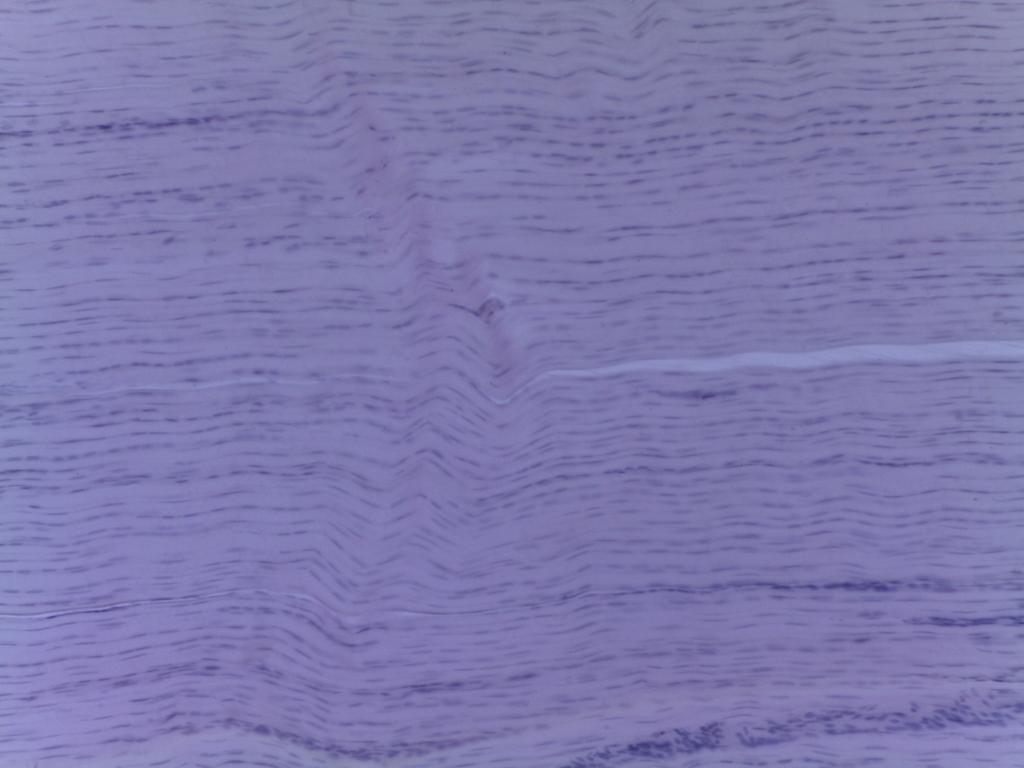
4 ARTERY EPITHELIAL TISSUES ARE EITHER GLANDULAR OR LINING. HERE WE RE LOOKING AT THE LINING AND WE LL USE THE ARTERY VEIN LOW

5 LINING! MED
. THAT IS THE SINGLE LAYER OF FLAT (SQUAMOUS) CELLS.")
6 SEE HOW THE TEXTURE OF THE LINING IS DIFFERENT THAN THE UNDERLYING STUFF (MUSCLE). THAT IS THE SINGLE LAYER OF FLAT (SQUAMOUS) CELLS. HIGH
7 SIMPLE CUBOIDAL EPI

8 medulla This is a whole kidney, which is divided into layers. The middle layer is called the medulla, and is where we ll find our simple cuboidal epithelium SCAN

9 SEE THAT THERE ARE DIFFERENT TEXTURES IN THIS SLIDE? THE NATURE OF THE KIDNEY IS THAT THESE TUBULES ALL SWEEP DOWN TOWARDS THE SAME PLACE TO EXIT THE KIDNEY. SO A SECTION THRU THE KIDNEY IS GOING TO CATCH SOME OF THE TUBULES IN CROSS SECTION AND SOME IN LONGITUDINAL SECTION. WE LL SEE HIGH POWER OF BOTH AREAS LOW

10 THIS IS MEDIUM POWER OF THE LEFT PORTION OF THE PREVIOUS PHOTO THE LONGITUDINAL SECTIONS MED

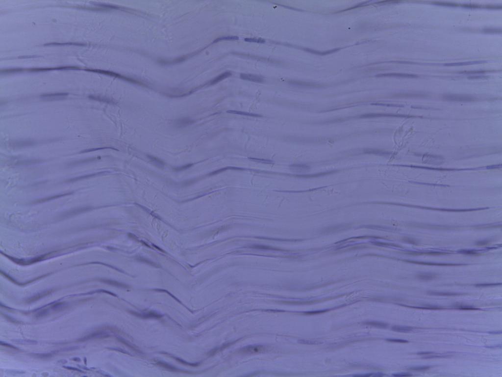
11 Simple cuboidal epi lining - a cross section of a tubule HIGH
12 Simple cuboidal epi lining a longitudinal section of a tubule HIGH

13 WHY DO THESE CELLS APPARENTLY HAVE NO NUCLEI? Simple cuboidal epi lining a longitudinal section of a tubule HIGH
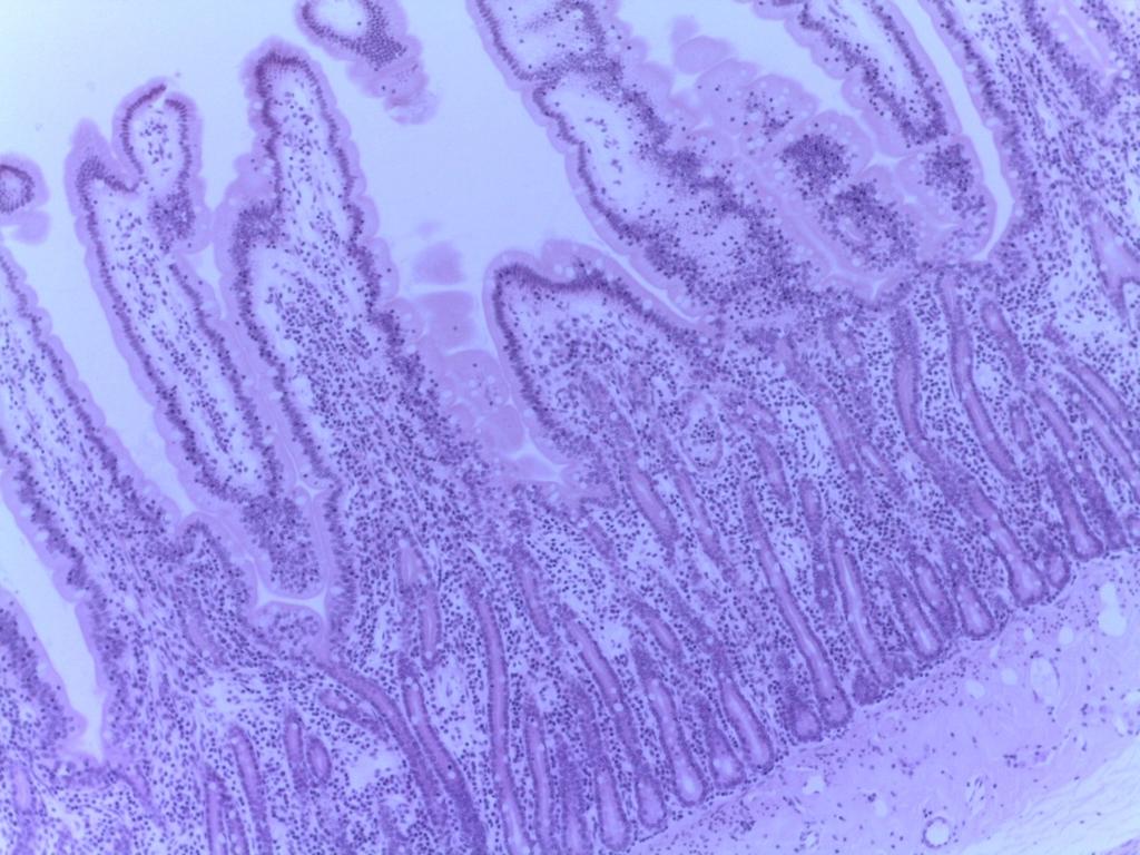
14 SIMPLE COLUMNAR EPI

15 THE STOMACH HAS BOTH LINING EPITHELIUM AND GLANDY EPITHELIUM. THE VERY EDGE IS THE LINING AND ALL THE PURPLE STUFF UNDER IT ARE THE GASTRIC GLANDS LOW

16 WE RE LOOKING HERE MED

17 SEE HOW THE LINING CELLS ARE TALL AND HAVE NUCLEI AT THE BASE, AND HOW THE NUCLEI ARE LINED UP IN A NICE ROW? HIGH
18 STRATIFIED SQUAMOUS EPI NON-KERATINIZED

19 Note the dark staining at the bottom of the tissue, reflecting the packed nuclei. LOW

20 As the cells are pushed away from the basal layer, they get flatter, and the nuclei get spread out too. MED
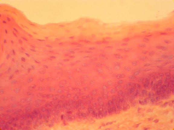
21 flattening HIGH

22 In this slide the flattening isn t as obvious, but we can still see the the density of the nuclei decrease as the cells are pushed to the surface HIGH
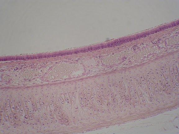
23 STRATIFIED SQUAMOUS EPI KERATINIZED
24 THIS IS ALL ADIPOSE TISSUE Look here for the epi LOW

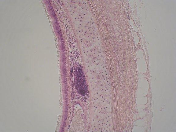
25 The basal layer of the st. sq epi curves around as the epidermis invaginates into the dermis. There are clearly layers here we ll get to them later. For now just note the concentrated, darkly staining nuclei at the base, thinning out a bit as they get pushed off the basal layer. Also note the thick superficial layer of pure keratin. MED


26 BORDER BETWEEN THE EPI AND THE UNDERLYING TISSUE HIGH
27 SIMPLE COLUMNAR EPI. W/ MICROVILLI


28 LINING THE LUMEN JEJUNUM BLOOD VESSELS A SNIPPET OF THE PANCREAS LOW
29 MED

30 THIS IS A SINGLE LAYER, SO LOOK AT HOW THE NUCLEI ARE LINED UP, AND LOOK AT THE EDGE OF THE CELLS, THAT DARKEaNED LINE ARE THE PACKED MICROVILLI (TOO SMALL TO BE SEEN ONE BY ONE, BUT SO DENSE THEY COLLECTIVELUY STAIN DARKER HIGH
31 PSUEDOSTRATIFIED, CILIATED COLUMNAR EPI.

32 NEXT TO THE LUMEN LOW
33 MED


34 WE CAN SEE THE CILIA THIS IS A BLOB OF LYMPHATIC TISSUE - WBCS THESE ARE COLUMNAR CELLS, BUT THE NUCLEI AREN T IN A NICE ROW THEY RE IN A JUMBLED MESS THIS IS MUSCUS GUNK ON THE SURFACE OF THE CILIA HIGH
35 TRANSITIONAL EPI.

36 Bladder slides are tough to make, the layer under the epithelial layer separates as the section is made, resulting in all those spaces, which are artifacts MED

37 Transitional epi is designed to be able to stretch and collapse as the bladder slowly fills and then quickly collapses. The nuclei are jumbled, but are clearly NOT columnar. HIGH
38 LOOSE CT
, AND THE CELLS ARE WIDELY SCATTERED.")
39 WHEREAS EPI CONSISTS OF TIGHTLY PACKED CELLS WITH ALMOST NO MATERIAL BETWEEN THEM, CT IS MOSTLY INSTITIAL STUFF (FIBERS AND SUCH), AND THE CELLS ARE WIDELY SCATTERED. EVERYWHERE YOU SEE A NUCLEUS IS A CELL LOW
40 MED
 its character This is loose ct, disorganized with the")
41 Look at how scattered the nuclei (and therefore the cells) are. Most of the mass of CT is extracellular material, made by the fibroblasts. All the pinkish and thin black strands are the fibers that give the tissue (along with the nature of the ground substance) its character This is loose ct, disorganized with the fibers not nearly as compacted as Dense CT HIGH
42 DENSE IRREGULAR CT
43 Back to the skin to see DICT. It s here, below the epithelial layer, but above the fatty layer LOW


44 There are a bunch of accessory structures here. Ignore them for now. Look at the disorganized stuff that is the stuff of this layer MED

45 Ignore all the extra stuff in the layer there are blood vessles and glands here and look at the thick pinkish collagen fibers. They re not regularly arranged and there are way more of them than in LCT HIGH
46 REGULAR DENSE CT

47 EVEN AT LOW POWER WE CAN SEE HOW THERE IS AN ORGANIZATION, REFLECTING THE FIBERS BEING PARALLEL TO EACH OTHER AND GIVING THE TISSUE POINT-TO-POINT STRENGTH AS OPPOSED TO DISTRIBUTED STRENGTH LOW
48 MED
49 HIGH
50 HYALINE CARTILAGE


51 Back to the trachea for hyaline cartilage LOW

52 MED

53 Note the chondrocytes in their lacunae, and the perichondrium HIGH
54 ELASTIC CARTILAGE

55 This is an animal ear. We can see the epidermis, then a layer of muscle, then the cartilage layer LOW


56 Elastic cartilage has much less interstitial area than any other CT, but what it has stains dark with elastic fibers MED


57 Chondrocyte in its lacunae Perichondrium HIGH
58 FIBROUS CARTILAGE

59 Part of an intervertebral disk, so the cartilage is mostly collagen with very little elastin; it s also layered for additional strength LOW

60 The chondrocytes are stacked between the layers. MED
61 HIGH
62 COMPACT BONE
 DOESN T WASH AWAY FROM SPACES, LEAVING THEM BLACK.")
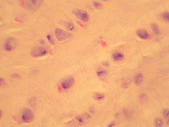
63 WE USE A DIFFERENT STAIN FOR BONE, THAT MAKE MINERALS LOOK GOLDISH. AND WHEN THE SLIDE IS MADE, THE STAIN (WHICH STARTS BLACKISH) DOESN T WASH AWAY FROM SPACES, LEAVING THEM BLACK. SO ALL THE BLACK AREAS ARE ACTUALLY EMPTY. LOW


64 THIS IS A HAVERSIAN SYSTEM, WITH A CANAL IN THE MIDDLE,, SURROUNDED BY 3 OR 4 HAVERSIAN LAMELLAE THESE LAMELLAE, WHICH DON T BELONG TO ANY SYSTEM, ARE INTERSTITIAL LAMELLAE VOLKMAN S CANAL MED


65 INTERSTITIAL LAMELLAE ALL THE SMALL BLACK SPOTS ARE LACUNAE WITH OSTEOCYTES ALL THE LITTLE LINES SPOKING OUT ARE CANALICULI HAVERSIAN CANAL HIGH
66 SKIN


67 HYPODERMIS WITH ADIPOSE TISSUE PACINIAN CORPUSULE TRUE SWEAT GLAND WITH PIECES OF ITS DUCT HEADINIGI TOWARDS THE SURFACE DERMIS WITH DICT EPIDERMIS WITH STRATIFIED SQUAMOUS EPI THIS IS A FINGERTIP WHICH HAS A THICK KERATIN LAYER LOW

68 THIS IS THE SCALP, WITH A THINNER KERATIN LAYER AND LOTS OF HAIR FOLLICLES ROOT SURROUNDED BY FOLLICLE SHAFT TRUE SWEAT GLANDS SEBACEOUS GLANDS LOW


69 ST CORNEUM ST GRANULOSUM ST SPINOSUM PAPILLARY LAYER OF DERMIS RETICULAR LAYER OF DERMIS ST BASALE THE ONE LAYER AT THE BASE OF THE EPIDERMIS DERMAL PAPILLA MED
70 PACINIAN CORPUSULE HIGH

71 THE STRATUM CORNEUM IS MUCH THINNER AND LESS STURDY ON THE SCALP, SO IT SEPARATES INTO ARTIFACTS WHEN THE SLIDE IS MADE ALL WE CAN REALLY POINT TO HERE IS THE ST BASALE, ST SPINOSUM AND ST CORNEUM DERMIS WITH DICT HIGH
72 SEBACEOUS GLAND HIGH


73 ARRECTOR PILI SUPERHIGH


74 MEISSNER S CORPUSULE WITHIN DERMAL PAPILLA SUPERHIGH
HISTOLOGY. Simple squamal lungs
 HISTOLOGY Lab Objectives: Students should be able to... 1. Visually identify each class of tissue and examples within each class 2. Indicate the location (in the human body and/or organ) and function of
Histology review. Histology. Slides. Epithelial tissue. Another example - kidney. Simple cuboidal epithelium. What to look for
 Histology review Histology What to look for Histology Practical = 50 pts Some slides set up on scopes (~10) Some Powerpoint pictures on the projector Questions I will ask: What kind of tissue? General
Mitosis Models 3-5. Chromosome. #1 Prophase. #2 Prophase. 2n = 4 4 Chromosomes 8 Chromatids. 2n = 4
 MITOSIS Mitosis Models 3-5 Chromosome #1 Prophase 2n = 4 4 Chromosomes 8 Chromatids #2 Prophase 2n = 4 4 Chromosomes 8 Chromatids Mitosis Models 3-5 Astral Rays Chromosomes Chromosome Chromosome Spindle
Basic Histology. By Mrs. Bailey
 Basic Histology By Mrs. Bailey Primary Tissues 1. Epithelial Tissue 2. Connective Tissue 3. Muscle Tissue 4. Nervous Tissue Very cellular Supported by underlying connective tissue Epithelial & connective
ACTIVITY 2: HISTOLOGY AND INTEGUMENT
 ACTIVITY 2: HISTOLOGY AND INTEGUMENT Objectives: 1) How to get ready: Read Chapter 4 and 5, McKinley et al., Human Anatomy, 5e. All text references are for this textbook. 2) Identify each tissue (26 tissues)
Dr Narmeen S. Ahmad. Lab 1
 Dr Narmeen S. Ahmad Lab 1 1 Tissues are groups of cells with a common structure (form) and function (job). There are (4) types of tissue: 1. Epithelial 2. Connective 3. Muscle 4. Nervous 2 Epithelial cells
TISSUES. Objectives. Tissues
 TISSUES Objectives Introduce the four major types of tissues Describe the general characteristics and functions of epithelial & connective tissue Name the major types of epithelial & connective tissues
Tissues. Tissues - Overview. Bio211 Laboratory 2. Epithelial and Connective Tissues
 Bio211 Laboratory 2 Epithelial and Connective Tissues 1 Tissues Tissues to be examined under the microscope Epithelial Tissue (p. 79 Lab Manual) [TODAY] Connective Tissue (p. 93 Lab Manual) [TODAY] Muscle/Nervous
Tissues Chapter 5...Tissue - a group or mass of similar cells working together to perform certain common functions
 Tissues Chapter 5...Tissue - a group or mass of similar cells working together to perform certain common functions There are 4 major types of tissue Epithelial Connective Muscle Nervous 1. Epithelial Tissue
Tissues. Tissues. Four basic tissues. A collection of cells with a common function. 1. Epithelial 2. Connective 3. Muscular 4.
 Tissues Tissues A collection of cells with a common function Four basic tissues 1. Epithelial 2. Connective 3. Muscular 4. Nervous Epithelia: cells in layers Types of epithelia 1) lining Layers of cells
ACTIVITY 2: HISTOLOGY AND INTEGUMENT
 ACTIVITY 2: HISTOLOGY AND INTEGUMENT Objectives: 1) How to get ready: Read Chapter 4 and 5, McKinley et al., Human Anatomy, 4e. All text references are for this textbook. 2) Identify each tissue (26 tissues)
BI 121 LAB. WEEK 2: Tissues (continued); Integumentary System
; Integumentary System") BI 121 LAB 2-1 WEEK 2: Tissues (continued); Integumentary System This week you will 1) Review the four major tissue types 2) Review the characteristics of epithelial tissues. 3) Learn the major characteristics
BI 121 LAB 2-1 WEEK 2: Tissues (continued); Integumentary System This week you will 1) Review the four major tissue types 2) Review the characteristics of epithelial tissues. 3) Learn the major characteristics
Tissues. Tissues - Overview. Bio 101 Laboratory 3. Epithelial Tissues and Integument
 Bio 101 Laboratory 3 Epithelial Tissues and Integument 1 Tissues Tissues to be examined under the microscope Epithelial Tissue Integument Connective Tissue **We will be doing muscle and nervous tissues
INTEGUMENTARY 1-Epidermis, 2-Dermis, Structure of thick and thin skin I- Epidermis . Stratum basale
 INTEGUMENTARY The skin (integument, cutis ) and its derivatives constitute the integumentary system. It form the external covering of the body and is the largest organ of the body. The skin consists of
Histology. There are four basic tissue types in the body are :-
 Histology Lab.I There are four basic tissue types in the body are :- 1- Epithelial tissues (Epithelium) 2- Connective tissues 3- Muscular tissues 4- Nervous tissues 1-Epithelial tissues epithelial tissues
Lab 7: Integumentary System Hamilton ANSWERS TO PRE- LAB ASSIGNMENTS
 Lab 7: Integumentary System Hamilton ANSWERS TO PRE- LAB ASSIGNMENTS Pre-Lab Activity 1: 1. a. epidermis b. dermis c. hypodermis d. adipose tissue e. hair f. sebaceous gland g. sweat gland 2. a Pre-Lab
Tissues. How do cells form tissues?
 Tissues How do cells form tissues? Using cell junctions Tissues Epithelial tissue Connective tissue Muscle tissue Nervous tissue Epithelial Tissue Closely packed cells in continuous sheets connected by
Chapter 4 Histology: The study of body tissues
 Chapter 4 Histology: The study of body tissues https://www.youtube.com/watch?v=zwxm2a0tfxm Body Tissues Cells are specialized for particular functions Tissues = groups of cells with similar structure and
Skin. Kristine Krafts, M.D.
 Skin Kristine Krafts, M.D. Skin Lecture Objectives Describe the functions of skin. Describe the structure, location and function of the cell types found in epidermis: keratinocytes, melanocytes, Langerhans
Anatomy and Physiology Tissue Review
 Anatomy and Physiology Tissue Review OVERVIEW Histology practicals can be rough, especially when access to slides is limited to the lab period. This resource provides an opportunity to learn or review
Epithelium. Four primary tissue types:
 Epithelium Four primary tissue types: Epithelial (covering) Connective (support) Nervous (control) Muscular (movement) Smooth muscle Cardiac muscle Skeletal muscle 1 Epithelial Tissue Features Epithelial
****************************************************************************************************** INTEGUMENTARY SYSTEM
 BIOLOGY 211: HUMAN ANATOMY & PHYSIOLOGY ****************************************************************************************************** INTEGUMENTARY SYSTEM ******************************************************************************************************
BIO 130 Anatomy and Physiology Spring, 2016 Exam 3 Name: Course ID Number. Section 1 Answer questions 1 40 on the scan sheet.
 BIO 130 Anatomy and Physiology Spring, 2016 Exam 3 Name: Course ID Number Section 1 Answer questions 1 40 on the scan sheet. 1. Which of the following is NOT a characteristic of epithelial tissue? a. It
SKIN. 3. How is the skin structured around the finger joints to allow for flexible movement of the fingers?
 SKIN Objectives for Exam #1: 1. List various skin structures and describe their functions. 2. Describe skin responses to increases and decreases in body temperature. 3. Provide examples of various skin
Skin (Integumentary System) Wheater, Chap. 9
 Wheater, Chap. 9") Skin (Integumentary System) Wheater, Chap. 9 Skin (Integument) Consists of skin and associated derivatives Largest organ of body (21 ft 2 ; 9 lbs.; has 11 miles of blood vessels) Functions: Protection
Skin (Integumentary System) Wheater, Chap. 9 Skin (Integument) Consists of skin and associated derivatives Largest organ of body (21 ft 2 ; 9 lbs.; has 11 miles of blood vessels) Functions: Protection
Chapter 05. Review. Copyright The McGraw-Hill Companies, Inc. Permission required for reproduction or display.
 Chapter 05 Review 5.1: Introduction Similar cells with a common function are called tissues. The study of tissues is called histology. There are four (4) primary or major tissue types: 1. Epithelial Tissue
The Integumentary System
 The Integumentary System Integument is skin Skin and its appendages make up the integumentary system A fatty layer (hypodermis) lies deep to it Two distinct regions Epidermis Dermis PHL 212 1 Function
THE TISSUE LEVEL OF ORGANIZATION PART I: EPITHELIAL TISSUE
 THE TISSUE LEVEL OF ORGANIZATION PART I: EPITHELIAL TISSUE 4 Main Tissue Types Epithelium Covers surfaces, lines cavities, forms glands Connective Tissue Support and protects body Muscular Tissue Movement
The Tissue Level of Organization
 Tissue The Tissue Level of Organization Chapter 3 Definition an aggregation of cells in which each cooperates with all others in the performance of a given function Examples of general functions Movement
Overview of the Integumentary System. Lab #7. Layers of the epidermis are known as strata. Organization of the Epidermis: Layers of the Epidermis
 Overview of the Integumentary System Lab #7 Integumentary System Organization of the Epidermis: Layers of the epidermis are known as strata Figure 5 2 Layers of the Epidermis Top: Free surface of skin
Chapter 5. Tissues. 4 Types of Body Tissues. Tissues
 Chapter 5 Tissues Tissues Tissues - groups of cells that are similar in structure & function RBC, WBC, & platelets are a group of cells working together to form BLOOD tissue Histology Pathohistology study
Tissues. tissue = many cells w/ same structure and function. cell shape aids function tissue shape aids function. Histology = study of tissues
 Tissues tissue = many cells w/ same structure and function cell shape aids function tissue shape aids function Histology = study of tissues 4 types of tissues Epithelial coverings contact openings Connective
2/5/2019. Organ System: Skin or Integumentary System. Hypodermis (or superficial fascia) Integumentary System - Learn and Understand
 Integumentary System - Learn and Understand") Integumentary System - Learn and Understand Skin is an organ comprised of all four tissues Each layer of the skin contributes to one or more of its numerous functions Skin is both strong and flexible Keratinization
Integumentary System - Learn and Understand Skin is an organ comprised of all four tissues Each layer of the skin contributes to one or more of its numerous functions Skin is both strong and flexible Keratinization
3. Dense connective tissue is found in skin, & surrounding blood vessels, nerves, and organs.
 Ch.4&5 Group Quiz True/False Indicate whether the statement is true or false. 1. There are 4 basic types of tissue in the human body. 2. Cartilage is also known as osseous tissue. 3. Dense connective tissue
Tissues are: group of similar or identical cells that share a common function. used to build organs
 Tissues: Four classes Epithelium Connective Muscle Nervous Tissues are: group of similar or identical cells that share a common function. used to build organs Overview: Epithelial o Line body cavities
If necessary, you should review the proper care and use of the light microscope before beginning this exercise.
 BIOLOGY 211: HUMAN ANATOMY & PHYSIOLOGY ********************************************************************************************************* HISTOLOGY THE EXAMINATION OF TISSUES **********************************************************************************************************
CHAPTER 5 INTEGUMENTARY
 CHAPTER 5 INTEGUMENTARY skin under the skin other stuff cutaneous layer hypodermis (subcutaneous) accessory structures Cutaneous layer = skin epithelial layers = connective tissue layer = dermis Subcutaneous
Chapter 4 Opener Pearson Education, Inc.
 Chapter 4 Opener Introduction The integumentary system is composed of: Skin Hair Nails Sweat glands Oil glands Mammary glands The skin is the most visible organ of the body Clinicians can tell a lot about
Anatomy Fall Semester Set 1: Organization and Tissues
 . 1. Which of the following describes anatomy? a. using devices to investigate parameters such as heart rate and blood pressure b. investigating human structure via dissections and other methods c. studying
Epithelia of Coverings and Linings. Tissues. Tissue
 Tissue Tissues Chapter 3 Definition an aggregation of cells in which each cooperates with all others in the performance of a given function Examples of general functions Movement Protection Support Production
Tissues 10/21/2016. Epithelial Tissue
 Tissues This is a generalized cell diagram. It shows the anatomy of a cell, but most cells do not actually look like this. Cells can have a wide variety of shapes and sizes, depending on their function.
What is a tissue? Points to ponder. Tissues Connective Tissue. 1. Connective tissue 2/23/2019. Organization and Regulation of Body Systems
 Organization and Regulation of Body Systems Chapter 04 Lecture Outline See separate PowerPoint slides for all figures and tables preinserted into PowerPoint without notes. Copyright 2016 McGraw-Hill Education.
The Integumentary System: An Overview
 The Integumentary System: An Overview Functions: Protective covering Helps regulate body temperature Retards water loss from deeper tissues Houses sensory receptors Synthesizes biochemicals Excretes small
Skin and Body Membranes Body Membranes Function of body membranes Cover body surfaces Line body cavities Form protective sheets around organs
 Skin and Body Membranes Body Membranes Function of body membranes Cover body surfaces Line body cavities Form protective sheets around organs Classification of Body Membranes Epithelial membranes Cutaneous
AP I f2014 E3 c_5 & 6
 AP I f2014 E3 c_5 & 6 Student: Multiple choice questions choose the best answer. True/false answer A for true and B for false 1. The layer within the epidermis that acts as the foundation providing new
Ex. 7: Integumentary
 Collin County Community College BIOL. 2401 Ex. 7: Integumentary. Skin or Integument Consists of three major regions Epidermis outermost superficial region Dermis middle region Hypodermis (superficial fascia)
Anatomy Fall Semester Set 1: Organization and Tissues
 . 1. Which of the following describes anatomy? a. using devices to investigate parameters such as heart rate and blood pressure b. investigating human structure via dissections and other methods c. studying
Basic Tissue Types and Functions
 Tissues Histology Basic Tissue Types and Functions 1) Epithelial tissue covering 2) Connective tissue support 3) Muscle tissue movement 4) Nervous tissue control Epithelial Tissue 1) Covers a body surface
Air sacs of lungs and the lining of the heart, blood vessels, and lymphatic vessels
 Cells Location Function Simple squamous epithelium Air sacs of lungs and the lining of the heart, blood vessels, and lymphatic vessels Allows materials to pass through by diffusion and filtration, and
Anatomy and Physiology 1 Chapter 4 Outline Tissues and Membranes
 Anatomy and Physiology 1 Chapter 4 Outline Tissues and Membranes 1 Tissue group of cells with similar structure and function o 4 major groups epithelial, connective, muscle, nerve Epithelial tissue (Fig
Unit II: Tissues and Integumentary System
 Unit II: Tissues and Integumentary System 2.1 - Tissues Chapter 4 Written Response #1 1. What is a tissue? 2. What are four major types of tissues? Tissue Definition: a group or mass of similar cells working
Lab Animal Tissue. LEARNING OBJECTIVES: To understand the relationship between the structure and function of different animal tissues
 Name: Bio A.P. PURPOSE: HYPOTHESIS: NONE Lab Animal Tissue BACKGROUND: In animals, groups of closely related cells specialized to perform the same function are called tissues. There are four general classes
The Integumentary System
 The Integumentary System The Integumentary System Integument is skin Skin and its appendages make up the integumentary system (See if you can name some appendages) A fatty layer (hypodermis) lies deep
Classification of Tissues
 6 R e v i e w S h e e t Exercise Classification of Tissues NAME LAB TIME/DATE Tissue Structure and Function General Review 1. Define tissue. A group of cells similar to one another in structure that perform
PowerPoint Lecture Slide Presentation by Patty Bostwick-Taylor, Florence-Darlington Technical College Skin and Body Membranes
 PowerPoint Lecture Slide Presentation by Patty Bostwick-Taylor, Florence-Darlington Technical College Skin and Body Membranes 4 Body Membranes Function of body membranes Cover body surfaces Line body cavities
Hole s Human Anatomy and Physiology
 Hole s Human Anatomy and Physiology 1 Chapter 5 Tissues Four major tissue types 1. Epithelial 2. Connective 3. Muscle 4. Nervous 2 Epithelial Tissues General characteristics - cover organs and the body
B. Classification of epithelium: by number of cell layers present and by shape of the superficial cell layers.
 I. Introduction - tissue: group of cells that are closely associated, similar in structure and function, and perform a common or related function. - four primary tissues: epithelial tissue, connective
A adipose cells. B capillary. C epithelium
 EPITHELIA Objective The objective of this class is to observe how different epithelia vary in terms of cell shape, size and number of cell layers enabling them to be well adapted for functions in different
Unit 4 - The Skin and Body Membranes 1
 Unit 4 - The Skin and Body Membranes 1 I. Unit 4: Skin and Body Membranes A. Body Membranes 1. Function of body membranes a) Cover body surfaces b) Line body cavities c) Form protective sheets around organs
Tissues organs system organism. pg151
 Histology is the study of tissues A TISSUE is a group of cells, usually of one kind, & their intercellular substance (e.g. intercellular matrix in animal) which are linked together & perform a particular
Ch. 4: Skin and Body Membranes
 Ch. 4: Skin and Body Membranes I. Body Membranes A. Function of body membranes 1. Cover body surfaces 2. Line body cavities 3. Form protective sheets around organs II. Classification of Body Membranes
Ch 4. Skin and Body Membranes
 Ch 4 Skin and Body Membranes TITLE HISTOLOGY SLIDES & NOTES ESSENTIAL QUESTION What tissues compose the integumentary system? Stratified Squamous Epithelium Stratified = several layers; Squamous = shape
Integumentary System
 Integumentary System Overview Functions 1. Protection 2. Excretion of wastes 3. Maintenance of T b 4. Synthesis of vitamin D 3 5. Storage of lipids 6. Detection of sensory stimuli Epidermis Tissue types
Epithelial Tissue lining, covering, glandular tissue > Function protect, absorption, filtration, secretion, excretion
 Chapter 4: TISSUES IX. Tissues Intro Epithelial Tissue lining, covering, glandular tissue > Function protect, absorption, filtration, secretion, excretion Connective Tissue most widespread tissue type
Epithelial Tissues. Types of Epithelial Tissues: Lining of Kidney
 Epithelial Tissues Covers the entire body surface and most of the body s inner cavities Outer epidermis (skin) protects from injury and drying out Inner epidermal tissue (on internal surfaces) often serves
Lab 1 ANIMAL TISSUES
 Lab 1 ANIMAL TISSUES Levels of Organization Animals are multicellular heterotrophs whose cells lack cell walls. Most animals exhibit a hierarchical level of organization: Cells are organized into tissues
Tissues, Glands, and Membranes. Chapter Five Mrs. Hornacek
 Tissues, Glands, and Membranes Chapter Five Mrs. Hornacek Objectives 1. Name the four main groups of tissues and give the location and general characteristics of each. 2. Differentiate between voluntary
Ch 5: Integumentary System
 Ch 5: Integumentary System You gotta have skin; All you really need is skin. Skin's the thing, that if you've got it outside, It helps keep your insides in. Alan Sherman (1924-1973) Developed by John Gallagher,
Histology 101! !! Name:! Block: Identify and describe the functions of major tissue types including their subclasses and varieties!
 Histology 101 Identify and describe the functions of major tissue types including their subclasses and varieties Name: Block: "1 Introduction to Tissues Histology Notes Tissue (living fabric) : groups
I. Introduction. Unit One. Tendons of the hand. The white glistening appearance results from the collagen of which tendons are composed.
 5 Tendons of the hand tendons The white glistening appearance results from the collagen of which tendons are composed. Chapter 5 Karen Webb Smith Unit One I. Introduction A. Cells are arranged in tissues
Histology. Study of body tissues
 Histology Study of body tissues 2 Introduction to Body Tissues 1. Composed of specialized cells of similar structure and perform a common function 2. Four major types (4 Cs) a. Epithelial - Cover b. Connective
Practical Histology. Lab 3: Connective tissue
 Practical Histology Lab 3: Connective tissue Connective tissues Connective tissue provides structural support for the body by binding cells and tissues together to form organs. It also provides metabolic
Tissues. tissue = many cells w/ same structure and function. cell shape aids its function tissue shape aids its function
 Tissues tissue = many cells w/ same structure and function cell shape aids its function tissue shape aids its function Histology = study of tissues 4 types of tissues Epithelial coverings contact openings
The purpose of this practical session is to demonstrate cartilage and bone as specialized connective tissues to the student.
 1 CARTILAGE AND BONE The purpose of this practical session is to demonstrate cartilage and bone as specialized connective tissues to the student. 1. Hyaline cartilage Slide 73 This is a cross section through
Cells are the basic unit of life
 Ch. 4 Tissues Cells are the basic unit of life Organism Organ System Organs Tissues Cells Living thing A group of organ systems working together Group of organs working together Each system has a specific
Classification of Tissues
 M06_MARI0000_00_SE_CH06.qxd 3/28/11 4:37 PM Page 35 NAME LAB TIME/DATE R E V I E W S H E E T EXERCISE 6 Classification of Tissues Tissue Structure and Function General Review 1. Define tissue. A group
Study of different tissues Abnormal cells and tissues can be compared to normal tissues to identify disease, such as cancer Being able to know and
 CHAPTER 4 Study of different tissues Abnormal cells and tissues can be compared to normal tissues to identify disease, such as cancer Being able to know and recognize normal tissues under the microscope
The Integumentary System
 The Integumentary System The Integumentary System Integument is skin Skin and its appendages make up the integumentary system A fatty layer (hypodermis) lies deep to it Two distinct regions Epidermis Dermis
Anatomy Ch 6: Integumentary System
 Anatomy Ch 6: Integumentary System Introduction: A. Organs are body structures composed of two or more different tissues. B. The skin and its accessory organs make up the integumentary system. Types of
Tissues and Structures to Know for the Lab Practical
 Ch. 3 - Cells and Tissues Tissues and Structures to Know for the Lab Practical Miss School, Miss Out! Simple squamous epithelium line and cover; site of diffusion Simple squamous epithelium apical surface
Integumentary System. Integumentary System
 1. General aspects a. The integumentary system consists of several organs major organ of the system is the skin other organs are relatively small and they can be considered as specialized structures of
Unit I Problem 9 Histology: Basic Tissues of The Body
 Unit I Problem 9 Histology: Basic Tissues of The Body - What is the difference between cytology and histology? Cytology: it is the study of the structure and functions of cells and their contents. Histology:
Integumentary System (Script) Slide 1: Integumentary System. Slide 2: An overview of the integumentary system
 Slide 1: Integumentary System. Slide 2: An overview of the integumentary system") Integumentary System (Script) Slide 1: Integumentary System Slide 2: An overview of the integumentary system Skin is the body s largest and heaviest organ making up 15% of body weight. Most skin is 1 to
Integumentary System (Script) Slide 1: Integumentary System Slide 2: An overview of the integumentary system Skin is the body s largest and heaviest organ making up 15% of body weight. Most skin is 1 to
Histology= the study of tissues
 Unit 3-Histology Histology= the study of tissues A tissue is a group of cells that have a similar shape and function. Different types of tissues can be found in different organs. In humans, there are four
Histology: Epithelial tissue
 Histology: Epithelial tissue Epithelial Tissue is presented in two forms: 1. Covering Epithelia: 2. Glandular Epithelia: 1. Simple Epithelium: contain only one layer of cells. 2. Stratified Epithelium:
Connective Tissue. Answer Choices(In CAPITAL BOLD): RETICULAR ELASTIC. IRREGULAR Spongy bone ELASTIC BLOOD
: RETICULAR ELASTIC. IRREGULAR Spongy bone ELASTIC BLOOD") Connective Tissue Answer Choices(In CAPITAL BOLD): Proper: Specialized: Loose- Cartilage- AREOLAR HYALINE ADIPOSE FIBROCARTILAGE RETICULAR ELASTIC Dense- Bone- REGULAR COMPACT BONE IRREGULAR Spongy bone
Connective Tissue Answer Choices(In CAPITAL BOLD): Proper: Specialized: Loose- Cartilage- AREOLAR HYALINE ADIPOSE FIBROCARTILAGE RETICULAR ELASTIC Dense- Bone- REGULAR COMPACT BONE IRREGULAR Spongy bone
Epithelial Tissue. Simple Cuboidal Function: secretion and absorption. Simple Squamous
 Epithelial Tissue General Functions: Lines and covers organs Absorbs / secretes substances Gas exchange Protection Special Characteristics: - have an apical surface on top - have a basement membrane below
UNIT 4 T I S S U E S
 UNIT 4 T I S S U E S WHAT IS A TISSUE Group of cells that work together to do a function Cells are similar Extracellular fluid around them is similar Histology EPITHELIAL TISSUE Also called epithelium
Cell Types in Epidermis
 Epidermis Stratified, squamous keratinized epithelium Appendages hair follicles nails sweat glands sebaceous glands mammary glands Dermis Dense, irregular connective tissue Hypodermis Superficial fascia
Histology. The study of tissues.
 Histology The study of tissues. Body Tissues Cells are specialized for particular functions Tissues Groups of cells with similar structure and function Four primary types Epithelium Connective tissue Nervous
Histology Final Exam Done by:maha AbuAjamieh
 Histology Final Exam Done by:maha AbuAjamieh 1) Which of the following is the least valuable when distinguishing between bone and hyaline cartilage?? A- lacunae B-canaliculi C-lamella D-cell nest E- harversian
Histology Notes -Part 1: Epithelial Tissues
 Introduction Group of cells w/ similar structure & function = TISSUE Four Basic Tissue Types 1. Epithelial-covers 2. Connective-supports 3. Muscular*-produces movement (will discuss in the muscular system
Body Tissues Pearson Education, Inc.
 Body Tissues Tissues Groups of cells with similar structure and function Four primary types: Epithelial tissue (epithelium).1 Connective tissue.2 Muscle tissue.3 Nervous tissue.4 Epithelial Tissues Locations:
Integumentary System and Body Membranes
 Integumentary System and Body Membranes The Skin and its appendages hair, nails, and skin glands Anatomy/Physiology NHS http://www.lab.anhb.uwa.edu.au/mb140/corepages/integumentary/integum.htm I. System
They cells can not function death.
 Jenna Hellack Jan 2001 Tissues What do you think happens when the cells use up their food and oxygen before there is time to replenish it? They cells can not function death. Blood Cell Cancer cell Plant
A. cells that perform related functions and are similar in structure. B. extracellular material - made by cells and secreted into interstitial space
 I. tissue components A. cells that perform related functions and are similar in structure B. extracellular material - made by cells and secreted into interstitial space II. tissue types A. epithelium (e.)
Cell and Tissue Types. Epithelial, Connective, Muscle, Nerve
 Cell and Tissue Types Epithelial, Connective, Muscle, Nerve Objectives Explain the major stages of the cell cycle and cellular division (mitosis). Describe specific events occurring in each of the phases
Biology 323 Human Anatomy for Biology Majors Lecture 3 Dr. Stuart S. Sumida. Integument Support Structures; Axial Skeleton
 Biology 323 Human Anatomy for Biology Majors Lecture 3 Dr. Stuart S. Sumida Integument Support Structures; Axial Skeleton Integumentary System Functions Protection Sensory Synthesis The Skin Epidermis
Hole s Essentials of Human Anatomy & Physiology
 Hole s Essentials of Human Anatomy & Physiology David Shier Jackie Butler Ricki Lewis Created by Dr. Melissa Eisenhauer Head Athletic Trainer/Assistant Professor Trevecca Nazarene University Chapter 6
Chapter 5. Integumentary System 5-1
 Chapter 5 Integumentary System 5-1 Structures that are part of the integument Skin Hair Nails Glands Overview of Functions Protection Sensation Temperature regulation Vitamin D production Excretion Immunity