SOME species of Nephtys, particularly those from the north-east Pacific, but
|
|
|
- Shanna McCormick
- 6 years ago
- Views:
Transcription
 SUMMARY The disposition of the prostomial mucus-glands in Nephtys picta, N. incisa, and N.")
1 505 The 'Posterior Lobes' of Nephtys: Observations on three New England Species By R. B. CLARK (From the Department of Zoology, University of Bristol) With one plate (fig. 2) SUMMARY The disposition of the prostomial mucus-glands in Nephtys picta, N. incisa, and N. bucera provides additional evidence that the posterior lobes, which are attached to the supra-oesophageal ganglion of species such as N. californiensis and N. caeca, have been formed by the posterior migration of prostomial epidermal mucus-cells. In N. picta there are few mucus-cells in the prostomium. In N. incisa there are many, and some at the sides of the prostomium occur in clumps and project into the prostomial cavity. In N. bucera there are numerous mucus-cells, but they are massed at the sides of the prostomium and open to the exterior over a narrow zone. The cell-bodies are in intimate contact with the sides of the supra-oesophageal ganglion and lie within the membranes investing the brain. Subsequent evolution of the prostomial mucus-glands has led to the accumulation of their cell-bodies in the posterior part of the supraoesophageal ganglion in the form of posterior lobes. SOME species of Nephtys, particularly those from the north-east Pacific, but including the common circumpolar species JV. caeca, have a pair of long lobes attached to the posterior margin of the supra-oesophageal ganglion. These lobes are filled with mucus-cells, the long necks of which run in a tract on each side of the ganglion and open to the exterior in the lateral walls of the prostomium by way of conspicuous lateral organs. The cells in the posterior lobes and the lateral tracts of cell processes are all enclosed within the sheath that invests the ganglion, and they and the lateral organs are separated from the ganglion-cells only by a layer of neuroglial tissue. In other species these lobes are missing and mucus-cells are confined to the lateral walls of the prostomium, more or less in the position occupied by the lateral organs in the former species, and have no connexion with the supra-oesophageal ganglion at all. In all species of Nephtys, whether they have posterior lobes or not, there is a mass of mucus-cells in the middle of the anterior edge of the prostomium. These generally open to the exterior on the ventral surface, and there may be other mucus-cells in the lateral walls of the anterior part of the prostomium. However, in one species, N. longosetosa, while most of the mucus-cells in the medial group open to the exterior ventrally, and most of those in the lateral walls open to the side, a few cells in the medial group open to the side by way of long necks, forming a small lateral organ in the anterior part of the prostomium. In some other species, most obviously in N. californiensis, most of the cells in the medial group open to the exterior by way of long necks which [Quarterly Journal of Microscopical Science, Vol. 99, part 4, pp , Dec ] L 1

2 506 Clark The 'Posterior Lobes' of Nephtys run to the lateral walls of the prostomium, forming a large anterior lateral organ on each side, which completely replaces the small mucus-cells in the epidermis at the sides of the prostomium, and which adjoins a similar posterior lateral organ associated with the posterior lobes. Anterior and posterior prostomial mucus-cell systems vary independently, and in N. cornuta there are well-developed anterior lateral organs, but no posterior lobes and, therefore, no posterior lateral organs. When describing the mucus-gland system of Nephtys (Clark, 1955), I suggested that mucus-glands in the lateral walls of the anterior part of the prostomium had migrated into the medial mass, retaining their connexion with the lateral walls of the prostomium. N. hombergi is an example of a species with a complete separation of the lateral and medial mucus-cells, N. longosetosa represents an intermediate stage in which a few cells from the lateral walls have migrated into the medial group, and N. californiensis an extreme case in which all the mucus-cells opening to the lateral walls of the prostomium lie in the medial group. By analogy, I argued that the mucus-cells in the lateral walls of the posterior part of the prostomium must have hypertrophied and migrated back into the brain capsule, but retained their connexion with the prostomial walls so that they opened to the exterior by way of the lateral tracts and the lateral organs. The chief weakness in this interpretation lay in the absence of any intermediate steps in the process. The mucus-cells were found in the lateral prostomial walls or in the posterior lobes, but never half-way between the two in any species I had examined. Recently I have had the opportunity of examining specimens of TV. bucera, kindly sent to me by Dr. Marion Pettibone from various localities in Maine and New Hampshire, and I have re-examined specimens of N. incisa and JV. picta from Massachusetts. This material has provided additional evidence about the evolution of posterior lobes in Nephtys and has provided the intermediate stages which were previously missing. MATERIAL AND METHODS Specimens of N. bucera Ehlers, N. incisa Malmgren, and N. picta Ehlers were fixed whole in Bouin's fluid. Frontal and transverse sections have been cut at io/x and stained with paraldehyde fuchsin (Clark, 1955), Altmann's fuchsin and methyl green picrate (Gabe, 1947), Heidenhain's iron haematoxylin, or by Holmes's silver impregnation technique (Nicol, 1948). OBSERVATIONS None of the three species possesses posterior lobes, and the mucus-cells in the anterior medial group open to the exterior ventrally. There are thus no lateral organs and we are concerned solely with the arrangement of the mucuscells in the lateral walls of the prostomium, particularly those in the posterior part of it that are in the neighbourhood of the supra-oesophageal ganglion. The supra-oesophageal ganglion of JV. picta lies mainly in the first body segment and only a small part of it is in the prostomium (fig. 1). The nuchal
3

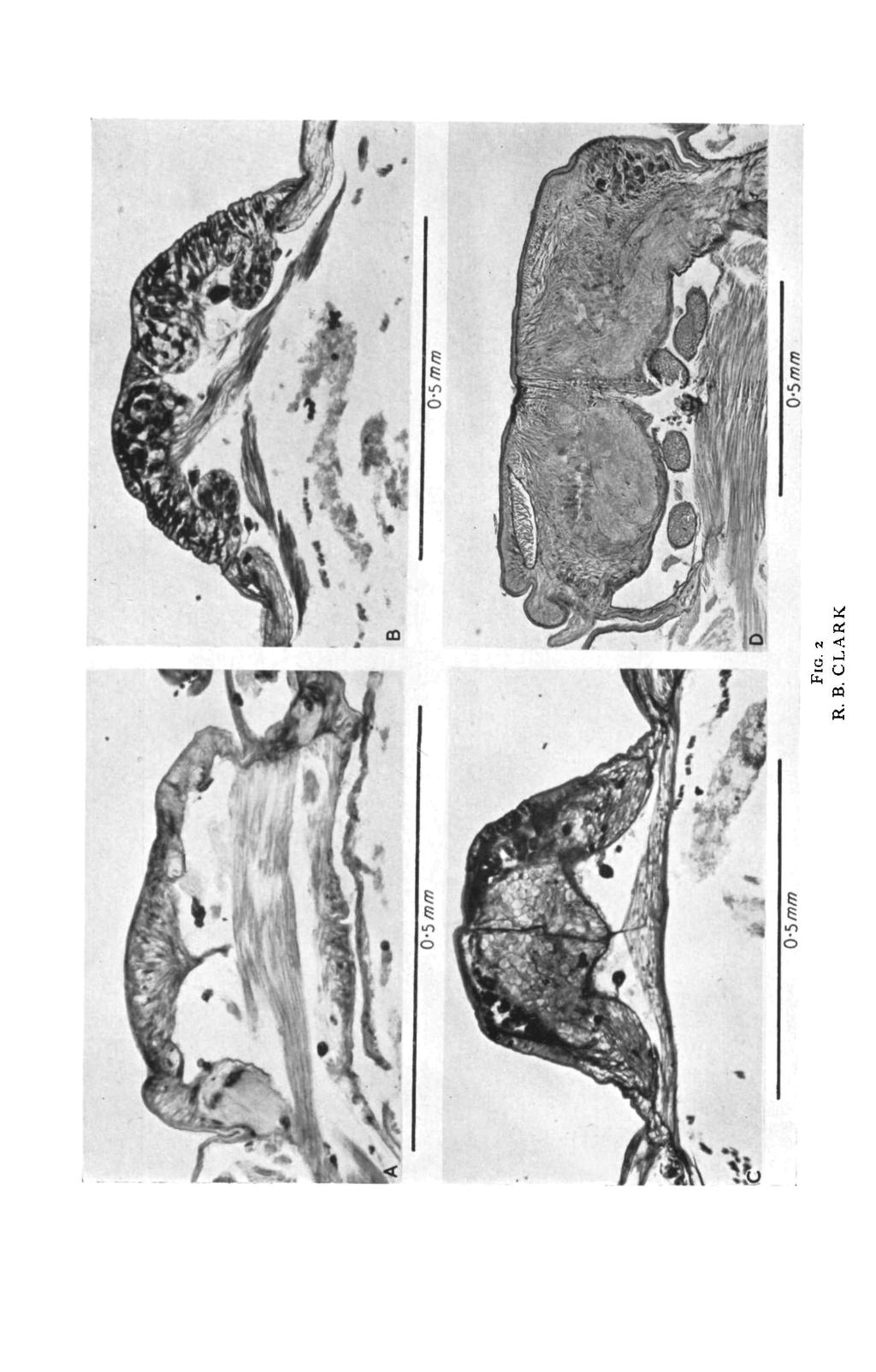
4 Clark The 'Posterior Lobes' of Nephtys 507 organs, which mark the posterior limit of the prostomium, are at the level of the most anterior ganglion-cells in the brain. While the ganglion is attached to the dorsal surface of the prostomium, it makes no contact with the sides of it, although the circum-oesophageal connectives in front of the brain do so. Nephtys picta Nephtys inciso Nephtys bucero Nephtys caeca FIG. 1. Evolution of prostomial mucus-glands in the Nephtyidae. In JV. picta there are only a few mucus-cells in the prostomial walls and these have no contact with the ganglion. In JV. incisa there are more mucus-cells, and clumps of them project into the prostomial cavity. In JV. bucera the mucus-cells are in contact with the ganglion, are enclosed within the ganglionic membranes, and are massed at the sides of the ganglion. In N. caeca there are far more mucus-cells, these have migrated into the posterior part of the ganglion but open to the exterior in the sides of the prostomium. Supra-oesophageal ganglion stippled, mucus-cells black. They run in the sub-epidermal basement membrane. Compared with most other species of Nephtys, there are few mucus-cells in the prostomial epidermis and those that there are, are in the lateral walls and have no contact with any part of the nervous system (fig. 2, A). The brain of N. incisa occupies a more anterior position than that of N. picta and most of it lies in the prostomium, so that the nuchal organs are near the back of the brain. As is the case in all species of Nephtys in which the brain is largely prostomial, it is attached not only to the dorsal part of the prostomium, but also to the sides of it. The circum-oesophageal connectives, as usual, run forwards and downwards from the brain in the sub-epidermal basement membrane of the prostomial walls. There are numerous mucus-cells in the prostomial walls (figs. 1; 2, c), most of which lie in the base of the epidermis and have narrow necks running directly to the cuticle through which they open by fine pores. There appear to be more and larger mucus-cells than can be accommodated in a single layer of epidermal cells, and a number of them are arranged in clumps at the sides of the prostomium and project into the prostomial cavity (fig. 2, B). These clumps of mucus-cells lie dorsal FlG. 2 (plate), A, transverse section of the prostomium of N. picta, cut a little anterior to the supra-oesophageal ganglion. Bouin, paraldehyde fuchsin. B, transverse section of the prostomium of JV. incisa, cut a little nnt«rior to the supra-oesophageal ganglion, showing a clump of mucus-cells projecting from thf* epidermis into the prostomial cavity on each side. Bouin, paraldehyde fuchsin. c, transverse section of the prostomium of JV. incisa, including the anterior part of the supra-oesophage.il ganglion. Numerous dark-staining mucus-cells occur in the epidermis. Bouin, paraldehyde fuchsin. D, slightly oblique transverse section of the prostomium of JV. bucera, showing massed mucus-cells at the right-hand side of the supraoesophageal ganglion. Bouin, paraldehyde fuchsin.

5 508 Clark The 'Posterior Lobes 1 of Nephtys to the circum-oesophageal connectives and occur only anterior to the supraoesophageal ganglion, so that they do not form any connexion with the nervous system. The walls of the prostomium of N. bucera are curious in that a thickened ridge of epidermis runs along the middle of each side and the prostomial mucus-cells are concentrated in it almost to the exclusion of other epidermal elements. The mucus-cells are large and have long, narrow, and slightly coiled necks. The cuticle along the line of the two ridges is peppered with the pores through which these mucus-cells open to the exterior. Massed mucus-cells occur at the sides of the prostomium from the level of the posterior antennae almost to the nuchal organs. The supra-oesophageal ganglion of N. bucera, like that of N. incisa, occupies a relatively anterior position and is largely prostomial. It extends from side to side of the prostomium and is in intimate contact with some of the prostomial mucus-glands (figs, i; 2 D). The latter extend anterior to the brain and lie dorsal to the circum-oesophageal connectives, but in the posterior part of the prostomium they lie within the membranes investing the ganglion (extensions of the sub-epidermal basement membrane) and are separated from the lateral groups of nerve-cells only by a barrier of neuroglial fibres. Some of these penetrate between the mucus-cells and there appears also to be a number of connective tissue elements in the zone between the ganglion proper and the cuticle; ordinary, structural epidermal cells are missing from this region of the prostomium. DISCUSSION The disposition and form of the mucus-cells in the prostomium of N. incisa and N. bucera suggest how posterior lobes may have evolved from purely epidermal mucus-cells. The function of the mucus is unknown, but evidently there is a need for it to be released in copious quantities at the sides of the prostomium in some species. N. picta is exceptional because it has very few mucus-cells in the prostomium, but in other species there is either a considerable number of them in the prostomial walls or else they fill the posterior lobes and open to the exterior by way of the lateral organs at the sides of the prostomium. An incipient hypertrophy of the prostomial mucus-gland system may be seen in N. incisa, in which mucus-cells are particularly numerous in the entire prostomial epidermis and, further, groups of them bulge inwards into the prostomial cavity, so permitting a greater number of mucus-cells than can be accommodated in the epidermis to open to the exterior. In N. bucera the arrangement of the prostomial mucus-cells is more specialized. These cells are restricted to ridges at the sides of the prostomium and there are relatively few elsewhere on the prostomial epidermis. The cells have long, narrow necks and so, although there are many of them, they open to the exterior over a narrow zone running along the sides of the prostomium. This concentration of mucus-cells brings them inevitably into contact with the supra-oesophageal ganglion. The brain of Nephtys is epidermal (Clark, 1958), as are the mucus-cells, and in the posterior half of the prostomium it

6 Clark The 'Posterior Lobes' of Nephtys 509 replaces the ordinary epidermal cells. There is thus no ganglionic sheath which, in N. bucera, might separate the lateral mucus-cells from the ganglion; both are invested by the sub-epidermal basement membrane and the mucuscells are separated from the nerve-cells only by neuroglial tissue. Once this stage of organization has been reached, further development of the mucusgland system must be influenced by the presence of the ganglion. The only way in which a substantial increase in the number and size of the mucus-cells can be achieved is by their migration in a posterior direction so that they can expand into the body cavity, retaining their connexion with the prostomial walls by long ducts. In N. incisa, to be sure, the mucus-cells have extended into the prostomial cavity, but there is space for only very limited development in that direction. In all the species that have a highly developed mucus-gland system, the extension of these cells has been into the body cavity. The mucus-cells of N. bucera have long, coiled necks and those of the more posterior cells run forwards, suggesting that even in this species there has been some posterior migration of the cell-bodies. A further migration in a posterior direction must inevitably result in the cells eventually lying in the posterior part of the ganglion (see fig. 1, p. 507). This is the situation in N. californiensis and N. caeca, and in N. californiensis there are a few mucus-cells occupying a lateral position in the lateral tract of ducts that runs from the cell-bodies in the posterior lobes to the lateral walls of the prostomium (Clark, 1955). No further information about the function of the mucus-gland system has been discovered since the original description of the posterior lobes. The factors which must be taken into account in any explanation of their function are now clearer. In N. picta, a species which appears to be primitive in several respects (Clark, 1957), there are virtually no prostomial mucus-glands. lnn. incisa there are many, but they are dispersed over the whole prostomial epidermis though the larger clumps of glands open laterally. In N. bucera the mucus-cells are concentrated and open in a narrow zone along the sides of the prostomium. In N. californiensis, N. caeca, and many other species, the mucusgland system is enormously developed but the cells still open to the exterior along the sides of the prostomium, and in a few species (e.g. N. californiensis and N. caecoides) this copious supply of mucus at the sides of the prostomium is supplemented by the discharge from mucus-cells in the anterior median part of the prostomium. In N. cirrosa the mucus-cells of the posterior lobes appear to have been highly modified and reduced in number, and it may be supposed that in this species they serve a different function. I am grateful to Mr. K. J. Wood for taking the photomicrographs. REFERENCES CLARK, R. B., 'The posterior lobes of the brain of Nephtys and the mucus-glands of the prostomium.' Quart. J. micr. Sci., 96, 545., 'The influence of size on the structure of the brain of Nephtys.' Zool. Jb., Abt. Physiol., 67, 261.

7 510 Clark The 'Posterior Lobes' of Nephtys CLARK, R. B., 'The gross morphology of the anterior nervous system of Nephtys.' Quart. J. micr. Sci., 99, 205. GABE, M., 'Sur l'emploi du picrate de vert de methyle pour la differenciation de la coloration d'altmann.' Bull. Hist, appl., 24, 5. NICOL, J. A. C, 'The giant nerve-fibres in the central nervous system of Myxicola (Polychaeta, Sabellidae).' Quart. J. micr. Sci., 89, 1.
SWANN (1950) figured and described major subcollar glands in Mercierella
 figured and described major subcollar glands in Mercierella") 421 Studies of Serpulid Tube Formation II. The Calcium-secreting Glands in the Peristomium of Spirorbis, Hydroides, and Serpula By R. H. HEDLEY (From the Department of Zoology, King's College, Newcastle
421 Studies of Serpulid Tube Formation II. The Calcium-secreting Glands in the Peristomium of Spirorbis, Hydroides, and Serpula By R. H. HEDLEY (From the Department of Zoology, King's College, Newcastle
The Blood Vascular System of Nephtys (Annelida, Polychaeta) By R. B. CLARK
 By R. B. CLARK") The Blood Vascular System of Nephtys (Annelida, Polychaeta) By R. B. CLARK (From the Department of Zoology, University of California, Berkeley, California) SUMMARY 235 The four longitudinal vessels of
The Blood Vascular System of Nephtys (Annelida, Polychaeta) By R. B. CLARK (From the Department of Zoology, University of California, Berkeley, California) SUMMARY 235 The four longitudinal vessels of
How to use this material
 !!!CAUTION!!! This power point presentation is intended to be used as an add on exercise to your standard lab experience. It is not intended to be used in lieu of the hands on lab time. In lab you will
Biology Earthworm Dissection
 Biology 521 - Earthworm Dissection Kingdom Phylum Class Order Genus Species Animalia Annelida Oligochaeta Haplotaxida Lumbricus L. terrestris PRELAB: The earthworm is an excellent organism to study as
The Ligamentary System and the Segmental Musculature of Nephtys
 149 The Ligamentary System and the Segmental Musculature of Nephtys By R. B. CLARK and M. E. CLARK (From the Department of Zoology, University of Bristol) With two plates (figs. 14 and 15) SUMMARY Nephtys
Essentials of Anatomy and Physiology, 9e (Marieb) Chapter 1 The Human Body: An Orientation. Short Answer. Figure 1.1
 Chapter 1 The Human Body: An Orientation. Short Answer. Figure 1.1") Essentials of Anatomy and Physiology, 9e (Marieb) Chapter 1 The Human Body: An Orientation Short Answer Figure 1.1 Using Figure 1.1, identify the following: 1) Label A points to the cavity. 2) Label B
Essentials of Anatomy and Physiology, 9e (Marieb) Chapter 1 The Human Body: An Orientation Short Answer Figure 1.1 Using Figure 1.1, identify the following: 1) Label A points to the cavity. 2) Label B
Posterior Triangle of the Neck By Prof. Dr. Muhammad Imran Qureshi
 Posterior Triangle of the Neck By Prof. Dr. Muhammad Imran Qureshi For the purpose of anatomical description the neck is sub divided into two major triangles, the Anterior and the Posterior by muscle bellies
PSYCHE THE FUSED THORACIC GANGLIA OF THE FIBER TRACTS OF THE ADULT EPHESTIA KUEHNIELLA ZELLER (LEPIDOPTERA: PYRALIDZE) BY ROBEttT W.
 BY ROBEttT W.") PSYCHE VOL. XLVIII DECEMBER, 1941 No. 4 THE FIBER TRACTS OF THE FUSED THORACIC GANGLIA OF THE ADULT EPHESTIA KUEHNIELLA ZELLER (LEPIDOPTERA: PYRALIDZE) BY ROBEttT W. PYLE Biological Laboratories, Harvard
PSYCHE VOL. XLVIII DECEMBER, 1941 No. 4 THE FIBER TRACTS OF THE FUSED THORACIC GANGLIA OF THE ADULT EPHESTIA KUEHNIELLA ZELLER (LEPIDOPTERA: PYRALIDZE) BY ROBEttT W. PYLE Biological Laboratories, Harvard
Digestive System. The group of organs which performs the function of digestion constitute digestive system.
 Digestive System Definition:- The active biological process by which food materials impermeable to the cell membrane is converted into permeable to the cell membrane is called digestion. The group of organs
Lecture 02 Anatomy of the LIVER
 Lecture 02 Anatomy of the LIVER BY Dr Farooq Khan Aurakzai Dated: 02.01.2018 Introduction to Liver Largest gland in the body. 2 nd largest organ of the body. Weight approximately 1500 gm, and is roughly
The arteries of the human kidney
 J. Anat. (1966), 100, 4, pp. 881-894 881 With 8 figures Printed in Great Britain The arteries of the human kidney BY H. FINE AND E. N. KEEN Department of Anatomy, University of Natal INTRODUCTION A study
Tikrit University collage of dentistry Dr.Ban I.S. head & neck anatomy 2 nd y. Lec [5] / Temporal fossa :
![Tikrit University collage of dentistry Dr.Ban I.S. head & neck anatomy 2 nd y. Lec [5] / Temporal fossa :](/thumbs/88/115294566.jpg "Tikrit University collage of dentistry Dr.Ban I.S. head & neck anatomy 2 nd y. Lec [5] / Temporal fossa :") Lec [5] / Temporal fossa : Borders of the Temporal Fossa: Superior: Superior temporal line. Inferior: gap between zygomatic arch and infratemporal crest of sphenoid bone. Anterior: Frontal process of the
Lec [5] / Temporal fossa : Borders of the Temporal Fossa: Superior: Superior temporal line. Inferior: gap between zygomatic arch and infratemporal crest of sphenoid bone. Anterior: Frontal process of the
Sense of Vision. Chapter 8. The Eye and Vision. The Eye Orbit. Eyebrows, Eyelids, Eyelashes. Accessory Organs 5/3/2016.
 Sense of Vision Chapter 8 Special Senses The Eye and Vision 70 percent of all sensory receptors are in the eyes Each eye has over 1 million nerve fibers Protection for the eye Most of the eye is enclosed
FACTORS CONTROLLING THE DIURNAL RHYTHM OF ACTIVITY OF PERIPLANETA AMERICANA L.
 [ 224 ] FACTORS CONTROLLING THE DIURNAL RHYTHM OF ACTIVITY OF PERIPLANETA AMERICANA L. BY JANET E. HARKER Department of Zoology, University of Cambridge {Received 7 October 1955) INTRODUCTION Two main
Glandular Epithelium. Dr. Heba Kalbouneh Associate Professor of Anatomy and Histology
 Glandular Epithelium Dr. Heba Kalbouneh Associate Professor of Anatomy and Histology Glands Glandular epithelia are tissues formed by cells specialized to produce secretion. Secretion: if substances produced
(a), in a discussion of Paget s disease of the nipple, has expressed
, in a discussion of Paget s disease of the nipple, has expressed") SKIN INVOLVEMENT IN BREAST CANCER WITH REFERENCE TO ITS BEARING ON THE INTERPRE- TATION OF APPEARANCES OF TRANSITION BE- TWEEN NORMAL EPITHELIUM AND CANCER ALSON R. KILGORE From tlw Surgiral Pathology
SKIN INVOLVEMENT IN BREAST CANCER WITH REFERENCE TO ITS BEARING ON THE INTERPRE- TATION OF APPEARANCES OF TRANSITION BE- TWEEN NORMAL EPITHELIUM AND CANCER ALSON R. KILGORE From tlw Surgiral Pathology
The Human Body: An Orientation
 The Human Body: An Orientation Body standing upright Anatomical Position feet slightly apart palms facing forward thumbs point away from body Directional Terms Superior and inferior toward and away from
The sebaceous glands (glands of Zeis) open directly into the eyelash follicles, ciliary glands (glands of Moll) are modified sweat glands that open
 open directly into the eyelash follicles, ciliary glands (glands of Moll) are modified sweat glands that open") The Orbital Region The orbits are a pair of bony cavities that contain the eyeballs; their associated muscles, nerves, vessels, and fat; and most of the lacrimal apparatus upper eyelid is larger and more
The Orbital Region The orbits are a pair of bony cavities that contain the eyeballs; their associated muscles, nerves, vessels, and fat; and most of the lacrimal apparatus upper eyelid is larger and more
Neuroanatomy. Assistant Professor of Anatomy Faculty of Medicine The University of Jordan Dr Maha ELBeltagy
 Neuroanatomy Dr. Maha ELBeltagy Assistant Professor of Anatomy Faculty of Medicine The University of Jordan 2018 Development of the Central Nervous System Development of the nervous system Development
PHYLUM NEMATODA. Introduction. Ascaris lubricoides. External anatomy - preserved specimen. Internal anatomy - preserved specimen
 PHYLUM NEMATODA Introduction Commonly called round worms the phylum includes both free living and parasitic forms. Distinctive features include: a nearly perfect cylindrical body, radial or biradial arrangement
INTRODUCTION. There are three main approaches to studying anatomy: 1. Systemic anatomy 2. Regional anatomy (topographic) 3.
 3.") INTRODUCTION Anatomy is the science of the structure and function of the body. It is the study of internal and external structures, and the physical relationships between the various body parts. INTRODUCTION
INTRODUCTION Anatomy is the science of the structure and function of the body. It is the study of internal and external structures, and the physical relationships between the various body parts. INTRODUCTION
INTRODUCTION: ****************************************************************************************************
 BIOLOGY 211: HUMAN ANATOMY & PHYSIOLOGY **************************************************************************************************** EYES AND VISION ****************************************************************************************************
Introduction in human anatomy
 Introduction in human anatomy Overview of Anatomy Anatomy is the study of the body structure and the relationships of the various parts of the body Gross or macroscopic (visible structures) Microscopic
Introduction. Study detail of structure - - Gross Anatomy. Study all structures in one part of body Study of internal structures as relate to skin
 Introduction What is Anatomy and Physiology? Anatomy study of the shape and structure of body parts and their relationships to one another Physiology study of how the body functions individually and cooperatively
Zoology Exercise #10: Phylum Nematoda Lab Guide
 Zoology Exercise #10: Phylum Nematoda Lab Guide All animals with bilateral symmetry, except the acoelomates, have a body cavity. They are either true coelomates (where peritoneum covers both the inner
8/4/2012. Causes and Cures. Nucleus pulposus. Annulus fibrosis. Vertebral end plate % water. Deforms under pressure
 Causes and Cures Intervertebral discs Facet (zygopophyseal) joints Inter body joints Spinal nerve roots Nerve compression Pathological conditions Video Causes of back pain Nucleus pulposus Annulus fibrosis
Remember from the first year embryology Trilaminar disc has 3 layers: ectoderm, mesoderm, and endoderm
 Development of face Remember from the first year embryology Trilaminar disc has 3 layers: ectoderm, mesoderm, and endoderm The ectoderm forms the neural groove, then tube The neural tube lies in the mesoderm
31-2. The Earthworm. . Relate the structure of systems. . Demonstrate dissection technique. . Identifythe major advancesof
 Name Class Date INVESTIGATION 31-2 The Earthworm Introduction The earthworm is a segmented worm. It exhibits more complex structures than any of the more primitive animals that you have studied thus far.
The Language of Anatomy. (Anatomical Terminology)
") The Language of Anatomy (Anatomical Terminology) Terms of Position The anatomical position is a fixed position of the body (cadaver) taken as if the body is standing (erect) looking forward with the upper
The Language of Anatomy (Anatomical Terminology) Terms of Position The anatomical position is a fixed position of the body (cadaver) taken as if the body is standing (erect) looking forward with the upper
Anatomy of the Nervous System. Brain Components
 Anatomy of the Nervous System Brain Components NERVOUS SYSTEM INTRODUCTION Is the master system of human body, controlling the functions of rest of the body systems Nervous System CLASSIFICATION A. Anatomical
LABORATORY EXERCISE 4 PHYLUM PLATYHELMINTHES
 GENUS PLANARIA Planaria sp. Lab 4, pg 1 LABORATORY EXERCISE 4 PHYLUM PLATYHELMINTHES With a soft brush, place a live Planaria in a small dish with a few millimeters of pond water. BEHAVIOR. Examine the
DURING investigations on the blood systems of serpulids (Hanson, 1949)
") Observations on the Branchial Crown of the Serpulidae (Annelida, Polychaeta) BY JEAN HANSON (From the Department of Zoology, Bedford College, University of London) CONTENTS I N T R O D U C T I O N 221
Observations on the Branchial Crown of the Serpulidae (Annelida, Polychaeta) BY JEAN HANSON (From the Department of Zoology, Bedford College, University of London) CONTENTS I N T R O D U C T I O N 221
THYROID & PARATHYROID. By Prof. Saeed Abuel Makarem & Dr. Sanaa Al-Sharawy
 THYROID & PARATHYROID By Prof. Saeed Abuel Makarem & Dr. Sanaa Al-Sharawy 1 OBJECTIVES By the end of the lecture, the student should be able to: Describe the shape, position, relations and structure of
THE sebaceous glands of the rabbit consist of clusters of about ten cells
 79 On the Relationship between Mammary, Sweat, and Sebaceous Glands By D. B. CARLISLE (From the Department of Zoology and Comparative Anatomy, Oxford, and the Plymouth Laboratory of the Marine Biological
Human Anatomy and Physiology (ANAT 5) Mrs. Fulton Phone: ext. 6049
 Mrs. Fulton Phone: ext. 6049") Human Anatomy and Physiology (ANAT 5) Mrs. Fulton Phone: 645-1300 ext. 6049 Please, read your green sheets. 1 Lecture #1 A. Definitions: anatomy, physiology B. Requirements of an Organism C. Homeostasis
Human Anatomy and Physiology (ANAT 5) Mrs. Fulton Phone: 645-1300 ext. 6049 Please, read your green sheets. 1 Lecture #1 A. Definitions: anatomy, physiology B. Requirements of an Organism C. Homeostasis
Lecture 01. The Thyroid & Parathyroid Glands. By: Dr Farooq Khan PMC Date: 12 th March. 2018
 Lecture 01 The Thyroid & Parathyroid Glands By: Dr Farooq Khan PMC Date: 12 th March. 2018 INTRODUCTION LAYERS OF THE NECK The neck has four major compartments or layer which are enclosed by an outer musculofascial
Chapter 1: The Human Organism
 Chapter 1: The Human Organism I. Anatomy and Physiology A. Anatomy - study of structure 1. Studying structural changes from conception to adulthood is called: 2. Embryology is the study of 3. The study
Name: Block: Date: Begin by clicking on the link entitled: Anatomical References. 2. What organs do you think the cranial region holds?
 Virtual Pig Dissection ABSENT Assignment. Name: Block: Date: Go to this website: http://www.whitman.edu/biology/vpd/main.html You will be doing a pig dissection through the internet and answering questions
Anatomy & Physiology. An Introduction
 Anatomy & Physiology An Introduction An Overview of Anatomy Anatomy - The study of the structure of the human body Physiology - The study of body function Branches of Anatomy Surface anatomy Gross anatomy
Glandular Epithelium. Dr. Heba Kalbouneh Assistant Professor of Anatomy and Histology
 Glandular Epithelium Dr. Heba Kalbouneh Assistant Professor of Anatomy and Histology Glands Gla dular epithelia are tissues for ed y ells spe ialized to produ e se retio. Secretion: if substances produced
Studies on the Nervous System of Crustacea.
 STUDIES ON THE NERVOUS SYSTEM OF CRUSTACEA. 33 Studies on the Nervous System of Crustacea. By Edgar.1. Allen, B.Sc, Director of the Plymouth Laboratory of the Marine Biological Association. With Plate
Biological Bases of Behavior. 3: Structure of the Nervous System
 Biological Bases of Behavior 3: Structure of the Nervous System Neuroanatomy Terms The neuraxis is an imaginary line drawn through the spinal cord up to the front of the brain Anatomical directions are
Differentiation of the facial-vestibulocochlear ganglionic complex in human embryos of developmental stages 13 15
 O R I G I N A L A R T I C L E Folia Morphol. Vol. 68, No. 3, pp. 167 173 Copyright 2009 Via Medica ISSN 0015 5659 www.fm.viamedica.pl Differentiation of the facial-vestibulocochlear ganglionic complex
Observations on the Genital System and Alimentary Canal of a Lamiid Beetle, Nupserha bicolor By N. DUTT
 393 Observations on the Genital System and Alimentary Canal of a Lamiid Beetle, Nupserha bicolor By N. DUTT (From the Jute Agricultural Research Institute, Barrackpore, West Bengal) With one plate (fig.
Diaphragm and intercostal muscles. Dr. Heba Kalbouneh Associate Professor of Anatomy and Histology
 Diaphragm and intercostal muscles Dr. Heba Kalbouneh Associate Professor of Anatomy and Histology Skeletal System Adult Human contains 206 Bones 2 parts: Axial skeleton (axis): Skull, Vertebral column,
Ch 1.1 An Overview of Anatomy and Physiology. Copyright 2009 Pearson Education, Inc., publishing as Benjamin Cummings
 Ch 1.1 An Overview of Anatomy and Physiology The Human Body An Orientation Anatomy Study of the structure and shape of the body and its parts Physiology Study of how the body and its parts work or function
Basic Body Structure
 Basic Body Structure The Cell All life consists of microscopic living structures called cells. They perform various functions throughout the body. All cells are similar in structure, but not identical.
Lesson 31. Lesson Outline:
 Lesson 31 Lesson Outline: The Urogenital System Urinary System o Embryonic Origins o Phylogenetic Trends Function Urinary Bladder Implications for the Evolution of the Vertebrates o Freshwater or Seawater
Dorsum of the tongue. Oral Part exhibit lingual papillae of the 4 types. Oral Part of Tongue divided into Left & right halves by shallow median groove
 Histology of TONGUE Figure 22.13 Dorsum of the tongue Oral Part of Tongue divided into Left & right halves by shallow median groove Oral Part exhibit lingual papillae of the 4 types a. filiform papillae,
The Chromosomes of Pour Species of Marsupials.
 The Chromosomes of Pour Species of Marsupials. By Stella C. A. Altmann, B.Sc, and Mavis E. W. Ellery, B.Sc, University of Melbourne. With Plates 37 and 38. THIS is a joint paper only in so far as the results
The Thoracic wall including the diaphragm. Prof Oluwadiya KS
 The Thoracic wall including the diaphragm Prof Oluwadiya KS www.oluwadiya.com Components of the thoracic wall Skin Superficial fascia Chest wall muscles (see upper limb slides) Skeletal framework Intercostal
Chapter 4 Opener Pearson Education, Inc.
 Chapter 4 Opener Introduction The integumentary system is composed of: Skin Hair Nails Sweat glands Oil glands Mammary glands The skin is the most visible organ of the body Clinicians can tell a lot about
An additional trigeminal system in certain snakes possessing infrared receptors
 340 Brain Research, 78 (l 9741 340-344 ;~:) Elsevier Scientific Publishing Company, Amsterdam - Printed in The Netherlands An additional trigeminal system in certain snakes possessing infrared receptors
1 Eyelids. Lacrimal Apparatus. Orbital Region. 3 The Orbit. The Eye
 1 1 Eyelids Orbital Region 2 Lacrimal Apparatus 3 The Orbit 4 The Eye 2 Eyelids The eyelids protect the eye from injury and excessive light by their closure. The upper eyelid is larger and more mobile
Overview of the Nervous System (some basic concepts) Steven McLoon Department of Neuroscience University of Minnesota
 Steven McLoon Department of Neuroscience University of Minnesota") Overview of the Nervous System (some basic concepts) Steven McLoon Department of Neuroscience University of Minnesota 1 Coffee Hour Tuesday (Sept 11) 10:00-11:00am Friday (Sept 14) 8:30-9:30am Surdyk s
Overview of the Nervous System (some basic concepts) Steven McLoon Department of Neuroscience University of Minnesota 1 Coffee Hour Tuesday (Sept 11) 10:00-11:00am Friday (Sept 14) 8:30-9:30am Surdyk s
Nervous System. Student Learning Objectives:
 Nervous System Student Learning Objectives: Identify the primary parts of the neuron Identify the major structures of the central nervous system Identify the major structures of the peripheral nervous
Dr. Heba Kalbouneh. Dr. Heba Kalbouneh. Dr. Heba Kalbouneh
 Dr. Heba Kalbouneh Dr. Heba Kalbouneh Dr. Heba Kalbouneh Basement membrane: What is the basement membrane? - It is a layer of ECM separating the epithelial cells from the underlying connective tissue Basement
THE SPECIFIC CHARACTERS OF SACCULINA ROTUNDATA MIERS AND SACCULINA YATSUI NOV. SPEC.
 THE SPECIFIC CHARACTERS OF SACCULINA ROTUNDATA MIERS AND SACCULINA YATSUI NOV. SPEC. by H. BOSCHMA In a previous paper (Boschma, 1935) I made some remarks concerning the parasites of Pachygrapsus crassipes
Studies on the Abdominal Musculature of the Subterranean Mysid, Lepidomysis /ongipes (Pillai and Mariammal C. N. NATH* INTRODUCTION
 Int. J. SpeleoI. 6 (1974), pp. 173-180. Studies on the Abdominal Musculature of the Subterranean Mysid, Lepidomysis /ongipes (Pillai and Mariammal by C. N. NATH* INTRODUCTION Subterranean life induces
Infratemporal fossa: Tikrit University college of Dentistry Dr.Ban I.S. head & neck Anatomy 2 nd y.
 Infratemporal fossa: This is a space lying beneath the base of the skull between the lateral wall of the pharynx and the ramus of the mandible. It is also referred to as the parapharyngeal or lateral pharyngeal
McClure, C. F. W., On the experimental production of edema in larval and adult Anura, J. Gen. Physiol., , i, 261.
 ON THE EXPERIMENTAL PRODUCTION OF EDEMA BY NEPHRECTOMY. BY W. W. SWINGLE. (From the Laboratory of Comparative Anatomy, Princeton University, Princeton.) (Received for publication, March 27, 1919.) The
The Spermatheca of Loligo vulgaris. I. Structure of the Spermatheca and function of its Unicellular Glands.
 The Spermatheca of Loligo vulgaris. I. Structure of the Spermatheca and function of its Unicellular Glands. By G. J. van Oordt. Department of Experimental Morphology, Zoological Institute, University of
The peritoneum. Prof. Oluwadiya KS, MBBS, FMCS(Orthop) Website:
 Website:") The peritoneum Prof. Oluwadiya KS, MBBS, FMCS(Orthop) Website: http://oluwadiya.com The peritoneum Serous membrane that lines the abdominopelvic cavity and invests the viscera The largest serous membrane
The peritoneum Prof. Oluwadiya KS, MBBS, FMCS(Orthop) Website: http://oluwadiya.com The peritoneum Serous membrane that lines the abdominopelvic cavity and invests the viscera The largest serous membrane
UNIT ACTIVITY IN THE MEDULLA OBLONGATA OF FISHES
 [218] UNIT ACTIVITY IN THE MEDULLA OBLONGATA OF FISHES BY S. WOLDRING AND M. N. J. DIRKEN Physiological Institute, Groningen, Netherlands (Received 17 July 1950) (With Plate 2 and one Text-figure) Adrian
Tissues. Tissues - Overview. Bio 101 Laboratory 3. Epithelial Tissues and Integument
 Bio 101 Laboratory 3 Epithelial Tissues and Integument 1 Tissues Tissues to be examined under the microscope Epithelial Tissue Integument Connective Tissue **We will be doing muscle and nervous tissues
Internal Insect Anatomy
 EEB 286 - Lab 4 (Internal insect anatomy) 1 Internal Insect Anatomy During today's lab we will look at the internal anatomy of Gromphadorhina portentosa, a tropical, Madagascan cockroach. Dissection of
&- 000,.21 9 '1 DESMODASYS PHOCOJDES GEN. ET SP. N., FAMILY TURBANELLIDAE (GASTROTRICHA :MACRODASYOIDEA)1
1") RepTinted f1'om SARSIA 21, 30 DecembeT 1965 & 000,.21 9 '1 DESMODASYS PHOCOJDES GEN. ET SP. N., FAMILY TURBANELLIDAE (GASTROTRICHA :MACRODASYOIDEA)1 By CLAUS CLAUSEN Zoological Laboratory, Bergen ABSTRACT
RepTinted f1'om SARSIA 21, 30 DecembeT 1965 & 000,.21 9 '1 DESMODASYS PHOCOJDES GEN. ET SP. N., FAMILY TURBANELLIDAE (GASTROTRICHA :MACRODASYOIDEA)1 By CLAUS CLAUSEN Zoological Laboratory, Bergen ABSTRACT
**Confirm accuracy of above with your instructor.** Revised 8/22/2017 1
 AP1 Lab 1 Cavities, Organs, Serous Membranes, Quadrants, Regions, and Directional Terms, Planes & Sections Project 1 Directional Terminology Step 1: Define/Describe what is known as the "ANATOMICAL POSITION."
Morphological characters of the pheretimoid earthworms in North America north of Mexico
 Morphological characters of the pheretimoid earthworms in North America north of Mexico This document is modified from the following paper: Chang, C.-H., Snyder, B., Szlavecz K. (2016) Asian pheretimoid
THE PENETRATION OF ACETYLCHOLINE INTO THE CENTRAL NERVOUS SYSTEM OF THE COCKROACH PERIPLANETA AMERICANA L.
 J Exp. Biol. (1967), 46, i53-»59 Printed in Great Britain I53 THE PENETRATION OF ACETYLCHOLINE INTO THE CENTRAL NERVOUS SYSTEM OF THE COCKROACH PERIPLANETA AMERICANA L. BY K. A. LORD, G. E. GREGORY AND
Phylum Platyhelminthes
 Phylum Platyhelminthes Class? Dugesia (planaria, non-parasitic flatworm) Class? Liver fluke Class? Tapeworm Phylum Platyhelminthes Class Turbellaria Dugesia (planaria, non-parasitic flatworm) Class Trematoda
Crayfish Dissection. Objectives: Describe the appearance of various organs found in a crayfish. Name the organs that make up systems of the crayfish.
 Crayfish Dissection Objectives: Describe the appearance of various organs found in a crayfish. Name the organs that make up systems of the crayfish. Background: Like all crustaceans, a crayfish has a fairly
ZOOLOGISCHE MEDEDELINGEN
 MINISTERIE VAN ONDERWIJS, KUNSTEN EN WETENSCHAPPEN ZOOLOGISCHE MEDEDELINGEN UITGEGEVEN DOOR HET RIJKSMUSEUM VAN NATUURLIJKE HISTORIE TE LEIDEN DEEL XXX, No. 13 6 April 1949 NOTES ON SACCULINA CARPILIAE
B17 instructions for 227. April 15, 2011
 Microviewer 227: Comparative Digestive Systems Introduction This set is one of a series of lessons examining comparative life function systems. In these sets, you will examine slides of different animals,
Gluteal region DR. GITANJALI KHORWAL
 Gluteal region DR. GITANJALI KHORWAL Gluteal region The transitional area between the trunk and the lower extremity. The gluteal region includes the rounded, posterior buttocks and the laterally placed
A New Terrestrial Prosobranch Family (Tutuilanidae) I from Samoa, with Descriptions of a New Genus and a New Species*
 I from Samoa, with Descriptions of a New Genus and a New Species*") OCCASIONAL PAPERS OF BERNICE P. BISHOP MUSEUM HONOLULU, HAWAII Volume XX June 13, 1952 Number 18 A New Terrestrial Prosobranch Family (Tutuilanidae) I from Samoa, with Descriptions of a New Genus and a
OCCASIONAL PAPERS OF BERNICE P. BISHOP MUSEUM HONOLULU, HAWAII Volume XX June 13, 1952 Number 18 A New Terrestrial Prosobranch Family (Tutuilanidae) I from Samoa, with Descriptions of a New Genus and a
Introduction. Chapter 1. Structure and Function. Introduction. Anatomy and Physiology Integrated. Anatomy and Physiology Integrated Anatomy
 Introduction Chapter 1 An Introduction to A&P Study strategies crucial for success Attend all lectures, labs, and study sessions Read your lecture and laboratory assignments before going to class or lab
THE THORACIC WALL. Boundaries Posteriorly by the thoracic part of the vertebral column. Anteriorly by the sternum and costal cartilages
 THE THORACIC WALL Boundaries Posteriorly by the thoracic part of the vertebral column Anteriorly by the sternum and costal cartilages Laterally by the ribs and intercostal spaces Superiorly by the suprapleural
GI Histology Lab 1. Prepared by: Zeina Kalaji
 GI Histology Lab 1 Prepared by: Zeina Kalaji Lip ORAL MUCOSA -Arrow shows labial salivary glands in the submucosa. VERMILLION transitional zone. SKIN Stratified Squamous epithelium, keratinized -Arrow
An electron microscope study of the tegument and associated structures of Dipylidium Caninum. By L. T. THREADGOLD
 An electron microscope study of the tegument and associated structures of Dipylidium Caninum By L. T. THREADGOLD (From the Department of Anatomy, Dalhousie University, Halifax, Nova Scotia, Canada. Present
Early Development of Neural Tube Development of Medulla Spinalis and Peripheral Nervous System. Assoc.Prof. E.Elif Güzel, M.D.
 Early Development of Neural Tube Development of Medulla Spinalis and Peripheral Nervous System Assoc.Prof. E.Elif Güzel, M.D. Third week of Embryogenesis Primitive streak/pit appears on the epiblast (day
Insect nervous system. Zoo 514 Dr. Reem Alajmi
 Insect nervous system Zoo 514 Dr. Reem Alajmi Nervous System The nervous system is the primary mechanism of conduction and control in the body. In insects it serves as an elaborate (complex) connecting
Intercostal Muscles LO4
 Intercostal Muscles LO4 4 List the structures, from superficial to deep, in an intercostal space. Describe their relationships to each other, to the associated neurovascular bundle and to the pleural cavity.
 Spinal Cord Tracts DESCENDING SPINAL TRACTS: Are concerned with somatic motor function, modification of ms. tone, visceral innervation, segmental reflexes. Main tracts arise form cerebral cortex and others
In this lab, you will observe the external structures of a crayfish and dissect it to study its internal structures and systems.
 Crayfish Dissection Objectives: Describe the appearance of various organs found in a crayfish. Name the organs that make up systems of the crayfish. Materials: safety goggles, gloves, magnifying glass,
The Knee Joint By Prof. Dr. Muhammad Imran Qureshi
 The Knee Joint By Prof. Dr. Muhammad Imran Qureshi Structurally, it is the Largest and the most complex joint in the body because of the functions that it performs: Allows mobility (flexion/extension)
Ex. 1 :Language of Anatomy
 Collin College BIOL 2401 : Human Anatomy & Physiology Ex. 1 :Language of Anatomy The Anatomical Position Used as a reference point when referring to specific areas of the human body Body erect Head and
OBSERVATIONS ON PIGMENTARY CO-ORDINATION IN ELASMOBRANCHS
 460 OBSERVATIONS ON PIGMENTARY CO-ORDINATION IN ELASMOBRANCHS BY URSULA WYKES (Laboratory of the Marine Biological Association, Plymouth) (With One Text-figure) (Received February 27, 1936) I. INTRODUCTION
HUMAN ANATOMY II STUDY NOTES. At the end of this chapter the student should be able to answer the following questions:
 HUMAN ANATOMY II STUDY NOTES CHAPTER ONE The Special Senses Learning objectives At the end of this chapter the student should be able to answer the following questions: 1. What is the gross and histological
Crayfish Observation and Dissection
 Name Period Date Crayfish Observation and Dissection Purpose: In this lab, you will observe the external structures of a crayfish and dissect it to study its internal structures and systems. Materials:
Bronchioles. Alveoli. Type I alveolar cells are very thin simple squamous epithelial cells and form most of the lining of an alveolus.
 276 Bronchioles Bronchioles continue on to form bronchi. The primary identifying feature is the loss of hyaline cartilage. The epithelium has become simple ciliated columnar, and there is a complete ring
Brain, Cranial Nerves, and Spinal Cord
 Bio101 Laboratory 13 Neuron/Spinal Cord Histology Brain Anatomy Ear & Eye Anatomy 1 Brain, Cranial Nerves, and Spinal Cord Objectives for today s lab Become familiar with the gross anatomy of the brain
Lab 9 Abdomen MUSCLES
 Lab 9 Abdomen MUSCLES External abdominal oblique continuous with the external intercostal muscle; its fibers point in a caudal direction as it moves anteriorly until it inserts on the linea alba via its
(Iteceived for publication December 3, 1915)
") TRANSPLANTABLE SARCOMATA OF THE RAT LIVER ARISING IN THE WALLS OF PARASITIC CYSTS G. L. ROHDENBURG, M.D., AND F. D. BULLOCK, M.D. From Colurnbia University, George Crocker Special Re-search Fund, F. C.
TRANSPLANTABLE SARCOMATA OF THE RAT LIVER ARISING IN THE WALLS OF PARASITIC CYSTS G. L. ROHDENBURG, M.D., AND F. D. BULLOCK, M.D. From Colurnbia University, George Crocker Special Re-search Fund, F. C.
Anatomy & Physiology (Part 2)
") Overview Endocrine System Skeletal System Muscle System Nervous System Endocrine System A system of glands & organs which produce hormones that biologically alter the function of the body Endocrine System
Overview Endocrine System Skeletal System Muscle System Nervous System Endocrine System A system of glands & organs which produce hormones that biologically alter the function of the body Endocrine System
Blue Crab Dissection
 Name: Blue Crab Dissection External Anatomy Examine your crab and note that, unlike more primitive decapods such as shrimps and crayfish, the body is very wide and is dorsoventrally flattened. Most of
A Frame of Reference for Anatomical Study. Anatomy and Physiology Mr. Knowles Chapter 1 Liberty Senior High School
 A Frame of Reference for Anatomical Study Anatomy and Physiology Mr. Knowles Chapter 1 Liberty Senior High School Anatomical Terms of Direction and Position Created for communicating the direction and
Accessory Glands of Digestive System
 Accessory Glands of Digestive System The liver The liver is soft and pliable and occupies the upper part of the abdominal cavity just beneath the diaphragm. The greater part of the liver is situated under
THIEME. Scalp and Superficial Temporal Region
 CHAPTER 2 Scalp and Superficial Temporal Region Scalp Learning Objectives At the end of the dissection of the scalp, you should be able to identify, understand and correlate the clinical aspects: Layers
- Tamara Wahbeh. - Fareed Khdair. 0 P a g e
 -1 - Tamara Wahbeh - - Fareed Khdair 0 P a g e GI Embryology Note: I included everything in the records and slides; anything in the slide not included in this sheet was not mentioned by the doctor during