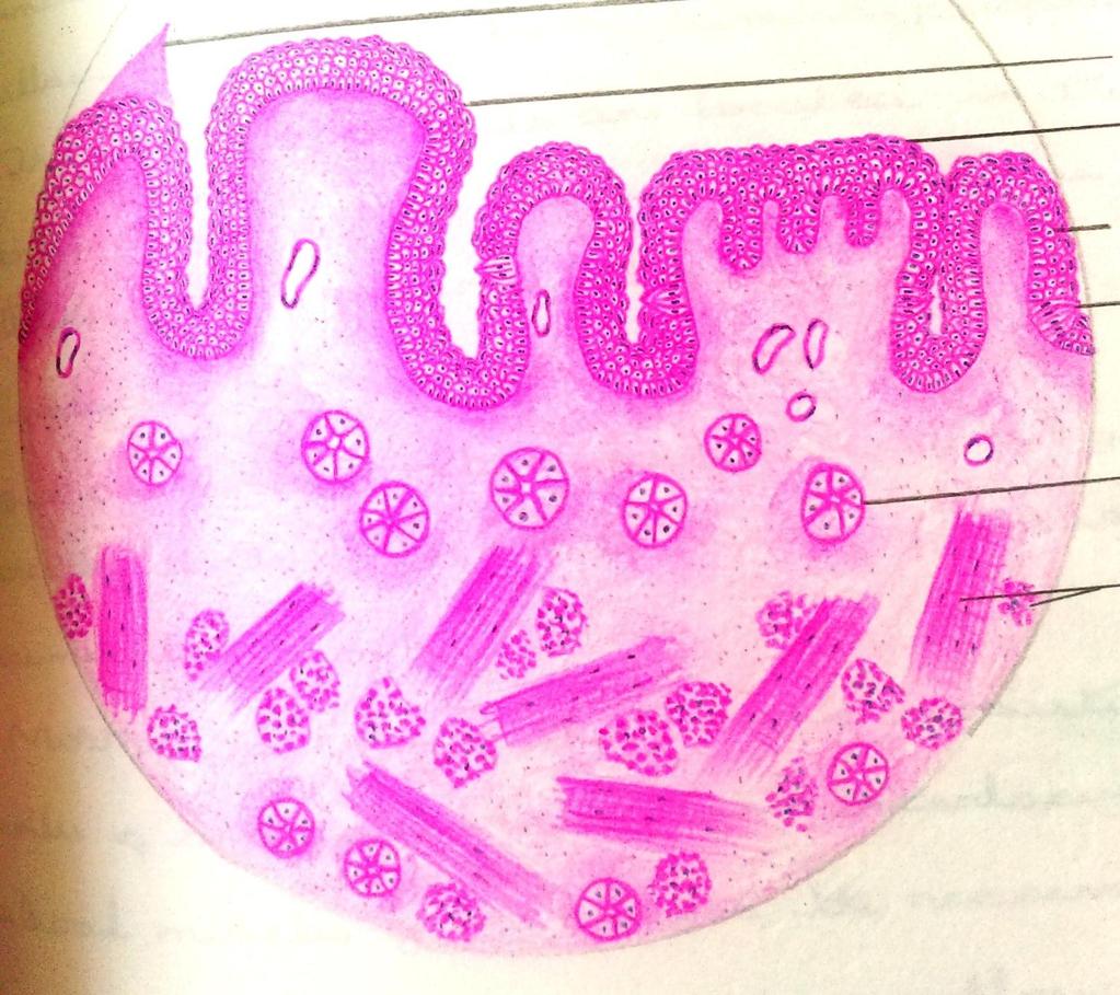
Dorsum of the tongue. Oral Part exhibit lingual papillae of the 4 types. Oral Part of Tongue divided into Left & right halves by shallow median groove
|
|
|
- Abner Jenkins
- 5 years ago
- Views:
Transcription

1 Histology of TONGUE

2 Figure 22.13
3 Dorsum of the tongue Oral Part of Tongue divided into Left & right halves by shallow median groove Oral Part exhibit lingual papillae of the 4 types a. filiform papillae, b. fungiform papillae, c. vallate papillae & d. foliate papillae Pharyngeal Part of Tongue devoid of papillae, contains lymphatic nodules - lingual tonsils
4 Most numerous is the filiform papillae it appears short and bristles macroscopically. Fungiform papillae are small red globular. Circumvallate papillae are arranged in rows in front of sulcus terminalis.

5 Tongue

6 Filliform Fungiform Circumvallate
7 Tongue
8 FILIFORM PAPILLAE Most numerous type. Entire dorsal surface of tongue. Lined stratified squamous keratinized epithelium No taste buds Increase the friction between tongue & food
9 Clinical correlation Hairy papillae Filiform papillae are hypertrophied and elongated. These hair (papillae) are stained black, brown, yellowish white by food or oral flora
10 FUNGIFORM PAPILLAE Rounded reddish elevations high vascular connective tissue core Looks like fungi narrow base & broad top Scattered all over anterior 2/3 rd of tongue most numerous in the tip of tongue Contains taste buds on surface.

11 Tongue
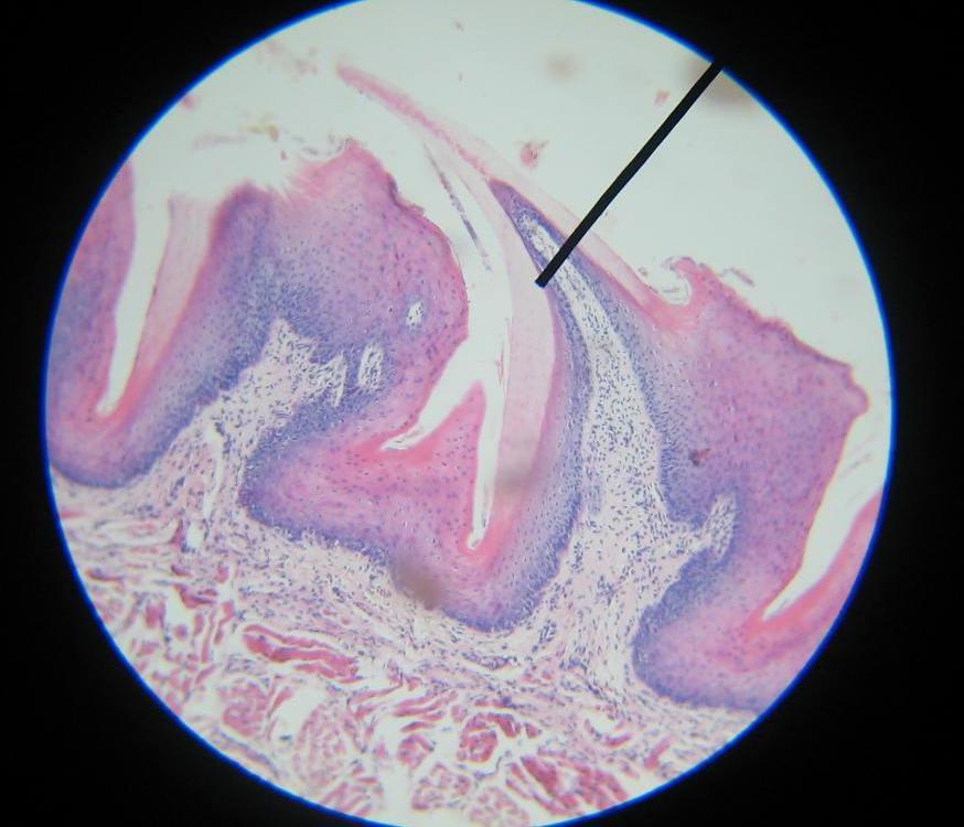
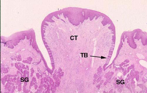
12 CIRCUMVALLET PAPILLAE Circumvallate papillae are the largest and least common type 8 to 12 in number. Arranged in a single row infront & parallel to sulcus terminalis Each papillae is surrounded by a circular sulcus/ groove (trench) called vellum which separates rest of the part
13 CIRCUMVALLET PAPILLAE These papillae has broad circular top and narrow base. Lined by stratified squamous non-keratinized epithelium Base of each papillae contains circular and longitudinal muscle fibers
14 CIRCUMVALLET PAPILLAE It also presents numerous of serous glands seen near the base and groove of papillae called serous glands of von ebner Serous secretions helps in appreciating taste of food by dissolving food particles in it Epithelium lining the walls of the sulcus shows numerous of taste buds
15 Foliate papillae Shape leaf like Rudimentary in humans It is functional in lower animals such has cows and buffaloes.
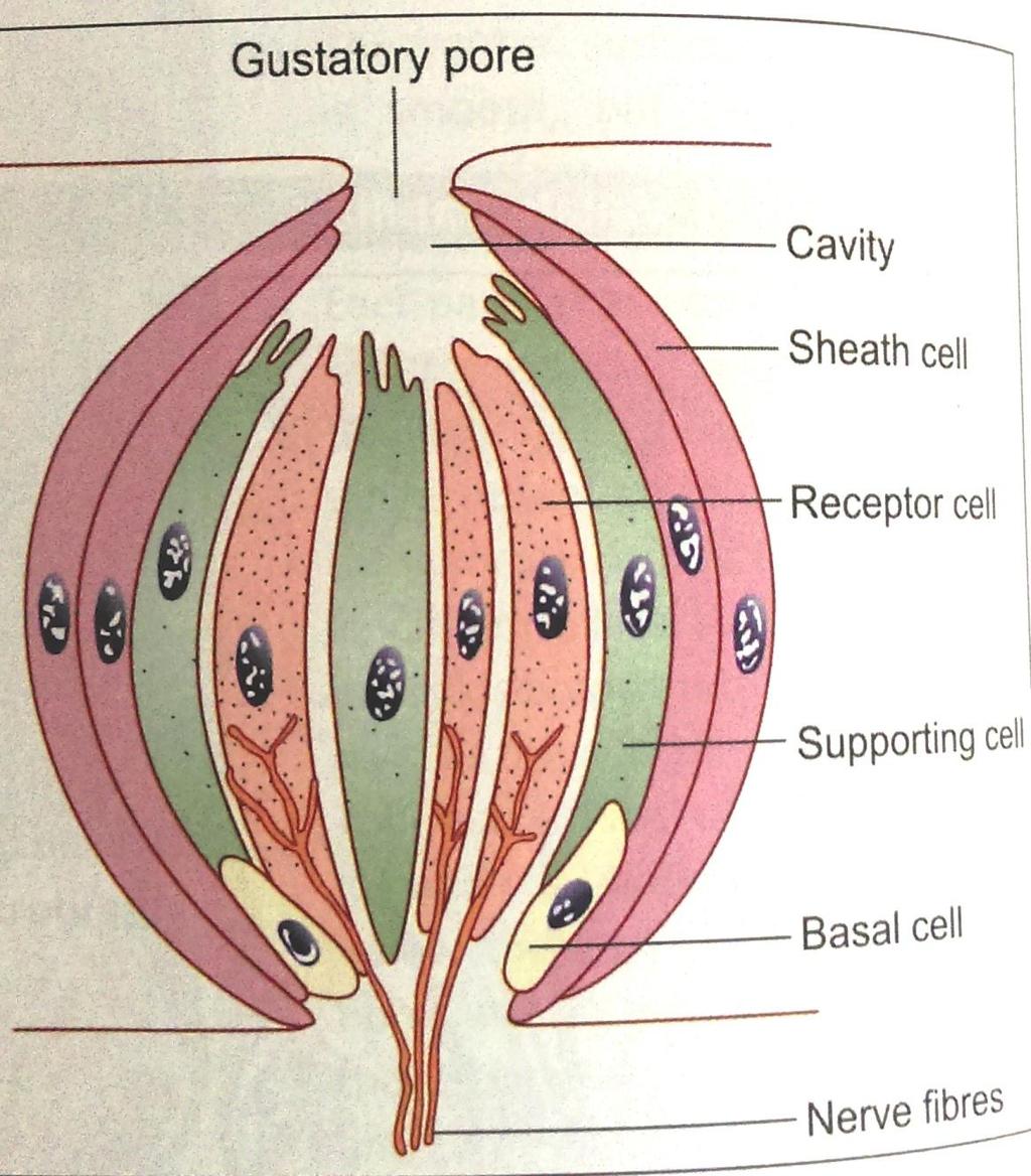
16 Taste bud
17 Taste bud It is a center for appreciation of taste Located in papillae of tongue In circumvallate it is located in groove In fungiform on surface or on its lateral part Filiform taste buds are absent Shape of taste bud is barrel.
18 Taste bud Made up modified epithelial cells Each bud has cavity which opens on the surface through gustatory pore. There are 5 types of cells
19 Taste bud There are 5 types of cells Type I Supportive cells Long elongated epithelial cells Darkly stained Upper pole bears fine microvilli Lower pole is close to basal cells
20 Taste bud Type II & III Gustatory cells or receptor cells It is the bipolar neuroepithelial cells Lightly stained Upper pole bears microvilli Lower pole is associated to sensory nerve fibers
21 Taste bud Type IV Basal cells or stem cells These pyramidal cells It is present close to the basement membrane of the epithelium Type V These cells forms the sheath or boundary
22

23 General plan of GIT Lamina propria Serosa/ Adventitia
24

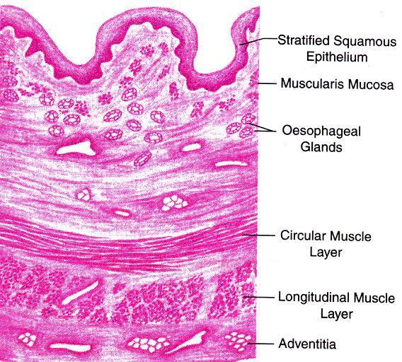
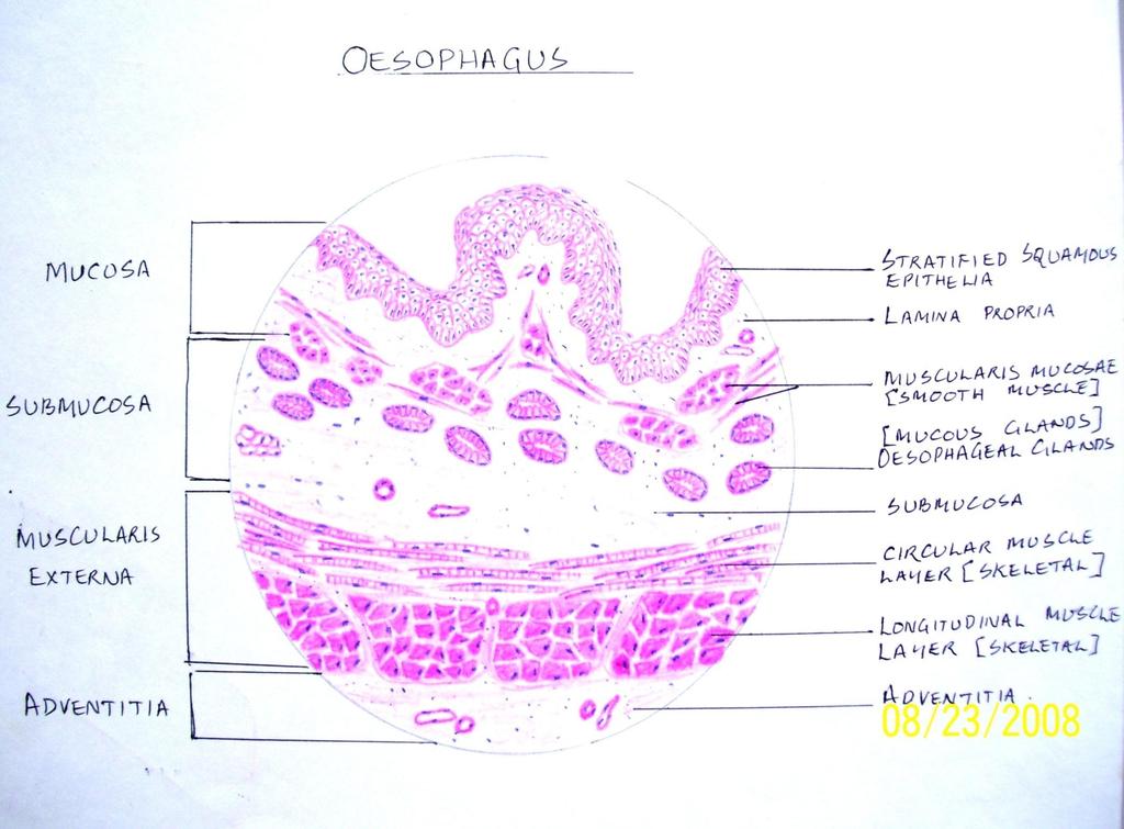
25 Mucosa - Stratified squamous nonkeratinized epithelium Papillae of lamina propria Muscularis mucosa longitudinal muscle bundles, mainly Submucosa :- Oesophageal glands tubulo - alveolar mucus acini in Muscularis externa: Upper one-third striated muscle Lower one-third smooth muscle Middle one-third striated & smooth muscle Adventitia/ Serosa

26 MUCOSA SUBMUCOSA Stratified squamous nonkeratinized epithelium Lamina propria Oesophageal glands Muscularis mucosa MUSCULARIS EXTERNA Inner circular layer Outer longitudinal layer SEROSA Connective tissue
27
28
29 Clinical anatomy: Oesophagitis : reflux of gastric contents causes heart burn Oesophageal varices: anastomosis dilate in lower end of oesophagus causes vomitting of blood
A deep groove encircles the body of the circumvallate papilla. Serous (von Ebner s) glands (serous) drain into the base of this groove.
 glands (serous) drain into the base of this groove.") By Dr. Raja Ali A deep groove encircles the body of the circumvallate papilla. Serous (von Ebner s) glands (serous) drain into the base of this groove. The flow of fluid from these glands serves to wash
By Dr. Raja Ali A deep groove encircles the body of the circumvallate papilla. Serous (von Ebner s) glands (serous) drain into the base of this groove. The flow of fluid from these glands serves to wash
HUMAN ANATOMY II STUDY NOTES. At the end of this chapter the student should be able to answer the following questions:
 HUMAN ANATOMY II STUDY NOTES CHAPTER ONE The Special Senses Learning objectives At the end of this chapter the student should be able to answer the following questions: 1. What is the gross and histological
Tongue In the buccal cavity of the digestive system
 Tongue In the buccal cavity of the digestive system same layers as those of tubular organs Mucosa, submucosa, and muscularis muscularis = the muscularis externa no muscularis mucosa 1 Tongue ling = tongue
GI Histology Lab 1. Prepared by: Zeina Kalaji
 GI Histology Lab 1 Prepared by: Zeina Kalaji Lip ORAL MUCOSA -Arrow shows labial salivary glands in the submucosa. VERMILLION transitional zone. SKIN Stratified Squamous epithelium, keratinized -Arrow
MICROSTRUCTURES LIPS TOOTH TONGUE OESOPHAGUS STOMACH, CARDIAC, PYLORIC FUNDIC GLANDS
 MICROSTRUCTURES LIPS TOOTH TONGUE OESOPHAGUS STOMACH, CARDIAC, PYLORIC FUNDIC GLANDS HUMAN ANATOMY: MICROSTRUCTURES CLASSIFICATION: LOCATION AND BOUNDARIES, FORM, FUNCTION, MICROSCOPIC STRUCTURE: A hollow
HISTOLOGY VIRTUAL LABORATORY GASTROINTESTINAL SYSTEM
 HISTOLOGY VIRTUAL LABORATORY GASTROINTESTINAL SYSTEM LIP (Slides GI 1, 2) Identify the outer portion lined by stratified squamous (keratinized) epithelium. Note the hair follicles and sebaceous glands
Objectives. Describe the cells of the GI tract and their function. Differentiate between different parts of the GI tract
 GI Histology 1 Objectives Describe the cells of the GI tract and their function Describe the histological features of each part of the GI tract. Differentiate between different parts of the GI tract Appreciate
Oral cavity Lab exercises
 Oral cavity Lab exercises Slide #190 (GT-1-32). Oral cavity, goat. large conical buccal papillae stratified squamous epithelium keratinized or non-keratinized no muscularis mucosae connective tissue represents
بسم هللا الرحمن الرحيم
 بسم هللا الرحمن الرحيم Today, we will leave all hormones and start with another topic which is GI system This lecture is talking about general histology of Gastrointestinal system.. The gastrointestinal
Epithelia will be discussed according to the following scheme: Type Number of layers Shape Line drawing. Squamous Cuboidal Columnar
 Epithelia Epithelia will be discussed according to the following scheme: Type Number of layers Shape Line drawing Simple Squamous Cuboidal Columnar Covering and Lining epithelium Pseudostratified Stratified
Digestive System. Presented by: Dr M. Arianmanesh PhD in Reproductive and Developmental Biology Dept. of Anatomical Sciences
 Digestive System Presented by: Dr M. Arianmanesh PhD in Reproductive and Developmental Biology Dept. of Anatomical Sciences Today we will discuss: Histological layers of alimentary canal Oral cavity Lip
HISTOLOGY OF THE RESPIRATORY SYSTEM I. Introduction A. The respiratory system provides for gas exchange between the environment and the blood. B.
 HISTOLOGY OF THE RESPIRATORY SYSTEM I. Introduction A. The respiratory system provides for gas exchange between the environment and the blood. B. The human respiratory system may be subdivided into two
General Structure of Digestive Tract
 Dr. Nabil Khouri General Structure of Digestive Tract Common Characteristics: Hollow tube composed of a lumen whose diameter varies. Surrounded by a wall made up of 4 principal layers: Mucosa Epithelial
Dr Nadine Gravett School of Anatomical Sciences Room 2B10B
 Dr Nadine Gravett School of Anatomical Sciences Room 2B10B Nadine.Gravett@wits.ac.za Oral cavity Mechanical breakdown Formation of bolus Oesophagus Conduit from mouth to stomach Stomach Digestion Temporary
Alimentary Canal (I)
") Alimentary Canal (I) Esophagus and Stomach (Objectives) By the end of this lecture, the student should be able to discuss the microscopic structure in correlation with the function of the following organs:
Alimentary Canal (I) Esophagus and Stomach (Objectives) By the end of this lecture, the student should be able to discuss the microscopic structure in correlation with the function of the following organs:
HISTOLOGY. GIT Block 432 Histology Team. Lecture 1: Alimentary Canal (1) (Esophagus & Stomach) Done by: Ethar Alqarni Reviewed by: Ibrahim Alfuraih
 (Esophagus & Stomach) Done by: Ethar Alqarni Reviewed by: Ibrahim Alfuraih") HISTOLOGY Lecture 1: Alimentary Canal (1) (Esophagus & Stomach) Done by: Ethar Alqarni Reviewed by: Ibrahim Alfuraih Color Guide: Black: Slides. Red: Important. Green: Doctor s notes. Blue: Explanation.
HISTOLOGY Lecture 1: Alimentary Canal (1) (Esophagus & Stomach) Done by: Ethar Alqarni Reviewed by: Ibrahim Alfuraih Color Guide: Black: Slides. Red: Important. Green: Doctor s notes. Blue: Explanation.
Connective tissue The Digestive System
 Connective tissue The Digestive System Part 1 Structure of digestive system Functions Basic Structure of the Alimentary Canal Wall Tube is made up of four layers: 1. Mucosa 2. Submucosa 3. Muscularis externa
DIGESTIVE. CHAPTER 17 Lecture: Part 1 Part 2 BIO 212: ANATOMY & PHYSIOLOGY II
 BIO 212: ANATOMY & PHYSIOLOGY II 1 CHAPTER 17 Lecture: DIGESTIVE Part 1 Part 2 Dr. Lawrence G. Altman www.lawrencegaltman.com Some illustrations are courtesy of McGraw-Hill. Processes of DIGESTION Mechanical
Organs Histology D. Sahar AL-Sharqi. Digestive System
 Digestive System The digestive system consists of the digestive tract oral cavity, esophagus, stomach, small and large intestines, and anus and its associated glands salivary glands, liver, and pancreas.
-Ibrahim Al-Naser. -Dr Al- Muhtaseb. 1 P a g e
 -1 -Ibrahim Al-Naser - -Dr Al- Muhtaseb 1 P a g e The Digestive System The doctor started the lecture by talking about the class rules. The GI system is an organ system, it is divided into: The Alimentary
Small Intestine, Large Intestine and anal cannel
 Small Intestine, Large Intestine and anal cannel 32409 Small intestine Large intestine Small intestine General Structure of the Digestive Tract rat 32409 Epithelium with goblet cells and absorptive cells
PRACTICAL ROADMAP EPITHELIUM A. JOVANOVIĆ
 PRACTICAL ROADMAP EPITHELIUM A. JOVANOVIĆ Epithelia Simple epithelia Stratified epithelia Simple squamous Simple cuboidal Simple columnar Pseudostratified Stratified squamous - non keratinized - keratinized
Connective tissue The Digestive System
 Connective tissue The Digestive System Part 1 Structure of digestive system Functions Basic Structure of the Alimentary Canal Wall Tube is made up of four layers: 1. Mucosa 2. Submucosa 3. Muscularis externa
Digestive system L 2. Lecturer Dr. Firdous M. Jaafar Department of Anatomy/Histology section
 Digestive system L 2 Lecturer Dr. Firdous M. Jaafar Department of Anatomy/Histology section objectives 1-Describe the general structure of digestive tract: a-mucosa. b-submucosa. c-muscularis externa d-adventitia
Lab activity manual Histology of the digestive system
 Lab activity manual Histology of the digestive system Jeanne Adiwinata Pawitan Prerequisite: Histology of the 4 basic tissues In this module we learn about the histology of the digestive system, from the
Tissues. tissue = many cells w/ same structure and function. cell shape aids its function tissue shape aids its function
 Tissues tissue = many cells w/ same structure and function cell shape aids its function tissue shape aids its function Histology = study of tissues 4 types of tissues Epithelial coverings contact openings
UNIVERSITY OF NAIROBI
 UNIVERSITY OF NAIROBI UNIVERSITY EXAMINATIONS 2013/2014 LEVEL I MID-SEMESTER II EXAMINATION FOR THE DEGREE OF BACHELOR OF SCIENCE IN NURSING (BScN) AND BACHELOR OF PHARMACY (B.PHARM) MARKING SCHEME HNS101/UPC106:
Small intestine. Small intestine
 General features Tubular organ longest part; 5-6 m most of chemical digestion absorption of nutrients reabsorption of H2O occurs. Two structural features; maximize the lumenal surface area villi microvilli
口腔生理學 ( 含顎咬合 ) Oral physiology (occlusion included) 學習目標. Oral moucosa 參考資料. Classification. Mucosa
 Oral physiology (occlusion included) 學習目標. Oral moucosa 參考資料. Classification. Mucosa") 口腔生理學 ( 含顎咬合 ) Oral physiology (occlusion included) Mucosa 臺北醫學大學牙醫學系張維仁老師 E-mail cweijen1@tmu.edu.tw 學習目標 1. let the student to understand the base knolwedge of oral physisology. 2.The student can firsther
口腔生理學 ( 含顎咬合 ) Oral physiology (occlusion included) Mucosa 臺北醫學大學牙醫學系張維仁老師 E-mail cweijen1@tmu.edu.tw 學習目標 1. let the student to understand the base knolwedge of oral physisology. 2.The student can firsther
DIGESTIVE TRACT ESOPHAGUS
 DIGESTIVE TRACT From the lower esophagus to the lower rectum four fundamental layers comprise the wall of the digestive tube: mucosa, submucosa, muscularis propria (externa), and adventitia or serosa (see
The Digestive System and Body Metabolism Premedical Biology
 The Digestive System and Body Metabolism Premedical Biology Copyright 2003 Pearson Education, Inc. publishing as Benjamin Cummings The Digestive System and Body Digestion Metabolism Breakdown of ingested
(b) Stomach s function 1. Dilution of food materials 2. Acidification of food (absorption of dietary Fe in small intestine) 3. Partial chemical digest
 Stomach s function 1. Dilution of food materials 2. Acidification of food (absorption of dietary Fe in small intestine) 3. Partial chemical digest") (1) General features a) Stomach is widened portion of gut-tube: between tubular and spherical; Note arranged of smooth muscle tissue in muscularis externa. 1 (b) Stomach s function 1. Dilution of food
(1) General features a) Stomach is widened portion of gut-tube: between tubular and spherical; Note arranged of smooth muscle tissue in muscularis externa. 1 (b) Stomach s function 1. Dilution of food
Upper Respiratory Histology
 Upper Respiratory Histology - Today we ll discuss the histology of larynx, trachea, primary, secondary, and tertiary bronchus. *First: The Larynx: -The picture below represents a section in the larynx,
Anatomy of Oral Cavity DR. MAAN AL-ABBASI
 Anatomy of Oral Cavity DR. MAAN AL-ABBASI By the end of this lecture you should be able to: 1. Differentiate different parts of the oral cavity 2. Describe the blood and nerve supply of mucosa and muscles
The Digestive System Laboratory
 The Digestive System Laboratory 1 The Digestive Tract The alimentary canal is a continuous tube stretching from the mouth to the anus. Liver Gallbladder Small intestine Anus Parotid, sublingual, and submaxillary
The Digestive System. Chapter 23 Anatomy of the Digestive System Part 1
 The Digestive System Chapter 23 Anatomy of the Digestive System Part 1 Overview Organs: Mouth, pharynx, esophagus, stomach, small intestine, and large intestine Overview Accessory Organs Teeth, tongue,
Histology Lab. looking at microscopic pictures of tissues, for more information use Junqueira book and you can use BlueHistolgy website
 Done By: Aseel Twaijer & Laith Sorour Histology Lab *These notes help in differentiating tissues and you must read them while looking at microscopic pictures of tissues, for more information use Junqueira
Digestive system 1. Oral cavity: Lips Tongue Palate soft - hard Tooth
 Digestive system 1 Oral cavity: Lips Tongue Palate soft - hard Tooth Common structure of the wall of GIT tube The mucosa epithelial linning lamina propria /loose connect. tissue/ the muscularis mucosae
Lecture Overview. Marieb s Human Anatomy and Physiology. Chapter 4 Tissues: The Living Fabric Epithelial Tissues Lecture 9. Introduction to Tissues
 Marieb s Human Anatomy and Physiology Marieb Hoehn Chapter 4 Tissues: The Living Fabric Epithelial Tissues Lecture 9 Lecture Overview Introduction to Tissues Epithelial Tissues Location General characteristics
Lab Activities 16, 17, & 18
 Lab Activities 16, 17, & 18 Olfaction & Taste Vision Hearing & Equilibrium Portland Community College BI 232 Lingual Papilla Papilla are epithelial projections on the superior surface of the tongue Circumvallate
Digestive Anatomy Lab
 Digestive Anatomy Lab In-Lab Exercises I have included the word list in this document. Any descrepencies between this document and the wordlist, you should default to this document. There is a lot of repetition
Anatomy & Histology of The Small intestine
 Anatomy & Histology of The Small intestine Prof. Abdulameer Al-Nuaimi E-mail: a.al-nuaimi@sheffield.ac.uk E. mail: abdulameerh@yahoo.com Jejunum Ileum Histology: Duodenum, jejunum, and ileum
Lecture Overview. Chapter 4 Epithelial Tissues Lecture 9. Introduction to Tissues. Epithelial Tissues. Glandular Epithelium
 Visual Anatomy & Physiology First Edition Martini & Ober Chapter 4 Lecture 9 Lecture Overview Introduction to Tissues Location General characteristics Functions Classification Glandular Epithelium 2 Where
Respiratory & Digestive Organs of the Head and Neck, Human;
 Name Date Lab Exercise 5: Lab Exercise 6: Lab Exercise 7: Lab Exercise 8: Respiratory & Digestive Organs of the Head and Neck, Human; Histology of the Respiratory System Digestive System Models, Human
Dana Alrafaiah. Dareen Abu Shalbak. Mohammad Almuhtaseb. 1 P a g e
 2 Dana Alrafaiah Dareen Abu Shalbak Mohammad Almuhtaseb 1 P a g e Esophagus: A muscular tube that is 25 cm long, but if measured from the incisors it would be 45cm long. Extends from C6 of cervical vertebra,
Sunday 29th January. Day 1: The Digestive Tract. From Anatomy to Treatment. A Nutritional Approach MPS.
 Sunday 29th January Day 1: The Digestive Tract From Anatomy to Treatment A Nutritional Approach MPS www.osteopathicea.com mike@osteopathicea.com AKA Digestive Tract, Alimentary Canal, Enteron, or Gut Body
Urinary Anatomy. Lab 40. Kidneys. Nephrons. Renal Corpuscle
 Urinary Anatomy Lab 40. Urinary Anatomy and Kidney Dissection Kidneys: filters blood, produces urine Ureters: convey urine to bladder Bladder: holding tank Urethra: carries urine to the outside for elimination
Organs Histology D. Sahar AL-Sharqi. Respiratory system
 Respiratory system The respiratory system provides for exchange of O2 and CO2 to and from the blood. Respiratory organs include the lungs and a branching system of bronchial tubes that link the sites of
Cell and Tissue Types. Epithelial, Connective, Muscle, Nerve
 Cell and Tissue Types Epithelial, Connective, Muscle, Nerve Objectives Explain the major stages of the cell cycle and cellular division (mitosis). Describe specific events occurring in each of the phases
Histology = the study of tissues. Tissue = a complex of cells that have a common function
 { EPITHELIAL TISSUE Histology = the study of tissues Tissue = a complex of cells that have a common function The Four Primary Tissue Types: Epithelium (epithelial tissue) covers body surfaces, lines body
Epithelium. Four primary tissue types:
 Epithelium Four primary tissue types: Epithelial (covering) Connective (support) Nervous (control) Muscular (movement) Smooth muscle Cardiac muscle Skeletal muscle 1 Epithelial Tissue Features Epithelial
Section 1.1: What is the function of digestion?
 Section 1.1: What is the function of digestion? When you have completed this section, you should be able to: Describe the overall function of the GI tract. Describe the processes involved in digestion.
On the Nerve Supply of the Radix Linguae of Newborn Dog. One of the present authors, KIKUCHI, has reported in detail on the innervation
 Arch. hist. jap. Vol. 19, n. 3 (May 1960). P. 437-446. Anat. Labor. of Prof. H. SETO, Tohoku Univ., Sendai. On the Nerve Supply of the Radix Linguae of Newborn Dog. Mizuho KIKUCHI, Shunsaku HATAKEYAMA,
General Human Histology. The Urinary System
 General Human Histology Lecture 8 Assist. Prof. Ahmed Anwar Albir The Urinary System Collecting Tubules & Ducts Urine passes from the distal convoluted tubules to collecting tubules that join each other
General principles of gastrointestinal motility
 General principles of gastrointestinal motility OBJECTIVES Physiological anatomy General Principles Circulation of blood through the GIT organs Control of all GIT functions by local, nervous, and hormonal
Basic Anatomy and Physiology of the Lips and Oral Cavity. Dr. Faghih
 Basic Anatomy and Physiology of the Lips and Oral Cavity Dr. Faghih It is divided into seven specific subsites : 1. Lips 2. dentoalveolar ridges 3. oral tongue 4. retromolar trigone 5. floor of mouth 6.
Lymphoid Organs. Dr. Sami Zaqout. Dr. Sami Zaqout IUG Faculty of Medicine
 Lymphoid Organs Dr. Sami Zaqout Cells of the Immune System Lymphocytes Plasma cells Mast cells Neutrophils Eosinophils Cells of the mononuclear phagocyte system Distribution of cells of the immune system
Topic and Aims. Contractile cells. Nervous system. Muscle tissue is one of the four basic tissue types.
 Topic and Aims Contractile cells Muscle tissue is one of the four basic tissue types. You should be able to: 1. Describe and identify the types of contractile cells, and summarise similarities and differences.
TASTE BUDS IN THE PITS AT THE POSTERIOR DORSUM OF THE TONGUE OF
 TASTE BUDS IN THE PITS AT THE POSTERIOR DORSUM OF THE TONGUE OF STENELLA COERULEOALBA FUSAO YAMASAKI, SHUNRO KOMATSU Department of Biology, Sapporo Medical College, Sapporo AND TOSHIRO KAMIYA Department
Digestive system. Dr. Sami Zaqout. IUG
 Digestive system Digestive system Digestive tract Associated glands Oral cavity Salivary glands Esophagus Liver Stomach Pancreas Small and large intestines Rectum and anus General Structure of the Digestive
Histology Urinary system
 Histology Urinary system Urinary system Composed of two kidneys, two ureters, the urinary bladder, and the urethra, the urinary system plays a critical role in: 1- Blood filtration,(filtration of cellular
SCPA602 Respiratory System
 SCPA602 Respiratory System Associate Professor Dr. Wannee Jiraungkoorskul Department of Pathobiology, Faculty of Science, Mahidol University Tel: 02-201-5563, E-mail: wannee.jir@mahidol.ac.th 1 Objectives
Epithelial Tissue. Functions include: 1. Protection 4. Absorption 2. Secretion 5. Filtration 3. Sensory reception
 Tissues There are 4 primary tissue types in the human body: 1. Epithelial (covering/lining) 2. Connective (support) 3. Muscle (movement) 4. Nervous (control) Epithelium Epithelial Tissue Covers the surface
Anatomy & Physiology Revealed Instructions. 1. From the Module dropdown menu, chose the 12. Digestive system.
 #10 - Objectives: Examine the histology of selected body organs using Anatomy & Physiology Revealed software and microscope slides. Be able to identify each organ and the specific structures indicated
Bio & 241 A&P Unit 1 / Lecture 3
 Bio & 241 A&P Unit 1 / Lecture 3 Tissues All body tissues arise from three fundamental embryonic tissues. Endoderm: forms epithelial tissues lining internal organs such as the GI tract Mesoderm: connective
The Tissue Level of Organization
 The Tissue Level of Organization Study of this lecture is to be accomplished in conjunction with the Histology Module on the Web!! 1. Introduction Cell Tissue Histology A. General Tissue Types i. Epithelial
Respiratory System. Functional Anatomy of the Respiratory System
 Respiratory System Overview of the Respiratory System s Job Major Duty Respiration Other important aspects ph control Vocalization Processing incoming air Protection Metabolism (ACE) What structures allow
From the Anat. Labor. of Prof. H. SETO, Tohoku University, Innervation of Radical Part of Tongue of Hedgehog.
 From the Anat. Labor. of Prof. H. SETO, Tohoku University, Sendai. Innervation of Radical Part of Tongue of Hedgehog. Many histological studies on the innervation, especially on the sensory innervation,
Common details: blood vessels, nerve trunks, ganglions, adipose tissue, lymphocytes
 Common details: blood vessels, nerve trunks, ganglions, adipose tissue, lymphocytes Outer lining of the organs: ADVENTITIA (connective tissue) or SEROSA (thin layer of connective tissue lined by simple
Tissues and organs PART 1
 Tissues and organs PART 1 Animals and plants are multicellular (made of many cells). Cells become specialised according to their function Tissues: Many cells that perform one or several functions; they
The doctor mentioned a few things about the esophagus from the previous lecture:
 السالم عليكم [HISOLOGY 2] April 27, 2014 The doctor mentioned a few things about the esophagus from the previous lecture: Esophagus - It is about 25 cm in length (from the incisor it is 45 cm) Histological
Subdivided into Vestibule & Oral cavity proper
 Extends from the lips to the oropharyngeal isthmus The oropharyngeal isthmus: Is the junction of mouth and pharynx. Is bounded: Above by the soft palate and the palatoglossal folds Below by the dorsum
Indo Asian Journal of Multidisciplinary Research (IAJMR) ISSN:
 ISSN:") Available online at www.jpsscientificpublications.com Volume 3; Issue - 1; Year 2017; Page: 969 973 Indo Asian Journal of Multidisciplinary Research (IAJMR) ISSN: 2454-1370 MORPHOLOGICAL AND HISTOLOGICAL
Available online at www.jpsscientificpublications.com Volume 3; Issue - 1; Year 2017; Page: 969 973 Indo Asian Journal of Multidisciplinary Research (IAJMR) ISSN: 2454-1370 MORPHOLOGICAL AND HISTOLOGICAL
14 Cardiovascular System
 14 Cardiovascular System The goal of this topic is to examine and understand the structure of the heart, blood vessels and the lymphatic vessels. You should aim to understand how the structure of blood
Mirror of Research in Veterinary Sciences and Animals (MRVSA)
") Mirror of Research in Veterinary Sciences and Animals (MRVSA) Original article Anatomical and histological studies of esophagus of one-humped camel (Camelus dromedarius) Adel Jabbar Hussein 1 ;Muntdhur
Mirror of Research in Veterinary Sciences and Animals (MRVSA) Original article Anatomical and histological studies of esophagus of one-humped camel (Camelus dromedarius) Adel Jabbar Hussein 1 ;Muntdhur
The Respiratory System. Prof. Dr.Mohammed Hisham Al-Muhtaseb
 The Respiratory System Prof. Dr.Mohammed Hisham Al-Muhtaseb Objectives (lecture + practical) 1. Identify the conduction part of the respiratory tract and analyze the function of each segment 2. Identify
bolus. The bolus is passed to the pharynx which will convey it to the esophagus, the start of the digestive tube proper. This muscular tube will
 Chapter 13 Digestive System (Oral Cavity and the Alimentary Canal) 13.1. Basic Concepts The digestive system is involved with the intake, mechanical and chemical breakdown, and absorption of food. It also
Slide 154: Pancreas, H&E
 Slide 154: Pancreas, H&E the pancreas, located adjacent to the duodenum, is a mixed exocrine and endocrine gland; it is usually readily identifiable by the presence of the interspersed endocrine pancreatic
Tissues. Definition. A group of similar cells and their intercellular substances specialized to perform a specific function.
 Chapter 4 - Tissues Tissues Definition A group of similar cells and their intercellular substances specialized to perform a specific function. Tissues Epithelial covers exposed surfaces, lines internal
Tissue: The Living Fabric: Part A
 PowerPoint Lecture Slides prepared by Janice Meeking, Mount Royal College C H A P T E R 4 Tissue: The Living Fabric: Part A Tissues Groups of cells similar in structure and function Types of tissues Epithelial
Tissues. Definition. A group of similar cells and their intercellular substances specialized to perform a specific function.
 Chapter 4 - Tissues Tissues Definition A group of similar cells and their intercellular substances specialized to perform a specific function. Tissues Epithelial covers exposed surfaces, lines internal
Epithelium Characteristics cont. 2. Apical Surface
 Epithelium Characteristics cont. 2. Apical Surface always has one exposed (apical) surface Some surfaces are smooth & slick, others may have: microvilli fingerlike extensions of the plasma membrane; increase
Tissues (Histology) Ch. 3 Human Anatomy lecture
 Ch. 3 Human Anatomy lecture") I. Histology the study of tissues A. 4 basic tissue types epithelial connective muscle nervous Tissues (Histology) Ch. 3 Human Anatomy lecture B. Usually found in combinations to form organs. C. As you
I. Histology the study of tissues A. 4 basic tissue types epithelial connective muscle nervous Tissues (Histology) Ch. 3 Human Anatomy lecture B. Usually found in combinations to form organs. C. As you
Lab 8: Digestive System
 BIOL 221 A&P II Lab 8: Digestive System Become familiar with the gross anatomy of the digestive system (Exercise 38) using the models, Fig. 38.1 (Activity 1), and the rat. Recognize and know the functions
(A) Diarrhea. (B) Stomach cramps. (C) Dehydration due to excess fluid loss. (D) A, B, and C are correct. (E) Only answer B is correct.
 Diarrhea. (B) Stomach cramps. (C) Dehydration due to excess fluid loss. (D) A, B, and C are correct. (E) Only answer B is correct.") Human Anatomy - Problem Drill 21: The Digestive System Question No. 1 of 10 1. A 26-year-old male is treated in the emergency department for severe gastrointestinal disturbance. Which of the following
Human Anatomy - Problem Drill 21: The Digestive System Question No. 1 of 10 1. A 26-year-old male is treated in the emergency department for severe gastrointestinal disturbance. Which of the following
Advances in Bioresearch
 Advances in Bioresearch Adv. Biores., Vol 5 (1) March 2014: 70-74 Print ISSN 0976-4585; Online ISSN 2277-1573 Journal s URL:http://www.soeagra.com/abr.html CODEN: ABRDC3 ICV 7.20 [Poland] Advances in Bioresearch
MH1001 Larynx, Monkey, H&E
 MH1001 Larynx, Monkey, H&E - False vocal fold : ciliated pseuodostratified columnar epithelium - True vocal fold : nonkeratinized stratified squamous epithelium vocalis muscle, vocal ligament - Thyroid
Lab activity manual - Histology of the digestive system. Lab activity 1: esophagus stomach - small intestines
 Lab activity manual - Histology of the digestive system Jeanne Adiwinata Pawitan Prerequisite: Histology of the 4 basic tissues In this module we learn about the histology of the digestive system, from
An overview of the digestive system. mouth pharynx esophagus stomach small intestine large intestine rectum anus
 An overview of the digestive system mouth pharynx esophagus stomach small intestine large intestine rectum anus Why GIT? What are the main steps in the digestive process? Ingestion intake of food via the
abstract Key words: Hard palate, Tongue, Buccal floor, Sambar deer, Gross anatomy
 Gross anatomical studies on the hard palate, tongue and buccal floor in sambar deer (Cervus unicolor) a.r sreeranjini 1, c.v rajani 2 and n. ashok 3 Department of Veterinary Anatomy & Histology College
Tissues. Tissues - Overview. Bio 101 Laboratory 3. Epithelial Tissues and Integument
 Bio 101 Laboratory 3 Epithelial Tissues and Integument 1 Tissues Tissues to be examined under the microscope Epithelial Tissue Integument Connective Tissue **We will be doing muscle and nervous tissues
Histology Notes -Part 1: Epithelial Tissues
 Introduction Group of cells w/ similar structure & function = TISSUE Four Basic Tissue Types 1. Epithelial-covers 2. Connective-supports 3. Muscular*-produces movement (will discuss in the muscular system
A Rough look at the tonsils and adenoids, for Bonny Peppa!
 A Rough look at the tonsils and adenoids, for Bonny Peppa! tonsils (two oval masses in the back of the throat) Lymphoid organs include: adenoids (two glands located at the back of the nasal passage) appendix
DIGESTIVE SYSTEJV1 OF MYSTUS CAVASIUS. Morphology and Anatomy Histology Physiology
 OBSERVATION .. DIGESTIVE SYSTEJV1 OF MYSTUS CAVASIUS Morphology and Anatomy Histology Physiology 43 OBSERVATIONS DIGESTIVE SYSTEM of Mystus cavasius MORPHOLOGY AND ANATOMY The alimentary can~1: The alimentary
Urinary system. Urinary system
 Distal convoluted tubule (DCT) Highly coiled, ~ 5 mm in length Last part of the nephron. Wall; simple cuboidal epithelium Less metabolically active than the PCT no brush border light eosinophilic cytoplasm
الله الر ح م ن الر ح يم مسب
 بسم رلا هللارلا هللا This is the second histology lecture in the GI system. In this lecture, we will discuss the histology of the esophagus, stomach, and small intestine so prepare yourself.this sheet
BIOL& 253 Lab Manual for Practical #2 Page 1 Rausch. For all slides, know a function for structures marked with a single asterisk (*).
.") BIOL& 253 Lab Manual for Practical #2 Page 1 Rausch Lab equipment: slides, models SLIDES For all slides, know a function for structures marked with a single asterisk (*). DIGESTIVE SYSTEM Layers of the
BIOL& 253 Lab Manual for Practical #2 Page 1 Rausch Lab equipment: slides, models SLIDES For all slides, know a function for structures marked with a single asterisk (*). DIGESTIVE SYSTEM Layers of the
I. The Alimentary Canal (GI track)
") A. About 9 meters long B. Passes through the ventral cavity. C.Movements of the Tube 1. Mixing movements- smooth muscles contract rhythmically. 2. Propelling movements- a wavelike motion called peristalsis.
A. About 9 meters long B. Passes through the ventral cavity. C.Movements of the Tube 1. Mixing movements- smooth muscles contract rhythmically. 2. Propelling movements- a wavelike motion called peristalsis.
Alimentary Canal (I) Salivatory Glands. (Esophagus and Stomach) Color index: Slides.. Important..Notes..Extra..
 Salivatory Glands. (Esophagus and Stomach) Color index: Slides.. Important..Notes..Extra..") Alimentary Canal (I) (Esophagus and Stomach) Salivatory Glands Color index: Slides.. Important..Notes..Extra.. Objectives: 1. By the end of this lecture, the student should be able to discuss the microscopic
Alimentary Canal (I) (Esophagus and Stomach) Salivatory Glands Color index: Slides.. Important..Notes..Extra.. Objectives: 1. By the end of this lecture, the student should be able to discuss the microscopic
Smell and taste sensation/ objectives of the lecture
 Smell and taste sensation/ objectives of the lecture Describe the basic features of the neural elements in the olfactory epithelium and olfactory bulb. Outline the pathway by which impulses generated in