Trophic peptide therapies for neonatal short bowel syndrome: actions and mechanisms studied in a preclinical model
|
|
|
- Godfrey Baldwin
- 5 years ago
- Views:
Transcription
1 Trophic peptide therapies for neonatal short bowel syndrome: actions and mechanisms studied in a preclinical model by David Wai Lim A thesis submitted in partial fulfillment of the requirements for the degree of Doctor of Philosophy in EXPERIMENTAL SURGERY Department of Surgery University of Alberta David Wai Lim, 2016
2 Abstract Short bowel syndrome (SBS) occurs when a significant length of intestine is surgically resected for both congenital and acquired intestinal abnormalities and remains a significant cause of morbidity and mortality in neonates. With an insufficient amount of intestine available for nutrient absorption, neonates with SBS are dependent on parenteral nutrition (PN) for survival but many continue to develop and succumb to PNassociated complications such liver disease and sepsis. In order for these children to survive, the remnant intestine must adapt and improve nutrient absorption over time. Glucagon-like peptide-2 (GLP-2) is a distal intestinal-derived peptide that is trophic to the intestine and stimulates intestinal adaptation after major resection. Diseases that lead to SBS in neonates, such as necrotizing enterocolitis, most commonly affect and require removal of the distal intestine, including ileum. Thus, GLP-2 may be the limiting factor for adaptation in human neonates with SBS. Furthermore, the intestinotrophic effects of GLP-2 may be augmented by the simultaneous delivery of either enteral nutrition (EN) or epidermal growth factor (EGF). We thus hypothesized that GLP-2 therapy stimulates intestinal growth and function in neonatal SBS, and that the intestinotrophic effects of GLP-2 are augmented when given in combination with either EN or EGF therapy. For these studies, neonatal piglets were block-randomized to either a 75% midintestinal resection (JI model) with jejunoileal anastomosis (leaving equal lengths of jejunum and ileum) or 75% distal-intestinal resection (JC model) (removing all ileum) with jejunocolic anastomosis or sham (no resection) control. Piglets also received a jugular venous catheter and a gastrostomy tube for the provision of PN and EN, respectively. Piglets were subsequently maintained for 7 days. In the first study, piglets ii
3 received either intravenous GLP-2 (42 g/kg/day) or saline control and either remained on total PN (0% EN) or received EN at 40% of nutritional requirement. In the second study, piglets received saline control, intravenous GLP-2 (42 g /kg/day), enteral EGF (80 µg/kg/day), or combined GLP-2 and EGF and all piglets received EN at 20% of nutritional requirement. Structural adaptation was assessed by the change in intestinal length, mucosal and intestinal weight, and histopathology. Functional adaptation was assessed by measuring several parameters: the relative gene expression of nutrient transporters, digestive enzymes and tight junction proteins, measurement of intestinal permeability using the Üssing chamber apparatus, fat absorption and weight gain. In the first study, we observed that in piglets maintained on total PN in the absence of treatment, the JI model demonstrated intrinsic structural adaptation, including increased intestinal weight and villus height, while the JC model did not. In this group that was not enterally fed, GLP-2 treatment induced histological adaptation in the JC model. In contrast, enteral feeding at 40% of nutritional requirement resulted in intestinal lengthening and increased intestinal weight in the JI model, while increased diarrhea and decreased weight gain were observed in the JC model. GLP-2 treatment in this fed group of piglets had no effect in the JI model but increased villus height in the JC model. We did not observe differences in the gene expression of nutrient transporters or tight junction proteins. In the second study, combined EGF and GLP-2 treatment increased intestinal length by 15%, regardless of surgical anatomy. Both GLP-2 alone and combination therapy increased intestinal weight in the JC model, and jejunal mucosal weight and villus height in both JI and JC models. Combination therapy decreased intestinal iii
4 permeability to both mannitol and polyethylene glycol in both surgical models. There was no difference in fat absorption or weight gain. Our results demonstrate the beneficial effects of exogenous GLP-2 treatment in the JC model, which anatomically represents most human infants with SBS. In contrast, the JI model demonstrated greater structural adaptation in response to enteral feeding. We further demonstrated a beneficial effect of combined GLP-2 and EGF treatment on increasing intestinal length and absorptive surface area in both models, which may lead to improved nutrient absorption. The benefit of decreased intestinal permeability with combination therapy translates to strengthened barrier function and decreased risk for bacterial translocation. GLP-2 therapy may thus benefit human infants with SBS, who commonly experience small intestinal bacterial overgrowth. Moving forward, our studies provide important preclinical data with regards to the translation of trophic peptide therapies in neonatal SBS. iv
5 Preface This thesis is an original work by David Wai Lim. The research project, of which this thesis is a part, obtained ethics approval from the University of Alberta Animal Care and Use Committee, study title Determining the impact of exogenous GLP-2 on intestinal adaptation in our short bowel syndrome neonatal piglet models, study ID AUP , May 30, 2013, and study title A pilot study of systemic glucagon-like peptide combined with oral epidermal growth factor and characterization of gut microbiome in short bowel piglets with and without ileum, study ID AUP , March 20, Portions of the research conducted for this research form part of an institutional and international research collaboration, led by Dr. Justine M. Turner at the University of Alberta and Dr. Paul W. Wales at the University of Toronto. The literature review in chapters 1-4 represents my original work. Chapter 3 of this thesis has been published as D.W. Lim, J.M. Turner, and P.W. Wales, Emerging Piglet Models of Neonatal Short Bowel Syndrome, Journal of Parenteral and Enteral Nutrition, vol. 39, issue 6, pages Chapter 4 of this thesis has been published as D.W. Lim, P.W. Wales, J.M. Turner, D.L. Bigam, and P.L. Brubaker, On the horizon: trophic peptide growth factors as therapy for neonatal short bowel syndrome, Expert Opinion on Therapeutic Targets, vol. 20, issue 7, pages Chapter 5 of this thesis has been published as D.W. Lim, A. Diané, M. Muto, D.F. Vine, P.N. Nation, P.R. Wizzard, D.L. Sigalet, D.L. Bigam, P.B. Pencharz, J.M. Turner, and P.W. Wales, Differential effects on intestinal adaptation following exogenous glucagon-like peptide-2 therapy with and without enteral nutrition in neonatal short v
6 bowel syndrome, Journal of Parenteral and Enteral Nutrition, doi: / [published online ahead of print, September 22 nd, 2016]. This study was designed by myself with the assistance of J.M. Turner, P.W. Wales and P.B. Pencharz at the University of Toronto. I was responsible for data collection (assisted by P.R. Wizzard) and analysis and manuscript composition. A. Diané performed the gene expression analyses supervised by D.F. Vine. M. Muto performed the fat absorption studies supervised by P.R. Wizzard. P.N. Nation performed the analysis of histological specimens. D.F. Vine, D.L. Sigalet. D.L. Bigam, J.M. Turner and P.W. Wales contributed to manuscript editing. Chapter 6 of this thesis has been submitted to and peer-reviewed by the American Journal of Physiology Gastrointestinal and Liver Physiology; revisions were requested by the journal and are currently being drafted for journal submission. This study was designed by myself with the assistance of J.M. Turner, P.W. Wales and C.L. Lévesque at South Dakota State University. I was responsible for data collection (assisted by P.R. Wizzard and M. Muto) and analysis and manuscript composition. The Üssing chamber experiments were performed by myself under the supervision of D.F.Vine. J.R. Koepke performed some of the gene expression analyses supervised by C.L. Lévesque at South Dakota State University. The gene expression analyses on intestinal growth factors and their receptors were performed by myself under the supervision of P.L. Brubaker at the University of Toronto. P.N. Nation performed the analysis of histological specimens. C.L. Lévesque, D.F. Vine, J. Li at the University of Guelph, P.L. Brubaker, D.L. Sigalet, D.L. Bigam, J.M. Turner and P.W. Wales contributed to manuscript editing. vi
7 Two roads diverged in a wood, and I I took the one less traveled by, And that has made all the difference. - Robert Frost (1916) vii
8 Dedication I dedicate this PhD thesis, for which I have devoted these past 4.5 years towards, to my loving family: My mother and father, Yuk Lin and Gue Duck Lim, for their never-ending support, And my brother Allan and sister Kim for always being there for me. viii
9 Acknowledgments First and foremost, thank you to Drs. Justine Turner and Paul Wales for providing me with the opportunity to pursue graduate research in their lab and for mentoring my personal and professional growth and development. I am forever grateful for their kindness, patience and enthusiasm. Thank you very much to Dr. David Bigam for his guidance and support throughout the duration of my studies and whose mentorship I value immensely. Thank you to Dr. Donna Vine for taking the time to share with me her passion for basic science and translational research. Thank you very much to Dr. Tom Churchill and Christina Smith in the Department of Surgery, who have strongly advocated for and ensured the well-being of graduate students like myself. Thank you to Janice Bowers, Tomiko McCall, Ida Seifeddine and Stephanie van Lieshout for their time and helpful assistance throughout the years. Thank you so much to the many collaborators whom I have had the pleasure to meet and work with. Thank you to Dr. Patricia Brubaker at the University of Toronto for so kindly hosting me at her laboratory on several occasions and sharing with me her passion for science and discovery. Thank you to Crystal Lévesque at South Dakota State University for our collaborative work. Thank you to Dr. David Sigalet for his guidance and insightful contributions to my thesis and his mentorship towards my career development. Thank you to Dr. Nick Nation for his kindness and contributions to my projects. Thank you sincerely to Drs. Diana Mager, Vera Mazurak, Jason Yap, Jonathan Curtis, Consolato Sergi and Ben Willing for the exciting collaborations that we undertook throughout my graduate studies. ix
10 I would further like to acknowledge all the support and help from the friends I made along my graduate journey. Thank you to Ms. Pamela Wizzard and Ms. Charlane Gorsak, at the Swine Research and Technology Center, for sharing with me many laughs, in addition to their passion for animal care. Thank you to Abha Dunichand-Hoedl in the Mager-Mazurak lab and Sandra Kelly in the Vine lab for their assistance and expertise. Thank you to the many graduate students and postdoctoral fellows who I have had the opportunity to collaborate with and learn from: Zheng Hua, Christine Pendlebury, Mitsuru Muto, Jessica Josephson, Amanda Soukvilay, Celeste Lavallee and Marihan Lansing (Turner-Wales lab), Kaori Yamada, Holly Stacey, Melanie Markovic and Bradley Smithers (Brubaker lab), and Abdoulaye Diané and Faye Borthwick (Vine lab). Finally, I would like to acknowledge the generous support of the following agencies and institutions towards the successful completion of my PhD thesis: Canadian Institutes of Health Research (Doctoral Research Award), Alberta Innovates Health Solutions (Clinician Fellowship), Killam Trusts (Izaak Walton Killam Memorial Scholarship), the Women`s and Children`s Health Research Institute (Graduate Studentship), and at the University of Alberta: Faculty of Medicine and Dentistry (75 th Anniversary Graduate Student Award), Faculty of Graduate Studies and Research (Queen Elizabeth II Graduate Scholarships, Killam Laureate), Department of Surgery, Division of General Surgery, the University of Alberta Clinician-Investigator Program, and the Graduate Students` Association. Thank you furthermore to the American Society for Parenteral and Enteral Nutrition and the American Society for Nutrition for awards supporting graduate student research. Finally, thank you to the Edmonton Civic x
11 Employees Charitable Assistance Fund for its continual support of resident and graduate student research in the Department of Surgery at the University of Alberta. xi
12 Table of Contents Chapter 1: Neonatal Short Bowel Syndrome page 1-25 Introduction.page 2 Short Bowel Syndrome and Intestinal Failure..page 2 4 Epidemiology and Etiology.....page 4 6 Pathophysiology of Short Bowel Syndrome... page 6 10 Intestinal Adaptation.....page Management......page Predictors of Outcome in SBS...page Parenteral Nutrition Associated Liver Disease.page Conclusion. page 17 References......page Chapter 2: Nutrient and Hormonal Regulation of Intestinal Adaptation.... page Introduction...page 27 Two Processes of Adaptation...page The Physiology of Intestinal Adaptation...page Dietary Regulation....page Dietary Protein....page Dietary Carbohydrate..page Dietary Lipids..page xii
13 Hormonal Regulation of Intestinal Adaptation.page Growth Hormone...page Insulin-like Growth Factor-1...page Epidermal Growth Factors...page Glucagon-like Peptide-2...page The Function of GLP-2...page Mechanism of GLP-2 action...page Role of GLP-2 in Animal Models of Intestinal Adaptation page Role of GLP-2 in Higher Mammals...page Conclusion.....page 53 References......page Chapter 3: Emerging Piglet Models of Neonatal Short Bowel Syndrome...page Abstract.....page 73 Introduction...page Short Bowel Syndrome: A Heterogeneous Disease..page Intestinal Length.. page Anatomy and Function of the Remnant Intestine.....page Presence or Absence of the Colon and Ileocecal Valve...page The Piglet as an Appropriate Model for the Developing Human Intestine...page Emerging Piglet Models of Neonatal Short Bowel Syndrome..page xiii
14 Translating Piglet Models of SBS to Human Neonates page Conclusions and Future Directions....page References page Chapter 4: On the Horizon: Trophic Peptide Growth Factors as Therapy for Neonatal Short Bowel Syndrome..page Abstract......page 102 Article Highlights Box page 103 Introduction...page Trophic peptides......page Glucagon-like peptide-2..page GLP-2 physiology..page GLP-2 signaling and secondary messengers.page GLP-2 in adult models of SBS..page GLP-2 in adult humans with SBS...page 111 Insulin-like growth factor family page IGF-I..page IGF-II.page Epidermal growth factor family page Other peptides...page Role of Trophic Peptides in Gastrointestinal Development..page Trophic peptide therapy in preclinical models of neonatal SBS...page Trophic peptides in human infants with SBS page xiv
15 Conclusion...page 125 Expert Opinion...page References.. page Chapter 5: Differential Effects on Intestinal Adaptation Following Exogenous Glucagon-like Peptide-2 Therapy With and Without Enteral Nutrition in Neonatal Short Bowel Syndrome..page Abstract page 148 Introduction... page Methods.page Results...page Discussion.page Conclusion...page 174 References.. page Chapter 6: Synergy of Glucagon-Like Peptide-2 and Epidermal Growth Factor Coadministration on Intestinal Adaptation in Neonatal Piglet with Short Bowel Syndrome..page Abstract......page Introduction... page Methods.page Results...page Discussion.page xv
16 References..page Chapter 7: The Role for Trophic Peptide Therapies in Neonatal Short Bowel Syndrome Summary and Future Directions.....page References..page xvi
17 List of Tables Table 1-1: Factors predictive of intestinal adaptation and outcome in short bowel syndrome... page 18 Table 4-1: Neonatal Short Bowel Syndrome: Key Facts...page 130 Table 4-2: Neonatal SBS: subtypes, anatomy and clinical sequelae..page 131 Table 4-3: Summary of Available Studies on Exogenous Trophic Peptide Therapies in Animal SBS Models and Adult and Pediatric Human Studies...page 133 Table 5-1: List of target and housekeeping genes for Chapter 5....page 192 Table 5-2: Jejunal and ileal mrna expression of nutrient transporters and tight junctional proteins in piglets on total PN (0% EN).page 193 Table 5-3: Jejunal and ileal mrna expression of nutrient transporters and tight junctional proteins in piglets receiving 40% EN.page 194 Table 6-1: Intestinal Growth and Function....page 237 Table 6-2: Intestinal Growth Factors and their Receptors....page 238 xvii
18 Table 6-3: Baseline data....page 239 xviii
19 List of Figures Figure 4-1: Distribution of GLP-2-producing L cells and GLP-2R expression along the gastrointestinal tract, and their consequential removal in the varying types of short bowel syndrome.....page 132 Figure 5-1: Study flow charts....pages Figure 5-2: Weight gain and gross intestinal morphology in 0% EN piglets page 177 Figure 5-3: Remnant intestinal histology in 0% EN piglets...page 178 Figure 5-4: Weight gain and gross intestinal morphology in 40% EN piglets..page 179 Figure 5-5: Remnant intestinal histology in 40% EN piglets.....page 180 Figure 5-6: Representative intestinal cross-sections pages Figure 5-7: Fat absorption......page 185 Figure 5-8: Weight gain and gross intestinal morphology comparing 0% EN versus 40% EN piglets pages xix
20 Figure 5-9: Remnant intestinal histology comparing 0% EN versus 40% EN piglets...pages Figure 5-10: Summary Tables..pages Figure 6-1: Study flow chart..page 235 Figure 6-2: Tissue Collection.page 236 Figure 6-3A-E: Weight Gain and Gross Morphology...page 240 Figure 6-3F: Change in length of remnant jejunum and ileum in JI piglets...page 241 Figure 6-4: Histopathology....page 242 Figure 6-5: Jejunal permeability....page 243 Figure 6-6: Electrical Parameters of Jejunum....page 244 Figure 6-7: Fat absorption......page 245 Figure 6-8: Intestinal growth and function.....page 246 xx
21 Figure 6-9: Intestinal gene expression of growth factors and their receptors.....pages xxi
22 Chapter 1 Neonatal Short Bowel Syndrome Sections adapted from: (1) Lim DW, Wales PW, Josephson JK, Nation PN, Wizzard P, Sergi CM, Field CJ, Sigalet DL, Turner JM. Glucagon-like peptide 2 improves cholestasis in parenteral nutrition associated liver disease. JPEN Journal of Parenteral and Enteral Nutrition. 2016; 40(1): doi: /
23 Introduction Short bowel syndrome (SBS) remains a commonly encountered clinical problem in human infants and continues to pose challenges from medical, surgical, nutritional and social perspectives. The incidence of neonatal SBS is expected to rise, given that SBS occurs more frequently in premature infants, and the global incidence of preterm births in increasing. 1 Neonatal SBS is the most common indication for intestinal transplantation and historically carried a greater than 30% mortality rate from secondary complications. 2 The rise of multi-disciplinary intestinal rehabilitation programs and medical and surgical advances have improved the survival rate to over 90% within the first 5 years of diagnosis. 3,4 However, as children with SBS are surviving longer, ongoing healthcare costs and quality of life have become important facets in managing this patient population. The following is a review of neonatal SBS, focusing on its etiology and epidemiology, pathophysiology, clinical presentation, treatment, and a consideration of factors associated with improved outcomes in SBS. Short Bowel Syndrome and Intestinal Failure One of the intricacies that beset the scientific literature on SBS is its varying nomenclature. In practical terms, SBS refers to the condition that occurs when a significant amount of small intestine is surgically resected for congenital and acquired intestinal lesions. 5 To be more specific, some authors have designated this definition as surgical or anatomical SBS, in contrast to functional SBS, whereby the intestine becomes functionally inadequate due to mucosal enteropathies or intestinal dysmotility syndromes. Regardless of whether having an anatomically shortened or functionally inadequate small intestine, patients with SBS exhibit inadequate fluid and/or nutrient 2
24 absorption for growth and/or survival, the state of which has been termed as intestinal failure. Patients with SBS and intestinal failure classically require parenteral nutrition (PN) support for some time or in the most severe cases, indefinitely. For this reason, the need for PN support (over a specified amount of time) is often included in the definition of SBS or intestinal failure. Moreover, SBS is generally viewed as the major cause of intestinal failure, with other causes being enteropathies and motility syndromes ( functional SBS ). The definition of short bowel syndrome put forth by the Canadian Association of Paediatric Surgeons in 2002 considers SBS as a functional condition, with patients needing PN support greater than 6 weeks in duration, but also incorporating an anatomical aspect, including patients with greater 75% intestinal resection. 6 This lack of standardized classification and overlapping nomenclature has resulted in the disparate reporting of SBS epidemiology and outcomes. Occasionally, the terms short bowel syndrome and intestinal failure are used synonymously in the literature. To address this, the European Society for Clinical Nutrition and Metabolism in 2015 released a formal definition and classification of intestinal failure. Intestinal failure was defined as the reduction of gut function below the minimum necessary for the absorption of macronutrients and/or water and electrolytes, such that intravenous supplementation is required to maintain health and/or growth. 7 Patients with reduced gut function but not requiring PN support are regarded to demonstrate intestinal insufficiency. A pathophysiological classification organized intestinal failure into its five main pathophysiological etiologies: SBS, intestinal fistula, intestinal dysmotility, mechanical obstruction and extensive intestinal mucosal disease. In this classification, SBS is regarded to occur in the event of extensive surgical resection or following 3
25 congenital anomalies that result in bowel length shorter than expected. A functional classification based on onset, metabolic stability and expected outcome further organized intestinal failure into: Type I (acute, short-term, self-limited), Type II (prolonged acute condition, often metabolically unstable, weeks to months of PN support) and Type III (chronic, metabolically stable, needing PN over months to years, can be either reversible or irreversible). By this classification, SBS most commonly results in Type III intestinal failure. 7 Thirdly, chronic (Type III) intestinal failure was organized into a clinical classification of 16 subtypes based on intravenous energy requirements and volume supplementation, although the practical utility of this classification is debated. 8 Epidemiology and Etiology Characterizing the epidemiology of SBS is challenged, not only by the varying definition of SBS and intestinal failure between studies, but also the rarity of disease, variations in study period and lengths of follow-up, and an inability of tertiary institutions to clearly define their study population due to complex referral patterns. 9 These factors have direct impact on the epidemiological data reported in the SBS literature and limit the generalizability of SBS patient series. Varied research questions also lead to differential inclusion and exclusion criteria of studies on SBS patients. Despite the limitations of reported SBS data, there are overarching trends that can be appreciated from the epidemiologic literature. Historically, congenital lesions such as intestinal atresia and midgut volvulus were the most common causes of SBS, as documented by Willmore et al in Since then, there has a been a shift from congenital anomalies to necrotizing enterocolitis (NEC) as the most prevalent cause of neonatal SBS, concomitant with the increasing likelihood of survival in extremely premature infants. 9,11 In most patient 4
26 series, NEC is by far is the most common cause of neonatal SBS, followed by intestinal atresia, abdominal wall defects (e.g. gastroschisis), intestinal volvulus, and so forth. 2,12 Depending on the complex referral patterns of some centers, there is also increased representation of some diagnoses such as gastroschisis in some patient series. To address research limitations and gaps in the literature, the Pediatric Intestinal Failure Consortium (PIFCon) was established in 2006, representing 14 pediatric centers with multidisciplinary intestinal rehabilitation programs, 9 of which are coupled to an intestinal transplantation program. The initial 2012 report and subsequent reports from the PIFCon illustrate that in 272 children with intestinal failure (defined as less than 1 year of age and receiving PN support for 60 out of 74 consecutive days) between , the causal etiologies were NEC (26%), gastroschisis (16%), intestinal atresia (10%), volvulus (9%), Hirschsprung disease (4%), tufting or microvillus inclusion disease (1%), other single diagnoses (5%) and 28% had multiple single diagnoses. 13 Accurate measures of SBS incidence and mortality are also difficult to ascertain, due to the challenges facing research on neonatal SBS. In a 2008 multicenter study involving 16 American tertiary neonatal centers, Cole reported an incidence in surgical SBS of 0.7% in very low birth-weight infants and 1.1% in extremely low birth-weight patients, of which 96% of cases were attributable to NEC. 14 This study however omits term infants, where congenital causes are more encountered. Also in 2008, a study involving 7 tertiary neonatal intensive care units in Italy identified an incidence of intestinal failure (defined as residual intestine measuring less than 25% of expected for gestational age or requiring PN support for more than 42 days following intestinal resection) in 0.1% of all live births and 0.5% of all NICU admissions. 15 Furthermore, the 5
27 Canadian Collaborative Study Group reported an incidence of SBS of 4.8/million/year across Canada, an estimate based on a sample size of only 11 infants 9, while studies from the intestinal transplantation literature extrapolates an estimated incidence of 2-3 patients per million per year, half of which are children. 16,17 To date, only one population-based study has investigated neonatal SBS incidence and mortality. This 2004 study by Wales et al. reported a population-based incidence of 24.5 per live births, and increasing to per live births in premature (< 37 weeks gestation) infants. 2 Mortality estimates in neonatal SBS are usually represented by the case fatality rate, which is the number of deaths that occur amongst all cases of that disease, a measure of disease severity. 9 The population-based study by Wales et al. reported a mortality rate of 37.5%, accounting for 1.4% of all deaths in children less than 4 years of age. 2 This relatively high mortality rate may be partly explained by the inclusion of immediate deaths, due to the SBS definition that was chosen for the study. The PIFCon studies report a mortality rate of 25% amongst the 272 infants with intestinal failure studied between 2000 to Recent case series demonstrate a decrease in mortality rate from 25% over 4 year to 10-15% over 4 years, which has been attributed to advances in the medical and surgical management of infants with SBS. 3,4 In 2016, Fullerton et al. determined that for patients with SBS (defined as need PN for greater than 90 days), the overall survival was 97% at one year and 94.4% at five years. 18 Pathophysiology of Short Bowel Syndrome In adults and children, the clinical manifestations of SBS are dependent on the extent of resection and remnant intestinal anatomy. Regarding the extent of resection, a greater impairment in overall intestinal nutrient processing and absorptive function 6
28 occurs with more extensive resection. However, the specific nutritional deficiencies that occur are more influenced by the anatomic location of the intestinal segment that is removed, which directly relates to the site-specific processing and absorption macronutrients, vitamins and minerals. Based on anatomic location of resection, three subtypes of SBS have been characterized. The first type is a proximal or mid-intestinal resection with jejunoileal anastomosis and colon-in-continuity (type 1 9 or Jejunoileal 19 ). The second type is a distal intestinal resection, generally removing all ileum and proximal colon, with a jejunocolic anastomosis and colon-in-continuity (type 2 9 or Jejunocolic 19 ). Finally, the third subtype involves distal intestinal resection with creation of a proximal jejunostomy (type 3 9 or Jejunostomy 19 ) and leaving the colon out of continuity with the remnant intestine. 9,19,20 In general, type I resections are better tolerated and managed because severe nutrient or electrolyte disturbances occur infrequently for several reasons. First, patients with type I resections usually retain duodenum and some jejunum, thereby reducing the likelihood of site-specific nutrient processing and absorption. 20 Second, in type I resections, the remnant ileum can accommodate the nutrient absorptive functions of the lost jejunum. Type 1 resections are therefore considered pro-adaptive, a property which may relate to intestinotrophic hormones, such as glucagon-like peptide-2 (GLP-2), that are uniquely synthesized in the ileum. Regarding fluid balance, the tight junctions in the ileum are less permeable than those in the jejunum, such that less water enters the ileal lumen (compared to the jejunum) following a hyperosmotic meal. Furthermore, the colon is able to increase its capacity for fluid absorption (from 1.9 L/d up to 5 L/d in adults). 19 Despite the infrequent development of nutrient deficiencies, type 1 resections 7
29 can result in a decrease in regulatory hormones that are synthesized in the jejunum. Disruption of cholecystokinin (CCK) and secretin feedback inhibition on gastrin and gastric acid secretion results in a transient gastric acid hypersecretion phase that decreases the luminal ph of the proximal intestine, which can lead to the denaturing of pancreatic enzymes and altered digestion. 11 In contrast, patients with type 2 resections are more likely to develop nutrient or electrolyte disturbances and are more difficult to manage clinically. Patients with type 2 resections usually lose a significant proportion of their ileum, such that nutrient processing and absorptive functions unique to the ileum (e.g. vitamin B12 and bile acid absorption) are compromised. 9 Unlike type I resections, the remnant jejunum cannot accommodate the unique absorptive functions of the lost ileum. Patients with distal intestinal resections also experience significant diarrhea because the residual jejunum is more permeable than the resected ileum and less water that enters the jejunum following a hyperosmotic meal is reabsorbed. Diarrhea is further exacerbated as the proximal colon is often resected, in addition to ileum, which diminishes the fluid absorptive capacity of the colon. 19 The reduction of bile salt reabsorption also predisposes patients with type 2 SBS to the malabsorption of fat and fat-soluble vitamins, chloretic diarrhea and steatorrhea. The ileum and proximal colon also harbors the enteroendocrine cells that synthesize and secrete neurotensin, glucagon-like peptides-1 (GLP-1) and -2 and polypeptide YY (PYY). GLP-1 and PYY inhibit gastric emptying and acid secretion and intestinal motility while neurotensin modulates motility, thereby acting as effectors of the ileal brake mechanism that allows sufficient contact time for nutrient absorption. GLP- 2 is a peptide hormone with intestinotrophic properties. Plasma levels of these peptides 8
30 are increased in type 2 resections that maintain a colon-in-continuity, such that gastric emptying and intestinal transit times are maintained. However, more extensive ileal and proximal colonic resection effectively removes the L-cell mass and abrogates the adaptive increase in these peptides after resection. 19,20 Patients with a jejunostomy or type 3 resection demonstrate marked nutrient and electrolyte deficiencies due to the absence of both ileum and colon. Consequently, they experience the same deficiencies (e.g. vitamin B12 and bile salts) as patients with type 2 SBS. Furthermore, they do not benefit from the excess fluid absorptive properties of an intact colon. The colon is also capable of producing an additional 4.2 mj/day of energy via SCFA production from malabsorbed carbohydrates reaching the colonic microbiota. 21 Magnesium deficiencies are also common in patients with jejunostomy as magnesium is normally absorbed in the ileum and colon. Patients with jejunostomy also demonstrate increased gastric emptying and intestinal transit time, due to the absence of peptide hormones (e.g. GLP-1, GLP-2, PYY) produced in the ileum and proximal colon that modulate intestinal transit. Furthermore, intestinal transit appears to be intrinsically faster in the jejunum relative to ileum. 19 In neonates, the diseases that typically lead to intestinal resection and SBS involve the distal intestine (e.g. NEC, congenital atresia) and therefore types 2 and 3 SBS are more frequently encountered in neonatal SBS. 9 Due to its ongoing development, the neonatal intestine is at further risk of nutritional deficiencies in the setting of SBS. Neonates have a transient physiologic insufficiency in duodenal amylase, which resolves by 1 year of age when the exocrine pancreas matures. Importantly, preterm infants have decreased bile acid pools (40 mg in full-term neonates) and reduced bile acid 9
31 reabsorption capacity, predisposing them to malabsorption of fat and fat-soluble vitamins. 22 Preterm infants also demonstrate a relative decrease in pancreatic lipase activity, further limiting their ability to digest fat. 23 In both Type 2 and 3 SBS anatomies, the lack of ileum and some or all colon has repercussions within and beyond the gastrointestinal tract. With worsening fat malabsorption, unabsorbed LCFA can precipitate with calcium and magnesium. Consequently, decreased circulating levels of calcium and magnesium leads to reduced formation of oxalate salts that are not readily absorbed in the colon. Free oxalate is however readily absorbed in the colon and leads to hyperoxaluria and risk for nephrolithiasis. 11 With a reduced ability to reabsorbed bile salts, patients with type 2 and 3 SBS are predisposed to developing cholelithiasis, as bile salts solubilize cholesterol in the gallbladder. 19 Removal of ileocecal valve, an anatomic barrier between the small intestine and colon, allows colonization of the small intestine with colonic bacteria. Patients can subsequently develop small intestinal bacterial overgrowth (SIBO) syndrome, characterized by diarrhea and steatorrhea, nausea, bloating, abdominal pain, anorexia, nutrient and vitamin deficiencies and failure to thrive. Resident bacteria can further lead to complications such as D-lactic acidosis. 11 Vitamin B12 deficiency may also be exacerbated, leading to macrocytic anemia and B12 neuropathy, as resident bacteria consume vitamin B12. Furthermore, these bacteria can secondarily deconjugate bile acids, leading to their excretion, further decreasing the bile acid pool. Intestinal Adaptation The gastrointestinal tract has the unique ability of adapting in response to a variety of internal and external pressures, such as major intestinal resection. As 10
32 previously alluded to, following a proximal- or mid-intestinal resection, the remnant ileum can functionally compensate in response to the loss of jejunum. The adaptive benefits of a colon-in-continuity include excess water re-absorption and the contribution of additional energy from SCFA fermentation of malabsorbed carbohydrates reaching the colonic microbiome. There are limits to adaptation, however. Remnant jejunum is less accommodating and cannot acquire the unique absorptive properties of the lost ileum in Type 2 and 3 SBS resections. Intestinal adaptation therefore refers to the intrinsic processes that occur in the remnant intestine and colon that allows nutrient and fluid absorption to improve over time. 24 Although a slow-occurring process, intestinal adaptation allows patients with SBS to wean off parenteral nutrition therapy and to achieve enteral autonomy, the end-goal in management for all patients with SBS. Intestinal adaptation encompasses the molecular, cellular and physiological changes that occur in remnant intestinal structure (structural adaptation), motility (motor adaptation) and function (functional adaptation) following major intestinal resection. These changes have been predominately described in experimental animal models of SBS. Structural adaptation, at this histologic level, is represented by an increase in intestinal villus height and diameter, and crypt deepening, allowing for an increase in mucosal absorptive surface area. There is both an increase in epithelial cell proliferative and apoptotic rates, which homeostasis favoring proliferation over apoptosis, which associated increases in intestinal DNA, RNA, and protein content. Macroscopic structural changes in rodents in include intestinal dilatation, thickening and lengthening, with hypertrophy of the muscularis propria. 25 Local angiogenesis also plays a role in structural adaptation of the remnant intestine. Intestinal resection is associated with an 11
33 increase in intestinal motility and severity is dependent on the location and extent of resection. In resections that do not remove ileum or proximal colon, motor adaptation develops over time, with the slowing of intestinal motility. This motor adaptation is mediated by ileal-brake hormones (GLP-1, GLP-2, PYY, neurotensin), which slow intestinal transit and gastric emptying time, secreted by enteroendocrine cells found in the distal ileum and proximal colon. 26 Consequently, motor adaptation is less observed following major resections that involve ileectomy and partial colon resection and/or removal of the colon from continuity with the remnant intestine. Functional adaptation of the intestinal refers to the increase in the absorptive capacity of the remnant intestine, which occurs as a result of both non-specific and specific mechanisms. Non-specific mechanisms include the increases in remnant intestinal mucosal mass and surface area that occurs as a result of structural adaptation, and results in the increased absorption of all nutrients. Specific mechanisms include alterations in nutrient transporters that augment nutrient absorption, such as an increase in transporter maximal transport (Vmax) or increase in the total number of transporters. 25 Increase in digestive enzyme activity also contributes to the functional aspects of adaptation. Furthermore, the permeability of nutrients absorbed passively, such short-, medium-, and long-chain fatty acids and cholesterol, are increased after resection due changes in composition of the brush-border membrane. Intestinal adaptation following major resection in humans is less well characterized. Reports of intestinal lengthening and dilatation and histological changes after surgical resection are inconsistent. Carbohydrate and xylose absorption have been shown to increase slowly over 2 years in humans after intestinal resection. Intestinal adaptation can be differentially regulated by 12
34 both dietary and hormonal factors. 25 Enteral nutrition is the most potent stimulus for intestinal adaptation after major resection, with dietary protein, carbohydrates and fats each having a stimulatory effect. An ever-growing list of trophic signals have been reported to modulate intestinal adaptation, including Bcl-2, CCK, EGF, EGF receptor, endothelin, enteroglucagon, erythropoietin, gastrin, GLP-2, growth hormone, insulin-like factors, their receptors and their binding proteins, L-glutamine, PYY, peroxisome proliferator-activated receptor-α, and prostanglandins. 11 Management The management of SBS commences with the acute phase (1-3 months) postresection that is characterized by gastric hypersecretion, intestinal dysmotility, decreased nutrient absorption and diarrhea. Goals of management are aimed at controlling gastric hyperacidity with histamine-2 (H2) receptor blockers or proton pump inhibitors (PPIs), correcting acid-base disturbances and maintaining fluid and electrolyte balance. 11 Following the acute phase, the adaptive phase of intestinal adaptation occurs, during which time the goals of management are to support and wean patients from PN and encourage enteral autonomy by optimizing intestinal growth and function. In adults, the long-held dogma is that intestinal adaptation lasts up to 2 years but recent evidence suggest that patients can be weaned off of PN beyond 2 years. 24 Medical therapy includes providing and optimizing parenteral and enteral nutrition, decreasing diarrhea and slowing intestinal motility with anti-diarrheal agents (narcotics, loperamide), controlling gastrointestinal secretions using anti-secretory agents (H2-receptor antagonists, PPIs, clonidine and octreotide), preventing and treating SIBO with prokinetics, probiotics and antibiotics. 26 In adults with SBS, treatment with hormones 13
35 and growth factors (e.g. teduglutide, growth hormone) is also an option. Surgical management of SBS is aimed at preserving the remnant intestine, improving gastrointestinal motility and increasing the intestinal surface area available for nutrient absorption. Both the Bianchi and serial transverse enteroplasty (STEP) procedures have been used to increase remnant intestinal length. The Bianci procedure entails longitudinal dissection of the mesentery supplying a dilated intestinal segment, with longitudinal division of the dilated segment of intestine and an end-to-end anastomosis of the two parallel intestinal segments. 27 The STEP procedure consists of sequentially applying a linear gastrointestinal stapler to a dilated intestinal segment in the plane perpendicular to the mesenteric axis in order to create a tapered, zigzagging intestinal tube. 28 The goals of the STEP procedure are to preserve mucosa while increase length and absorptive surface and improving intestinal peristalsis. 29,30 Intestinal transplantation serves as a last resort in the management of patients with SBS due to the associated elevated morbidity and mortality. The indications for intestinal transplantation, with or without concomitant liver transplantation, include growth failure, loss of central venous access, permanent PN dependence, recurrent sepsis and irreversible PN-associated liver disease. 31 Since 2002, both transplant waitlist mortality and the number of new pediatric patients waitlisted for combined liver-intestinal transplantation has steadily declined, especially in the neonatal age group. 32 Predictors of Outcome in SBS In SBS, there are several modifiable and non-modifiable factors that can influence successful adaptation of the remnant intestine, promotion of enteral autonomy and weaning of PN and improving patient outcomes. These factors are listed in Table
36 Patient age relates to the intrinsic gut growth potential of the remnant intestine. Neonates have a significant potential for gut growth, in comparison to adult humans, whose gut growth potential has been attained. The initial diagnosis and disease burden can have an impact on the remnant intestine, such as Crohn s disease (which may recur) or dysmotility that make affect function of the intestinal remnant. Remnant intestinal length has been a long-recognized determinant of outcome in SBS. While absolute length is highly correlated with outcome in adult SBS 34, remnant intestinal length as a percentage relative to the gestational norm is a better predictor of outcome in neonates and infants. 35,36 This latter observation relates to the fact the intestinal length doubles in the third trimester and the preterm infant may possess greater gut growth potential compared to the term infant. 33,35 Spencer et al. previously reported that mortality was 5.57-fold greater in patients with a remnant intestinal length less than 10% of expected compared to patients with greater than 10% of expected intestinal length. 36 As previously discussed, remnant intestinal anatomy is a significant predictor of successful intestinal adaptation and outcome in SBS. Patients with a type 1 or mid-intestinal resection, retaining ileum, are more likely to undergo successful adaptation due to the intrinsic adaptive properties of the ileum. Patients with type 2 SBS or distal-intestinal resection or type 3 SBS or jejunostomy demonstrate a decreased propensity for intestinal adaptation, due to the perturbed intestinal physiology of this remnant anatomy and the absence of intestinotrophic hormones such as GLP-2. Two important anatomic considerations that are intimately related to remnant intestinal anatomy are the presence of the ileocecal valve (ICV) and the presence of a colon in-continuity. The ICV serves an anatomical barrier between the small intestine and colon, thereby permitting adequate luminal 15
37 contact time with nutrients and preventing the reflux of colonic bacteria into the terminal ileum. Resection of the ICV has not been shown to affect outcome in adult SBS. 26 In children, Spencer et al. reported that ICV presence strongly predicted PN weaning but not patient survival 36 while Quiros-Tejeira found no benefit. 37 Recently, the PIFCon did associate a preserved ICV with achieving enteral autonomy. 38 The presence of an ICV may serve as a proxy for the presence of retained ileum, and relate to the intestinotrophic properties of the ileum, which are often lost in an ICV resection. The benefits of retaining a colon in-continuity have been discussed above. In adults with SBS, the presence of the colon is associated with improved enteral energy intake and weaning from PN. 21,39 The impact of a colon in pediatric is SBS remains unclear. Quiros-Tejeira et al. observed improved weaning of PN in children with a colonic remnant greater than 50% of its original length 37 whereas Diamond et al. found no difference in the weaning of PN in patients with or without a remnant colon. 40 The presence of nutrients (proteins, carbohydrates and lipids) and hormones also regulate intestinal adaptation and outcomes in SBS and are discussed in the subsequent chapter. Parenteral Nutrition Associated Liver Disease PN remains a lifesaving measure for infants with intestinal failure, who are unable to absorb adequate nutrition for growth and development. 41 Long-term PN exposure, however, is associated with severe complications such as sepsis and parenteral nutrition associated liver disease (PNALD). 42 PNALD deserves special mention because it is the most significant cause of morbidity and mortality in infants with intestinal failure. In neonates, PNALD occurs early, with PN use greater than two weeks, and manifests primarily as cholestasis (a direct bilirubin 2 mg/dl). 43 PNALD occurs in 40-60% of 16
38 neonates with intestinal failure, with 25% further progressing to end-stage liver disease. 44 The etiology of PNALD is not clearly understood but is felt to be multifactorial. Immature liver function, lack of enteral nutrition, extended duration of PN, recurrent episodes of sepsis, nutritional deficiencies or excesses and the presence of phytosterols in soy-based lipid emulsions are all believed to play a role in pathogenesis. 45 Clinicians involved in the care of neonates and infants with intestinal failure are faced with limited treatment strategies, mainly advancing enteral feeding, specialized lipid emulsions and isolated small bowel or combined liver and small bowel transplantation, when all else fails. 46,47 PNALD is in fact the most significant cause of mortality in patients with intestinal failure, with a high proportion of pediatric patients dying while awaiting transplantation, while PNALD further remains a significant cause of morbidity. 48 Conclusion Short bowel syndrome remains a significant cause of morbidity in neonates, especially in those who fail to adapt and achieve enteral autonomy. With decreasing mortality rates, children with SBS are surviving longer and are at increased risk of developing PNassociated complications with increased duration of PN dependence. Adaptation of the remnant intestine allows infants with SBS to increase their nutrient absorptive function and wean off PN therapy over time. Currently, medical management is mainly supportive, via PN therapy and preventing complications, with surgical lengthening and intestinal transplantation considered as last-resort options in children who fail to adapt. Therapies and strategies aimed at supporting and augmenting intestinal adaptation are therefore desired. 17
39 Table 1-1: Factors predictive of intestinal adaptation and outcome in short bowel syndrome. Patient age Initial diagnosis and disease burden Remnant intestinal length Remnant intestinal anatomy Function of the small and large intestinal remnants Adaptive capacity of the intestinal remnant Presence or absence of the ileocecal valve Exposure to enteral nutrients Exposure to pancreaticobiliary secretions Exposure to growth factors and intestinotrophic hormones 18
40 References 1. Blencowe H, Cousens S, Oestergaard MZ, et al. National, regional, and worldwide estimates of preterm birth rates in the year 2010 with time trends since 1990 for selected countries: A systematic analysis and implications. Lancet. 2012;379(9832): Wales PW, de Silva N, Kim J, Lecce L, To T, Moore A. Neonatal short bowel syndrome: Population-based estimates of incidence and mortality rates. J Pediatr Surg. 2004;39(5): Modi BP, Langer M, Ching YA, et al. Improved survival in a multidisciplinary short bowel syndrome program. J Pediatr Surg. 2008;43(1): Javid PJ, Malone FR, Reyes J, Healey PJ, Horslen SP. The experience of a regional pediatric intestinal failure program: Successful outcomes from intestinal rehabilitation. Am J Surg. 2010;199(5): Goulet O, Ruemmele F. Causes and management of intestinal failure in children. Gastroenterology. 2006;130(2 Suppl 1):S Sigalet DL. Short bowel syndrome in infants and children: An overview. Semin Pediatr Surg. 2001;10(2): Pironi L, Arends J, Baxter J, et al. ESPEN endorsed recommendations. definition and classification of intestinal failure in adults. Clin Nutr. 2015;34(2):
41 8. Nightingale JM, Small M, Jeejeebhoy K. Intestinal failure definition and classification comments: Good in parts but could be better. Clin Nutr Wales PW, Christison-Lagay ER. Short bowel syndrome: Epidemiology and etiology. Semin Pediatr Surg. 2010;19(1): Wilmore DW. Factors correlating with a successful outcome following extensive intestinal resection in newborn infants. J Pediatr. 1972;80(1): Carlson SJ, Chang MI, Nandivada P, Cowan E, Puder M. Neonatal intestinal physiology and failure. Semin Pediatr Surg. 2013;22(4): Fallon EM, Mitchell PD, Nehra D, et al. Neonates with short bowel syndrome: An optimistic future for parenteral nutrition independence. JAMA Surg. 2014;149(7): Squires RH, Duggan C, Teitelbaum DH, et al. Natural history of pediatric intestinal failure: Initial report from the pediatric intestinal failure consortium. J Pediatr. 2012;161(4):723-8.e Cole CR, Hansen NI, Higgins RD, Ziegler TR, Stoll BJ, Eunice Kennedy Shriver NICHD Neonatal Research Network. Very low birth weight preterm infants with surgical short bowel syndrome: Incidence, morbidity and mortality, and growth outcomes at 18 to 22 months. Pediatrics. 2008;122(3):e Salvia G, Guarino A, Terrin G, et al. Neonatal onset intestinal failure: An italian multicenter study. J Pediatr. 2008;153(5):674-6, 676.e
42 16. DeLegge M, Alsolaiman MM, Barbour E, Bassas S, Siddiqi MF, Moore NM. Short bowel syndrome: Parenteral nutrition versus intestinal transplantation. where are we today? Dig Dis Sci. 2007;52(4): Buchman AL. Etiology and initial management of short bowel syndrome. Gastroenterology. 2006;130(2 Suppl 1):S5-S Fullerton BS, Sparks EA, Hall AM, Duggan C, Jaksic T, Modi BP. Enteral autonomy, cirrhosis, and long term transplant-free survival in pediatric intestinal failure patients. J Pediatr Surg. 2016;51(1): Tappenden KA. Pathophysiology of short bowel syndrome: Considerations of resected and residual anatomy. JPEN J Parenter Enteral Nutr. 2014;38(1 Suppl):14S- 22S. 20. Nightingale JM. Management of patients with a short bowel. Nutrition. 1999;15(7-8): Nordgaard I, Hansen BS, Mortensen PB. Importance of colonic support for energy absorption as small-bowel failure proceeds. Am J Clin Nutr. 1996;64(2): Boehm G, Braun W, Moro G, Minoli I. Bile acid concentrations in serum and duodenal aspirates of healthy preterm infants: Effects of gestational and postnatal age. Biol Neonate. 1997;71(4): Fanaro S. Feeding intolerance in the preterm infant. Early Hum Dev. 2013;89 Suppl 2:S
43 24. Tappenden KA. Intestinal adaptation following resection. JPEN J Parenter Enteral Nutr. 2014;38(1 Suppl):23S-31S. 25. Drozdowski L, Thomson AB. Intestinal mucosal adaptation. World J Gastroenterol. 2006;12(29): Nightingale JM. Management of patients with a short bowel. World J Gastroenterol. 2001;7(6): Bianchi A. Intestinal loop lengthening--a technique for increasing small intestinal length. J Pediatr Surg. 1980;15(2): Kim HB, Fauza D, Garza J, Oh JT, Nurko S, Jaksic T. Serial transverse enteroplasty (STEP): A novel bowel lengthening procedure. J Pediatr Surg. 2003;38(3): Javid PJ, Kim HB, Duggan CP, Jaksic T. Serial transverse enteroplasty is associated with successful short-term outcomes in infants with short bowel syndrome. J Pediatr Surg. 2005;40(6): ; discussion Jones BA, Hull MA, Potanos KM, et al. Report of 111 consecutive patients enrolled in the international serial transverse enteroplasty (STEP) data registry: A retrospective observational study. J Am Coll Surg. 2013;216(3): Iyer KR. Surgical management of short bowel syndrome. JPEN J Parenter Enteral Nutr. 2014;38(1 Suppl):53S-59S. 22
44 32. Khan KM, Desai CS, Mete M, et al. Developing trends in the intestinal transplant waitlist. Am J Transplant. 2014;14(12): Koffeman GI, van Gemert WG, George EK, Veenendaal RA. Classification, epidemiology and aetiology. Best Pract Res Clin Gastroenterol. 2003;17(6): Sondheimer JM, Cadnapaphornchai M, Sontag M, Zerbe GO. Predicting the duration of dependence on parenteral nutrition after neonatal intestinal resection. J Pediatr. 1998;132(1): Spencer AU, Neaga A, West B, et al. Pediatric short bowel syndrome: Redefining predictors of success. Ann Surg. 2005;242(3):403-9; discussion Struijs MC, Diamond IR, de Silva N, Wales PW. Establishing norms for intestinal length in children. J Pediatr Surg. 2009;44(5): Quiros-Tejeira RE, Ament ME, Reyen L, et al. Long-term parenteral nutritional support and intestinal adaptation in children with short bowel syndrome: A 25-year experience. J Pediatr. 2004;145(2): Khan FA, Squires RH, Litman HJ, et al. Predictors of enteral autonomy in children with intestinal failure: A multicenter cohort study. J Pediatr. 2015;167(1):29-34.e Nightingale JM, Lennard-Jones JE, Gertner DJ, Wood SR, Bartram CI. Colonic preservation reduces need for parenteral therapy, increases incidence of renal stones, but does not change high prevalence of gall stones in patients with a short bowel. Gut. 1992;33(11):
45 40. Diamond IR, Struijs MC, de Silva NT, Wales PW. Does the colon play a role in intestinal adaptation in infants with short bowel syndrome? A multiple variable analysis. J Pediatr Surg. 2010;45(5): Squires RH, Duggan C, Teitelbaum DH, et al. Natural history of pediatric intestinal failure: initial report from the Pediatric Intestinal Failure Consortium. J Pediatr. 2012;161(4): e Rangel SJ, Calkins CM, Cowles RA, et al. Parenteral nutrition-associated cholestasis: an American Pediatric Surgical Association Outcomes and Clinical Trials Committee systematic review. J Pediatr Surg. 2012;47(1): Slicker J, Vermilyea S. Pediatric parenteral nutrition: putting the microscope on macronutrients and micronutrients. Nutr Clin Pract. 2009;24(4): Diamond IR, Sterescu A, Pencharz PB, Kim JH, Wales PW. Changing the paradigm: omegaven for the treatment of liver failure in pediatric short bowel syndrome. J Pediatr Gastroenterol Nutr. 2009;48(2): Tillman EM. Review and clinical update on parenteral nutrition-associated liver disease. Nutr Clin Pract. 2013;28(1): Xu ZW, Li YS. Pathogenesis and treatment of parenteral nutrition-associated liver disease. Hepatobiliary Pancreat Dis Int. 2012;11(6):
46 47. Wales PW, Allen N, Worthington P, George D, Compher C, the American Society for Parenteral and Enteral Nutrition, Teitelbaum D. A.S.P.E.N. Clinical Guidelines: Support of pediatric patients with intestinal failure at risk of parenteral nutrition-associated liver disease. JPEN J Parenter Enteral Nutr. 2014;38(5): Chungfat N, Dixler I, Cohran V, Buchman A, Abecassis M, Fryer J. Impact of parenteral nutrition-associated liver disease on intestinal transplant waitlist dynamics. J Am Coll Surg. 2007;205(6):
47 Chapter 2 Nutrient and Hormonal Regulation of Intestinal Adaptation 26
48 Introduction The small intestine plays a pivotal role in mammalian growth and development and homeostasis. In addition to its major role in nutrient absorption, this organ also has the unique property of adapting in response to internal and external environmental stimuli. 1 For example, intestinal mucosal hypoplasia occurs in animals that are either starved or completely fed by PN 2,3 while mucosal hypertrophy is demonstrated in animals that are made hyperphagic from hyperthermia. 4 In animal models, intestinal adaptation occurs following extensive intestinal resection, as in SBS. Within a clinical context, this is relevant for many neonatal diseases (e.g. NEC) where surgical removal of a potentially significant amount of bowel is inevitable. When a significant portion of intestine is removed, the remnant intestine may not be able to adequately absorb the amount of nutrients required for growth and development, leading to malabsorption and malnutrition. However, depending on the amount of intestine removed, the remaining bowel can potentially regain absorptive capacity via changes in brush-border membrane fluidity and permeability and alterations in carrier-mediated transport. 5 Intestinal adaptation is a complex process and occurs at several levels: physiological, cellular and molecular. The following will review the basic science of intestinal adaptation and factors known to play a role in adaptation physiology. We begin first a review of the anatomy and histology of intestinal adaptation. Two Processes of Adaptation The process of intestinal adaptation has been previously characterized in experimental rodent models. Animals were subjected to extensive intestinal resection (>70%) and the 27
49 resulting pattern of morphological and functional changes were described. 6 The morphological changes that occur in the small intestine following intestinal resection are collectively referred to as structural adaptation and were first described by Dowling and Booth. At the macroscopic level, these changes are dilatation, thickening and lengthening. 7 Histologically, the intestinal remnant is hyperplastic. There is an increase in mucosal surface area due to both an increase in villous height and diameter, and crypt depth and elongation. The process of adaptation begins with the stem cell at the base of the crypts. Alterations in epithelial cell homeostasis favor cellular proliferation over apoptosis and this is reflected in the increase in dynamic morphologic parameters such as the crypt cell production rate. 8 This occurs with concomitant increases in intestinal DNA, RNA, and protein content. 9,10 There is preferred cytodifferentiation of the stem cell towards cell lines of an absorptive nature. In rodent models, there is also hypertrophy of the muscularis propria in the intestinal remnant. These changes are more pronounced with increasing extent of resection. Furthermore, the response to resection is much greater in the distal small bowel and ileum than the proximal small bowel. 11 The functional adaptation that occurs following intestinal resection in the intestinal remnant results in an increase in absorptive capacity. Per unit length of bowel, there is increasing segmental uptake of carbohydrates (mono-, di- and oligosaccharides), amino acids, water and electrolytes. 12 This modification in nutrient uptake kinetics is mediated by both non-specific and specific mechanisms. Specific mechanisms are reflected by an increase in the value of the maximal transport rate (Vmax) of specific carbohydrate and amino acid transporters. 13 The increased Vmax is due to either an upregulation of the total number of transporters, such as the sodium-glucose co-transporter, 28
50 the main mechanism behind fluid and electrolyte absorption in the enterocyte, 14 or an increase in the number of transporting mucosal cells or an increase in the intrinsic activity of the transporter. 15 There is no change to the Michaelis affinity constant (Km) such that simply supplying more substrate will not lead to increased absorption. It is important to note, however, that following ileal resection, the jejunum does not acquire the ability to absorb vitamin B12 and bile acids. 16 Non-specific mechanisms include changes in intestinal mucosal mass and villous surface area, which effectively results in the increased uptake of all nutrients, including those absorbed passively. 17 The uptake of nutrients absorbed passively is also affected by alterations to the passive permeability properties of the brush-border membrane (BBM). The passive permeability coefficients of nutrients transported passively, such as short-, medium- and long-chain fatty acids and cholesterol, are increased following intestinal resection. 18 This altered permeability is not due to changes in mucosal surface area or the effective resistance of the intestinal unstirred water layer but rather changes in lipid content of the BBM, which in turn alters the lipophilic properties of the BBM. 19 In that study, rabbits were subjected to an ileal resection and jejunal mucosal scrapings were collected and analyzed. There was a 53% increase in jejunal BBM protein after resection. The actual lipid composition of the BBM (total free fatty acids, total bile acids, total cholesterol, total phospholipids, individual phospholipids, and the ratio of total phospholipids/total cholesterol) was similar in controls and resected rabbits, suggesting that quantitative changes in the BBM composition was responsible for the transport changes seen in resected animals. 29
51 The intestinal adaptive response in humans is less well characterized. Intestinal dilatation and lengthening have been characterized in patients with SBS, which suggests possible structural adaptation in humans. 20 However, the mucosal changes typically seen in rodent models such as increased villous height and crypt depth have not been uniformly observed in the human adaptive response. 21 Our knowledge regarding structural adaptation in humans is actually quite limited compared to the knowledge gained from animal (mostly rodent) models. The evidence for functional adaptation in humans is also limited. The oligopeptide transporter, PepT1, the H+ dependent transporter of di- and tri-peptides, has been found to be up-regulated in the colon but not the small intestine. 22 The absorption of calcium and xylose is also increased following resection and this increase continues for at least two years. 23 Segmental glucose uptake and sucrose hydrolysis are also increased following small intestinal resection. 24 Indirect evidence for intestinal adaptation in human stems from the observation that patients with very short bowel lengths experience a gradual decline in diarrhea and regain autonomous absorptive function. Some patients can be successfully weaned from PN and this depends on many factors including the length of intestinal remnant 25, the presence of colon 26,27, the amount of time dependent on PN and enteral tolerance 28. Further indirect evidence is derived from patients with SBS who received segmental small bowel transplants. In these patients, transplanted ileal grafts demonstrated structural and functional adaptation, with an increase in villous area up to 50% 29 and normal carbohydrate and fat absorption tests by six months 30. The Physiology of Intestinal Adaptation 30
52 The processes that regulate intestinal adaptation are complex and classically categorized under: dietary regulation, hormone and peptide growth factors, and pancreato-biliary secretions. Here we discuss the roles of dietary regulation and hormonal factors, with an emphasis on GLP-2, the peptide of interest in our studies. Dietary Regulation The presence of dietary constituents in the gastrointestinal tract provides constant external stimulation to intestinal mucosal cells. These nutrient sources provide a signal that activates a myriad of gene expression that allows the intestine to adapt to varying degrees of diet load and composition. 31,32 In this regard, the presence of luminal nutrition is the most potent stimulator of intestinal adaptation. In the absence of luminal nutrients, the adaptive process following extensive bowel resection is limited, although not entirely abolished. 33 In rodent models using total PN as the sole source of nutrition, the resulting small bowel displayed hypoplasia with lower mucosal DNA and protein content, decreased mitoses in the crypts and villi, and increased rates of apoptosis. 34 In humans, where data is limited, total PN is associated with subtle mucosal changes (increase in intestinal permeability and decreased mucosal thickness secondary to decreased villous cell count) that can be reversed with the addition of glutamine to PN formulation. 35,36 Glutamine is a unique amino acid in gut physiology as it is the primary fuel for enterocytes and is one of the only nutrients that stimulates ornithine decarboxylase, the rate-limiting enzyme for enterocyte proliferation. The intestinotrophic effect of luminal nutrition is mediated by several mechanisms. First, there is a direct local effect on mucosal cell proliferation following contact between the epithelial cell and luminal nutrients. It has been previously shown 31
53 that infusing nutrients into a portion of bowel that has been isolated from gastrointestinal continuity will stimulate mucosal growth. 37 An interesting concept that has emerged from the literature is that the local stimulatory effect of luminal nutrition occurs independently of substrate metabolism or active absorption. Non-metabolized substrates have been previously shown to also promote mucosal cell proliferation 38, which led to the hypothesis that epithelial workload was a factor in mucosal cell proliferation, with complex and non-metabolizable substrates increasing the workload of the epithelial cell. 39 Absorption in itself is necessary, as evidenced by diets high in non-absorbable kaolin have no stimulatory effect. 40 However, passively absorbed carbohydrates like mannitol have been shown to stimulate mucosal growth 41, which precludes active absorption as a necessity. The other mechanisms underlying the stimulatory effect of luminal nutrition include stimulation of the release of upper gastrointestinal (pancreaticobiliary) secretions that are trophic to the small intestine as well as the stimulation of trophic gastrointestinal hormones from the distal small intestine and proximal colon, as will be discussed in subsequent sections. 42 The role of nutritional regulation in intestinal adaptation may also provide insight as to why the distal intestine has greater adaptive capacity than the proximal intestine. It has been hypothesized that altered luminal nutrition is the prime stimulus to adaptive change and this accounts for differences in adaptive capacity between jejunum and ileum. 43 Following jejunectomy, the ileum is exposed to significantly greater amounts of chyme than usual, whereas following ileectomy, the luminal contents in the jejunum are not significantly changed. 32
54 Each of the constituent dietary macronutrients (carbohydrates, amino acids, lipids) also has individual effects on the intestinal adaptative process due to the regulation of their corresponding transporters in the gastrointestinal tract. 44 The following will review these interactions. Dietary Protein Dietary protein has an impact on amino acid transport activity in the intestine and subsequent morphology. 45 In rats, in vitro and in vivo experiments have demonstrated an increase in amino acid uptake in the jejunum with a high protein diet. 45,46 The adaptive response depends on the type of amino acid and the needs of the animal. 47 When mice are given a high-protein diet, there is an 80% increase in non-essential amino acid uptake and a marginal 30-60% increase for essential amino acids. Conversely, when given a protein-deficient diet, there is a reduction in non-essential amino acid uptake, while the uptake of essential amino acids is maintained or increased. 48 The amino acid, glutamine, deserves special attention as it is the major fuel for mitochondrial respiration in enterocytes (as opposed to glucose). 49 Following an 80% intestinal resection in rodents, there is an increase in glutamine and amino acid uptake per gram of tissue within 24 hours. 50 However, given the decrease in overall intestinal mass and tissue, the net glutamine consumption is less than controls. 51 The evidence for the stimulatory effect of oral glutamine on adaptation is inconsistent 52,53 Parenteral glutamine administration does, however, circumvent the mucosal atrophy seen in rats fed parenterally after intestinal resection. 54 The differences in effect on mucosal proliferation may suggest that differences in the method of glutamine administration may impact the adaptive response. 33
55 Other amino acids may have varying effects on adaptation. Oral and/or parenteral administration of arginine to rats after a 75% intestinal resection was associated with decreased cell proliferation and increased enterocyte apoptosis, leading to the conclusion that arginine inhibits structural adaptation. 55,56 Ornithine alpha-ketoglutarate (OKG) is not an amino acid but a ketone, which is a derivative of a fatty acid (glutaric acid) and an amino acid (glutamine). One study has reported that supplementing enteral feeds with OKG impacted positively on structural adaptation and mucosal polyamine synthesis. 57 The role of polyamines in intestinal adaptation has also been previously investigated. Polyamines are organic compounds having two or more primary amino groups and play an important role in eukaryotic growth and development. 58 Polyamines are supplied either directly from the diet or indirectly via synthesis from ornithine. 59 The luminal perfusion of polyamines was found to increase glucose uptake in rats by up-regulating the BBM SGLT1 protein. 60 Studies demonstrating that enteral supplementation with OKG (a precursor for polyamines) enhances adaptation further highlight the potential importance of polyamines in stimulating adaptation. Subsequent studies have identified the enzyme ornithine decarboxylase, a key enzyme in polyamine synthesis, as a possible mediator of adaptation in rats after intestinal resection. 61,62 The enzyme can be stimulated by administration of glucocorticoids, which are known to be trophic to the developing gut, or short-chain fatty acids, which are further recognized as promoters of adaptation. Dietary Carbohydrate Dietary carbohydrate may play a role in intestinal adaptation by stimulating an increase in hexose transporters, which effectively promotes increased carbohydrate absorption. 63 Carbohydrates must first be digested into monosaccharides before being 34
56 absorbed by the enterocyte. Absorption of monosaccharides occurs via both active transport (via the SGLT1 transporter) and facilitative transport down concentration gradients (via the GLUT2 and GLUT5 transporters). 64 Many animal models have characterized the effect of dietary carbohydrate on increasing numbers of transporters. In animals fed a high-carbohydrate diet, there is an increase in the SGLT1 transporter in the BBM and GLUT2 transporter in the basolateral membrane, with an associated increase in glucose absorption. 65,66 Similarly, when given high-fructose diets, an increase in the abundance of the corresponding GLUT5 transporter was observed, associated with increased fructose absorption. 67 Furthermore, the expression of SGLT1 was found to be transiently increased followed experimental intestinal resection. 14,64 The induction of the adaptive response to dietary carbohydrate begins in the intestinal crypts, where the programming of nutrient transport capacities occurs. 68 In this murine model, phlorizin binding was used a surrogate measure of glucose transporter site density. When animal diets were changed from a high- to a low-carbohydrate one, there was a decrease in the abundance of glucose transporters, as measured by the density of phlorizin binding. This change in phlorizin binding density was first demonstrated in the crypt cells and subsequently observed in the villous tip cells three days later. The authors postulated that in the presence of a high carbohydrate diet, crypt enterocytes respond by increasing glucose transporter abundance (and in effect, phlorizin binding density), and those cells then migrate up the villous to enhance glucose uptake. This model highlights several mechanisms whereby enterocytes adapt to a high carbohydrate diet: increasing the crypt cell proliferation rate, increasing the enterocyte migration rate or 35
57 reprogramming the intrinsic capacity of the glucose transporters in order to accommodate the higher carbohydrate load. Luminal enzymes may also mediate the effect of dietary carbohydrate in intestinal adaptation. Polysaccharides are digested by amylase into oligosaccharides and disaccharides, which are further hydrolyzed to monosaccharides by intestinal BBM enzymes such as disaccharidases. Following intestinal resection, disaccharidase activity increases significantly. 69 Short chain fatty acids (SCFA), such as butyrate, are the product of bacterial hydrolysis and fermentation of carbohydrates and proteins that reach the colon undigested. In models with a preserved colon, SCFAs can be absorbed by colonocytes as a source of energy. 70 Complex carbohydrates such as fiber are a dietary source of SCFA. In a rodent model of SBS, when given a diet high in fiber and butyrate, there was an increase in the content of DNA, RNA, and protein per unit weight of intestinal mucosa. 71 Supplementing the diet with fiber or SCFAs has also been found to increase GLUT2 transporter expression and glucose uptake in both rodents after intestinal resection 72 and dogs 73. Among SCFAs, butyrate is believed to be most potent in stimulating GLUT2 mrna, over acetate and propionate. 74 Furthermore, SCFAs are readily metabolized by intestinal epithelium and have a high caloric content such that when absorbed, both water and electrolyte absorption is stimulated. 75 In a neonatal piglet model of intestinal resection, the administration of SCFAs locally and parenterally was associated with a trophic effect on the remnant intestine. 76 There are theoretical advantages to giving a high-carbohydrate diet to human patients who have undergone extensive intestinal resection. In adult SBS patients, there 36
58 appears to be no upper limit for carbohydrate absorption, as fecal carbohydrate (energy) loss is not increased with increasing carbohydrate in the diet 77. This renders highcarbohydrate diets potentially more attractive than diets high in lipids. There is however controversy with administering high-carbohydrate diets because of several untoward effects. In humans, the colonic fermentation of non-digested carbohydrate to SCFAs causes a decrease in luminal ph and promotes the overgrowth of D-lactate-producing bacteria (Lactobacillus acidophilus, Lactobacillus fermentum, streptococcus). 78 Patients subsequently develop flatulence, abdominal pain and D-lactic acidosis. 79 The osmotic diarrhea associated with a high carbohydrate diet also impairs the absorption of bile salts, lipids, and fat-soluble vitamins. 80 The situation is even more precarious in infants, where carbohydrate malabsorption and subsequent fermentation increases the risk of NEC. 81 Preterm infants are at especially high risk of carbohydrate malabsorption, given that lactase development and lactose digestion occurs in the late gestational phase of development. 82 Dietary Lipids Dietary lipid plays a pivotal role in intestinal adaptation. 83 Dietary lipid can alter the fatty acid composition of membrane phospholipids, which in turn can alter the activity of membrane nutrient transporters. 84 Furthermore, the fluidity of the BBM is influenced by dietary cholesterol, ganglioside/glycosphingolipid content, and the dietary ratio of unsaturated to saturated fatty acids. 85 Membrane fluidity in turns affects membrane permeability and the expression of binding sites for proteins. At the molecular level, these processes are thought to be mediated by the activation of genes coding for peroxisome proliferator-activated receptors, hepatic nuclear factor-4, nuclear factor 37
59 kappa-b and sterol response element binding protein 1c. 31 Dietary lipids therefore affect the expression of nutrient transporters by binding to the aforementioned transcriptional factors. 86 In essence, the composition of dietary fatty acids impacts the uptake of luminal nutrition. For example, dietary polyunsaturated fatty acids are associated with a decrease in glucose and galactose uptake, in comparison to saturated fatty acids, suggesting that saturated fatty acids promote intestinal adaptation. 87 Furthermore, when rats are fed a diet deficient in fatty acids following intestinal resection, the intestinal adaptive response is blunted in comparison to controls. 88,89 Dietary fat also increases carbohydrate and lipid uptake in rabbit jejunum following ileal resection. 90 In a rodent model of SBS where the distal half of the small intestine was resected, rats that were fed saturated fatty acids had twice the in vitro jejunal uptake of glucose in comparison to rats that were fed polyunsaturated fatty acids. 91 The absorption of lipid is mainly mediated by passive diffusion, although proteinfacilitated transfer also appears to play a role. 92 Following intestinal resection in rodents, early feeding of a high-fat diet is associated with an increase in lipid absorptive capacity of the intestinal remnant, attributable to an acceleration of structural intestinal adaptation and an increased number of enterocytes. 93 At the molecular level, a high-fat diet was associated with a decrease in mucosal mrna levels of the lipid-binding protein, intestinal fatty acid translocase (FAT/CD36), and decreased oleic acid uptake. In contrast, others have demonstrated that FAT/CD36 is up-regulated in rodents following intestinal resection, 94 indicating that the role of FAT/CD36 in intestinal adaptation needs further characterization. A high-fat diet is also associated with increased intestinal levels of liver fatty acid-binding protein (L-FABP), a cytosolic lipid-binding protein, but not 38
60 intestinal fatty acid binding protein. 95 Ornithine decarboxylase and proglucagon are also thought to be involved in the adaptive response to dietary lipids in animals following intestinal resection. 96 There is evidence that LCFAs such as arachiodonic acid and eicosapentaenoic acid are the most trophic amongst lipids regarding intestinal adaptation. 97 An increase in mucosal weight, DNA and protein was observed following 70% jejunoileal resection when rats were given a diet supplemented with LCFAs. 98 However, patients with extensive distal bowel resection cannot completely absorb LCFAs. A loss of ileum (where normal bile salt re-absorption occurs) leads to a reduced duodenal bile salt concentration, which leads a decrease in micellar solubilization and LCFA malabsorption. 99 In one study, Thiesen et al. demonstrated no increase in the intestinal uptake of LCFAs despite an up-regulation of fatty acid-binding protein and FAT/CD36 following intestinal resection. 100 Preterm infants are at even higher risk with distal bowel resection due to having lower luminal concentrations of pancreatic lipase and bile salt pools. 101 The adaptive effect of LCFAs is potentially mediated by metabolites of arachiodonic acid such as prostaglandins 102, as prostaglandin inhibition by administration of a cyclooxygenase inhibitor reduces the degree of intestinal adaptation in the distal ileum following an 80% mid-intestinal resection. 97 Hormonal Regulation of Intestinal Adaptation Circulating hormones and peptide growth factors play an important role in normal intestinal development, homeostasis and repair from injury. Due to their innate intestinotrophic properties, these factors have been studied within the context of intestinal regeneration and adaptation. 39
61 Growth Hormone Growth hormone (GH) is a 191-amino acid, single-chain peptide hormone that plays an important role in animal and human growth, reproduction and regeneration. GH is produced, stored and secreted by the somatotropic cells of the anterior pituitary gland. GH secretion is regulated by the hypothalamus via the release of the peptides, growth hormone-releasing hormone and growth hormone-inhibiting hormone. The effects of GH are largely anabolic and are mediated via its interaction with its specific growth hormone receptor on target cells. These effects include the regulation of growth in long bones, carbohydrate and lipid metabolism, and metabolic functions in the liver. 103,104 The mitogenic effect of GH in the duodenal crypts of Lieberkuhn was first observed in hypophysectomized rats. 105 In the intestine, GH displays both a trophic effect and a protective role. The trophic effects of GH are largely mediated by insulin-like growth factor 1 (IGF-1). By binding to its receptor in the liver, GH stimulates the hepatic production of IGF-1. However, the GH receptor is also found throughout the intestinal epithelium and in the lamina propria, muscularis mucosa, submucosa and muscularis propria. The finding of GH receptors in crypt and villus epithelial cells of rats has suggested a potential direct cellular effect of GH on small intestinal growth. 106 The actions of GH are therefore thought to be mediated via both a direct stimulatory effect and an indirect effect via IGF In rodent models of intestinal resection, exogenous administration of GH induces structural and functional adaptation. Following an 80% mid-intestinal resection in rat, GH administration increased weight gain and villus height and diameter. 108 Conversely, hypophysectomized rats develop intestinal mucosal hypoplasia and a reduced adaptive 40
62 response to intestinal resection. 105 In contrast, trangenic mice overexpressing GH display small intestinal hypertrophy. 109 However, studies on the post-resection intestinal adaptive effects of GH administration are inconsistent, as some studies have reported contradictory data. 110,111 In humans, GH administration inhibits glutamine release from muscle during catabolic states 112, which led to the idea of a combined GH and glutamine treatment in intestinal adaptation. Studies in rats exploring this hypothesis are conflicting; some studies demonstrate a positive synergistic effect with co-administration of GH and glutamine in post-resection intestinal adaptation 113 while others do not 114. Administration of exogenous GH has been investigated in clinical trials of human SBS with varying results. A literature review on the study of GH administration in humans by Matarese et al. observed those studies were marked by significant variation in remnant anatomy (such as the presence of colonic remnant), dietary compliance, nutritional compliance, the presence of mucosal disease and diagnosis within and between studies 115. When total PN-dependent patients with SBS are given low doses of GH (0.05 mg/kg/day) in combination with an ad libitum hyperphagic diet for three weeks, there was improvement in the absorption of energy, nitrogen and carbohydrate, and increases in body weight, lean body mass, IGF-1 and insulin-like growth factor binding protein In 8 patients with SBS, treatment with GH, oral glutamine and high carbohydrate-low fat diet for 3 weeks modestly improved electrolyte absorption and delayed gastric emptying but did not improve remnant intestinal morphology, macronutrient absorption or fecal losses. 117 Insulin-like Growth Factor-1 41
63 Insulin-like growth factor 1 (IGF-1) is a 70-amino acid protein encoded by the IGF-1 gene and is a hormone similar in structure to insulin. IGF-1 is produced mainly in the liver as an endocrine hormone but also in target issues in a paracrine manner. The synthesis of hepatic IGF-1 is positively regulated by GH. IGF-1 is always bound to one of six insulin growth factor-binding proteins (IGFBPs), the most abundant of which is IGFBP-3 (80%). 118 IGF-1 exerts its actions via the type I insulin-like growth factor receptor (IGF-1R), which is found on epithelial cells throughout the small intestine. 119 Both IGF-1 and its receptor are expressed locally at the intestine level and therefore IGF- 1 may act via endocrine or paracrine mechanisms at the gastrointestinal level. The insulin-like growth factor binding proteins also each have their own independent effects on cell proliferation. GH stimulates IGF-1 production in both the liver and the intestine, which increases both serum and intestinal IGF-1 levels. 120 Following a 70-80% jejuno-ileal resection, administration of IGF-1 for 7 days increased total bowel weight by up to 21% and improved amino acid and lipid malabsorption. 121 Furthermore, IGF-1R expression was up-regulated in both the remnant jejunum and colon following resection. In a separate study, administration of both IGF-1 and glutamine to rodents for 7 days following 80% mid-intestinal resection increased total ileal weight, and DNA and protein content. 122 Following a 60% jejunoileal resection with cecetomy, IGF-1 co-infusion with PN induces jejunal growth that is associated with increased circulating IGFBPs as well as increased jejunal IGFBP-5 mrna, which correlated with jejunal growth. There were no changes in IGF-1 mrna or IGF-1R density with IGF-1 treatment. 123 In contrast, administration of GH alone is associated with only modest increases in IGFBP-5 mrna 42
64 localized to the muscularis. 124 Furthermore, in isolated human intestinal smooth muscle cells, IGFBP-5 has been found to stimulate proliferation independently of IGF The intestinal adaptive mechanisms of IGF-1 treatment involve an alteration in enterocyte kinetics. 126 Following a mid-intestinal resection, IGF-1 treatment significantly increases the concentrations of protein by 36% and DNA by 33% above the response to resection alone. These increases translate to an increased crypt cell proliferation rate with no decrease in apoptosis. There was no difference in enterocyte migration rates, as they travel from the base of crypt up toward the villous tip while they differentiate from crypt stem cells to become absorptive cells. Transgenic mice have also proven useful in the study of IGF-1 treatment in bowel physiology. Mice overexpressing the IGF-1 transgene demonstrate increased small bowel weight and length, with increases in crypt depth and villus height. 127 In a more recent study, male transgenic mice with targeted smooth muscle IGF-1 overexpression were subjected to 50% proximal resection, and in comparison to their non-transgenic littermates who underwent the same procedure, IGF-1 overexpression was associated with a persistent increase in bowel length and mucosal surface area, suggesting that IGF- 1 signaling from the intestinal muscle layer may play a role in resection-induced intestinal adaptation. 128 IGF-1 treatment may also promote weaning from PN to EN following intestinal resection. In rats that underwent 60% jejuno-ileal resection and cecectomy and received total PN for the first 4 days, treatment with human IGF-1 for 7 days resulted in persistent jejunal hyperplasia and maintenance of greater body weight and serum IGF-1 levels after being transitioned to EN in comparison to rats that did not receive IGF
65 The study of the in vivo effect of IGF-1 in human SBS has been limited, as there are concerning reports of a link between increased serum IGF-1 levels (in the presence of low levels of the pro-apoptotic IGFBP-3) and increased risks of breast 130, prostate 131, colorectal 132 and lung 133 cancers. Future human trials of IGF-1 may therefore not be ethically feasible. Therefore, strategies aimed at increasing local intestinal IGF-1 without altering circulating hepatic-derived IGF-1 may offer the greatest potential translation benefit as therapy in SBS. Insulin-like growth factor-ii (IGF-II) is also known to play a role in normal growth and development. There is, however, conflicting evidence that IGF-II plays a role in intestinal adaptation. Although IGF-II administration has been associated with intestinal proliferation in vivo, increased levels of circulating IGF-II in mice expressing a human IGF-II transgene did not result in increased bowel weight. 134 In healthy rats, IGF- II treatment did not increase crypt depth, villus height or small bowel weight but in actuality resulted in decreased weight gain lost weight compared to baseline and controls. 135 Epidermal Growth Factors The family of epidermal growth factors includes epidermal growth factor (EGF), transforming growth factor α (TGFα), and heparin binding epidermal-like growth factor (HB-EGF). 136 They all bind the main epidermal growth factor receptor (EGFR or c- erbb-1) that is found on the basolateral membrane of the intestinal epithelial cells. 137 Collectively, this family is often commonly referred to as the Erb ligands. EGF is found in saliva, secretions from Brunner s glands and the exocrine pancreas, breast milk, and in small amounts in plasma. 138 EGF is normally involved in the maturation of many organ 44
66 systems including the gastrointestinal tract. 139 In the adult rat, intravenous (but not intragastric) administration of EGF stimulates intestinal epithelial crypt cell proliferation throughout the bowel and colonocytes. 140 EGF has been shown to up-regulate glucose absorption 141, which is mediated by protein kinase C and phosphoinositide 3-kinase 142 directing microsomal stores of SGLT1 to the BBM. 143 EGF has been shown to augment the adaptive response (increased body weight, intestinal villus height and crypt depth and DNA and protein content) following a 50% proximal intestinal resection in transgenic mice overexpressing EGF. 144 In rabbits undergoing 60% proximal intestinal resection, oral EGF treatment for 5 days was associated with an increase in brush border surface area and total absorptive area. 145 In mice following 50% proximal intestinal resection, endogenous EGF levels increase in saliva and decrease in urine, with increased activation of the ileal enterocyte EGFR, suggesting increased intestinal utilization of EGF. 146 In rabbits following intestinal resection, there is a redistribution of the EGFR from the basolateral membrane to the BBM with no change in the total amount of EGFR, the mechanism of which remains unknown. 147 When EGFR activation is disrupted by the administration of an oral EGFR inhibitor in a murine model of type 1 SBS, there is abrogation of the normal postresection increases in ileal wet weight, villus height and crypt depth with a two-fold reduction in enterocyte proliferation. 148 Similarly, the reduction of circulating levels of endogenous EGF via sialoadenectomy reduces intrinsic ileal adaptation in rodent models of proximal intestinal resection. 149 Following proximal intestinal resection, crypt cells in the proliferative zone exhibit the highest quantity of EGFR mrna expression in comparison to cells from the villi, muscularis and mesenchyme, 150 further supporting the 45
67 notion that EGFR signaling plays a role in intestinal adaptation. In a rodent model of SBS, male rats that underwent ileocecal resection (leaving behind only a 20-cm jejunal remnant) and given recombinant human EGF lost significantly less weight, absorbed more 3-0 methylglucose, and had reduced intestinal permeability (lactulose/mannitol ratio) as compared to rats that received placebo. 151 The importance of EGFR signaling in intestinal physiology is further highlighted in studies of transgenic mice with targeted disruption of the EGFR gene. These EGFRnull mice display decreased proliferation of jejunal enterocytes, decreased number of bowel loops, shorter and numerically less villi and thinning of the intestinal muscle layer. 152 TGFα is produced by mature villus enterocytes and also stimulates intestinal epithelial crypt cell proliferation. 153 In rats that underwent 75% intestinal resection and anastomosis, intraperitoneal administration of TGFα from post-operative days 9 to 15 increases intestinal and mucosal weights and villus height. 154 HB-EGF is produced in many tissues, including the intestinal epithelium, and is a potent mitogen for epithelial cells. 155 The expression of HB-EGF is up-regulated in vitro in intestinal epithelial cells following injury. 156 HB-EGF may also mediate some of the effects of IGF-1, as its expression is also up-regulated via the IGFR in stimulated pre-adipocytes. 157 Glucagon-like Peptide-2 Glucagon-like peptide-2 (GLP-2) is a 33-amino acid member of the pituitary adenylate cyclase-activating peptide glucagon superfamily. 158 GLP-2 is synthesized and secreted by the intestinal L cell found in the terminal ileum and colon. The L cells are a type of enteroendocrine cell found in the epithelial layer throughout the small intestine 46
68 and colon but with most prevalence in the most distal ileum and proximal colon. 159 L cells are derived as one of the four epithelial cell lineages of the crypt progenitor cells and function as a subtype of taste cell. L-cells have a unique cellular morphology with a main cell body found on the mucosal basal membrane and an apical extension into the intestinal lumen used to sense intra-luminal contents. The L cells responds to a variety of stimuli, with the most potent stimulus being LCFAs 18 carbons and longer. 160 GLP-2 is produced by expression of the proglucagon gene, followed by tissuespecific posttranslational modification. In the pancreas, proglucagon transcripts are processed to form glucagon. In intestinal L cells, proglucagon mrna processing leads to the formation of GLP-1, GLP-2, glicentin, and oxytinomodulin. 161 These products are stored within vesicles in the L cell and are released in response to both proximal enteric neuronal signaling and direct stimulation from intraluminal nutrition. 162 Neuronal signals may mediate the early rise in circulating GLP-1 and GLP-2 concentrations following the ingestion of a meal but the most significant rise in concentrations occur with direct L cell stimulation by luminal nutrition. 163 The Function of GLP-2 GLP-2 functions as both an endocrine and paracrine hormone, with a specific receptor found on enteroendocrine cells, enteric neurons, and pericryptal myofibroblasts. The early effects of stimulation of the GLP-2 receptor (GLP-2R) are delayed gastric emptying and a decrease in proximal intestinal motility. 164 Later effects of GLP-2R stimulation include the increase of crypt cell proliferation rate. In this regard, GLP-2 stimulates the necessary processes for intestinal adaptation. These observations were first reported by Drucker et al. after studying tumor cells lines producing peptides of the 47
69 glucagon family. 159 One of these tumors produced a peptide that had significant trophic effects on the gastrointestinal tract in a murine model and this peptide was characterized to be GLP-2. The peptide had been predicted from the sequencing of the pro-glucagon gene done previously but its function had not been known. 161 GLP-2 sequences are highly conserved across vertebrates and expressed in all mammalians. GLP-2 has also been shown to increase the activity of specific transport proteins, resulting in increased glucose uptake, 165 and to increase intestinal blood flow. 166 In addition, GLP-2 decreases intestinal permeability, which in turn prevents bacterial translocation and subsequent enteric-induced infections. 167 Lastly, GLP-2 has also been shown to have anti-inflammatory property mediated by vasoactive intestinal polypeptide (VIP)-producing enteric neurons. 168 Mechanism of GLP-2 Action Following initial reports of the intestinotrophic effects of GLP-2, it was assumed that the GLP-2R was found on the intestinal mucosa. Subsequent investigation revealed that this was not the case and that the GLP-2R is discretely found in a specific group of enteroendocrine cells, enteric neurons and the myofibroblast. The trophic effects of GLP-2 have been shown to require activation of enteric neurons 169 ; however, the relationship between GLP-2R activation of enteric neurons and mucosal proliferation remains uncertain. The GLP-2R is a 550-amino acid, 7-transmembrane G proteincoupled receptor and the human GLP-2R gene maps to chromosome 17p The effect of GLP-2R signaling on the crypt cell is an increase in proliferation, which leads to increases in villus height, crypt depth, mucosal mass and intestinal weight and length. 171 In parenterally-fed preterm pigs, exogenous administration of GLP-2 decreases 48
70 enterocyte apoptosis and proteolysis. 172 The exact pathways of GLP-2-induced intestinal adaptation are unclear. Since the expression of GLP-2R is not found on crypt cells or enterocytes, the functions of GLP-2 must be mediated indirectly via paracrine and/or neural pathways. 173 Some data suggests that GLP-2-mediated inhibition of cellular apoptosis occurs via a cyclic AMP-dependent pathway. 174 The pheochromocytoma cell- 4/TPA Induced Sequence 7 (PC4/TIS7) genes, a member of the fibroblast genes, are critical for intracellular signaling in intestinal adaptation, particularly for cell division cessation and cytodifferentiation. 175 Importantly, PC4/TIS7 expression is up-regulated in the remnant intestine following 70% mid-intestinal resection 176 and in vitro, GLP-2 has a potent effect on inducing PC4/TIS7 expression. 175 The intestinal growth effects of GLP-2 are also dependent on the relationship between GLP-2 and other growth factors, namely IGF-1, the ErbB ligands and the IGF-1 and ErbB receptors. For instance, Dubé et al. demonstrated that IGF-1 expression was necessary for GLP-2 to exert its trophic effects on the small intestine, as the intestinotrophic effects of GLP-2 were abolished in an IGF-knockout mouse. 177 The cellular source of these co-mediators is unclear but likely involves pericryptal subepithelial myofibroblasts. 178 The location of these myofibroblasts is such that they physically cradle the crypt zone and are in a position to significantly regulate crypt cell proliferation and differentiation. The anti-inflammatory and blood flow effects of GLP-2 appear to be mediated via VIP, as GLP-2 stimulates VIP-expressing neurons in the intestinal submucosa. 168 Inhibition of VIP activation disrupts the anti-inflammatory effects of GLP-2 but does not impacts the intestinal growth effects of GLP
71 GLP-2 normally has a short half-life, being 7 minutes in humans. Circulating GLP-2 is rendered inactive by the enzyme dipeptidylpeptidase IV, found in endothelium and kidney. The enzyme cleaves the active GLP-2 (1-33) at its N-terminus by removing the first two amino acids to form the inactive GLP-2 (3-33). 180,181 The Role of GLP-2 in Animal Models of Intestinal Adaptation To understand the relationship between GLP-2 and the intestinal adaptive process, the serum GLP-2 profile in rodent models of intestinal resection was characterized. In these studies, the amount of EN delivered was regulated such that nutrient load was a constant variable. The postprandial GLP-2 response was compared between animals undergoing intestinal resection and control animals undergoing a sham operation. At 3 days following a 90% proximal intestinal resection, a time when nutrient malabsorption was occurring, GLP-2 production was significantly and persistently elevated in resected animals. Baseline GLP-2 concentrations are elevated even before nutrient loading and the rise in GLP-2 persisted for a longer period of time with enteral feeding. This increased GLP-2 response persists to even 30 days after resection, when nutrient absorption of the intestine has normalized. The peak postprandial concentrations of GLP-2 in resected animals was double that of normal animals and there was a 210 times increase in exposure to GLP-2 in resected animals. 182 In subsequent studies, a similar resection model was used but rats were kept on total PN and no EN. 183 In this regard, the major stimulus for adaptation, being luminal nutrition, was abolished. The adaptive response of the intestinal mucosa was compared between enterally-fed rodents and rodents maintained solely on TPN, with and without GLP-2 at 10 ug/kg/hour. In PNdependent rodents given GLP-2, there was an increase in the crypt cell proliferation rate 50
72 and increase in villous height and crypt depth that was similar to that observed in the enterally-fed animals post-resection. Furthermore, rodents that received GLP-2 experienced a significant increase in remnant bowel length which exceeded that which was seen in the enterally-fed animals. The overall effect of these changes amount to an increase in mucosal area for absorption. These initial rodent studies suggest that GLP-2 may augment intestinal adaptation in the setting of SBS. It is to note, however, that the augmented adaptation that occurs with GLP-2 administration does not completely restore the spontaneous adaptation as seen with enteral feeding, which implies a multifactorial nature to the adaptive process, involving both hormones and EN. 183 Role of GLP-2 in the Intestinal Adaptation of Higher Mammals While there is evidence of a role for GLP-2 in intestinal resection-induced adaptation in rodents, the relevance to higher mammals is unclear. Similar physiologic systems and processes are believed to exist across species such as the piglet and the human. 184 The action of GLP-2 in humans is thus believed to be similar to the effects in the equivalent development stage of other higher-order mammals. 185 The patterns of GLP-2 expression in the pig are also very similar to those seen in human studies and both differ from the pattern described in rodents. 186,187 In the developing pig in utero, GLP-2 concentrations are reduced and quickly up-regulated near parturition. Tissue expression of the GLP-2R decreases after birth. A similar GLP-2 profile occurs in the developing human in utero, where concentrations appear to be relatively low 188, followed by a steady increase in GLP-2 level near birth, suggesting an important role for GLP-2 on the ontogeny of the gastrointestinal tract. 189 In humans, the highest potential GLP-2 production is during the final weeks of development in utero (and also postnatally), such 51
73 that if the child is born premature, increased concentrations of GLP-2 are produced. This contrasts to the juvenile rat, where the greatest GLP-2 and GLP-2 expression occurs after birth in the late weaning phase. Further study is required for understanding the role of GLP-2 in normal intestinal growth in utero, and the ensuing maturation of the gastrointestinal tract in higher mammals. The GLP-2 response profile has since been determined in infants who have undergone major intestinal resections. In a pilot study, infants with anatomic short bowel (less than 25% of expected intestinal length for gestational age) had low or very low GLP-2 concentrations, especially in the absence of ileum. 190 Furthermore, patients who were unable to produce a postprandial GLP-2 concentration greater than 15 pmol/l (normal: pmol/l) became unable to mount intestinal adaptation and had a poor prognosis. In subsequent studies, it has been shown that in infants who had intestinal resection within six months of resection, there was a uniform increase in postprandial GLP-2 production (on average, 116 pmol/l of GLP-2 production, compared to 60 pmol/l in normal patients). Those patients whose postprandial GLP-2 concentration never went above 40 pmol/l were never able to weaned from total parenteral nutrition. 191 The implications from these studies highlight the role of GLP-2 in regulating nutrient absorptive capacity in human infants. Infants who cannot produce a critical GLP- 2 concentration greater than 40 pmol/l are unlikely to undergo intestinal adaptation and wean off from parenteral nutrition. 189 These studies also highlight a potential role for exogenous GLP-2 therapy to encourage intestinal adaptation in this patient population. Furthermore, the timing of exogenous GLP-2 administration remains to be determined. One important difference between animal models of intestinal resection and the human 52
74 condition of short bowel syndrome is that in humans, the degree of pathologic disease is often so significant such that the remnant intestine may not respond appropriately to normal physiological stimuli (such as GLP-2), in comparison to animal models that retain sensitivity to such physiological signals. 189 However, there is evidence that with time, even with significant injury, the bowel does regain the sensitivity to all endogenous regulatory pathways. 190 In animal models, it seems that the optimal timing for GLP-2 administration is immediately after surgery but this may not be the case for disease states where a recovery phase is needed from the more widespread injury in order to regain sensitivity to regulatory pathways. Conclusions Intestinal adaptation following major intestinal resection is a complex process marked by structural and physiologic changes that allow nutrient absorption to improve over time. Both dietary and hormonal factors regulate and impact the adaptive process and modulation of these factors may positively impact and augment the adaptive process. 53
75 References 1. Jenkins PA, Thompson RPH. Mechanisms of Small Intestinal Adaptation. Dig Dis Sci. 1994;12: Altmann GG. Influence of starvation and refeeding on mucosal size and epithelial renewal in the rat small intestine. Am J Anat. 1972;133: Hughes CA, Dowling RH. Speed of onset of adaptive mucosal hypoplasia and hypofunction in the intestine of parenterally fed rats. Clin Sci. 1980;59: Jacobs LR, Bloom SR, Harsoulis P, et al. Intestinal adaptation in the hypothermic hyperphagia. Clin Sci. 1975;48:14P. 5. Drozdowski L, Thomson ABR. Intestinal mucosal adaptation. World J Gastroenterol. 2006;12(29): Weale AR, Edwards AG, Bailey M, Lear PA. Intestinal adaptation after massive intestinal resection. Postgrad Med J. 2005;81: Nygaard K. Resection of the small intestine in rats. 3. Morphological changes in the intestinal tract. Acta Chir Scand. 1967;133: Thomson AB, Cheeseman CI, Keelan M, Fedorak R, Clandinin MT. Crypt cell production rate, enterocyte turnover time and appearance of transport along the jejunal villus of the rat. Biochim Biophys Acta. 1994;1191: Williamson RC. Intestinal adaptation (first of two parts). Structural, functional and cytokinetic changes. N Engl J Med. 1978;298: Williamson RC. Intestinal adaptation (second of two parts). Mechanisms of control. N Engl J Med. 1978;298: Dowling RH, Booth CC. Structural and functional changes following small intestinal resection in the rat. Clin Sci. 1967;32: Morita A, Pellegrini CA, Kim YS. Functional changes and protein composition in the brush border membranes following small bowel resection in the rat; in Robinson JWL, Dowling RH, Riecken E-O (eds): Mechanisms of Intestinal Adaptation. Lancaster, MTP Press, 1982, pp Ferraris RP, Diamond JM. Specific regulation of intestinal nutrient transporters by their dietary substrates. Annu Rev Physiol. 1989;51:
76 14. Hines OJ, Bilchik AJ, Zinner MJ, et al. Adaptation of the Na+/glucose cotransporter following intestinal resection. J Surg Res. 1994;57: Helliwell PA, Richardson M, Affleck J, Kellett GL. Regulation of GLUT5, GLUT2, and intestinal brush-border fructose absorption by the extracellular signal-regulated kinase, p38 mitogen-associated kinase and phosphatidylinositol 3-kinase intracellular signaling pathways: implications for adaptation to diabetes. Biochem J. 2000;350 Pt 1: Dowling RH. Small bowel adaptation and its regulation. Scand J Gastroenterol. 1982;17(suppl 74): Rand EB, Depaoli AM, Davidson NO, Bell GI, Burant CF. Sequence, tissue distribution, and functional characterization of the rat fructose transporter GLUT5. Am J Physiol. 1993;264:G Thomson AB, McIntyre Y, MacLeod J, Keelan M. Dietary fat content influences uptake of hexoses and lipids into rabbit jejunum following ileal resection. Digestion. 1986;35: Keelan M, Walker K, Thomson AB. Resection of rabbit ileum: effect on brush border membrane enzyme markers and lipids. Can J Physiol Pharmacol. 1985;63: Thompson JS, Langnas AN, Pinch LW, Kaufman S, Quigley EM, Vanderhoof JA. Surgical approach to short-bowel syndrome. Experience in a population of 160 patients. Ann Surg. 1995;222: Alpers DH. How adaptable is the intestine in patients with short-bowel syndrome? Am J Clin Nutr. 2002;75: Ziegler TR, Fernandez-Estivariz C, Gu LH, et al. Distribution of the H+/peptide transporter PepT1 in human intestine: up-regulated expression in the colonic mucosa of patients with short-bowel syndrome. Am J Clin Nutr. 2002;75: Gouttebel MC, Saint AB, Colette C, et al. Intestinal adaptation in patients with short bowel syndrome. Measurement by calcium absorption. Dig Dis Sci. 1989; 34:
77 24. Schmitz J, Rey F, Bresson JL, et al. Perfusion study of disaccharide absorption after extensive intestinal resection; in Robinson JWL, Dowling RH, Riecken E-O (eds): Mechanisms of Intestinal Adaptation. Lancaster, MTP Press, 1982, pp Messing B, Crenn P, Beau P, et al. Long-term survival and parenteral nutrition dependence in adult patients with the short bowel syndrome. Gastroenterology. 1999;117: Nordgaard I, Hansen BS, Mortensen PB. Colon as a digestive organ in patients with short bowel syndrome. Lancet. 1994;343: Nightingale JM, Lennard-Jones JE, Gertner DJ, et al. Colonic preservation reduces need for parenteral therapy, increases incidence of renal stones, but does not change high prevalence of gallstones in patients with a short bowel. Gut. 1992;33: Sondheimer JM, Cadnapaphornchai M, Sontag M, et al. Predicting the duration of dependence on parenteral nutrition after neonatal intestinal resection. J Pediatr. 1998;132: Jao W, Sileri P, Holaysan J, et al. Morphologic adaptation following segmental living related intestinal transplantation. Transplant Proc. 2002;34: Benedetti E, Baum C, Cicalese L, et al. Progressive functional adaptation of segmental bowel graft from living related donor. Transplantation. 2001;71: Jump DB, Clarke SD. Regulation of gene expression by dietary fat. Annu Rev Nutr. 1999;19: Sanderson IR, Naik S. Dietary regulation of intestinal gene expression. Annu Rev Nutr. 2000;20: Koruda MJ, Rolandelli RH, Settle RG, Zimmaro DM, Rombeau JL. Effect of parenteral nutrition supplemented with short-chain fatty acids on adaptation to massive small bowel resection. Gastroenterology. 1998;95: Dahly EM, Guo Z, Ney DM. Alterations in enterocyte proliferation and apoptosis accompany TPN-induced mucosal hypoplasia and IGF-1-induced hyperplasia in rats. J Nutr. 2002;132:
78 35. Buchman AL, Moukarzel AA, Ament ME, et al. Effects of total parenteral nutrition on intestinal morphology and function in humans. Transplant Proc. 1994;26: van der Hulst RR, van Kreel BK, von Meyenfeldt MF, et al. Glutamine and the preservation of gut integrity. Lancet. 1993;341: Jacobs LR, Taylor BR, Dowling RH. Effect of luminal nutrition on the intestinal adaptation following Thiry-Vella by-pass in the dog. Clin Sci. 1975;49:26P. 38. Richter GC, Levine GM, Shiau Y-F. Effects of luminal glucose versus nonnutritive infusates on jejunal mass and absorption in the rat. Gastroenterology. 1983;85: Clarke RM. Luminal nutrition versus functional work-load as controllers of mucosal morphology and epithelial replacement in the rat small intestine. Digestion. 1977;15: Dowling RH, Riecken E-0, Law JW, et al. The intestinal response to high bulk feeding in the rat. Clin Sci. 1967;32: Weser E, Tawil T, Fletcher JT. Stimulation of small bowel mucosal growth by gastric infusion of different sugars in rats maintained on total parenteral nutrition; in Robinson JWL, Dowling RH, Riecken E-O (eds): Mechanisms of Intestinal Adaptation. Lancaster, MTP Press, 1982, pp Williamson RCN, Buchholtz TW, Malt RA. Humoral stimulation of cell proliferation in small bowel after transection and resection in rats. Gastroenterology. 1978;75(2): Bristol JB, Williamson RCN. Postoperative adaptation of the small intestine. World J Surg. 1985;9(6): Ferraris RP, Diamond JM. Specific regulation of intestinal nutrient transporters by their dietary substrates. Annu Rev Physiol. 1989;51: Casirola DM, Vinnakota RR, Ferraris RP. Intestinal amino acid transport in mice is modulated by diabetes and diet. J Nutr. 1994;124: Lis MT, Crampton RF, Matthews DM. Effect of dietary changes on intestinal absorption of L-methionine and L-methionyl-L-methionine in the rat. Br J Nutr. 1972;27:
79 47. Ziegler TR, Mantell MP, Chow JC, Rombeau JL, Smith RJ. Gut adaptation and the insulin-like growth factor system: regulation by glutamine and IGF-1 administration. Am J Physiol. 1996;271:G Karasov WH, Solberg DH, Diamond JM. Dependence of intestinal amino acid uptake on dietary protein or amino acid levels. Am J Physiol. 1987;252:G Windmueller HG, Spaeth AE. Identification of ketone bodies and glutamine as the major respiratory fuels in vivo for postabsorptive rat small intestine. J Biol Chem. 1978;253: Welters CF, Dejong CH, Deutz NE, et al. Intestinal function and metabolism in the early adaptive phase after massive small bowel resection in the rat. J Pediatr Surg. 2001;36: Klimberg VS, Souba WW, Salloum RM, et al. Intestinal glutamine metabolism after massive small bowel resection. Am J Surg. 1990;159; Wiren ME, Permert J, Skullman SP, Wang F, Larsson J. No differences in mucosal adaptive growth one week after intestinal resection in rats given enteral glutamine supplementation or deprived of glutamine. Eur J Surg 1996; 162: Michail S, Mohammadpour H, Park JH, et al. Effect of glutamine-supplemented elemental diet on mucosal adaptation following bowel resection in rats. J Pediatr Gastroenterol Nutr. 1995;21: Tamada H, Nezu R, Matsuo Y, Imamura I, Takagi Y, Okada A. Alanyl glutamine-enriched total parenteral nutrition restores intestinal adaptation after either proximal or distal massive resection in rats. JPEN Journal Parenter Enter Nutr. 1993;17: Sukhotnik I, Mogilner JG, Lerner A, Coran AG, Lurie M, Miselevich I, Shiloni E. Parenteral arginine impairs intestinal adaptation following massive small bowel resection in a rat model. Pediatr Surg Int. 2005;21: Sukhotnik I, Lerner A, Sabo E, et al. Effects of enteral arginine supplementation on the structural intestinal adaptation in a rat model of short bowel syndrome. Dig Dis Sci. 2003;48:
80 57. Czernichow B, Nsi-Emvo E, Galluser M, et al. Enteral supplementation with ornithine alpha ketoglutarate improves the early adaptive response to resection. Gut. 1997;40: Heby O. Role of polyamines in the control of cell proliferation and differentiation. Differentiation. 1981;19(1): Dall Asta V, Gazzola GC, Franchi-Gazzola R, Bussolati O, Longo N, Guidotti GG. Pathways of L-glutamic acid transport in cultured human fibroblasts. J Biol Chem. 1983;258: Uda K, Tsujikawa T, Ihara T, Fujiyama Y, Bamba T. Luminal polyamines upregulate transmural glucose transport in the rat small intestine. J Gastroenterol. 2002;37: Thiesen A, Wild GE, Keelan M, Clandinin MT, Agellon LB, Thomson AB. Locally and systemically active glucocorticoids modify intestinal absorption of lipids in rats. Lipids. 2002;37: Tappenden KA, Thomson AB, Wild GE, McBurney MI. Short-chain fatty acids increase proglucagon and ornithine decarboxylase messenger RNAs after intestinal resection in rats. JPEN Journal Parenter Enter Nutr. 1996;20: Diamond JM, Karasov WH, Cary C, Enders D, Yung R. Effect of dietary carbohydrate on monosaccharide uptake by mouse small intestine in vitro. J Physiol. 1984;349: Thiesen AL, Tappenden KA, McBurnery MI, et al. Dietary lipids alter the effect of steroids on the transport of glucose after intestinal resection: Part I. Phenotypic changes and expression of transporters. J Pediatr Surg. 2003;38: Cheeseman CI, Maenz DD. Rapid regulation of D-glucose transport in basolateral membrane of rat jejunum. Am J Physiol. 1989;256:G Brasitus TA, Dudeja PK, Bolt MJ, Sitrin MD, Baum C. Dietary triacylglycerol modulates sodium-dependent D-glucose transport, fluidity and fatty acid composition of rat small intestinal brush-border membrane. Biochim Biophys Acta. 1989;979:
81 67. Shu R, David ES, Ferraris RP. Dietary fructose enhances intestinal fructose transport and GLUT5 expression in weaning rats. Am J Physiol. 1997;272:G Ferraris RP, Diamond J. Crypt-villus site of glucose transporter induction by dietary carbohydrate in mouse intestine. Am J Physiol. 1992;262:G Vanderhoof JA, Blackwood DJ, Mohammadpour H, et al. Effects of oral supplementation of glutamine on small intestinal mucosal mass following resection. J Am Coll Nutr. 1992;11: Royall D, Wolever TMS, Jeejeebhoy KN. Evidence for colonic conservation of malabsorbed carbohydrate in short bowel syndrome. Am J Gastroenterol. 1992;87: Koruda MJ, Rolandelli RH, Settle RG, et al. The effect of a pectin-supplemented elemental diet on intestinal adaptation to massive small bowel resection. JPEN Journal Parenter Enter Nutr. 1986;10: Tappenden KA, Thomson AB, Wild GE, McBurney MI. Short-chain fatty acidsupplemented total parenteral nutrition enhances functional adaptation to intestinal resection in rats. Gastroenterology. 1997;112: Massimino SP, McBurney MI, Field CJ, Thomson ABR, Keelan M, et al. Fermentable dietary fiber increases GLP-1 secretion and improves glucose homeostasis despite increased intestinal glucose transport capacity in healthy dogs. J Nutr. 1998;128: Mangian HF, Tappenden KA. Butyrate increases GLUT2 mrna abundance by initiating transcription in Caco2-BBe cells. JPEN Journal of Parenter Enter Nutr. 2009;33: Rombeau JL, Kripke SA. Metabolic and intestinal effects of short-chain fatty acids. JPEN Journal of Parenter Enter Nutr. 1990;14:181S-185S. 76. Barnes JL, Hartmann B, Holst JJ, Tappenden KA. Intestinal adaptation is stimulated by partial enteral nutrition supplemented with the prebiotic short-chain fructooligosaccharide in a neonatal intestinal failure piglet model. JPEN J Parenter Enteral Nutr. 2012;36:
82 77. Nordgaard I, Hansen BS, Mortensen PB. Colon as a digestive organ in patients with short-bowel. Lancet. 1994;343: Zhang DL, Jiang ZW, Jiang J, et al. D-lactic acidosis secondary to short bowel syndrome. Postgrad Med J. 2003;79: Bongaerts GPA, Severijnen R. Arguments for a lower carbohydrate-higher fat diet in patients with a short small bowel. Med Hypotheses 2006;67(2): Tilg H. Short bowel syndrome: Searching for the proper diet. Eur J Gastroenterol Hepatol. 2008;20: Kien CL. Colonic fermentation of carbohydrate in the premature infant: possible relevance to necrotizing enterocolitis. J Pedatr. 1990;117(1Pt2):S52-S Mobassaleh M, Montgomery RK, Biller JA, Grand RJ. Development of carbohydrate absorption in the fetus and neonate. Pediatrics. 1985;75: Kollman KA, Lien EL, Vanderhoof JA. Dietary lipids influence intestinal adaptation after massive bowel resection. J Pediatr Gastroenterol Nutr. 1999;28: Spector AA, Yorek MA. Membrane lipid composition and cellular function. J Lipid Res. 1985;26: Bertoli E, Masserini M, Sonnino S, Ghidoni R, Cestaro B, et al. Electronparamagnetic resonance studies on the fluidity and surface dynamics of egg phosphatidylcholine vesicles containing gangliosides. Biochim Biophys Acta. 1981;647: Poirier H, Niot I, Monnot MC, Braissant O, Meunier-Durmort C, Costet P, Pineau T, Wahli W, Willson TM, Besnard P. Differential involvement of peroxisomeproliferator-activted receptors alpha and delta in fibrate and fatty-acid-binding protein in the liver and the small intestine. Biochem J. 2001;355: Thomson ABR, Keelan M, Clandinin MT, Walker K. A high linoleic acid diet diminishes enhances intestinal uptake of sugars in diabetic rats. Am J Physiol. 1987;252:G262-G Hart HM, Grandjean CJ, Park JH, et al. Essential fatty acid deficiency and postresection mucosal adaptation in the rat. Gastroenterology. 1988;94:
83 89. Sukhotnik I, Shiloni E, Krausz MM, et al. Low-fat diet impairs postresection intestinal adaptation in a rat model of short bowel syndrome. J Pediatr Surg. 2003;38: Thomson AB, McIntyre Y, MacLeod J, Keelan M. Dietary fat content influences uptake of hexoses and lipids into rabbit jejunum following ileal resection. Digestion. 1986;35: Keelan M, Cheeseman CI, Clandinin MT, Thomson AB. Intestinal morphology and transport after ileal resection in rats is modified by dietary fatty acids. Clin Invest Med. 1996;19: Hajri T, Abumrad NA. Fatty acid transport across membranes: relevance to nutrition and metabolic pathology. Annu Rev Nutr. 2002;22: Sukhotnik I, Gork AS, Chen M, Drongowski RA, Coran AG, Harmon CM. Effect of a high fat diet on lipid absorption and fatty acid transport in a rat model of short bowel syndrome. Pediatr Surg Int. 2003;19: Chen M, Yang Y, Braunstein E, et al. Gut expression and regulation of FAT/CD36: possible role in fatty acid transport in rat enterocytes. Am J Physiol Endocrinol Metab. 2001;281:E Niot I, Poirier H, Besnard P. Regulation of gene expression by fatty acids: special reference to fatty acid-binding protein (FABP). Biochimie. 1997;79(2-3): Thiesen A, Tappenden KA, McBurney MI, Clandinin MT, Keelan M, Thomson BK, Agellon L, Wild G, Thomson AB. Dietary lipids alter the effect of steroids on the uptake of lipids following intestinal resection in rats. Dig Dis Sci. 2002;47: Kollman-Bauerly KA, Thomas DL, Adrian TE, et al. The role of eicosanoids in the process of adaptation following massive bowel resection in the rat. JPEN Journal of Parenter Enter Nutr. 2001;25: Vanderhoof JA, Park JH, Herrington MK, et al. Effects of dietary menhaden oil on mucosal adaptation after small bowel resection in rats. Gastroenterology. 1994;106:
84 99. Buchman AL, Scolapio J, Fryer J. AGA technical review on short bowel syndrome and intestinal transplantation. Gastroenterology. 2003;124: Thiesen A, Wild GE, Tappenden KA, et al. Intestinal resection- and steroidassociated alterations in gene expression were not accompanied by changes in lipid uptake. Digestion. 2002;66: Lindquist S, Hernell O. Lipid digestion and absorption in early life: An update. Curr Opin Clin Nutr and Metab Care. 2010;13: Vanderhoof JA, Grandjean CJ, Baylor JM, et al. Morphological and functional effects of 16, 16-dimethyl-prostaglandin-E2 on mucosal adaptation after massive distal small bowel resection in the rat. Gut. 1988;29: Choi HK, Waxman DJ. Pulsatility of growth hormone (GH) signaling in liver cells: role of the JAK-STAT5b pathway in GH action. Growth Horm IGF Res. 2000;10:S1-S Lobie PE, Breipohl W, Lincoln DT, Garcia-Aragon J, Waters MJ. Localization of the growth hormone receptor/binding protein in skin. J Endocrinol 1990; 126: Taylor B, Murphy GM, Dowling RH. Pituitary hormones and the small bowel: effect of hypophysectomy on intestinal adaptation to small bowel resection in the rat. Eur J Clin Invest 1979; 9: Lobie PE, Breipohl W, Waters MJ. Growth hormone receptor expression in the rat gastrointestinal tract. Endocrinology. 1990;126: Green H, Morikawa M, Nixon T. A dual effector theory of growth-hormone action. Differentiation. 1985;29: Benhamou PH, Canarelli JP, Leroy C, et al. Stimulation by recombinant human growth hormone of growth and development of remaining bowel after subtotal ileojejunectomy in rats. J Pediatr Gastroenterol Nutr. 1994;18: Ulshen MH, Dowling RH, Fuller CR, Zimmermann EM, Lund PK. Enhanced growth of small bowel in transgenic mice overexpressing bovine growth hormone. Gastroenterology. 1993;104: Park JH, Vanderhoof JA. Growth hormone did not enhance mucosal hyperplasia after small bowel resection. Scand J Gastroenterol. 1996;31:
85 111. Ljungmann K, Grofte T, Kissmeyer-Nielsen P, Flyvbjerg A, Vilstrup H, Tygstrup N, Laurberg S. GH decreases hepatic amino acid degradation after small bowel resection in rats without enhancing bowel adaptation. Am J Physiol Gastrointest Liver Physiol. 2000;279: G Biolo G, Iscra F, Bosutti A, Toigo C, Ciocchi B, Geatti O, Gullo O, Guarnieri G. Growth hormone decreases muscle glutamine production and stimulations protein synthesis in hypercatabolic patients. Am J Physiol Endocrinol Metab. 2000;279:E Zhou X, Li YX, Li N, Li JS. Glutamine enhances the gut trophic effect of growth hormone in rat after massive small bowel resection. J Surg Res. 2001;99: Gu Y, Wu ZH. The anabolic effects of recombinant human growth hormone and glutamine on parenterally fed, short bowel rats. World J Gastroenterol. 2002;8: Matarese LE, Seidner DL, Steiger E. Growth hormone, glutamine, and modified diet for intestinal adaptation. J Am Diet Assoc. 2004;104: Seguy D, Vahedi K, Kapel N, Souberbielle JC, Messing B. Low-dose growth hormone in adult home parenteral nutrition-dependent short bowel syndrome patients: a positive study. Gastroenterology. 2003;124: Scolapio JS, Camillieri M, Fleming CR, Oenning LV, Burton DD, Sebo TJ, Batts KP, Kelly DG. Effect of growth hormone, glutamine, and diet of adaptation in short-bowel syndrome: a randomized controlled study. Gastroenterology. 1997;113: Lund PK. Molecular basis of intestinal adaptation: the role of the insulin-like growth factor system. Ann NY Acad Sci. 1998;859: Ryan J, Costigan DC. Determination of the histological distribution of insulin like growth factor 1 receptors in the rat gut. Gut. 1993;34: Peterson CA, Carey HV, Hinton PL, et al. GH elevates serum IGF-1 levels but does not alter mucosal atrophy in parenterally fed rats. Am J Physiol. 1997;272:G
86 121. Lemmey AB, Ballard FJ, Martin AA, Tomas FM, Howarth GS, Read LC. Treatment with IGF-1 peptides improves function of the remnant gut following small bowel resection in rats. Growth Factors 1994;10: Ziegler TR, Mantell MP, Chow JC, et al. Gut adaptation and the insulin-like growth factor system: regulation by glutamine and IGF-1 administration. Am J Physiol 1996;271(5Pt1):G866-G Gillingham MB, Kritsch KR, Murali SG, Lund PK, Ney DM. Resection upregulates the IGF-1 system of parenterally fed rats of jejunocolic anastomosis. Am J Physiol Gastrointest Liver Physiol. 2001;281:G Peterson CA, Gillingham MB, Mohapatra NK, et al. Enterotrophic effect of insulin-like growth factor-1 but not growth hormone and localized expression of insulin-like growth factor-1, insulin-like growth factor binding protein-3 and -5 mrnas in jejunum of parenterally fed rats. JPEN J Parenter Enteral Nutr. 2000;24: Kuemmerle JF, Zhou H. Insulin-like growth factor-binding protein-5 (IGFBP-5) stimulates growth and IGF-1 secretion in human intestinal smooth muscle by Rasdependent activation of p38 MAP kinase and Erk1/2 pathways. J Biol Chem. 2002;277: Dahly EM, Guo Z, Ney DM. IGF-1 augments resection-induced mucosal hyperplasia by altering enterocyte kinetics. Am J Physiol Regul Integr Comp Physiol. 2003;285:R800-R Ohneda K, Ulshen MH, Fuller CR, D Ercole J, Lund PK. Enhanced growth of small bowel in transgenic mice expressing human insulin-like growth factor I. Gastroenterology. 1997;112: Knott AW, Juno RJ, Jarboe MD, et al. Smooth muscle overexpression of IGF-1 induces a novel adaptive response to small bowel resection. Am J Physiol Gastrointest Liver Physiol. 2004;287:G Gillingham MB, Dahly EM, Murali SG, Ney DM. IGF-1 treatment facilitates transition from parenteral to enteral nutrition in rats with short bowel syndrome. Am J Physiol Regul Integr Comp Physiol. 2003;284:R
87 130. Hankinson SE, Willett WC, Colditz GA, et al. Circulating concentrations of insulin-like growth factor-1 and risk of breast cancer. Lancet. 1998;351: Chan JM, Stampfer MJ, Giovannucci E, et al. Plasma insulin-like growth factor-1 and prostate cancer risk: a prospective study. Science. 1998;279: Ma J, Pollak MN, Giovannucci E, et al. Prospective study of colorectal cancer risk in men and plasma levels of insulin-like growth factor (IGF)-1 and IGFbinding protein-3. J Natl Cancer Inst. 1999;91: Lukanova A, Toniolo P, Akhmedkhanov A, et al. A prospective study of insulinlike growth factor-1, IGF-binding proteins-1, -2 and-3 and lung cancer risk in women. Int J Cancer. 2001;92: Wolf E, Kramer R, Blum WF, Foll J, Brem G. Consequences of postnatally elevated insulin-like growth factor-ii in transgenic mice: endocrine changes and effects on body and organ growth. Endocrinology. 1994;135: Drucker DJ, DeForest L, Brubaker PL. Intestinal response to growth factors administered alone or in combination with human glucagon-like peptide 2. Am J Physiol. 1997;273:G Iwamoto R, Mekada E. Heparin-binding EGF-like growth factor: a juxtacrine growth factor. Cytokine Growth Factor Rev. 2000;11: Howarth GS, Shoubridge CA. Enhancement of intestinal growth and repair by growth factors. Curr Opin Pharmacol. 2001;1: Konturek SJ, Bielanski W, Konturek JW, et al. Release and action of epidermal growth factor on gastric secretion in humans. Scand J Gastroenterol. 1989;24: Marti U, Burwen SJ, Jones AL. Biological effects of epidermal growth factor, with emphasis on the gastrointestinal tract and liver: an update. Hepatology. 1989;9: Goodlad RA, Wilson TJG, Lenton W, et al. Intravenous but not intragastric urogastrone-egf is trophic to the intestine of parenterally fed rats. Gut. 1987;28:
88 141. Opleta-Madsen K, Meddings JB, Gall DG. Epidermal growth factor and postnatal development of intestinal transport and membrane structure. Pediatr Res. 1991;20: Millar GA, Hardin JA, Johnson LR, Gall DG. The role of PI3-kinase in EGFstimulated jejunal glucose transport. Can J Physiol Pharmacol. 2002;80: Chung BM, Wallace LE, Winkfein RK, O Loughlin EV, Hardin JA, Gall DG. The effect of massive small bowel resection and oral epidermal growth factor therapy on SGLT-1 distribution in rabbit distal remnant. Pediatr Res. 2004;55: Erwin CR, Helmrath MA, Shin CE, et al. Intestinal overexpression of EGF in transgenic mice enhances adaptation after small bowel resection. Am J Physiol. 1999;277:G Hardin JA, Chung B, O Loughlin EV, Gall DG. The effect of epidermal growth factor on brush border surface area and function in the distal remnant following resection in the rabbit. Gut. 1999;44: Shin CE, Falcone RA Jr, Duane KR, Erwin CR, Warner BW. The distribution of endogenous epidermal growth factor after small bowel resection suggests increased intestinal utilization during adaptation. J Pediatr Surg. 1999;34: Avissar NE, Wang HT, Miller JH, Iannoli P, Sax HC. Epidermal growth factor receptor is increased in rabbit intestinal brush border membrane after small bowel resection. Dig Dis Sci. 2000;45: O Brien DP, Nelson LA, Williams JL, Kemp CJ, Erwin CR, Warner BW. Selective inhibition of the epidermal growth factor receptor impairs intestinal adaptation after small bowel resection. J Surg Res. 2002;105: Helmrath MA, Shin CE, Fox JW, et al. Adaptation after small bowel resection is attenuated by sialoadenectomy: the role for endogenous epidermal growth factor. Surgery. 1998;124: Knott AW, Erwin CR, Profitt SA, Juno RJ, Warner BW. Localization of postresection EGF receptor expression using laser capture microdissection. J Pediatr Surg. 2003;38:
89 151. Sham J, Martin G, Meddings JB, Sigalet DL. Epidermal growth factor improves nutritional outcome in a rat model of short bowel syndrome. J Pediatr Surg. 2002; 37: Miettinen PJ, Berger JE, Meneses J, Phung Y, Pedersen RA, Werb Z, Derynck R. Epithelial immaturity and multiorgan failure in mice lacking epidermal growth factor receptor. Nature. 1995;376: Malden LT, Novak U, Burgess AW. Expression of transforming growth factor alpha messenger RNA in the normal and neoplastic gastrointestinal tract. Int J Cancer. 1989;43: Sukhotnik I, Yakirevich E, Coran AG, et al. Effect of transforming growth factor-alpha on intestinal adaptation in a rat model of short bowel syndrome. J Surg Res. 2002;108: Xia G, Martin AE, Michalsky MP, et al. Heparin-binding EGF-like growth factor preserves crypt cell proliferation and decreases bacterial translocation after intestinal ischemia/reperfusion injury. J Pediatr Surg. 2002;35: Ellis PD, Hadfield KM, Pascall JC, et al. Heparin-binding epidermal-growthfactor-like growth factor gene expression is induced by scrape-wounding epithelial cell monolayers: involvement of mitogen-activated protein kinase cascades. Biochem J. 2001;354: Mulligan C, Rochford J, Denyer G, et al. Microarray analysis of insulin and insulin-like growth factor-1 (IGF-1) receptor signaling reveals the selective upregulation of the mitogen heparin-binding EGF-like growth factor by IGF-1. J Biol Chem. 2002;277: Mayo KE, Miller LJ, Bataille D, Dalle S, Goke B, Thorens B, Drucker DJ. International Union of Pharmacology. XXXV. The glucagon receptor family. Pharmacol Rev. 2003;55: Drucker DJ, Ehrlich P, Asa SL, Brubaker PL. Induction of intestinal epithelial proliferation by glucagon-like peptide 2. Proc Natl Acad Sci USA. 1996;93:
90 160. Brubaker PL and Anini Y. Direct and indirect mechanisms for regulating secretion of glucagon-like peptide-1 and glucagon-like peptide-2. Can J Physiol Pharmacol. 2003;81: Orskov C, Holst JJ, Knuhtsen S, Baldissera FG, Poulsen SS, Nielsen OV. Glucagon-like peptides, GLP-1 and GLP-2, predicted products of the glucagon gene are secreted separately from pig small intestine but not pancreas. Endocrinology. 1986;119: Roberge JN, Brubaker PL. Secretion of proglucagon-derived peptides in response to intestinal luminal nutrients. Endocrinology. 1991;128: Xiao Q, Boushey RP, Drucker DJ, Brubaker PL. Secretion of the intestinotrophic hormone glucagon-like peptide 2 is differentially regulated by nutrients in humans. Gastroenterology. 1999;117: Meier JJ, Nauck MA, Pott A, Heinze K, Goetze O, Bulut K, Schmidt WE, Gallwitz B, Holst JJ. Glucagon-like peptide 2 stimulates glucagon secretion, enhances lipid absorption, and inhibits gastric acid secretion in humans. Gastroenterology. 2006;130: Cheeseman CI. Upregulation of SGLT-1 transport activity in rat jejunum induced by GLP-2 infusion in vivo. Am J Physiol. 1997;273:R Guan X, Stoll B, Lu X, Tappenden KA, Holst JJ. GLP-2-mediated upregulation of intestinal blood flow and glucose uptake is nitric oxide-dependent in TPN-fed piglets. Gastroenterology. 2003;125: Kouris GJ, Liu Q, Rossi H, Djuricin G, Gattuso P, et al. The effect of glucagonlike peptide 2 on intestinal permeability and bacterial translocation in acute necrotizing pancreatitis. Am J Surg. 2001;181: Sigalet DL, Wallace LE, Holst JJ, et al. Enteric neural pathways mediate the antiinflammatory actions of glucagon-like peptide 2. Am J Physiol Gastrointest Liver Physiol. 2007;293(1):G211-G Bjerknes M, Cheng H. Modulation of specific intestinal epithelial progenitors by enteric neurons. Proc Natl Acad Sci USA. 2001;98:
91 170. Munroe DG, Gupta AK, Kooshesh F, Vyas TB, Rizkalla G, et al. Prototypic G protein-coupled recptor for the intestinotrophic factor glucagon-like peptide-2. Proc Natl Acad Sci USA. 1999;96(4): Brubaker PL, Izzo A, Hill M, Drucker DJ. Intestinal function in mice with small bowel growth induced by glucagon-like peptide-2. Am J Physiol. 1997:272:E Burrin DG, Stoll B, Jiang R, Petersen Y, Elnif J, et al. GLP-2 stimulates intestinal growth in premature TPN-fed pigs by suppressing proteolysis and apoptosis. Am J Physiol. 2000;279:G1249-G Estall JL, Drucker DJ. Tales beyond the crypt: Glucagon-like peptide-2 and cytoprotection in the intestinal mucosa. Endocrinology. 2005;146: Yusta B, Boushey RP, Drucker DJ. The glucagon-like peptide-2 receptor mediates direct inhibition of cellular apoptosis via a camp-dependent protein kinase-independent pathway. J Biol Chem. 2000;275: Swietlicki E, Iordanov H, Fritsch C, Yi L, Levin MS, et al. Growth factor regulation of pc4/tis7, an immediate early gene expressed during gut adaptation after resection. JPEN J Parenter Enter Nutr. 2003;27: Rubin DC, Swietlicki E, Wang JL, Levin MS. Regulation of pc4/tis7 expression in adapting remnant intestine after resection. Am J Physiol. 1998;275:G506-G Dubé PE, Forse CL, Bahrami J, Brubaker PL. The essential role of insulin-like growth factor-1 in the intestinal trophic effects of glucagon-like peptide-2 in mice. Gastroenterology. 2006;131: Rowland KJ, Brubaker PL. The cryptic mechanism of action of glucagon-like peptide-2. Am J Physiol Gastrointest Liver Physiol. 2011;301:G Yusta B, Holland D, Waschek JA, Drucker DJ. Intestinotrophic glucagon-like peptide-2 (GLP-2) activates intestinal gene expression and growth factor-dependent pathways independent of the vasoactive intestinal peptide gene in mice. Endocrinology. 2012;153(6): Drucker DJ, Shi Q, Crivici A, et al. Regulation of the biological activity of glucagon-like peptide-2 in vivo by dipeptidyl peptidase IV. Nat Biotechnol. 1997;15(7):
92 181. Tavares W, Drucker DJ, Brubaker PL. Enzymatic- and renal-dependent catabolism of the intestinotrophic hormone glucagon-like peptide-2 in rats. Am J Physiol Endocrinol Metabol. 2000;278(1):E Martin GR, Beck PL, Sigalet DL. Gut hormones, and short bowel syndrome: The enigmatic role of glucagon-like peptide-2 in the regulation of intestinal adaptation. World J Gastroenterol. 2006;12: Martin GR, Wallace LE, Sigalet DL. Glucagon-like peptide-2 induces intestinal adaptation in parenterally fed rats with short bowel syndrome. Am J Physiol Gastrointest Liver Physiol. 2004;286:G964-G Burrin DG, Stoll B, Guan X, Cui L, Chang X, Hadsell D. GLP-2 rapidly activates divergent intracellular signaling pathways involved in intestinal cell survival and proliferation in neonatal piglets. Am J Physiol Endocrinol Metab. 2007;292:E281- E Sangild PT. Gut responses to enteral nutrition in preterm infants and animals. Exp Biol Med (Maywood). 2006;231: Burrin DG, Stoll B, Guan X, Cui L, Chang X, Holst JJ. Glucagon-like peptide 2 dose-dependently activates intestinal cell survival and proliferation in neonatal piglets. Endocrinology. 2005;146: Petersen YM, Burrin DG, Sangild PT. GLP-2 has differential effects on small intestine growth and function in fetal and neonatal pigs. Am J Physiol Regul Integr Comp Physiol. 2001;281:R1986-R Bodé S, Hartmann B, Holst JJ, Greisen G. Glucagon-like peptide-2 in umbilical cord blood from mature infants. Neonatology. 2007;91: Sigalet DL, Martin G, Meddings J, Hartman B, Holst JJ. GLP-2 levels in infants with intestinal dysfunction. Pediatr Res. 2004;56: Sigalet DL, Boctor D, Holst J, Lam V, Wallace L. Delayed Development of the enteric hormone GLP-2 response in infants with gastroschisis. (Abstr) Gastroenterology. 2008;247: Sigalet D, Boctor D, Brindle M, Lam V, Robertson M. Elements of successful intestinal rehabilitation. J Pediatr Surg. 2011;46:
93 Chapter 3 Emerging Piglet Models of Neonatal Short Bowel Syndrome. Adapted from: Lim DW, Turner JM, Wales PW. Emerging Piglet Models of Neonatal Short Bowel Syndrome. JPEN Journal of Parenteral and Enteral Nutrition. 2015; 39(6):
94 Abstract Short bowel syndrome (SBS) is a growing problem in the human neonatal population. In infants, SBS is the leading cause of intestinal failure, the state of being unable to absorb sufficient nutrients for growth and development. Neonates with SBS are dependent on long-term parenteral nutrition therapy, but many succumb to the complications of sepsis and liver disease. Research in neonatal SBS is challenged by the ethical limits of studying sick human neonates and the heterogeneous nature of the disease process. Outcomes in SBS vary depending on residual intestinal anatomy, intestinal length, patient age, and exposure to nutrition therapies. The neonatal piglet serves as an appropriate translational model of the human neonate because of similarities in gastrointestinal ontogeny, physiological maturity, and adaptive processes. Re-creating the disease process in a piglet model presents a unique opportunity for researchers to discover novel insights and therapies in SBS. Emerging piglet models of neonatal SBS now represent the entire spectrum of disease seen in human infants. This review aims to contextualize these emerging piglet models within the context of SBS as a heterogeneous disease. We first explore the factors that account for SBS heterogeneity and then explore the suitability of the neonatal piglet as an appropriate translational animal model. We then examine differences between the emerging piglet models of neonatal SBS and how these differences affect their translational potential to human neonates with SBS. 73
95 Introduction Short bowel syndrome (SBS) remains a significant problem in the human neonatal population. The incidence of neonatal SBS is 24.5 per live births, with 100 times greater incidence in premature babies. 1 In infants and children, SBS is the most common cause of intestinal failure, defined as the impaired absorption of nutrients resulting in the failure to meet requirements for growth and development. 2 In order to survive, these children require long-term parenteral nutrition (PN); however, 25-50% of children with SBS will subsequently develop complications from long-term PN therapy, notably PN-associated liver disease (PNALD) and infection with sepsis. Many patients are therefore listed for intestinal transplantation or combined liver and small bowel transplant. In Canada, nearly 75% of all patients listed for intestinal transplantation are children, of which 50% are younger than 5 years of age. 3,4 In order to avoid death or transplantation, children with SBS must be weaned off of PN therapy. For this to occur, the remaining intestine must undergo a process known as intestinal adaptation, whereby structural and functional changes occur in the remnant intestine that allows nutrient absorption to improve. 5 The unfortunate reality is that many children do not successfully adapt and languish for months to years before dying from complications. 6 In fact, half of all children listed for intestinal transplantation die waiting and ultimately, 37.5% of infants with SBS succumb. 1,4 Clearly, there is an urgent need to develop novel therapies for neonatal SBS. However, SBS is a complex disease with many etiologies that confer varying residual intestinal anatomy, a range of complications and many therapeutic strategies. The heterogeneity of the disease impacts patient outcomes and limits the ability to study SBS 74
96 in the clinical setting. In addition, genetic diversity, methodological constraints, small sample size and ethical considerations limit clinical research in human neonates. 7 Animal models are therefore invaluable in the study of neonatal SBS, as they overcome the limitations encountered in research on human neonates. Controlled experimental conditions can be applied to an animal model such the use of a homogeneous genetic population, equivalent disease burden (such as similar surgical anatomy in an SBS model), and uniform delivery of therapy. Furthermore, animal models serve as an attractive alternative to clinical research given their rapid growth and development, the ability to obtain a large sample size, conduct invasive blood sampling and perform euthanasia to obtain tissue samples. 7,8 While earlier animal models of SBS utilized mature adult rodents, the piglet has emerged as a translational model for the clinical disease seen in human neonates. In the last fifteen years, several novel piglet SBS models have been established; however, no two models are alike, which stirs the debate for the ideal piglet SBS model. The following will examine currently established piglet models of neonatal SBS under the context of SBS as a heterogeneous disease. We first examine SBS through this lens, then review the development and physiology of the piglet that makes it an appropriate translational model and finally examine how varying piglet models will impact future translational research in neonatal SBS. Short Bowel Syndrome: A Heterogeneous Disease Short bowel syndrome (SBS) occurs following the massive resection of intestine, leaving a shortened and functionally inadequate intestinal remnant for growth, development and hydration. 9 In a Canadian population-based study, the incidence of neonatal SBS was reported at 24.5 per live births in term infants (born after 37 75
97 weeks of gestation) and per live births in pre-term infants (born prior to 37 weeks of gestation). 1 The most common causes of SBS in children are necrotizing enterocolitis (27%), jejunoileal atresia (23%), intestinal volvulus (23%), gastroschisis (14%) and Hirschsprung disease (4%). 10 There are several notable factors that impact successful intestinal adaptation with weaning of PN and improved patient outcomes. These are: (1) remnant intestinal length, (2) the adaptive capacity of the remnant intestine (a function of residual anatomy), (3) the presence or absence of the ileocecal valve and colon, (4) the age of the patient, (5) initial diagnosis and disease burden, and (6) exposure to enteral nutrients, pancreatobiliary secretions, hormones and growth factors. 11,12 These factors account for the vast heterogeneity encountered in SBS and are important in considering animal models of SBS. The first three factors are key determinants that are manipulated in the various piglet models of SBS and as such, will be considered here. For the purposes of this review, we are specifically looking at SBS models aiming to study the neonatal age group. A discussion regarding initial diagnosis and disease burden will be deferred, as animal models of the various etiologies of SBS (e.g. necrotizing enterocolitis) are beyond the scope of this review. The last determinant relates to the variety of potential therapies (enteral feeding, trophic hormones, nutrient supplementation) that are actively being studied using piglet models of neonatal SBS. (1) Intestinal Length The length of the remnant intestine in SBS has traditionally been regarded as an important predictor of outcome In pediatric SBS, a shortened intestine refers to either an absolute length of less than 75 cm or shorter than 30% of the predicted length 76
98 for a given gestational age. 16 Quirós-Tejeira et al further classified pediatric SBS in terms of absolute length: short intestinal remnant (>38 cm), very short intestinal remnant (15-38 cm) and ultrashort intestinal remnant (<15 cm) and showed that these categories were strongly correlated with mortality and successful weaning of PN therapy. 17 While absolute intestinal length is strongly correlated with outcome in adult SBS 10, remnant intestinal length as a percentage relative to the gestational norm is a better determinant of outcome in neonates and infants This is due to the fact that a neonatal infant has tremendous potential for gut growth, with the intestinal length more than doubling during late gestation. As such, the percentage of expected length for gestation is a better predictor of neonatal SBS outcomes that takes into account the rapid intestinal growth that occurs in utero in late gestation. In particular, Spencer et al. reported that patients with a remnant intestinal length less than 10% of expected for gestational age had 5.57 times greater mortality and 11.8 times less likelihood of weaning off PN therapy than those patients with greater than 10% of expected intestinal length. 20 (2) Anatomy and Function of the Remnant Intestine The anatomical location of the resected intestine is an important factor that impacts clinical outcome in SBS. The small intestinal mucosa is characterized by unique morphological features that increase the luminal surface area for nutrient absorption: mucosal folds or valves of Kerckring or valvulae conniventes, villi and microvilli. 21 In contrast to the ileum, the jejunum has longer villi, greater absorptive surface area, higher concentration of brush-border enzymes and nutrient transporters and is the site of carbohydrate, amino acid and water-soluble vitamin absorption. 22, 23 Resection of jejunum, however, only results in a transient reduction in nutrient absorption, due to the 77
99 highly adaptive capacity of the ileum to increase nutrient absorption and take over 12, absorptive functions of the jejunum. The ileum is the site of vitamin B12 and bile salt absorption, with slower gastrointestinal transit time than the jejunum, allowing greater luminal contact time for nutrient absorption. The terminal ileum also contains resident L cells, enteroendocrine cells that release several hormones that regulate appetite (glucagon-like peptide-1), 11, 12, 16, intestinal motility (peptide YY), and nutrient absorption (glucagon-like peptide-2). 24 Compared to the jejunum, the mucosal intracellular junctions in the ileum are tight and water secreted into the jejunum following a hypertonic meal is reabsorbed in the ileum. Resection of ileum is less favorable compared to jejunal resection. In the absence of ileum, water secreted from the proximal intestine after a meal is not reabsorbed and results in significant diarrhea, electrolyte and fluid losses. Ileal resection results in Vitamin B12 deficiency, necessitating lifelong replacement therapy, and the loss of bile salts. The decrease in the overall bile salt pool leads to the malabsorption of fat and fat- soluble vitamins (A, D, E, K), further exacerbating symptoms of diarrhea. Malabsorbed fat also binds intraluminal calcium and decreases calcium absorption and contributes to hypocalcemia. Hypocalcemia in turn decreases fecal oxalate excretion, leading to oxaluria and nephrolithiasis. 11, 16, 26 Bile salt malabsorption also leads to disturbances in bile homeostasis, ultimately predisposing patients to gallstone formation. There is also an overall decrease in intestinal transit time, therefore nutrient contact time, with ileal resection. This is due to the fact that transit time is slower in the ileum, mediated by the secretion of hormones by L-cells that decrease proximal gastrointestinal motility: peptide 12, 24, 25 YY and glucagon-like peptide 1 (so-called ileal-brake hormones). 78
100 (3) Presence or Absence of the Colon and Ileocecal Valve The importance of the colon and ileocecal valve in influencing outcomes in SBS is a source of debate. The colon absorbs fluids and electrolytes, increases intestinal transit time and augments energy absorption through bacterial fermentation of unabsorbed carbohydrates to short-chain fatty acids. 9, 24, 26 The proximal colon also harbors intestinal L-cells that secrete hormones regulating nutrient absorption (glucagon-like peptide 2) and intestinal motility (peptide YY). 27, 28 In adult SBS, the presence of a colon is associated with weaning of PN and enteral independence. 29, 30 The impact of a remnant colon in pediatric SBS is unclear. While Quirós-Tejeira et al found improved weaning of PN in patients with a remnant colon greater than 50% of its original length, Diamond et al found no difference in the weaning of PN between patients with or without a remnant colon. 17, 31 This may potentially be due to the limited intestinal growth potential in adults, so the colon may have a greater impact. In neonates and infants, the intrinsic high growth potential of remnant small intestine may overshadow the colonic benefit in statistical analyses. The importance of a present ileocecal value (ICV) is another point of contention in SBS outcomes. The ICV prolongs intestinal transit and prevents the reflux of colonic luminal contents back into the ileum. 11 Willmore in 1972 suggested that in the absence of an ICV, 40 cm of remnant intestine is required for survival while only 15 cm is necessary when the ICV is retained. 32 More recently, the adult literature has shown that resection of the ICV does not impact clinical outcomes. 24 In pediatric SBS, the impact of a present ICV remains controversial. Spencer et al found that a present ICV strongly predicts weaning of PN but does not improve overall survival. 20 Quirós-Tejeira et al 79
101 found no difference in weaning of PN between children with and without an intact ICV, but those children with a remnant intestinal length less than 15 cm were more likely to successfully wean off PN if they had a present ICV. 17 The debate is further confounded because the ICV is often resected in conjunction with the terminal ileum, and any positive impact of a present ICV in improving SBS outcomes may be more related to the intestinotrophic properties of the resected terminal ileum. 26 The Piglet as an Appropriate Model for the Developing Human Intestine Traditional animal models of SBS have utilized mature adult rodents. While the use of rodents has been successful in deciphering mechanistic pathways underlying gastrointestinal physiology, there are key differences that preclude successful clinical translation between rodents and humans. Humans and rodents have different lifespan and body size, different food intake ( meal eaters versus nibblers ), energy expenditure, enteric flora, intestinal morphology and rodents practice coprophagia while humans do not. 8, 33 The gestational length of rodents (19-22 days) is also much less than that of humans (37 weeks). Premature birth in humans occurs at 70-90% of gestation (28-36 weeks) and at 94-97% of gestation in rats (21 days). Rodents also have a very immature gastrointestinal tract at birth compared to humans. Term human neonates can digest both milk and non-milk carbohydrates and proteins, while rodents can only tolerate adult diets in the late postnatal period. 34 Unlike human neonates, the majority of gastrointestinal maturation in rodents occurs after birth, with gradual maturation during lactation (0-21 days after birth) and rapid gastrointestinal development during the weaning period (transition to solid foods)
102 In contrast to the young of other animals like the dog, cat, sheep and goat, the piglet is most similar to the human neonate. Humans are born with an average birth weight of 3 kg while piglets are born with an average weight of 2 kg. In addition to similar gastrointestinal anatomy and physiology, neonatal piglets also have similar respiratory, renal and hematologic systems. Fetal development in the pig also occurs similarly to human fetal development, such that the piglet has become the standard animal model for embryology research. 35 The pig also has hepatic features similar to humans and, unlike the rat, a gallbladder. For these reasons, the neonatal piglet has emerged as an appropriate model for studying many clinical diseases of human neonates. Looking more closely at gastrointestinal development, the intestines originate as a folding of the endoderm surrounded by mesenchyme in both humans in piglets. This process commences at 10% of gestation in humans and slightly later (12% of gestation) in piglets. Following organogenesis and the development of stratified epithelium, villi begin to form. Cellular proliferation and enterocyte differentiation follows, starting only at the crypts and migrating along the crypt-villus unit until the epithelium lining the villi becomes simple columnar. Piglet intestines become morphologically similar to the adult pig at 8 weeks (49% of gestation), whereas this occurs at 22 weeks (53% gestation) in humans. At birth, the lengths of the small and large intestine are similar in pigs and humans: about 200 cm in vivo (small intestine) and 50 cm in vivo (large intestine) in humans and cm (small intestine) and cm (large intestine) in piglets. 36 Functionally, lactase activity and glucose and leucine transport is detected in fetal pigs at 7 weeks (43% of gestation), whereas they are detected in human fetuses at 25-30% of gestation. However, the piglet intestine does not attain the functionality of an adult 81
103 intestine until much later. The enzyme sucrase, for example, appears only at birth in pigs. 37 In the last 4 weeks of gestation, there is a significant increase in intestinal weight, protein content, hydrolase activity and nutrient transport rates as the intestine prepares for birth and suckling. Furthermore, there is a developmental shift in glucose transporters that occurs in pigs, similar to humans. A lower affinity glucose transporter system is lost while a higher affinity glucose transporter system is retained. 38 Intestinal transit time is also similar in pigs and humans. 8 During the neonatal period, rapid growth of the intestine occurs in response to the onset of suckling. This is accompanied by increased motor activity and functional changes in digestive enzyme activity, nutrient absorption and immune function. Protein deposition is very rapid, owing to enterocyte proliferation, protein synthesis and pinocytosis of milk proteins, especially immunoglobulins. 39 While fetal human and fetal pig ontogeny is very similar, there are some differences. Anatomically, pigs have a gastric diverticulum and a spiral-shaped ascending colon (while humans have a square-shaped colon). Pigs also lack the vermiform appendix and the pancreatic and bile ducts in pigs enter the duodenum at separate entry points. 8, 36 Regarding development, in humans, a relatively greater amount of time in utero is spent on the late stages of fetal development and accelerated growth. At birth, the piglet has yet to reach its peak growth rate and is slightly less mature than a newborn human with respect to the digestive system and body composition (low fat content). 7 Neonatal piglets also have the same limitations as preterm human infants with regards to lipid digestion and absorption. Preterm human infants, lacking adequate amounts of lipolytic enzymes and having small bile acid pools, have difficulty absorbing the high percentage of long-chain and saturated fatty acids in milk. Similarly, the bile 82
104 acid pool is small per weight in very young, suckling pigs compared to weaned pigs and piglets also do not absorb long-chain saturated fatty acids efficiently. For these reasons, the neonatal piglet intestine has been argued to serve as an appropriate model for preterm human infants. 40 In addition, many of the diseases that cause SBS are seen in preterm infants, such as necrotizing enterocolitis (NEC), which validates the use of a neonatal piglet model. More importantly, the process of intestinal adaptation following surgical resection, as in SBS, is similar between the piglet and the human infant. Intestinal adaptation refers the structural and functional changes in the remnant intestine following massive resection that allows nutrient absorption to improve. Structural changes include alterations in morphology (increased remnant intestinal length and diameter) and histology (increased enterocyte proliferation and decreased apoptosis, with increased crypt depth and villus height), while functional changes are reflected in increased nutrient transporter expression and digestive enzyme activity, and slowed intestinal transit. 41 Intestinal adaptation is the end goal for all patients with SBS that allows for enteral autonomy and successful weaning of PN. In humans, intestinal adaptation occurs immediately following resection and takes months to years for the process to complete. 41 Although similar, the adaptive process occurs much more quickly in piglets (over weeks). In contrast, adult rodents are naturally pro-adaptive and can adapt within days to a 90% intestinal resection. Adult rodents also exhibit significant villus hypertrophy in the adaptive process, which is not seen in humans or swine. 44 For these reasons, the piglet represents a more appropriate model to study intestinal adaptation in SBS and translate findings to the clinical disease in humans. 83
105 There are inherent advantages to using pigs in the study of human nutrition and disease. Pigs have a relatively short reproductive cycle and rapid growth rates, with the ability to obtain a large litter size. They also consume an omnivorous diet similar to humans, and can be weaned at birth and bottle-fed, and maintained in metabolic cages. 36 The piglet also doubles its birth weight after 7 days and its intestinal length after 10 days while the human neonate doubles its birth weight after 6 months and its intestinal length after 2-3 years. 42 Owing to the vast similarities in physiology and metabolism, and their rapid postnatal growth, the neonatal piglet can therefore be regarded as an accelerated model of human neonatal growth and development. 7 Emerging Piglet Models of Neonatal Short Bowel Syndrome Given the developmental and physiological similarities between the piglet and human neonate, it is no surprise that piglet models of neonatal short bowel syndrome are emerging. As an animal model of neonatal SBS, piglets are attractive because they can tolerate abdominal surgery with intestinal resection and anastomosis, unlike neonatal rodents. Piglet models of SBS have been validated for the clinical syndrome seen in human neonates by exhibiting similar clinical signs (diarrhea, malnutrition, failure to 44, 47, 49, 51, 64 thrive), and structural and functional evidence of intestinal adaptation. However, there are striking differences in the various piglet models of SBS, which are in direct relation to the various factors that impact SBS outcomes, especially residual anatomy of the remnant intestine. Three types of SBS are encountered in the clinical setting. 9, 24 The first type, Type 1, is a SBS due to a proximal or mid-intestinal (predominantly jejunum) resection with jejuno-ileal anastomosis, preservation of some ileum and an intact ICV and colon. The second, Type 2, is a SBS due to distal-intestinal 84
106 (predominantly ileal and partial colon) resection with jejuno-colic anastomosis and preservation of some colon (and lacking an ICV). The third type, Type 3, involves partial resection of the jejunum and creation of a high-output end-jejunostomy with either complete resection of the ileum, ICV and colon or deliberate bowel discontinuity with colon left in-situ for delayed re-establishment of continuity. Children with the Jejunoileal type have the most favorable outcomes, as they retain ileum, which is naturally pro-adaptive, and can increase nutrient absorption and acquire functions of the jejunum. They are, however, less commonly seen clinically and most commonly used in animal models. Patients with a Jejunostomy are most difficult to manage, owing to large fluid and electrolyte losses and have the worst outcomes. 43 In neonatal SBS, the most common etiologies affect the terminal ileum and colon and so the type 2 subtype is most frequently encountered clinically. 9 The first piglet models to emerge from 1990 onwards were invariably SBS models of the Jejunoileal type (Type 1), with resection involving 75-80% of the small intestine In 2011, a distal-intestinal resection model (Jejunocolic type, Type 2) emerged and within the last year, a piglet SBS Jejunotomy model (Type 3) was reported The delay in emergence of the Jejunocolic and Jejunostomy models is related to the level of technical expertise required to establish these more advanced models. Furthermore, piglets with Jejunocolic and Jejunostomy anatomies are difficult to maintain due to significant dehydration from diarrheal losses, and injury to the skin from leakage of jejunostomy contents in the latter model. The development of these latter models are especially relevant as they represent the types of SBS seen most in human neonates, with resection of the ileum and/or creation of an end-jejunostomy, and offer the 85
107 greatest translational benefit. Interestingly, a recent technical review of piglet models of short bowel syndrome noted a limitation that current piglet models of SBS were inconsistent and there was a lack of a standardized model. 71 We believe that differences in neonatal piglet models of SBS relate to the fact that SBS is a heterogeneous disease. However, we exclude piglet models of SBS that are not designed to study intestinal adaptation in the neonate. This specifically refers to pig models designed to study surgical interventions such as small bowel transplantation and bowel lengthening procedures (Bianchi, serial transverse enteroplasty (STEP), intestinal bypass, bowel interpositioning), which often use older piglets (> 1 week old) and longer observation periods to study the success rates of procedures. In addition to the inability to standardize a piglet model of SBS, it is also difficult to select which model is the most appropriate for the study of neonatal SBS, as all three types of SBS are encountered clinically. In effect, all three subtypes of SBS merit study as each type differs fundamentally from one another in anatomic etiology, pathophysiology, and outcomes. Due to differences in pathophysiology, potential therapies may have greater effects in some models, depending on remnant anatomy and function. This underscores the importance of advancing studies in all three anatomical models of neonatal SBS, in order to successfully translate to human neonates with varying intestinal anatomy and function. In addition to remnant intestinal anatomy, the various piglet models of neonatal SBS differ in piglet age, which is relevant when developing a model to approximate normal physiological function in the human neonate. Initial piglet models utilized the juvenile pig, at 4 to 5 weeks of age , While juvenile pigs may approximate normal physiology in young children, it has been put forth that pigs in this age range are 86
108 too old for neonatal bowel adaptation studies. 49 As such, term neonatal piglets, ranging 1 12 days old, have subsequently been used in models of neonatal SBS , 61-67, The difference in piglet age between models may serve to highlight important physiological differences between neonatal and juvenile piglets. For example, the administration of the intestinotrophic hormone glucagon-like peptide-2 (GLP-2) was found to be highly proadaptive with better clinical outcomes in a neonatal model of SBS 67 while it paradoxically lead to more adverse clinical outcomes in the juvenile pig 54, suggesting that piglet age in the SBS models is an important physiological determinant. Given the ontogeny of the piglet gastrointestinal tract, the neonatal piglet may serve as a model of the gastrointestinal tract of the preterm human neonate, with the youngest neonatal piglets (1-5 days old) being a better approximation than neonatal piglets greater than a week old. 35 More recently, preterm piglet models of SBS and gastrointestinal disease have been developed, utilizing piglets born at greater than 92% of gestation via caesarian delivery. 68, 70, 72 Preterm pigs delivered at less than 95% of gestation exhibit similar organ immaturities as preterm infants. 73 However, preterm piglet models require an extraordinary amount of care including constant 24-hour observation and specialized intensive care incubators with environmental controls. Once stabilized, preterm piglets serve as an excellent model for diseases affecting prematurity such as necrotizing enterocolitis. 72, 74 A natural extension of this model is that preterm pigs may also model the physiological process of intestinal adaptation in preterm human neonates and as such, preterm piglet models of SBS have emerged. 68, 70 Proponents of the preterm piglet model highlight that this model is a superior model of SBS because it mirrors the preterm human infant. 73 While this is true for many organ immaturities associated with preterm 87
109 delivery, we have previously discussed that the gastrointestinal tract of the term piglet neonate is already immature compared to the term human neonate and therefore still serves as a model of the premature human intestine. We believe there is merit to studying intestinal adaptation in the preterm piglet as it may translate to the most premature human infants, with relatively immature gastrointestinal tracts compared to their term counterparts. However, preterm piglets are also more physiologically unstable (more prone to develop hypoglycemia, respiratory distress syndrome, dehydration and circulatory compromise) compared to term piglets, such that normal physiological function may be blunted by this physiologic instability. This phenomenon was observed in a recent preterm piglet Jejunostomy model of SBS, where functional adaptation was decreased in preterm piglets compared to term piglets. However, it remains unknown if this observation represented normal gastrointestinal physiology as a result of intestinal immaturity or the consequence of physiological instability, and whether the same phenomenon occurs in the most premature human infants. 70 Using term piglets as a model of the premature human intestine allows us to circumvent the physiologic instability encountered with preterm piglets, as well as negating the ethical cost of increased piglet mortality. Ultimately, both models will continue to serve as a model of the preterm human intestine, as it allows the study of potential therapies not otherwise possible in preterm human neonates. Together, they will offer insights into intestinal adaptation that are most translatable to the preterm human infants, the ones most at risk of developing SBS. Translating Piglet Models of SBS to Human Neonates 88
110 The various piglet models of neonatal SBS are indeed heterogeneous, varying in the extent and location of resection and the age of the piglet. This reflects the very heterogeneous nature of neonatal SBS. Diseases of the human neonate that culminate in massive intestinal resection often require removal of the distal intestine, which have prompted some to consider the Type II piglet model a more clinically representative model. 64 Others have suggested that a majority of neonates with SBS undergo distal intestinal resection with placement of an end-jejunostomy, rendering the Type III piglet model most appropriate. 69 Furthermore, given SBS occurs more frequently in preterm human infants, it has been suggested that the preterm piglet models of SBS are most valid for clinical translation. 68, 70 Epidemiologic studies on human infants with SBS were historically flawed in design due to the small number of patients treated at individual institutions, leading to long recruitment times, as well as a lack of consensus for the definition of SBS. 9 Recent prospective population-based epidemiologic studies have instead used contemporary cohorts with short enrolment periods, thereby minimizing concerns for introducing bias when using historical patients in an outcomes analysis. In 2005, Wales et al performed a cohort study of the neonates requiring laparotomy for abdominal pathology at the Hospital for Sick Children in Toronto, Canada from January 1, 1997 to December 31, Patients were defined to have short bowel syndrome if they had a remnant intestinal length less that 25% of predicted for gestational age or required postoperative PN for longer than 42 days. Patients who developed SBS were significantly more premature than patients without SBS (30.7 vs weeks gestational age). Patients with SBS also had a higher proportion of jejunostomies, ileostomies and ICV resections, suggesting that infants with SBS had the distal intestine and terminal 89
111 ileum resected more frequently. 18 An update of this population was performed in 2005, where the mean gestational age of infants that developed SBS were less than 35 weeks of gestation and the mean percentage of remnant small intestine was approximately 70%. More importantly, there was a newfound trend towards increased preservation of the ileum and ICV, serving as a proxy for the terminal ileum. As such, ileostomies and colostomies made up a greater percentage of stomas than jejunostomies. 75 Taken together, this data justifies the need for studying preclinical piglet models in all three subtypes of SBS, in order to fully understand the spectrum of disease. Certainly, diseases that lead to neonatal SBS affect the ileum predominately, justifying the need for the Type II and III models. However, given the increasing trend towards ileal preservation, there is a need to continue to study the traditional Type 1 model. As the advancement of hormonal and nutritional therapies for neonatal SBS progresses, attention will turn to the timing of treatment administration (immediately postoperatively or delayed? before or after bowel continuity is re-established?) and this may be strongly influenced by the significant commercial costs of therapies, thereby justifying the need to study potential therapies in all three model subtypes. Furthermore, 20% of pediatric SBS occurs outside the neonatal period, due largely to volvulus and trauma. The emerging juvenile piglet models of SBS are therefore an important model to study intestinal adaptation and 44-48, potential therapies in this sub-population of pediatric SBS. The significance of the presence or absence of colon in SBS continues to be refined. As such, it is an important factor in emerging neonatal piglet model of SBS. Healey et al determined that following a 75% proximal intestinal resection in a juvenile piglet SBS model, the colon does indeed undergo structural and functional adaptive 90
112 changes. 57 More importantly, the colon is home to resident L-cells that endogenously secrete GLP-2. The significance of this endogenous colonic GLP-2 production remains to be elucidated. While establishing bowel continuity in piglet models overcome the challenges of dehydration and stoma leakage with a jejunostomy model, some authors put forth that endogenous colonic GLP-2 production may stimulate adaptation. However, even with a colon in continuity in a neonatal piglet model with a 75% distal intestinal resection, Turner et al found that endogenous colonic GLP-2 is, in fact, not sufficient to stimulate intestinal adaptation in the remnant jejunum. 64 Clearly, both piglet models of Type II and III SBS have merit; the latter represents a common anatomy following massive intestinal resection and the former representing bowel continuity, which occurs in 25% of SBS infants during the primary surgery and eventually in many patients with stomas, at a mean of 46 days post-initial surgery. 18 Conclusions and Future Directions Neonatal short bowel syndrome remains a significant problem and the incidence is expected to rise. The challenges of studying neonatal SBS include the inherent heterogeneity of disease (due to differing residual anatomy) and ethical limitations. The neonatal piglet has emerged as an appropriate translational model for the study of neonatal SBS that overcomes the limitations of studying human neonates. Piglet models of SBS have been developed and vary in piglet age and residual intestinal anatomy, representing the entire spectrum of disease seen in human neonates. Researchers are now in a prime position to use these piglet models to discover novel insights into neonatal intestinal adaptation, in both term and preterm human infants. The study and development of potential therapies is now more possible than ever; recent examples 91
113 include the hormones glucagon-like peptide-2 and growth hormone and enteral supplementation with colostrum or butyrate. The summation of knowledge gained from studying these emerging piglet SBS models will provide a unique and meaningful translational benefit to all human neonates with short bowel syndrome. 92
114 References 1. Wales PW, de Silva N, Kim J, Lecce L, To T, Moore A. Neonatal short bowel syndrome: population-based estimates of incidence and mortality rates. J Pediatr Surg. 2004;39(5): Goulet O, Ruemmele F, Lacaille F, Colomb V. Irreversible intestinal failure. J Pediatric Gastroenterol Nutr. 2004;38(3): Grant D, Abu-Elmagd K, Reyes J, et al report of the intestine transplant registry: a new era has dawned. Ann Surg. 2005;241(4): Fecteau A, Atkinson P, Grant D. Early referral is essential for successful pediatric small bowel transplantation: the Canadian experience. J Pediatr Surg. 2001;36(5): Spencer AU, Kovacevich D, McKinney-Barnett M, et al. Pediatric short-bowel syndrome: the cost of comprehensive care. Am J Clin Nutr. 2008;88(6): doi: /ajcn Pereira PM, Bines JE. New growth factor therapies aimed at improving intestinal adaptation in short bowel syndrome. J Gastroenterol Hepatol. 2006;21(6): Puiman P, Stoll B. Animal models to study neonatal nutrition in humans. Curr Opin Clin Nutr Metab Care. 2008;11(5): doi: /MCO.0b013e32830b5b Patterson JK, Lei XG, Miller DD. The pig as an experimental model for elucidating the mechanisms governing dietary influence on mineral absorption. Exp Biol Med (Maywood). 2008;233(6): doi: /0709-MR Wales PW, Christison-Lagay ER. Short bowel syndrome: epidemiology and etiology. Semin Pediatr Surg. 2010;19(1):3-9. doi: /j.sempedsurg Koffeman GI, van Gemert WG, George EK, Veenendaal RA. Classification, epidemiology, and aetiology. Best Prac Res Clin Gastroenterol. 2003;17(6): Utter S, Duggan C. Short Bowel Syndrome. In: Hendricks K, Duggan C, eds. Manual of Pediatric Nutrition. Hamilton, ON: BC Decker Inc; 2005:
115 12. Cisler JJ, Buchman AL. Intestinal adaptation in short bowel syndrome. J Investig Med. 2005;53(8): Weaver LT, Austin S, Cole TJ. Small intestinal length: a factor essential for gut adaptation. Gut. 1991;32(11): Georgeson KE, Breaux CW Jr. Outcome and intestinal adaptation in neonatal short-bowel syndrome. J Pediatr Surg. 1992;27(3); Andorsky DJ, Lund DP, Lillehei CW, et al. Nutritional and other postoperative management of neonates with short bowel syndrome correlates with clinical outcomes. J Pediatr. 2001;139(1): Hollwarth ME. Short Bowel Syndrome. In: Puri P, Hollwarth ME, eds. Pediatric Surgery: Diagnosis and Management. Berlin, Germany: Springer-Verlag Inc: 2009: Quirós-Tejeira RE, Ament ME, Reyen L, et al. Long-term parenteral nutritional support and intestinal adaptation in children with short bowel syndrome: a 25-year experience. J Pediatr. 2004;145(2): Wales PW, de Silva N, Kim JH, Lecce L, Sandhu A, Moore AM. Neonatal short bowel syndrome: a cohort study. J Pediatr Surg. 2005;40(5): Sondheimer JM, Cadnapaphornchai M, Sontag M, Zerbe GO. Predicting the duration of dependence on parenteral nutrition after neonatal intestinal resection. J Pediatr. 1998;132(1): Spender AU, Neaga A, West B, et al. Pediatric short bowel syndrome: redefining predictors of success. Ann Surg. 2005;242(3): Gramlich TL, Petras RE. Small Intestine. In: Mills SE, ed. Histology for Pathologists. Philadelphia, PA: Lippincott Williams & Wilkins Inc; Goodman BE. Insights into digestion and absorption of major nutrients in humans. Adv Physiol Educ. 2010;34(2): doi: /advan Chapter 3, Digestion and Absoprtion. In: Insel P, Ross D, McMahon K, Bernstein M, eds. Nutrition, Fourth Edition. Burlington, MA: Jones and Bartlett Publishers, LLC; Nightingale JM. Management of patients with a short bowel. World J Gastroenterol. 2001;7(6):
116 25. Jeppesen PB, Mortensen PB. Enhancing bowel adaptation in short bowel syndrome. Curr Gastroenterol Rep. 2002;4(4): Thompson JS, Rochling FA, Weseman RA, Mercer DF. Current management of short bowel syndrome. Curr Probl Surg. 2012;49(2): doi: /j.cpsurg Eissele R, Göke R, Willemer S, et al. Glucagon-like peptide-1 cells in the gastrointestinal tract and pancreas of rat, pig and man. Eur J Clin Invest. 1992;22(4): Varndell IM, Bishop AE, Sikri KL, Uttenthal LO, Bloom SR, Polak JM. Localization of the glucagon-like peptide (GLP) immunoreactants in human gut and pancreas using light and electron microscopic immunocytochemistry. J Histochem Cytochem. 1985;33(10): Jeppesen PB, Mortensen PB. Significance of a preserved colon for parenteral energy requirements in patients receiving home parenteral nutrition. Scand J Gastroenterol. 1998;33(11): Nightingale JM, Lennard-Jones JE, Gertner DJ, Wood SR, Bartram CI. Colonic preservation reduces need for parenteral therapy, increases incidence of renal stones, but does not change high prevalence of gallstones in patients with a short bowel. Gut. 1992;33(11): Diamond IR, Struijs MC, de Silva NT, Wales PW. Does the colon play a role in intestinal adaptation in infants with short bowel syndrome? A multiple variable analysis. J Pediatr Surg (5): doi: /j.jpedsurg Willmore DW. Factors correlating with a successful outcome following extensive intestinal resection in newborn infants. J Pediatr. 1972;80(1): Baker DH. Animal models in nutrition research. J Nutr. 2008;138(2): Sangild PT. Gut responses to enteral nutrition in preterm infants and animals. Exp Biol Med (Maywood). 2006;231(11): Book SA, Bustad LK. The fetal and neonatal pig in biomedical research. J Anim Sci. 1974;38(5):
117 36. Mouhjan PJ, Birtles MJ, Cranwell PD, Smith WC, Pedraza M. The piglet as a model animal for studying aspects of digestion and absorption in milk-fed human infants. World Rev Nutr Diet. 1992;67: Buddington RK, Diamond JM. Ontogenetic development of intestinal nutrient transporters. Annu Rev Physiol. 1989;51: Malo C, Berteloot, A. Analysis of kinetic data in transport studies: new insights from kinetic studies of Na(+)-D-glucose cotransport in human intestinal brushborder membrane vesicles using a fast sampling, rapid filtration apparatus. J Membr Biol. 1991;122(2): Simmen FA, Cera KR, Mahan DC. Stimulation by colostrum or mature milk of gastrointestinal tissue development in newborn pigs. J Anim Sci. 1990;68(11): Carnagey KM, Lewis DS, Stewart JW, Beitz DC. (2004) Improvement of Lipid Absorption in Young Pigs as a Model for Preterm Infants. Animal Industry Report: AS 650, ASL R1958. Available at: Tappenden KA. Intestinal adaptation following resection. JPEN J Parenter Enteral Nutr. 2014;38(1Suppl):23S-31S. doi: / Miller ER, Ullrey DE. The pig as a model for human nutrition. Ann Rev Nutr. 1987;7: Tappenden KA. Pathophysiology of short bowel syndrome: considerations of resected and residual anatomy. JPEN J Parenter Enteral Nutr. 2014;38(1Suppl): 14S-22S. doi: / Sigalet DL, Lees GM, Aherne F, et al. The physiology of adaptation to small bowel resection in the pig: an integrated study of morphological and functional changes. J Pediatr Surg. 1990;25(6): Benhamou PH, Canarelli JP, Richard S, et al. Human recombinant growth hormone increases small bowel lengthening after massive bowel resection in piglets. J Pediatr Surg. 1997;32(9): Sacher P, Stauffer UG. An animal model for short-bowel syndrome in piglets to assess the efficiency of bowel-lengthening procedures. Eur J Pediatr Surg. 1997; 7(4):
118 47. Lauronen J, Pakarinen MP, Kuusanmäki P, et al. Intestinal adaptation after massive proximal small-bowel resection in the pig. Scand J Gastroenterol. 1998; 33(2): Welters CF, Deutz NE, Dejong CH, Soeters PB, Heineman E. Supplementation of enteral nutrition with butyrate leads to increased portal efflux of amino acids in growing pigs with short bowel syndrome. J Pediatr Surg. 1996;31(4): Heemskerk VH, van Heurn LW, Farla P, et al. A successful short-bowel syndrome model in neonatal piglets. J Pediatr Gastroenterol Nutr. 1999;29(4): Heemskerk VH, van Heurn LW, Farla P, et al. Effect of IGF-rich colostrum on bowel adaptation in neonatal piglets with short bowel syndrome. J Pediatr Gastroenterol Nutr. 2002; 34(1): Bines JE, Taylor RG, Justice F, et al. Influence of diet complexity on intestinal adaptation following massive small bowel resection in a preclinical model. J Gastroenterol Hepatol. 2002;17(11): Paris MC, Fuller PJ, Carstensen B, et al. Plasma GLP-2 levels and intestinal makers in the juvenile pig during intestinal adaptation: effects of different diet regimens. Dig Dis Sci. 2004;49(10): Nagy ES, Paris MC, Taylor RG, et al. Colostrum protein concentrate enhances intestinal adaptation after massive small bowel resection in juvenile pigs. J Pediatr Gastroenterol Nutr. 2004;39(5): Pereira-Fantini PM, Nagy ES, Thomas SL, et al. GLP-2 administration results in increased proliferation but paradoxically an adverse outcome in a juvenile piglet model of short bowel syndrome. J Pediatr Gastroenterol Nutr. 2008;46(1): Pereira-Fantini PM, Thomas SL, Taylor RG, et al. Colostrum supplementation restores insulin-like growth factor-1 levels and alters muscle morphology following massive small bowel resection. JPEN J Parenter Enteral Nutr. 2008;32(3): doi: / Stephens AN, Pereira-Fantini PM, Wilson G, et al. Proteomic analysis of the intestinal adaptation response reveals altered expression of fatty acid binding proteins following massive small bowel resection. J Proteome Res. 2010;9(3): doi: /pr900976f. 97
119 57. Healey KL, Bines JE, Thomas SL, et al. Morphological and functional changes in the colon after massive small bowel resection. J Pediatr Surg. 2010;45(8): doi: /j.jpedsurg Pereira-Fantini PM, Thomas SL, Wilson G, Taylor RG, Sourial M, Bines JE. Short- and long-term effects of small bowel resection: a unique histological study in a piglet model of short bowel syndrome. Histochem Cell Biol. 2011;135(2): doi: /s Lapthorne S, Pereira-Fantini PM, Fouhy F, et al. Gut microbial diversity is reduced and is associated with colonic inflammation in a piglet model of short bowel syndrome. Gut Microbes. 2013;4(3): doi: /gmic Pereira-Fantini PM, Lapthorne S, Joyce SA, et al. Altered FXR signaling is associated with bile acid dysmetabolism in short bowel syndrome-associated liver disease. J Hepatol. 2014;pii:S (14) doi: /j.jhep Dodge ME, Bertolo RF, Brunton JA. Enteral feeding induces early intestinal adaptation in a parenterally fed neonatal piglet model of short bowel syndrome. JPEN J Parenter Enteral Nutr. 2012;36(2): doi: / Bartholome AL, Albin DM, Baker DH, Holst JJ, Tappenden KA. Supplementation of total parenteral nutrition with butyrate acutely increases structural aspects of intestinal adaptation after an 80% jejunoileal resection in neonatal piglets. JPEN J Parenter Enteral Nutr. 2004;28(4): Barnes JL, Hartmann B, Holst JJ, Tappenden KA. Intestinal adaptation is stimulated by partial enteral nutrition supplemented with the prebiotic short-chain fructooligosaccharide in a neonatal intestinal failure piglet model. JPEN J Parenter Enteral Nutr. 2012;36(5): doi: / Turner JM, Wales PW, Nation PN, et al. Novel neonatal piglet models of surgical short bowel syndrome with intestinal failure. J Pediatr Gastroenterol Nutr. 2011;52(1):9-16. doi: /MPG.0b013e3181f18ca0. 98
120 65. Hua Z, Turner JM, Sigalet DL, et al. Role of glucagon-like peptide-2 deficiency in neonatal short-bowel syndrome using neonatal piglets. Pediatr Res. 2013;73(6): doi: /pr Hua Z, Turner JM, Mager DR, et al. Effects of polymeric formula vs elemental formula in neonatal piglets with short bowel syndrome. JPEN J Parenter Enteral Nutr. 2014;38(4): doi: / Suri M, Turner JM, Sigalet DR, et al. Exogenous glucagon-like peptide-2 improves outcomes of intestinal adaptation in a distal-intestinal resection neonatal piglet model of short bowel syndrome. Pediatr Res doi: /pr Vegge A, Thymann T, Lund P, et al. Glucagon-like peptide-2 induces rapid digestive adaptation following intestinal resection in preterm neonates. Am J Physiol Gastrointest Liver Physiol. 2013;305(4):G277-G285. doi: /ajpgi Thymann T, Stoll B, Mecklenburg L, et al. Acute effects of the glucagon-like peptide-2 analogue, teduglutide, on intestinal adaptation in short bowel syndrome. J Pediatr Gastroenterol Nutr. 2014;58(6): doi: /MPG Aunsholt L, Thymann T, Qvist N, Sigalet D, Husby S, Sangild PT. Prematurity reduces functional adaptation to intestinal resection in piglets. JPEN J Parenter Enteral Nutr doi: / Weih S, Nickkholgh A, Kessler M, et al. Models of short bowel syndrome in pigs: a technical review. Eur Surg Res. 2013:51(1-2): doi: / Sangild PT, Thymann T, Schmidt M, Stoll B, Burrin DG, Buddington RK. Invited review: the preterm pig as a model in pediatric gastroenterology. J Anim Sci. 2013;91(10): doi: /jas Sangild PT, Petersen YM, Schmidt M, et al. Preterm birth affects the intestinal response to parenteral and enteral nutrition in newborn pigs. J Nutr. 2002;132(12):
121 74. Osterloo BC, Premkumar M, Stoll B, et al. Dual purpose use of preterm piglets as a model of disease in pediatric GI disease. Vet Immunol Immunopathol. 2014;159(3-4): doi: /j.vetimm Diamond IR, de Silva N, Pencharz PB, Kim JH, Wales PW; Group for the Improvement of Intestinal Function and Treatment. Neonatal short bowel syndrome outcomes after the establishment of the first Canadian multidisciplinary intestinal rehabilitation program: preliminary experience. J Pediatr Surg. 2007;42(5):
122 Chapter 4 On the Horizon: Trophic Peptide Growth Factors as Therapy for Neonatal Short Bowel Syndrome. Adapted from: Lim DW, Wales PW, Turner JM, Bigam DL, Brubaker PL. On the horizon: trophic peptide growth factors as therapy for neonatal short bowel syndrome. Expert Opinion on Therapeutic Targets. 2016; 20(7): doi: /
123 Abstract Introduction: Short bowel syndrome (SBS) occurs more commonly in human neonates than in adults. There are currently no approved therapeutic agents aimed directly at stimulating intestinal adaptation in this population. Areas Covered: A brief review of SBS and intestinal adaptation is first presented. We then present candidate peptide growth factors that are suggested to augment intestinal adaptation in SBS, with a particular focus on glucagon-like peptide-2, as well as insulinlike growth factor-1 and epidermal growth factor. The normal physiology of these peptides and our understanding of their roles in intestinal adaptation are discussed. We further consider the roles of these peptides in the ontogeny of the gastrointestinal tract and we present the limited preclinical data on the effects of administering these peptides in neonatal SBS. Expert Opinion: The clinical translation of trophic peptide therapies in neonatal SBS will require several challenges to be overcome. The optimal dose, timing and route of administration for the likely peptide, or combination of peptides, to be administered will be paramount. Despite their cost to patient care, trophic peptides have shown promise in preclinical models of neonatal SBS and may be especially beneficial for neonates that lack remnant ileum and suffer from irreversible intestinal failure. 102
124 Article Highlights Box Short bowel syndrome (SBS) and consequent intestinal failure occurs more frequently in neonates and children, and diseases leading to SBS in neonates often require removal of ileum. Unlike adults, there are currently no approved therapies aimed at stimulating intestinal adaptation in children with short bowel syndrome. Intestinotrophic peptides, such as glucagon-like peptide-2, insulin-growth factor-1 and epidermal growth factor have shown therapeutic benefit in preclinical models of adult short bowel syndrome. The limited preclinical studies investigating the potential efficacy of trophic peptide therapies in neonatal short bowel syndrome show promise with glucagonlike peptide-2 therapy, notably in short bowel syndrome without remnant ileum. The clinical translation of trophic peptide therapies targeted for neonates and children with short bowel syndrome will require the determination of the ideal growth factor or combination of growth factors, and of the optimal dose, route and timing of administration. Trophic peptides may become an important option for neonates with short bowel syndrome that lack remnant ileum and are most at risk for irreversible intestinal failure. 103
125 1. Introduction The intestine is vital for absorption of fluid and nutrients in the neonate, allowing for normal health and development. When the neonatal intestine becomes compromised, either quantitatively or functionally, adequate absorption of nutrients may become jeopardized, resulting in varying degrees of intestinal insufficiency. 1,2 Intestinal failure refers to a compromise in the small intestine s normal absorptive capacity, such that intravenous fluid or nutrient support is required. 3 This may result from anatomical (e.g. short bowel syndrome; SBS), mucosal (e.g. mucosal enteropathy) or neuromuscular (e.g. dysmotility) intestinal diseases. 3 In human neonates, SBS is the most common cause of intestinal failure and is, therefore, the focus of the present review. All neonates with intestinal failure require total or supplemental nutrition support via parenteral nutrition (PN) to maintain the child s growth and development whilst permitting structural and functional adaptation of the remnant intestine. SBS occurs following the massive resection of intestine, which, in neonates, is often consequent to necrotizing enterocolitis (NEC; Table 1). SBS is a heterogeneous condition, marked by variation in etiology, age of occurrence, and the length, anatomy and motor function of the remnant intestine. 1,2 The remnant intestinal length and anatomy in particular directly impact disease severity and prognosis. 2,4 Although three subtypes have been described based on anatomical differences in the region of resection (Table 2; Figure 1), the pathologies that lead to intestinal resection and subsequent SBS in neonates most commonly involve the ileum (e.g. NEC), thus resulting most frequently in Type 2 and 3 SBS. 3 Both Type 2 and 3 SBS result in severe diarrhea, fluid and 104
126 electrolyte imbalance, and greater malabsorption than in Type 1 SBS due to complete loss of the ileum (Table 2). Adaptation of the remnant intestine in SBS is a process whereby structural and functional changes occur, enhancing nutrient absorptive capacity. 5 In animal models, structural adaptation results in increased surface absorptive area, secondary to increases in intestinal villus height and crypt depth as a result of crypt cell proliferation. Functional adaptive changes include the up-regulation of digestive enzymes and nutrient transporters and reductions in intestinal permeability. 5 In humans, the observed mechanisms of adaptation include intestinal dilatation, mucosal hyperplasia, and remnant mucosal hyperfunction. Expansion of intestinal stem cells following intestinal resection has been suggested as the initiating event in the adaptive process. 6 In contrast, suppression of enterocyte apoptosis does not appear to be a mechanism underlying such intestinal adaptation. 5 Indeed, following resection, rates of apoptosis have been reported to increase. 7 Furthermore, when this increase in apoptosis is abolished, as observed in p38- intestinal knockout mice, greater increases in cell proliferation, and crypt and villus lengths are observed after a 50% Type 1 resection. 8 Finally, intestinal adaptation following massive intestinal resection is variable and can take months to years. As intestinal adaptation occurs and intestinal nutrient absorption improves, infants with SBS can gradually wean off PN as they achieve enteral autonomy. As intestinal adaptation is a slow-occurring process, many infants with SBS require PN support for extended periods of time. This increases the risk for developing the two major co-morbidities associated with long-term PN therapy: intestinal failureassociated liver disease and central venous catheter-related thrombosis, infection and 105
127 sepsis, both of which are associated with high mortality (Table 1). 4,9 Furthermore, a significant limitation in the management of infants with SBS is the lack of treatment of options. Currently, PN is utilized as the mainstay of therapy to support growth and development and replenish fluid and electrolyte losses. 1,9 At this time, there are no therapeutic agents that augment intestinal adaptation in neonates with SBS. Recent attention has focused on the development of peptide factors with intestinotrophic actions as possible therapy for adults with SBS. Although several growth factors are believed to stimulate and augment intestinal adaptation, interest in the intestinal hormone, glucagon-like peptide-2 (GLP-2), has come into focus by approval of a synthetic analogue (teduglutide) for adult SBS. Yet, despite the fact that SBS is far more prevalent in the neonatal population, with higher rates of comorbidities and more often leading to consideration of intestinal transplantation, trophic peptides remain inadequately studied and none are approved for this vulnerable population Trophic peptides The list of peptides that exert growth and/or adaptive effects on the gut continues to grow. The purpose of this review is to examine those factors that have garnered the most attention and research, and particularly those with greatest potential for translation to use in pediatric patients with SBS. We begin by exploring the normal physiology of key intestinotrophic factors, including GLP-2, insulin-like growth factor (IGF) and epidermal growth factor (EGF), followed by a discussion of their known benefits in animal models of SBS and the available data from preclinical models of neonatal SBS, including the limited clinical trials involving children. 2.1 Glucagon-like peptide-2 106
128 2.1.1 GLP-2 physiology GLP-2 is a peptide hormone released by enteroendocrine L cells in response to oral nutrient ingestion. 11 L cells are found along the length of the gastrointestinal tract, but predominate in the ileum and proximal colon (Figure 1). Circulating GLP-2 is inactivated by dipeptidylpeptidase IV (DPP-IV)-mediated removal of the first two amino acids. 12 Active GLP-2 (1-33) has a half-life of 7 minutes in humans, whereas that of GLP-2 (3-33) is 27 minutes. GLP-2 is also cleared by glomerular filtration. 12 The intestinotrophic effects of an unidentified glucagon-related peptide were first described in rare individuals who developed intestinal mucosal hyperplasia secondary to proglucagon-expressing tumors. However, it was not until 1996 that GLP-2 was identified as the proglucagon-derived peptide capable of exerting intestinotrophic effects in mice. 11 Subsequent studies in healthy rodents demonstrated that administration of either native GLP-2 or a DPP-IV resistant GLP-2 analog (i.e. human Gly 2 -GLP-2, used synonymously with native GLP-2 in this discussion; teduglutide refers to recombinant human Gly 2 -GLP-2) increases small and large intestinal weight, mrna and protein, induces villus hypertrophy and crypt expansion, and increases epithelial cell proliferation as well as decreasing apoptosis. 11,13,14 Of key importance to the potential clinical use of GLP-2 therapy, the intestinotrophic actions of GLP-2 are lost in mice after discontinuation of treatment. 14 Finally, while little is known about the role of endogenous GLP-2, several studies have indicated that it mediates the intestinal adaptive growth response to oral refeeding after a prolonged fast. 15,16 In addition to its trophic effects, acute treatment with GLP-2 increases glucose transport via activation of SGLT-1 and GLUT-2 transporters. 17 Chronic administration 107
129 of GLP-2 (for days) further increases carbohydrate, protein and lipid absorption through enhanced expression of epithelial nutrient transporters and increased activity of brush-border digestive enzymes. 13,17 GLP-2 also increases plasma triglyceride and apob48 lipoprotein levels in rodents and humans given an oral fat load. The intestinotrophic effects of GLP-2 have also been studied in animal models of total PN (TPN) feeding. In piglets, GLP-2 administration reverses TPN-associated mucosal hypoplasia and reduction in intestinal function. Administration of human GLP-2 increases intestinal weight, lengthens the villi, deepens the crypts and decreases apoptosis. 18 GLP-2 treatment also enhances intestinal function in these animal models, via increased expression of epithelial nutrient transporters and digestive enzymes. 13,17,19 In addition to direct effects to promote intestinal function, GLP-2 administration to animals stimulates mesenteric blood flow. Interestingly, this effect appears to be sitespecific, with significant increases in blood supply to the duodenum and jejunum and to the intestinal serosa (versus mucosa), but no effect in the ileum or colon. 20 Increased mesenteric blood flow has also been observed in human patients with SBS with GLP-2 therapy. 21 Furthermore, in mice, pigs on PN, and humans, GLP-2 administration decreases gastric emptying and proximal intestinal motility. 22 Collectively, these effects are thought to further enhance the actions of GLP-2 to increase nutrient absorption. Finally, additional beneficial actions of GLP-2 administration include increases in intestinal barrier function and decreases in intestinal inflammation. In both healthy mice and animal models of impaired barrier function, GLP-2 decreases intestinal permeability in association with enhanced tight junctional protein expression. 23 In addition, GLP-2 108
130 exerts local intestinal anti-inflammatory effects that are mediated by vasoactive intestinal polypeptide (VIP), as will be subsequently discussed GLP-2 signaling and secondary messengers GLP-2 receptor (GLP-2R) expression is mainly localized to the gut, with only limited expression in the lung and hypothalamus, thereby conferring high specificity of its actions to the intestinal tract. Importantly, although receptor desensitization following ligand activation has been demonstrated in vitro, the GLP-2R does not appear to exhibit tachyphylaxis in vivo. 14,24 The greatest abundance of GLP-2R expression occurs in the jejunum relative to other portions of the gastrointestinal tract, with its trophic actions limited to the mucosa (Figure 1). However, the mechanism of action of GLP-2 is not fully understood, due to expression of the GLP-2R in multiple gut cell types that do not include the crypt or villus epithelium Numerous studies in rodents, pigs and humans have now localized the GLP-2R to intestinal epithelial enteroendocrine cells, enteric neurons and subepithelial myofibroblasts (SEMFs), as well as vagal afferents and colonic submucosal glia, suggesting the involvement of indirect mediators in the trophic effects of GLP-2. These second messengers are believed to be released by the cell types expressing the GLP-2R and include IGF-1 and -2 (SEMFs), fibroblast growth factors (SEMFs) 28, VIP (enteric neurons) 29, and ErbB ligands (SEMFs) 27, as discussed in the following section. Studies in PN-fed neonatal piglets also suggest that endothelial nitric oxide synthase (enos) may mediate the GLP-2-associated intestinotrophic response to enteral nutrition (EN), including changes in portal blood flow. 27,30 Finally, numerous studies have shown that GLP-2R activation leads to cyclic AMP generation and protein kinase A signaling. 24,25 However, GLP-2 has also been 109
131 reported to activate Erk1/2 in several cell models. 26 Furthermore, the effects of GLP-2 on both SEMFs and enteric neurons appear to be mediated by the Akt pathway. 29,31,32 In addition, GLP-2 administration has been reported to increase mucous cell number as well as VIP expression in enteric neurons via a mechanism requiring phosphatidylinositol-3 kinase-gamma. 29 Our limited understanding of the various intracellular signaling pathways that lie downstream of GLP-2R activation has been recently reviewed GLP-2 in adult models of SBS In animal models of Type 1 SBS (Table 2), proximal intestinal resection itself augments intestinal adaptation in the remnant intestine, characterized by increases in villus height, crypt depth, enterocyte proliferation, nutrient transporters and digestive enzymes. 34 Furthermore, elevated plasma GLP-2 levels are associated with this adaptation, suggesting a role for endogenous GLP-2 in this response. Consistent with this hypothesis, structural adaptation is diminished upon GLP-2 immunoneutralization in rats with a 75% proximal intestinal resection. 35 In contrast, the literature on GLP-2 levels in Type 2 SBS is conflicting. In rodents, there was no difference in plasma GLP-2 following a 60% distal intestinal resection with jejunocolic anastomosis; however, there was also no adaptation in the remnant jejunum. 36 In contrast, higher GLP-2 levels, with morphologic but not functional adaptation, have been reported in rodents after a distal intestinal resection leaving cm of jejunum. 37 The adaptive potential following a distal intestinal resection may therefore be decreased in Type 2 SBS compared to a proximal intestinal resection due to the absence of ileum and removal of a significant proportion of the intestinal L cell mass, although up-regulation of colonic GLP-2 release in this setting cannot be discounted (Figure 1). Finally, the potential benefits of 110
132 exogenous GLP-2 therapy in SBS have also been studied in rodent models. Hence, PNfed rats with 70-90% proximal intestinal resection demonstrate increases in the growth, and digestive, absorptive and barrier functions of the remnant intestine after chronic treatment with GLP GLP-2 in adult humans with SBS Although parameters of growth are more difficult to study in humans, administration of teduglutide for 21 days increased wet weight absorption, and decreased fecal wet weight and energy excretion. 39 These positive findings led to full randomized, placebo-controlled trials, in which teduglutide treatment for 24 weeks was found to reduce PN volume by >20%, as well as to increase small bowel villus height and surface area. 40,41 Progressive decreases in PN volume were also found in a 28-week doubleblind extension study. Furthermore, chronic teduglutide treatment increased citrulline levels, a marker of intestinal growth. 40,41 Whether these changes are associated with alterations in epithelial cell apoptosis is not known. Nonetheless, exogenous treatment with a long-acting GLP-2 analog has been found to improved measures of intestinal growth and function in adult patients with SBS Insulin-like growth factor family The IGF family encompasses three structurally similar ligands (IGF-I, IGF-II and insulin) and two high-affinity cell surface receptors: the IGF-I receptor (R) and the IGF- IIR, the latter of which is largely a clearance receptor for IGF-II. 42 The IGFs are widelyexpressed growth factors with important functions in tissue, organ and whole body growth and differentiation, from development to adulthood. IGF-I and IGF-II are synthesized and secreted by many cells in the body, including the gastrointestinal tract. 111
133 In vivo, the majority of circulating IGF-I is bound to IGF-binding proteins (IGFBPs) that modulate the activity of IGF-I. In humans, six IGFBPs both protect the IGFs from degradation and regulate interactions between the IGFs and their receptors IGF-I Circulating IGF-I is predominantly synthesized in the liver, where its secretion is regulated by growth hormone (GH), insulin and protein and caloric intake. 42 Studies in liver-specific IGF-I null mice have suggested that hepatic IGF-I exerts many of the endocrine functions related to GH-mediated somatic growth, as well as negatively feeding-back on GH synthesis in the pituitary. IGF-I binds with highest affinity to IGF- IR, which signals through activation of the Akt and Erk1/2 signaling pathways. 31,42,43 Whole-body overexpression of IGF-I or exogenous administration of either IGF-I or LR3IGF-I, an IGF-I analog with decreased affinity for IGFBPs, induces significant intestinal lengthening, and increases intestinal wet weight, villus height, crypt depth and crypt cell proliferation in the jejunum, but does not affect disaccharidase activities. 44,45 However, IGF-I is also locally produced in the intestine by SEMFs, consistent with expression of the IGF-IR on the basolateral membrane of the enterocyte. 42,46,47 IGF-I also promotes intestinal fibrogenesis by increasing collagen synthesis and secretion from these cells. 42 Locally-expressed IGF-I is thus believed to act in an autocrine and/or paracrine manner to regulate intestinal mucosal growth responses via the intestinal epithelial IGF-IR. Furthermore, IGF-I is also produced by intestinal myofibroblast cells, with both over- and under-expression studies indicating a role to stimulate proliferation and reduce apoptosis of the submucosa and muscularis propria. 45,47 Thus, unlike the 112
134 intestinal mucosa-specific actions of exogenously administered GLP-2, the growth effects of IGF-1 are considered to be pleiotrophic. Within the intestine, IGF-I has been found to exert many similar biological effects to GLP-2. Thus, in PN-dependent rats, IGF-I administration reverses structural intestinal hypoplasia, normalizes ion transport and permeability, and increases jejunal glucose absorption. 48 IGF-I may also play a role in the adaptive response to re-feeding, as mucosal growth in re-fed rats is associated with increased IGF-I mrna expression. 49 Finally, in neonatal pigs, oral administration of IGF-I increases small intestinal weight, DNA and protein content and villus height although, unlike GLP-2, IGF-I levels do not increase following intestinal resection in this model. 50 Similar to GLP-2, IGF-I administration also augments the adaptive response following intestinal resection. After a 70-80% Type 1 resection in rats, treatment with either native or long-acting IGF-I stimulates intestinal crypt cell proliferation in the remnant intestine, and increases digestive enzyme activities in the ileum. 51 Similarly, intravenous administration of IGF-I to rats with a 60% Type 2 resection promotes small intestinal growth, while IGF-I given orally increases colonic adaptation. 52 However, unlike the mucosal-specific actions of GLP-2, mice that over-express IGF-I in the myofibroblasts demonstrate a 50% increase in intestinal length after resection, suggesting that IGF-I expression in the muscularis plays a role in intestinal lengthening postresection. 47 The findings of a concomitant decrease in longitudinal muscle thickness, with unchanged cellularity, suggests that longitudinal stretching of individual smooth muscle cells may be a mechanism underlying the intestinal lengthening
135 Finally, both IGF-I and long-acting IGF-I increase circulating IGFBP-1-5 levels after 70-80% Type 1 intestinal resection in rats, although IGFBP4 levels are not increased in rats with a Type 2 resection. 51,52 Conversely, decreased IGFBP-3 mrna expression has been suggested as an adaptive mechanism for increasing post-resection local IGF-I bioavailability for intestinal adaptation this model. 42 These discrepancies may relate to the autocrine/paracrine effects of IGF-I within the intestine as compared to the hormonal effects exerted by circulating IGF-I. Given the similar biological effects of GLP-2 and IGF-I in the intestine, and the differential expression of GLP-2R in SEMF cells as compared to the IGF-IR in the intestinal epithelial cells (IECs), IGF-I has been proposed to serve as a downstream mediator of the intestinotrophic actions of GLP-2. Hence, the intestinal growthpromoting effects of GLP-2 are reduced in IGF-I knockout mice, while the growth and barrier effects of GLP-2 are impaired in IEC-IGF-IR null animals. 43,53,54 GLP-2 also increases the expression and secretion of intestinal IGF-I from SEMFs through a phosphatidylinositol 3 kinase (PI3K)/Akt-dependent pathway. 27,28,32,43 Finally, IGF-I mediates the effects of GLP-2 on crypt cell beta-catenin nuclear translocation and activation of Akt, both of which play key roles in crypt cell proliferation. 27,31 Collectively, these findings are consistent with a paracrine role for IGF-I as a mediator of the intestinal growth and functional effects of GLP IGF-II In contrast to the role of IGF-I in crypt cell proliferation, endogenous IGF-II appears to regulate crypt fission, thereby increasing overall crypt number. IGF-II also exhibits parental imprinting and, when maternal IGF-II silencing is lost, there is an 114
136 increase in IGF-II that is associated with tumor development including colorectal cancer. 55 However, the intestinal adaptive effects of GLP-2 are only partially abrogated by loss of IGF-II, and it has been surmised that loss of IGF-II reduces the total number of crypts that can respond to other growth factors such as GLP-2 or IGF-I. 27,43 Furthermore, while IGF-II administration to normal mice does not affect small bowel length or weight, villus height or crypt depth, colon weight and crypt depth are actually decreased. 56 The potential clinical utility of IGF-II for the treatment of either adult or neonatal SBS therefore remains uncertain. 2.3 Epidermal growth factor family The EGF family of peptides, also known as the ErbB ligands, includes a number of intestinotrophic peptides: EGF, transforming growth factor-alpha (TGF-α), amphiregulin, heparin-binding EGF (HB-EGF), epiregulin, betacellulin, neuregulin and neuregulin-2. All of these growth factors have been shown to stimulate crypt cell proliferation and inhibit apoptosis. 57 The major sites of EGF synthesis are the salivary glands and kidney, with immunoreactive EGF detectable in most biological fluids, including plasma. EGF is not synthesized by the normal small intestinal epithelium (except in duodenal Brunner s glands and, possibly, in response to injury), whereas most gastrointestinal cells synthesize and secrete TGF-α. 58 Amphiregulin is also detectable in differentiated, surface colonocytes. The main EGF receptor (EGFR; ErbB1) is present on the majority of epithelial and stromal cells, as well as some smooth muscle and glial cells, and is critical for the development and physiology of the gastrointestinal tract. 59 Within the gut, EGFR is expressed on the basolateral surface of enterocytes, where it plays a role in intestinal repair and homeostasis. Activation of the EGFR stimulates its 115
137 intrinsic tyrosine kinase activity, leading to activation of multiple downstream cellular substrates involved in proliferation, apoptosis and gene expression including, most notably, the Akt and Erk pathways. 60 Increased salivary EGF production, decreased urinary EGF excretion and increased ileal EGFR activation have been observed following a 50% Type 1 proximal resection in mice, suggesting increased intestinal utilization of EGF and a possible role for endogenous salivary EGF in intestinal adaptation. 60 Consistent with this notion, removal of the submandibular glands significantly reduces circulating EGF levels and attenuates the intestinal adaptive response to massive intestinal resection, and this is rescued equally by oral and systemic exogenous administration of EGF. 58 EGF therapy has also been shown to promote both structural and functional adaptation in rodent models of SBS. In a rat Type 1 resection model, enteral EGF administration increases villus height and crypt depth and attenuates enterocyte apoptosis, with greatest effects when given early after resection. 61 In a rat Type 2 resection model, enteral EGF administration prevents weight loss and improves both carbohydrate absorption and intestinal barrier function. 62 The adaptive effects of EGF are also dependent on a functional EGFR, as oral administration of a selective EGFR inhibitor (ZD1839) abrogates the adaptive response after massive resection. 63 Similar to IGF-I, the ErbB signaling pathway has been suggested to be a downstream mediator of GLP-2 effects in the intestine. In mice, treatment with either GLP-2 or EGF up-regulates expression of amphiregulin, epiregulin, and HB-EGF, as well as of their downstream immediate-early gene transcriptional targets, c-fos, egf-1 and Phlda-1, in jejunum and colon. These effects are reduced in Waved-2 mice that express a 116
138 mutated EGFR, and are absent in GLP-2R knockout mice. Nevertheless, Waved-2 mice still exhibit a trophic intestinal response to GLP However, treatment with a pan- ErbB receptor tyrosine kinase inhibitor, Cl-1033, abrogates the intestinal response to both exogenous GLP-2 and EGF administration. Subsequent studies using GLP-2R knockout mice revealed that EGF and not IGF-I administration rescues adaptive jejunal mucosal regrowth during re-feeding after a 24-hour fasting period. Furthermore, Cl-1033 administration inhibits the adaptive crypt cell proliferation and increase in ErbB ligand gene expression following re-feeding, further supporting a role for the ErbB pathways in mediating the adaptive effects of endogenous GLP Collectively, these findings indicate that signaling by the EGF family of ligands and receptors is required for the trophic effects of GLP-2, although the exact contribution of each family member remains to be fully elucidated, as does the exact relationship between the EGF and IGF families of trophic peptides in this pathway. In contrast, Hare et al. suggested that the intestinotrophic effects of GLP-2 occur independently of ErbB signaling because GLP-2 administration reverses the atrophy observed in the proximal intestine with administration of gefitinib, an EGFR tyrosine kinase inhibitor Other Peptides Vasoactive intestinal peptide (VIP), a 28-amino acid peptide widely expressed in the enteric nervous system and that regulates gut motility, has been suggested as a downstream mediator of the anti-inflammatory actions of GLP-2. Thus, GLP-2 treatment increases the number of VIP-expressing neurons and administration of a VIP antagonist abrogates the anti-inflammatory effects of GLP Yusta et al. has shown that VIP knockout mice exhibit abnormal villus morphology with increased crypt cell proliferation 117
139 and enhanced expression of both IGF-I and keratinocyte growth factor (KGF). However, the growth-promoting actions of exogenous GLP-2 administration are not affected in VIP knockout mice, suggesting that VIP is not required for these effects of GLP Several members of the fibroblast growth factor (FGF) family have also been implicated in the remnant intestinal adaptive response after resection. Administration of FGF-7 (also known as KGF) to rats after 55-85% Type 1 resection augments both structural (morphology and histology) and functional (basic ion transport and glucose and alanine absorption) indices of adaptation. 67 FGF-10 expression is also increased in the base of the crypts after a 50% Type 1 resection, and rat IECs treated with recombinant FGF-10 in vitro demonstrate increased proliferation and phosphorylation of Raf and Akt. 68 Finally, KGF has been implicated as a potential mediator of some of the trophic actions of GLP-2, as administration of a KGF antibody abolishes the growth effects of GLP-2 in the colon. 28 Notwithstanding, there is only limited evidence suggesting a potential clinical role for any of the FGF family members in the treatment of SBS. Finally, hepatocyte growth factor (HGF) and its receptor, c-met, are expressed in the small intestine, and HGF been shown to be trophic for the small intestinal epithelium. More importantly, HGF administration to rats following an 80% Type 1 resection augments both structural and functional measures of adaptation. 69 Vascular endothelial growth factor (VEGF) has also been suggested as a downstream mediator of GLP-2, and delivery of VEGF to rats following an 80% Type 1 resection increases villus height and sucrase activity in the remnant intestine. 70 Finally, given its trophic effect on the intestinal mucosa, oral insulin was found to augment indices of structural adaptation in rodent models of Type 1 SBS
140 3. Role of Trophic Peptides in Gastrointestinal Development Within the context of SBS in neonates, it is critical to consider the role that trophic factor peptides play in gut development and neonatal intestinal physiology. Although our understanding of these complex processes remains limited, many of the candidate trophic peptides being studied for a role in intestinal adaptation are either present in breast milk or have known roles in gut development or neonatal gut physiology. Neonates, with their innate intestinal growth potential, may thus have an advantage in recovering from SBS in comparison with adults. A better understanding of the role of trophic peptides in gut development and neonatal intestinal physiology, including GLP-2, IGF-I and EGF, may aid in the application of these growth factors to neonatal SBS, as well as serve as a foundation to assess the roles of other candidate peptides that harbor intestinotrophic potential. Enteroglucagon, an L cell proglucagon-derived peptide and, thus, a surrogate marker for GLP-2, is found in low concentrations in the human fetus at 8-11 weeks of gestation, but circulating concentrations are two-fold greater than those in the maternal blood by weeks of gestation. 72 Coincidently, the earliest time at which premature human fetuses are able to support themselves by oral feeding is 25 weeks of gestation, suggesting that enteroglucagon may play a role in human intestinal development. Although L cells appear late during development in rodent models, in the last third of gestation, the functionality of these cells is demonstrated by secretion of GLP-2 in vitro. Furthermore, as in humans, neonatal rats exhibit high circulating levels of GLP- 2, which then fall over several months to adult levels. 73 GLP-2R mrna is also highly expressed in the fetal and neonatal rat intestine, declining to adult levels by 21 days of 119
141 life. However, more importantly, administration of GLP-2 to neonatal rats increases intestinal weight and both small bowel and colon length. Altogether, these studies reveal that the developing and newborn rat intestine express a functional GLP-2-GLP-2R axis. However, the biological role of GLP-2 in fetal gastrointestinal development remains to be determined, as mice lacking either GLP-2 or its receptor demonstrate normal macroscopic and histologic intestinal development. 16 The neonatal piglet has served as an important translational model of the human infant gastrointestinal system due to its greater similarities in anatomy, ontogeny and physiology, as compared to rodents. 74 Notably, term neonatal piglets secrete GLP-2 in response to EN, and both circulating GLP-2 concentrations and intestinal mucosal growth correlate with the percentage of EN intake. 30 In TPN-dependent premature piglets, intravenous GLP-2 increases intestinal growth by suppressing proteolysis and apoptosis rather than by stimulating protein synthesis and cellular proliferation, particularly in the jejunum. 18 However, although the GLP-2R is expressed in fetal and neonatal pig intestine, there is no detectable circulating GLP-2 until the last 2 weeks of gestation. Furthermore, administration of GLP-2 to fetuses or premature piglets does not induce any trophic effects in the mucosa, suggesting that the preterm GLP-2R is either not functionally coupled to pathways involved in epithelial proliferation or is under tonic inhibition. These findings suggest that GLP-2 does not play a pivotal role in piglet intestinal development. 19 However, GLP-2 administration increases mucosal mass, villus height, digestive enzyme expression and Akt signaling in neonatal piglets, indicating maturation or activation of the GLP-2R signaling pathway at parturition. 120
142 The relevance of the findings in the piglet model, as compared to the human neonate, remains undetermined. However, early studies revealed that, as in these animal models, the human intestinal L cell is stimulated by first-feeding in human neonates. Hence, 5 ml/kg of breast milk given to term infants increases circulating levels of enteroglucagon. In contrast, these changes are not observed when 2.5 ml/kg of breast milk is given to preterm infants. 75 Conversely, significant increases in plasma enteroglucagon were observed in both orally fed term and preterm infants but not in preterm infants on TPN. Whether the human GLP-2R is expressed and functionally coupled to trophic signaling pathways at this time remains to be established. However, in human infants (mostly premature) undergoing intestinal resection, there is a strong correlation between GLP-2 levels and residual small bowel length. When a meal is given, circulating GLP-2 levels also correlate with tolerance to EN and nutrient absorption, although not with indicators of intestinal permeability. GLP-2 levels also correlate with likelihood for infants to achieve TPN independence, with a postprandial GLP-2 level of 15 pmol/l being discriminatory. Collectively, these findings suggest that the human GLP-2 GLP-2R axis is functional in the human neonate. 10 A role for IGF-I in intestinal development is suggested by the expression of IGF-I and, to a greater extent, IGF-II in the human fetal (16-20 weeks gestation) intestine. 76 Similarly, both TGF-α and EGF mrna are detectable in the human fetal intestine at weeks of gestation. The presence of all of these growth factors in milk also suggests a role in the postnatal development of the neonate and the neonatal gastrointestinal tract Trophic peptide therapy in preclinical models of neonatal SBS 121
143 The neonatal piglet and the human infant share similar features of the intestinal adaptive process following surgical resection, although this occurs much faster in piglets (weeks versus months to years). This is in contrast to adult rodents, wherein the adaptive process occurs within days following even a 90% proximal intestinal or 60% distal intestinal resection. 74 Furthermore, rats adapt via increased mucosal surface area with decreased brush border enzyme activity, whereas humans and piglets adapt via villus hypertrophy leading to increased nutrient uptake. 78 These findings further suggest that preclinical studies utilizing piglets provide an important translational model for the potential use of trophic peptide therapy in neonatal humans with SBS. 74,79 Similar to the three subtypes of SBS encountered in humans (Table 2), piglets with Type 2 and 3 anatomies are difficult to maintain due significant fluid and electrolyte losses, malabsorption and reduced intestinal adaptation. However, as in humans, piglets with Type 1 SBS anatomy are easier to maintain due to a highly pro-adaptive remnant intestine, which may be secondary to the presence of the retained ileum and L cell mass producing endogenous GLP-2 (Figure 1). Importantly, in a juvenile (4-week old) piglet model with Type 1 SBS anatomy, GLP-2 therapy increases the number of proliferating cells in the remnant ileal epithelium. However, GLP-2 treatment was associated with several negative outcomes in this model, including decreases in weight, serum albumin levels and ileal digestive enzyme expression, as well as ileal villus atrophy. 80 One of the caveats of this study was that weaned juvenile pigs were used, which precludes translation to human neonates with SBS, and subsequent studies have therefore utilized piglets at an earlier age, either neonatal (2-5 days old) or preterm (92% of gestation) to better approximate the age group in humans most commonly affected by SBS. 122
144 The effects of intravenous human GLP-2 therapy have recently been examined in neonatal piglets (2-5 days old) with Type 1 and Type 2 SBS anatomies consequent to 75% intestinal resection. 81 Following GLP-2 therapy for two weeks, Type 2 SBS piglets exhibit fewer days on PN, more days on EN alone with more EN overall, and fewer days of diarrhea in comparison to saline-treated surgical controls. These improvements in clinical outcomes were not observed in Type 1 SBS piglets following the same GLP-2 therapy regimen, although GLP-2 therapy increased intestinal length, jejunal villus height, crypt depth and markers of cell survival in both models. The effect of exogenous GLP-2 has also been studied in a piglet model of Type 3 SBS, an anatomy that reflects the clinical scenario commonly seen in newly-resected infants with SBS. In this setting, preterm (92% gestation) piglets on TPN do not display remnant intestinal adaptation and there is an absence of compensatory endogenous GLP-2 production in response to both resection and enteral stimulation. However, continuous infusion of GLP-2 for 5 days increases the relative absorption of fluid, energy and macronutrients, commensurate with increases in intestinal mass, villus height and crypt depth (but not proliferation) and digestive enzyme activity. 82 These results are in a contrast to a similar study using neonatal piglets in which subcutaneous teduglutide increased bowel weight per length, and protein synthesis, but decreased DNA concentration and had no effect on digestive enzyme activity, intestinal permeability or nutrient absorption. 83 Finally, whether the adaptive effects of teduglutide are complemented or enhanced by concomitant administration of EN was examined in 2-day old piglets with a Type 1 anatomy and on either PN alone or 80% PN with 20% EN. Interestingly, the adaptive effects of teduglutide and EN were complementary with regards to increased glutamine transport, 123
145 while synergistic effects on intestinal and colonic structural growth responses were observed. Through principal component analysis, crypt depth emerged as a highly predictive outcome of neonatal intestinal adaptation. However, teduglutide alone had a positive effect only on ileal mass and cell differentiation and the only long-term functional benefit was an increase in ileal peptide transport at 7 days. 84 Collectively, these are the first studies to show clinically-relevant benefits of GLP-2 therapy in preclinical models of SBS that mimic human neonatal SBS. These studies further suggest that continuous infusion of GLP-2 may provide significant benefits as compared to intermittent administration. The potential benefits of other growth factor therapies such as EGF and IGF-I, either alone or in combination with GLP-2, using these relevant preclinical models remains to be undertaken. 5. Trophic peptides in human infants with SBS Clinical trials and human studies investigating growth factor therapies in infants and children with SBS remain extremely limited. GH administration, in combination with glutamine, was the initial therapy studied in children with SBS. A prospective study in children determined the intestinal adaptive effect of GH therapy to be transient, and a subsequent prospective randomized open-label multicenter study found that 4 months of GH therapy had no effect on the weaning of PN in children with SBS. 85,86 The potential utility of other intestinotrophic growth factors also remains largely unexplored in children with SBS. One small study in children found that oral EGF improves carbohydrate absorption and EN tolerance. 87 Conversely, an open-label observational study did not observe a consistent decrease in PN requirement following oral insulin therapy in children with SBS. 88 Recent focus has thus been on the potential utility of GLP-2 124
146 therapy in pediatric clinical trials, given its recent approval for adults SBS. Sigalet et al. reported a Phase I-II safety study in children, revealing that subcutaneous GLP-2 treatment of children with SBS, at a single dose, appeared to be well tolerated with minimal side effects and a pharmacokinetic profile similar to adults with SBS Conclusion Short bowel syndrome and consequent intestinal failure remains a common condition seen in human neonates. Children who do not regain intestinal autonomy require long-term PN therapy but many succumb to complications. There are currently no approved therapies aimed at augmenting adaptation of the remnant intestine. Growth factors, such as GLP-2, IGF-I and EGF, are trophic to the intestinal mucosa and stimulate increases in functional capacity. The exogenous administration of such peptides has shown promise in inducing both structural and functional adaptation in rodent models of SBS. The neonatal piglet serves as a translational animal model for the human neonate and the limited studies of GLP-2 administration in piglet SBS models suggest that GLP-2 therapy may be beneficial to neonates with SBS. The study of trophic peptide therapies in preclinical models of SBS and human SBS trials is summarized in Table Expert Opinion A number of different growth factors have been investigated with respect to their potential for the treatment of adult SBS, although there remains only limited data regarding the utility of such factors in neonates or children with SBS. The majority of preclinical models studied also have limited relevance to the neonate. Nevertheless, GLP-2 therapy has shown promise in translational piglet models of neonatal SBS. Notably, piglets with Types 2 and 3 SBS respond to GLP
147 There are limitations to trophic peptide therapies to consider. The high cost of treatment may restrict the use of growth factor peptides to a last-resort therapy, when children are long-term PN-dependent and show no signs of continuing adaptation (most seen with Types 2 and 3 SBS). Furthermore, the potential requirement for life-long treatment if PN weaning is not completely successful will also be a consideration. Given the mitogenic properties of growth factors, a significant concern must also be the potential for dysplasia. Preclinical studies suggest that, while GLP-2 does not induce carcinogenesis, it accelerates the growth of preexisting tumours. 90 However, new dysplastic lesions were not detected in 77 adults with SBS receiving teduglutide for 6 months and only a single hyperplastic polyp was found in a 7-month extension study with 52 patients. 91 Considering other peptides, both the EGF/EGFR and IGF/IGF-IR axes have been implicated in the development and biology of a variety of cancers. The evidence therefore underlines the importance of carefully selecting candidate patients for growth factor therapy, potentially excluding those with pre-existing malignancy and suggesting a requirement for frequent, pre-emptive screening. Several aspects of treatment that are unique to the pediatric SBS population will need to be considered as GLP-2 therapy is advanced from current approval in adult SBS to pediatric trials. The timing of GLP-2 administration relative to the onset of SBS is one factor to consider. The landmark randomized clinical trials in adult patients with SBS selected patients that were several years post-major intestinal resection who were considered to be stable in terms of PN requirements, the so-called chronic phase of intestinal adaptation. 40,41 However, the preclinical studies suggest that exogenous administration of either GLP-2 or EGF is most effective when given immediately after 126
148 intestinal resection, to augment the acute phase of intestinal adaptation. 6,61 Early administration of therapy may also have more potent adaptive effects in neonates and children than in adults, given the intrinsic developmental potential of the neonatal and pediatric intestine for growth. The route and duration of administration will also be important factors to consider. Preclinical studies have shown that continuous GLP-2 exposure is required for the maintenance of intestinal adaptation in rodents, as cessation of GLP-2 therapy leads to reversal of the adaptive effects. 14,92 Similarly, the intermittent subcutaneous teduglutide administration employed in the trial with adult SBS patients reduced PN volume dependence and enhanced structural adaptation but, 4 weeks after drug discontinuation, there was a need to increase PN volume. 39,41,91 Furthermore, preclinical studies using piglets have demonstrated that continuous intravenous GLP-2 therapy stimulates both structural and functional adaptation, whereas intermittent subcutaneous teduglutide administration stimulates structural adaptation only In contrast to teduglutide, which has only been administered parenterally, both intravenous and oral administration of EGF to rats augment intestinal adaptation, consistent with the basolateral localization of the EGFR on the enterocyte and the detection of EGFR activity on the apical surface. 61 In further support of these findings, oral administration of EGF improved tolerance of enteral feeds and carbohydrate absorption in 5 children with SBS. 87 Despite its candidate role as a secondary mediator of GLP-2 actions, EGF is the least studied trophic peptide. Given the widespread effects of IGF-I in the body, oral administration of this growth factor may be preferred in order to restrict its mitogenic effects to the 127
149 intestine. In addition, a recent study investigating a role for oral insulin therapy in intestinal adaptation demonstrated extensive digestion and proteolysis, which will be a concern for any potential oral growth factor treatment unless efforts are made to protect the peptide. 88 Nevertheless, preclinical studies utilizing oral IGF-I administration have demonstrated a concomitant rise in plasma IGF-I suggesting that the oral route may be feasible. 52 For drugs such as teduglutide, that are eliminated by glomerular filtration, careful determination of the appropriate dose in infants who are at varying stages of renal maturation is also pertinent. The notion of administering several growth factors in combination remains a further consideration of potential trophic peptide therapies. The rationale behind combination therapy is that growth factors may act synergistically or the actions of individual peptides may complement one another. Thus, in mice, long-acting GLP-2 in combination with either GH or IGF-I augments intestinal growth as compared to GLP-2 alone. 56 EGF and IGF-I administered in combination has also been shown to have a synergistic effect. 93 Finally, due to the variant pathophysiology, growth factor therapies have differential effects depending on the SBS type in piglet models. This leads to the possibility that growth factors given in combination may have a synergistic effect, depending on SBS type, although this remains unexplored. In summary, the successful translation of peptide growth factor therapies for neonatal SBS will require a number of challenges to be overcome. Therapeutic considerations for the neonate, especially timing, target anatomy and indication, cannot be based on data from mature adult patients. Determination of the growth factor or combination of growth factors, optimal dose, route and timing, will most likely be 128
150 dependent on developmental stage. Better understanding of neonatal gut development is therefore paramount. In reality, these beneficial therapeutic opportunities will have to be carefully balanced against the high costs for patient care and the potential for the development of dysplasia in the setting of potential life-long treatment with these orphan intestinotrophic agents. In this regard, trophic peptide therapies may become an important option in the armamentarium for infants with Types 2 and 3 SBS, the types most commonly encountered in human neonates and most at risk for developing irreversible intestinal failure. 129
151 Table 4-1. Neonatal Short Bowel Syndrome: Key Facts 1,3,4,9 CAUSES: congenital intestinal malformations (e.g. intestinal atresia), midgut volvulus or thrombosis, post-natal ischemia or necrosis (e.g. necrotizing enterocolitis, NEC) PREVALENCE: 24.5 per 100, 000 live births (353 per 100, 000 premature (<37 weeks gestation) births) 94 MORTALITY: 1.4% of all deaths in children less than 4 years of age 94 CURRENT TREATMENT OPTIONS: Mainly supportive. Parenteral nutrition: to support growth and development, replenish lost fluids and electrolytes Parenteral lipid minimization strategies Use of fish oil rather than soy oil-based lipid emulsions Introduce enteral nutrition as early as tolerated Severe cases may require isolated bowel or combined liver/intestinal transplantation 130
152 Table 4-2. Neonatal SBS: subtypes, anatomy and clinical sequelae. 1-4 Subtype Anatomy Schematic Clinical sequelae Type 1 Removal of proximal or mid-small intestine (e.g. predominately jejunum and some ileum). Jejunoileal Anastomosis. Self-limited diarrhea. Generally welltolerated due to remnant ileal adaptation to enhance nutrient absorption. Type 2 Removal of distal small intestine (predominantly ileum). Jejunocolic Anastomosis. Vitamin B12 and bile salt malabsorption. Severe diarrhea, fluid and electrolyte disturbances associated with greater ileal loss. Type 3 Removal of some jejunum, all ileum. Creation of a jejunostomy. Lack of colonic continuity. Same as type 2. Significant output losses with higherlevel jejunostomy. Schemas adapted with permission from: Suri M. (2013). Exogenous glucagon-like peptide-2 in neonatal piglet models of short bowel syndrome: does the intestinal adaptive response vary with remnant intestinal anatomy (Master s thesis). Retrieved from 131
153 Figure 4-1. Distribution of GLP-2-producing L cells and GLP-2R expression along the gastrointestinal tract, and their consequential removal in the varying types of short bowel syndrome. S: stomach; D: duodenum; J: jejunum; I: ileum; C: colon Intestinal L cells, which produce GLP-2, are found along the entire gastrointestinal tract but the majority of the L cell mass resides in the distal ileum and proximal colon. In contrast, GLP-2 receptor (R) expression also occurs the entire gastrointestinal tract, with the greatest expression occurring the jejunum relative to other portions of the intestine. The three types of SBS have varying effects on the removal of L cell mass, which impacts pathophysiology. Type 1 SBS maintains ileum and retained L cells may secrete endogenous GLP-2 that promotes remnant intestinal adaptation. In contrast, Type 2 and Type 3 SBS involves removal of most or all ileum and the L cell mass, leading to a deficiency of endogenous GLP-2 and the lack of an endogenous adaptive response in the remnant intestine. 132
154 Table 4-3. Summary of Available Studies on Exogenous Trophic Peptide Therapies in Animal SBS Models and Adult and Pediatric Human Studies. Peptide Rodent SBS Models Piglet SBS Models Adult SBS Trials Pediatric SBS Trials GLP-2-38, , 91 In progress IGF-I EGF VIP 1 OL study 87 FGF-7/KGF HGF VEGF Insulin 1 OL study 88 SBS: short bowel syndrome; GLP-2: glucagon-like peptide-2; IGF-I: insulin-like growth factor-1; EGF: epidermal growth factor; VIP: vasoactive intestinal polypeptide; FGF-7: fibroblast growth factor-7; KGF: keratinocyte growth factor; HGF: hepatocyte growth factor; VEGF: vascular endothelial growth factor; OL: open-label 133
155 References: 1. Gutierrez IM, Kang KH, Jaksic T. Neonatal short bowel syndrome. Semin Fetal Neonatal Med. 2011;16(3): Tappenden KA. Pathophysiology of short bowel syndrome: Considerations of resected and residual anatomy. JPEN J Parenter Enteral Nutr. 2014;38(1 Suppl):14S-22S. 3. Wales PW, Christison-Lagay ER. Short bowel syndrome: Epidemiology and etiology. Semin Pediatr Surg. 2010;19(1): Goulet O, Ruemmele F. Causes and management of intestinal failure in children. Gastroenterology. 2006;130(2 Suppl 1):S Tappenden KA. Intestinal adaptation following resection. JPEN J Parenter Enteral Nutr. 2014;38(1 Suppl):23S-31S. 6. Garrison AP, Dekaney CM, von Allmen DC, Lund PK, Henning SJ, Helmrath MA. Early but not late administration of glucagon-like peptide-2 following ileo-cecal resection augments putative intestinal stem cell expansion. Am J Physiol Gastrointest Liver Physiol. 2009;296(3):G Helmrath MA, Erwin CR, Shin CE, Warner BW. Enterocyte apoptosis is increased following small bowel resection. J Gastrointest Surg. 1998;2(1): Wakeman D, Guo J, Santos JA, et al. p38 MAPK regulates bax activity and apoptosis in enterocytes at baseline and after intestinal resection. Am J Physiol Gastrointest Liver Physiol. 2012;302(9):G
156 9. Guglielmi FW, Boggio-Bertinet D, Federico A, et al. Total parenteral nutrition-related gastroenterological complications. Dig Liver Dis. 2006;38(9): Sigalet DL, Martin G, Meddings J, Hartman B, Holst JJ. GLP-2 levels in infants with intestinal dysfunction. Pediatr Res. 2004;56(3): Drucker DJ, Erlich P, Asa SL, Brubaker PL. Induction of intestinal epithelial proliferation by glucagon-like peptide 2. Proc Natl Acad Sci U S A. 1996;93(15): Tavares W, Drucker DJ, Brubaker PL. Enzymatic- and renal-dependent catabolism of the intestinotropic hormone glucagon-like peptide-2 in rats. Am J Physiol Endocrinol Metab. 2000;278(1):E Brubaker PL, Izzo A, Hill M, Drucker DJ. Intestinal function in mice with small bowel growth induced by glucagon-like peptide-2. Am J Physiol. 1997;272(6 Pt 1):E Tsai CH, Hill M, Asa SL, Brubaker PL, Drucker DJ. Intestinal growth-promoting properties of glucagon-like peptide-2 in mice. Am J Physiol. 1997;273(1 Pt 1):E Shin ED, Estall JL, Izzo A, Drucker DJ, Brubaker PL. Mucosal adaptation to enteral nutrients is dependent on the physiologic actions of glucagon-like peptide-2 in mice. Gastroenterology. 2005;128(5): Lee SJ, Lee J, Li KK, et al. Disruption of the murine Glp2r impairs paneth cell function and increases susceptibility to small bowel enteritis. Endocrinology. 2012;153(3): Cheeseman CI. Upregulation of SGLT-1 transport activity in rat jejunum induced by GLP-2 infusion in vivo. Am J Physiol. 1997;273(6 Pt 2):R
157 18. Burrin DG, Stoll B, Jiang R, et al. GLP-2 stimulates intestinal growth in premature TPN-fed pigs by suppressing proteolysis and apoptosis. Am J Physiol Gastrointest Liver Physiol. 2000;279(6):G Petersen YM, Burrin DG, Sangild PT. GLP-2 has differential effects on small intestine growth and function in fetal and neonatal pigs. Am J Physiol Regul Integr Comp Physiol. 2001;281(6):R Stephens J, Stoll B, Cottrell J, Chang X, Helmrath M, Burrin DG. Glucagon-like peptide-2 acutely increases proximal small intestinal blood flow in TPN-fed neonatal piglets. Am J Physiol Regul Integr Comp Physiol. 2006;290(2):R Hoyerup P, Hellstrom PM, Schmidt PT, et al. Glucagon-like peptide-2 stimulates mucosal microcirculation measured by laser doppler flowmetry in end-jejunostomy short bowel syndrome patients. Regul Pept. 2013;180: Nagell CF, Wettergren A, Pedersen JF, Mortensen D, Holst JJ. Glucagon-like peptide-2 inhibits antral emptying in man, but is not as potent as glucagon-like peptide-1. Scand J Gastroenterol. 2004;39(4): Cani PD, Possemiers S, Van de Wiele T, et al. Changes in gut microbiota control inflammation in obese mice through a mechanism involving GLP-2-driven improvement of gut permeability. Gut. 2009;58(8): Walsh NA, Yusta B, DaCambra MP, Anini Y, Drucker DJ, Brubaker PL. Glucagon-like peptide-2 receptor activation in the rat intestinal mucosa. Endocrinology. 2003;144(10):
158 25. Munroe DG, Gupta AK, Kooshesh F, et al. Prototypic G protein-coupled receptor for the intestinotrophic factor glucagon-like peptide 2. Proc Natl Acad Sci U S A. 1999;96(4): Yusta B, Huang L, Munroe D, et al. Enteroendocrine localization of GLP-2 receptor expression in humans and rodents. Gastroenterology. 2000;119(3): Dube PE, Brubaker PL. Frontiers in glucagon-like peptide-2: Multiple actions, multiple mediators. Am J Physiol Endocrinol Metab. 2007;293(2):E Orskov C, Hartmann B, Poulsen SS, Thulesen J, Hare KJ, Holst JJ. GLP-2 stimulates colonic growth via KGF, released by subepithelial myofibroblasts with GLP-2 receptors. Regul Pept. 2005;124(1-3): de Heuvel E, Wallace L, Sharkey KA, Sigalet DL. Glucagon-like peptide 2 induces vasoactive intestinal polypeptide expression in enteric neurons via phophatidylinositol 3-kinase- gamma signaling. Am J Physiol Endocrinol Metab. 2012;303(8):E Burrin DG, Stoll B, Jiang R, et al. Minimal enteral nutrient requirements for intestinal growth in neonatal piglets: How much is enough? Am J Clin Nutr. 2000;71(6): Dube PE, Rowland KJ, Brubaker PL. Glucagon-like peptide-2 activates beta-catenin signaling in the mouse intestinal crypt: Role of insulin-like growth factor-i. Endocrinology. 2008;149(1): Leen JL, Izzo A, Upadhyay C, et al. Mechanism of action of glucagon-like peptide-2 to increase IGF-I mrna in intestinal subepithelial fibroblasts. Endocrinology. 2011;152(2):
159 33. Drucker DJ, Yusta B. Physiology and pharmacology of the enteroendocrine hormone glucagon-like peptide-2. Annu Rev Physiol. 2014;76: Martin GR, Wallace LE, Hartmann B, et al. Nutrient-stimulated GLP-2 release and crypt cell proliferation in experimental short bowel syndrome. Am J Physiol Gastrointest Liver Physiol. 2005;288(3):G Perez A, Duxbury M, Rocha FG, et al. Glucagon-like peptide 2 is an endogenous mediator of postresection intestinal adaptation. JPEN J Parenter Enteral Nutr. 2005;29(2): Koopmann MC, Liu X, Boehler CJ, Murali SG, Holst JJ, Ney DM. Colonic GLP-2 is not sufficient to promote jejunal adaptation in a PN-dependent rat model of human short bowel syndrome. JPEN J Parenter Enteral Nutr. 2009;33(6):629-38; discussion Topstad D, Martin G, Sigalet D. Systemic GLP-2 levels do not limit adaptation after distal intestinal resection. J Pediatr Surg. 2001;36(5): Martin GR, Wallace LE, Sigalet DL. Glucagon-like peptide-2 induces intestinal adaptation in parenterally fed rats with short bowel syndrome. Am J Physiol Gastrointest Liver Physiol. 2004;286(6):G Jeppesen PB, Sanguinetti EL, Buchman A, et al. Teduglutide (ALX-0600), a dipeptidyl peptidase IV resistant glucagon-like peptide 2 analogue, improves intestinal function in short bowel syndrome patients. Gut. 2005;54(9): Jeppesen PB, Gilroy R, Pertkiewicz M, Allard JP, Messing B, O'Keefe SJ. Randomised placebo-controlled trial of teduglutide in reducing parenteral nutrition and/or intravenous fluid requirements in patients with short bowel syndrome. Gut. 2011;60(7):
160 41. Jeppesen PB, Pertkiewicz M, Messing B, et al. Teduglutide reduces need for parenteral support among patients with short bowel syndrome with intestinal failure. Gastroenterology. 2012;143(6): e Kuemmerle JF. Insulin-like growth factors in the gastrointestinal tract and liver. Endocrinol Metab Clin North Am. 2012;41(2):409-23, vii. 43. Dube PE, Forse CL, Bahrami J, Brubaker PL. The essential role of insulin-like growth factor- 1 in the intestinal tropic effects of glucagon-like peptide-2 in mice. Gastroenterology. 2006;131(2): Ohneda K, Ulshen MH, Fuller CR, D'Ercole AJ, Lund PK. Enhanced growth of small bowel in transgenic mice expressing human insulin-like growth factor I. Gastroenterology. 1997;112(2): Steeb CB, Shoubridge CA, Tivey DR, Read LC. Systemic infusion of IGF-I or LR(3)IGF-I stimulates visceral organ growth and proliferation of gut tissues in suckling rats. Am J Physiol. 1997;272(3 Pt 1):G Powell DW, Mifflin RC, Valentich JD, Crowe SE, Saada JI, West AB. Myofibroblasts. I. paracrine cells important in health and disease. Am J Physiol. 1999;277(1 Pt 1):C Knott AW, Juno RJ, Jarboe MD, et al. Smooth muscle overexpression of IGF-I induces a novel adaptive response to small bowel resection. Am J Physiol Gastrointest Liver Physiol. 2004;287(3):G Peterson CA, Ney DM, Hinton PS, Carey HV. Beneficial effects of insulin-like growth factor I on epithelial structure and function in parenterally fed rat jejunum. Gastroenterology. 1996;111(6):
161 49. Nelson DW, Murali SG, Liu X, Koopmann MC, Holst JJ, Ney DM. Insulin-like growth factor I and glucagon-like peptide-2 responses to fasting followed by controlled or ad libitum refeeding in rats. Am J Physiol Regul Integr Comp Physiol. 2008;294(4):R Burrin DG, Wester TJ, Davis TA, Amick S, Heath JP. Orally administered IGF-I increases intestinal mucosal growth in formula-fed neonatal pigs. Am J Physiol. 1996;270(5 Pt 2):R Vanderhoof JA, McCusker RH, Clark R, et al. Truncated and native insulinlike growth factor I enhance mucosal adaptation after jejunoileal resection. Gastroenterology. 1992;102(6): Mantell MP, Ziegler TR, Adamson WT, et al. Resection-induced colonic adaptation is augmented by IGF-I and associated with upregulation of colonic IGF-I mrna. Am J Physiol. 1995;269(6 Pt 1):G Rowland KJ, Trivedi S, Lee D, et al. Loss of glucagon-like peptide-2-induced proliferation following intestinal epithelial insulin-like growth factor-1-receptor deletion. Gastroenterology. 2011;141(6): e Dong CX, Zhao W, Solomon C, et al. The intestinal epithelial insulin-like growth factor-1 receptor links glucagon-like peptide-2 action to gut barrier function. Endocrinology. 2014;155(2): Cui H, Cruz-Correa M, Giardiello FM, et al. Loss of IGF2 imprinting: A potential marker of colorectal cancer risk. Science. 2003;299(5613):
162 56. Drucker DJ, DeForest L, Brubaker PL. Intestinal response to growth factors administered alone or in combination with human [Gly2]glucagon-like peptide 2. Am J Physiol. 1997;273(6 Pt 1):G Barnard JA, Beauchamp RD, Russell WE, Dubois RN, Coffey RJ. Epidermal growth factor- related peptides and their relevance to gastrointestinal pathophysiology. Gastroenterology. 1995;108(2): Helmrath MA, Shin CE, Fox JW, Erwin CR, Warner BW. Adaptation after small bowel resection is attenuated by sialoadenectomy: The role for endogenous epidermal growth factor. Surgery. 1998;124(5): Hare KJ, Hartmann B, Kissow H, Holst JJ, Poulsen SS. The intestinotrophic peptide, glp-2, counteracts intestinal atrophy in mice induced by the epidermal growth factor receptor inhibitor, gefitinib. Clin Cancer Res. 2007;13(17): Shin CE, Falcone RA,Jr, Duane KR, Erwin CR, Warner BW. The distribution of endogenous epidermal growth factor after small bowel resection suggests increased intestinal utilization during adaptation. J Pediatr Surg. 1999;34(1): Shin CE, Helmrath MA, Falcone RA,Jr, et al. Epidermal growth factor augments adaptation following small bowel resection: Optimal dosage, route, and timing of administration. J Surg Res. 1998;77(1): Sham J, Martin G, Meddings JB, Sigalet DL. Epidermal growth factor improves nutritional outcome in a rat model of short bowel syndrome. J Pediatr Surg. 2002;37(5):
163 63. O'Brien DP, Nelson LA, Williams JL, Kemp CJ, Erwin CR, Warner BW. Selective inhibition of the epidermal growth factor receptor impairs intestinal adaptation after small bowel resection. J Surg Res. 2002;105(1): Yusta B, Holland D, Koehler JA, et al. ErbB signaling is required for the proliferative actions of GLP-2 in the murine gut. Gastroenterology. 2009;137(3): Bahrami J, Yusta B, Drucker DJ. ErbB activity links the glucagon-like peptide-2 receptor to refeeding-induced adaptation in the murine small bowel. Gastroenterology. 2010;138(7): Yusta B, Holland D, Waschek JA, Drucker DJ. Intestinotrophic glucagon-like peptide-2 (GLP-2) activates intestinal gene expression and growth factor-dependent pathways independent of the vasoactive intestinal peptide gene in mice. Endocrinology. 2012;153(6): Yang H, Wildhaber BE, Teitelbaum DH harry M. vars research award. keratinocyte growth factor improves epithelial function after massive small bowel resection. JPEN J Parenter Enteral Nutr. 2003;27(3): ; discussion Tai CC, Curtis JL, Sala FG, et al. Induction of fibroblast growth factor 10 (FGF10) in the ileal crypt epithelium after massive small bowel resection suggests a role for FGF10 in gut adaptation. Dev Dyn. 2009;238(2): Kato Y, Yu D, Schwartz MZ. Hepatocyte growth factor up-regulates SGLT1 and GLUT5 gene expression after massive small bowel resection. J Pediatr Surg. 1998;33(1): Lei NY, Ma G, Zupekan T, Stark R, Puder M, Dunn JC. Controlled release of vascular endothelial growth factor enhances intestinal adaptation in rats with extensive small intestinal resection. Surgery. 2011;150(2):
164 71. Ben Lulu S, Coran AG, Shehadeh N, Shamir R, Mogilner JG, Sukhotnik I. Oral insulin stimulates intestinal epithelial cell turnover following massive small bowel resection in a rat and a cell culture model. Pediatr Surg Int. 2012;28(2): Adrian TE, Soltesz G, MacKenzie IZ, Bloom SR, Aynsley-Green A. Gastrointestinal and pancreatic hormones in the human fetus and mother at weeks of gestation. Biol Neonate. 1995;67(1): Lovshin J, Yusta B, Iliopoulos I, et al. Ontogeny of the glucagon-like peptide-2 receptor axis in the developing rat intestine. Endocrinology. 2000;141(11): Sangild PT, Ney DM, Sigalet DL, Vegge A, Burrin D. Animal models of gastrointestinal and liver diseases. animal models of infant short bowel syndrome: Translational relevance and challenges. Am J Physiol Gastrointest Liver Physiol. 2014;307(12):G Lucas A, Bloom SR, Aynsley-Green A. Metabolic and endocrine events at the time of the first feed of human milk in preterm and term infants. Arch Dis Child. 1978;53(9): Freier S, Eran M, Reinus C, et al. Relative expression and localization of the insulin-like growth factor system components in the fetal, child and adult intestine. J Pediatr Gastroenterol Nutr. 2005;40(2): Donovan SM, Odle J. Growth factors in milk as mediators of infant development. Annu Rev Nutr. 1994;14: Sigalet DL, Lees GM, Aherne F, et al. The physiology of adaptation to small bowel resection in the pig: An integrated study of morphological and functional changes. J Pediatr Surg. 1990;25(6):
165 79. Turner JM, Wales PW, Nation PN, et al. Novel neonatal piglet models of surgical short bowel syndrome with intestinal failure. J Pediatr Gastroenterol Nutr. 2011;52(1): Pereira-Fantini PM, Nagy ES, Thomas SL, et al. GLP-2 administration results in increased proliferation but paradoxically an adverse outcome in a juvenile piglet model of short bowel syndrome. J Pediatr Gastroenterol Nutr. 2008;46(1): Suri M, Turner JM, Sigalet DL, et al. Exogenous glucagon-like peptide-2 improves outcomes of intestinal adaptation in a distal-intestinal resection neonatal piglet model of short bowel syndrome. Pediatr Res. 2014;76(4): Vegge A, Thymann T, Lund P, et al. Glucagon-like peptide-2 induces rapid digestive adaptation following intestinal resection in preterm neonates. Am J Physiol Gastrointest Liver Physiol. 2013;305(4):G Thymann T, Stoll B, Mecklenburg L, et al. Acute effects of the glucagon-like peptide 2 analogue, teduglutide, on intestinal adaptation in short bowel syndrome. J Pediatr Gastroenterol Nutr. 2014;58(6): Naberhuis JK, Deutsch AS, Tappenden KA. Teduglutide-stimulated intestinal adaptation is complemented and synergistically enhanced by partial enteral nutrition in a neonatal piglet model of short bowel syndrome. JPEN J Parenter Enteral Nutr Goulet O, Dabbas-Tyan M, Talbotec C, et al. Effect of recombinant human growth hormone on intestinal absorption and body composition in children with short bowel syndrome. JPEN J Parenter Enteral Nutr. 2010;34(5):
166 86. Peretti N, Loras-Duclaux I, Kassai B, et al. Growth hormone to improve short bowel syndrome intestinal autonomy: A pediatric randomized open-label clinical trial. JPEN J Parenter Enteral Nutr. 2011;35(6): Sigalet DL, Martin GR, Butzner JD, Buret A, Meddings JB. A pilot study of the use of epidermal growth factor in pediatric short bowel syndrome. J Pediatr Surg. 2005;40(5): Shamir R, Kolacek S, Koletzko S, et al. Oral insulin supplementation in paediatric short bowel disease: A pilot observational study. J Pediatr Gastroenterol Nutr. 2009;49(1): Sigalet DL, Brindle M, Boctor D, et al. A safety and dosing study of glucagon-like peptide 2 in children with intestinal failure. JPEN J Parenter Enteral Nutr Trivedi S, Wiber SC, El-Zimaity HM, Brubaker PL. Glucagon-like peptide-2 increases dysplasia in rodent models of colon cancer. Am J Physiol Gastrointest Liver Physiol. 2012;302(8):G O'Keefe SJ, Jeppesen PB, Gilroy R, Pertkiewicz M, Allard JP, Messing B. Safety and efficacy of teduglutide after 52 weeks of treatment in patients with short bowel intestinal failure. Clin Gastroenterol Hepatol. 2013;11(7): e Koopmann MC, Chen X, Holst JJ, Ney DM. Sustained glucagon-like peptide-2 infusion is required for intestinal adaptation, and cessation reverses increased cellularity in rats with intestinal failure. Am J Physiol Gastrointest Liver Physiol. 2010;299(6):G Duncan MD, Korman LY, Bass BL. Epidermal growth factor primes intestinal epithelial cells for proliferative effect of insulin-like growth factor I. Dig Dis Sci. 1994;39(10):
167 94. Wales PW, de Silva N, Kim J, Lecce L, To T, Moore A. Neonatal short bowel syndrome: Population-based estimates of incidence and mortality rates. J Pediatr Surg. 2004;39(5):
168 Chapter 5 Differential Effects on Intestinal Adaptation Following Exogenous Glucagon-like Peptide-2 Therapy With and Without Enteral Nutrition in Neonatal Short Bowel Syndrome. Adapted from: Lim DW, Diané A, Muto M, Vine DF, Nation PN, Wizzard PR, Sigalet DL, Bigam DL, Pencharz PB, Turner JM, Wales PW. Differential effects on intestinal adaptation following exogenous glucagon-like peptide-2 therapy with and without enteral nutrition in neonatal short bowel syndrome. JPEN Journal of Parenteral and Enteral Nutrition Sept 22; doi: / [Epub ahead of print]. 147
169 Abstract Background: We aim to study the efficacy of exogenously administered GLP-2 on intestinal adaptation in two preclinical models of neonatal SBS according to remnant intestinal anatomy, with and without ileum. Further, we aim to determine if this adaptive effect was potentiated with enteral nutrition (EN). Methods: Neonatal piglets were block randomized to either 75% mid-intestinal (JI group, retains ileum) or distal-intestinal (JC group, has no ileum) resection or no resection (sham control) and either GLP-2 treatment (11 nmol/kg/day) or saline control for 7 days. Piglets received nutritional support, either 100% parenteral nutrition (0% EN, n=32 in total) or 80% PN + 40% EN (n=28 in total). Adaptation was assessed by morphological and histological changes, fat absorption and RT-qPCR of nutrient transporters and tight junctional proteins. Data are analyzed by 3-way ANOVA and 2- way ANOVA per EN level. Results: GLP-2 treatment lengthened villi, deepened crypts and improved intestinal weight in the remnant intestine of JC pigs. EN was a more potent adaptive stimulus for JI piglets. Small intestinal lengthening occurred only in the JI group, when given EN. There was no difference in total fat absorption and mrna expression of nutrient transporters and tight junctional proteins. Conclusions: GLP-2 administration augmented structural adaptation in JC piglets with distal intestinal resection. Given JI anatomy, further stimulation by GLP-2 treatment over innate adaptation and stimulation by EN was modest and restricted to ileum. The differential effect of GLP-2 in neonatal SBS, depending on remnant anatomy, has important implications for clinical translation and future planning of clinical trials. 148
170 INTRODUCTION Short bowel syndrome (SBS) in neonates remains a significant clinical challenge. In neonates, SBS occurs following major intestinal resection for the repair of congenital intestinal anomalies, such as complicated gastroschisis, intestinal atresia, malrotation, or necrotizing enterocolitis (NEC). 1-4 Neonates with SBS experience varying degrees of intestinal insufficiency and intestinal failure requiring parenteral nutrition (PN) therapy Following major intestinal resection, the remnant intestine undergoes intestinal adaptation to improve nutrient and fluid absorption to meet the demands of the infant for health and growth. 1,11 This includes both structural (e.g. intestinal dilatation, mucosal hyperplasia) and functional changes (e.g. remnant mucosal hyperfunction) and is a gradual process, taking months to years It is established that remnant intestinal anatomy is relevant to this process, with the ileum intrinsically more pro-adaptive than jejunum. 3-5,8,14-17 Remnant intestinal anatomy, especially the presence or absence of ileum and/or ileocecal valve, strongly influences the pathophysiology and clinical outcomes of intestinal failure, including its complications such as intestinal failureassociated liver disease and sepsis. 10,18-21 In a 2004 study, Wales et al. determined the mortality rate for neonatal SBS at 1.4% of all deaths in children less than 4 years of age. 22 Case-fatality rates vary in the literature depending on the SBS definition and criteria used to select cases, with rates ranging from 1% to as high as 37.5% For long-term survivors that remain PN-dependent, the cost of care is substantial. 26 Therapies to enhance adaptation and promote PN autonomy are highly desired. Glucagon-like peptide-2 (GLP-2) is an intestinotrophic peptide synthesized in the ileum and proximal colon and stimulates intestinal adaptation Teduglutide, the long-acting 149
171 GLP-2 analogue, has now been approved for treatment of adult SBS, but there remains no approved therapies for augmenting intestinal adaptation in children with SBS. 31 In addition, there is only limited data available regarding the utility of GLP-2 in neonatal SBS, with the majority of preclinical models studied having limited relevance to infants. 32 However, recent studies using translational neonatal piglet SBS models suggest that GLP-2 may also be a promising treatment for this population These studies further suggest that GLP-2 treatment may have differential effects on adaptation, depending on remnant intestinal anatomy and whether the ileum and/or ileocecal valve has been retained. 33,34 Thus, the benefit of GLP-2 in neonatal SBS requires further characterization. Preclinical SBS studies in rodents and piglets also suggest that the intestinotrophic effects of GLP-2 may be enhanced when given in combination with enteral nutrients, the most potent stimulus for intestinal adaptation. 36,37 The possible synergy between GLP-2 treatment and enteral nutrition (EN) administration warrants investigation because of a conflict between preclinical data and clinical realities when considering EN administration in neonates with SBS. On one hand, Burrin et al. has shown, in unresected parenterally-fed neonatal piglets, that supplying EN alone (without hormonal therapy) at less than 40% of total caloric intake does not induce intestinal adaptation. 38 On the other, the provision of EN, beyond minimal feeding volumes, can be problematic for many infants with SBS especially those with extensive or distal intestinal resection. 39 Given the heterogeneity of neonatal SBS with respect to clinical outcomes, differential benefit from GLP-2, and the uncertain synergy of EN and GLP-2 treatment, 150
172 we aim to answer the following questions: (1) does GLP-2 stimulate intestinal adaptation in neonatal SBS; (2) if an adaptive effect is present, will it vary depending on remnant intestinal anatomy and the presence of remnant ileum; and (3) will an adaptive effect be potentiated by the addition of EN. We studied the effect of exogenously administered GLP-2 on structural and functional adaptation in neonatal piglet models of SBS, with and without ileum, that we have previously shown to represent the spectrum of disease seen in human infants. 40 In addition, we studied adaptation given no EN (total PN, designated 0% EN ) or given EN at the level reported to stimulate adaptation (40% of total caloric intake, designated 40% EN ). 38 METHODS Study Design Piglets were first randomized to one of three surgical models: (1) JI anatomy - 75% mid-intestinal resection with jejunoileal anastomosis, leaving equal lengths of remnant jejunum and ileum, (2) JC anatomy - 75% distal-intestinal resection, including all ileum and the first 5 cm of colon, with jejunocolic anastomosis or (3) sham control (no intestinal resection or transection). Piglets were then randomized to one of 4 treatment groups: (1) 0% EN + saline control, (2) 0% EN + GLP-2, (3) 40% EN + saline control or (4) 40% EN + GLP-2. Animal Care and Surgical Procedures All study procedures were approved by the Faculty of Agricultural, Life and Environmental Sciences Animal Policy and Welfare Committee at the University of Alberta and conducted in accordance with the Canadian Council of Animal Care guidelines. Neonatal male Landrace/Large White cross piglets (Hypor, Regina, 151
173 Saskatchewan, Canada), age 2-5 days old, were supplied from the Swine Research and Technology Centre at the University of Alberta. Surgical procedures have been previously described. 34,40,41 Briefly, piglets underwent general anesthesia with isofluorane (2-3%; Bensen Medical Industries Inc., Markham, Ontario, Canada) in order to first implant a 5-French central venous catheter (Braintree Scientific Inc., Braintree, MA, USA) in the left jugular vein (for provision of PN). Piglets then underwent a laparotomy and the length of the small intestine (from the ligament of Treitz to the ileocecal valve) was measured along the antimesenteric border using a 60-cm 0 silk suture. Piglets then underwent the surgical procedure to which they were randomized. All piglets received a Stamm gastrostomy using 10-French silastic tubing to allow for delivery of EN and the abdomen was closed. Following surgery, piglets were nursed in individual metabolic cages lined with acrylic glass (Plexiglas ) and secured to a swivel-tether system (Lomir Biomedical Inc., Notre-Dame-de-l Île-Perrot, Québec, Canada), allowing for freedom of movement, in a room maintained at 25 o C with a 12-hour light/dark cycle. For the control of postoperative pain, piglets received intravenous buprenorphine hydrochloride (Buprinex; Rekitt and Colman Pharmaceutical, Richmond, VA, USA) at an initial dose of 0.02 mg/kg followed by mg/kg every 8 hours for 2 days, and oral meloxicam (0.1 mg/kg; Metacam; Boehringer Ingelheim, Burlington, Ontario, Canada) for 3 days. Piglets also received routine intravenous antibiotics on study days 0-3 for the prevention of venous catheter sepsis: ampicillin sodium (10 mg/kg; Sandoz, Boucherville, Québec, Canada) and trimethoprim-sulfadoxine 40/200 (0.5 ml; Borgal; Merck Animal Health, Kirkland, Québec, Canada). 152
174 Piglets were weighed daily, and daily nutrient intake and urine output measurements were recorded to monitor fluid balance. Piglets demonstrating an inadequate (<400 ml/day) fluid balance, significant diarrhea, and/or clinical signs and symptoms of dehydration were given intravenous fluid boluses of saline (0.9% sodium chlorine; Baxter, Mississauga, Ontario, Canada), as required. If piglets demonstrated clinical signs of ill health suggestive of sepsis (fever, vomiting, lethargy), blood cultures were drawn. If clinical deterioration began after study day 3, antibiotics (ampicillin and borgal) were immediately resumed. If the piglet did not improve within 24 hours or were on antibiotics already (study days 0-2), intravenous enrofloxacin (Baytril; 5 mg/kg; Bayer Inc., Animal Health, Toronto, Ontario, Canada) was added. If there was still no improvement after another 24 hours, intravenous clindamycin (3 mg/kg; Sandoz Canada Inc., Boucherville, Québec, Canada) was subsequently added. Piglets that improved with antibiotic treatment and did not have a positive blood culture were not excluded from the final analysis. Parenteral Nutrition Delivery Following surgery, all piglets commenced PN, as previously described, 40 via infusion through the central venous catheter by a pressure-sensitive Alaris infusion pump (CareFusion Corporation, San Diego, CA, USA) at 100% of total caloric intake. As previously described, the PN solution was prepared in our laboratory with amino acid content modeled after a total PN solution based on human milk protein (Vaminolact; Fresenius Kabi, Bad Hömburg, Germany). Target energy intake was 1100 kj/kg/day, with amino acids providing 27% of energy, carbohydrate 37% and fat 36%. Target nutrient intakes were derived from proof-of-concept studies on the daily nutritional 153
175 requirements of orally fed piglets, sow milk composition and nutrient metabolism in our laboratory. 42 Glucagon-Like Peptide-2 Delivery GLP-2 was administered as intravenous human GLP-2 (Human GLP-2 (1-33); Catalog #CS9065; Lot I074 with 96.83% purity confirmed by HPLC; CS Bio, Menlo Park, CA, USA) at 11 nmol/kg/day (~42 μg/kg/day) beginning immediately postoperatively to piglets randomized to receive GLP-2 (e.g. sham-glp-2, JI-GLP-2, and JC-GLP-2). This dose was selected based on findings by Burrin et al., who demonstrated that intravenous GLP-2 administered at this dose increases villus height, crypt depth and intestinal epithelial cell proliferation, as well as decreases intestinal epithelial cell apoptosis, in unresected premature neonatal piglets. 43 As previously described, this dose is twice higher than that used in most adult human trials. 44 GLP-2 was delivered continuously through the jugular venous catheter by a syringe pump (NE-300 Just Infusion Syringe Pump; New Era Pump Systems, Farmingdale, NY, USA) at a rate of 1.42 ml/kg/hour. Piglets randomized to the vehicle group (e.g. sham-saline, JI-saline, and JC-saline) received normal saline (0.9% sodium chlorine; Baxter, Mississauga, Ontario, Canada). Enteral Nutrition Delivery The EN solution was prepared to be identical to the PN solution, with the exception that in order to reduce the osmolarity of the EN solution and potential osmotic diarrhea, glucose was replaced with a glucose polymer module (Polycose; Abbott Nutrition Canada, Saint-Laurent, Québec, Canada). EN delivery commenced on postoperative day 2 through the gastric catheter at 20% of the total nutritional fluid rate 154
176 (2.7 ml/kg/hour) for all piglets randomized to receive 40% EN. EN was similarly delivered by a pressure-sensitive Alaris infusion pump (CareFusion Corporation, San Diego, CA, USA). After 12 hours, the EN delivery rate was increased to 40% of the total nutritional fluid rate (5.4 ml/kg/hour), ensuring that all piglets receiving EN were pairfed for the duration of the study. PN was concomitantly decreased to 80% of the total nutritional fluid rate, rather than to 60%, in order maintain hydration and prevent malnutrition in piglets given the diarrheal losses anticipated with the introduction of EN in surgical groups (JI and JC anatomy). Piglets randomized to receive no EN continued to receive total PN at 100% of target nutrient intake. Enteral Fat Absorption On study day 5, the fecal effluent of 40% EN-fed pigs was collected for 48 hours into drainable ostomy appliances (Two-Piece Pouch System; Hollister, Aurora, Ontario, Canada). Stoma bags were emptied every 6-8 hours into collection containers. At the start and finish of each 6- to 8-hour interval, EN bags were weighed in order to determine the exact amount of enteral lipid delivered. Fecal effluent was then freeze-dried for 6 days and then underwent fat extraction by petroleum ether distillation in a Goldfisch apparatus for 6 hours (method Aa 4-38, AoAC 2000). 45 Fecal samples were analyzed in duplicate, with samples weighing 2 g each, and the fat content of each sample was determined by the mass of the fat extract. Enteral fat absorption was calculated by subtracting the fecal fat content of all feces collected from the total amount of lipid infused during the 48-hour fecal collection period and adjusted for the total duration of fecal collection (expressed as grams per kilogram per day). Terminal Laparotomy, Specimen Collection and Measurement of Gross Morphology 155
177 On study day 7, piglets underwent general anesthesia and terminal laparotomy. The remnant intestinal length was measured using a 60-cm 0 silk suture to determine the final intestinal length. In sham and JI surgical groups, intestinal length was measured from the ligament of Treitz to the ileocecal valve. In the JC surgical group, intestinal length was measured from the ligament of Treitz to the jejunocolic anastomosis. Piglets were then euthanized with pentobarbital sodium (Schering, Pointe-Claire, Québec, Canada). The entire small intestine distal to the ligament of Treitz was removed, emptied of its contents and weighed. Cross-sectional samples were collected in each intestinal segment: 60 cm distal to the ligament of Treitz (jejunum; in all piglets), and 20 cm proximal to the ileocecal valve (terminal ileum; in JI and sham surgical groups). Tissue specimens were preserved in 10% buffered formaldehyde (Histoprep; Fisher Scientific, Ottawa, Ontario, Canada) for histological analysis. Concurrently, mucosal scrapings from 20-cm segments of jejunum and ileum were collected, flash frozen in liquid nitrogen, and stored at -80 C for gene expression analyses. Histological Analysis As previously described, two to three millimeter intestinal cross-sections were obtained from the formaldehyde-preserved intestinal specimens. 34,40,41 These were embedded in paraffin for the preparation of 5-μm sections that were stained with H&E using standard procedures. Histological specimens were prepared and analyzed by a certified veterinary pathologist (P.N.N.) who was blinded to the surgical and treatment groups. A micrometer eyepiece (Nikon Eclipse 80i; Nikon, Tokyo, Japan) was used to measure villus height and crypt depth. Ten well-oriented villi and crypts were measured on two to three H&E-stained sections per piglet. 156
178 Quantitative Real-time PCR Total RNA was isolated from frozen jejunal or ileal mucosal scrapings using TRIzol (Invitrogen, Burlington, Ontario, Canada) as described in the manufacturer s protocol, and further cleaned up using RNEasy MinElute Cleanup kit columns (Qiagen, Toronto, Ontario, Canada). RNA integrity was checked with Bioanalyzer chip analysis (Agilent, Mississauga, Ontario, Canada). RNA (1μg) was reverse transcribed into cdna using the High-Capacity cdna Reverse Transcription kit (Applied Biosystems, Mississauga, Ontario, Canada). Expression levels of nutrient transporter (SGLT1 and GLUT5 for carbohydrate transport, and CD36, and FATP4 for fatty acid transport) and tight junction (zona occludens-1, occludin, and claudins-3, -7, and -15) genes were assessed using quantitative real-time PCR (qpcr) using SYBR Green (Power SYBR Green PCR master mix; Applied Biosystems) and porcine-specific primers and corresponding universal probe library (Roche Applied Science, Laval, Québec, Canada) probes designed with the Roche UPL design centre software (Table 5-3). Target gene expression was normalized to the housekeeping gene β-actin and fold changes relative to sham-saline control at each EN level were calculated using the comparative CT (2 -ΔΔCt ) method. All assays were performed in triplicate. Statistical analysis Results are presented as means ± SE and were plotted using Microsoft Excel TM (Microsoft Corporation, Redmond, WA, USA). At each EN level, data were compared by two-way ANOVA for a 2x2 factorial design of surgery and treatment factors, followed by student s t-test or one-way ANOVA with Bonferroni s post hoc analysis, as appropriate, using StataSE v (StataCorp L.P., College Station, TX, USA). 157
179 Morphological and histological data were further compared by three-way ANOVA to determine the interactions between surgical anatomy, treatment and EN, using SPSS software, version 23 (IBM Corporation, Armonk, NY). Some data were transformed to meet ANOVA assumptions. P values <.05 were considered significant. RESULTS 0% Enteral Nutrition (Total PN) group Sixty-seven piglets underwent initial surgery. All thirty-two piglets allocated to receive no EN (0% EN) and remain on total PN completed the study (Figure 5-1A). The JI and JC piglets receiving no EN experienced diarrhea for the first few days postoperatively that usually resolved by study day 3. Piglets receiving no EN were noticeably active and healthy-appearing throughout the trial. Nutritional Outcomes In piglets receiving 0% EN, there was no difference in weight gain with respect to surgical groups and GLP-2 treatment (P =.66) (Figure 5-2A). PN was delivered at 76% to 88% of target with no differences between groups (P =.37). Gross Intestinal Morphology Piglets receiving 0% EN experienced no increase in remnant intestinal length and there was no difference between groups, regardless of GLP-2 treatment (P =.79) (Figure 5-2B). Intestinal weight was normalized by adjusting for body weight. There was a significant interaction between surgical anatomy and treatment on normalized intestinal weight; thus an analysis of simple main effects was performed with statistical significance using a Bonferroni adjustment, being accepted at the P <.017 level for surgical anatomy and P <.025 level for treatment. GLP-2 treatment had no significant 158
180 effect on normalized intestinal weight in all surgical groups. Due to the study design, sham piglets had greater normalized intestinal weight than JI and JC piglets since they did not undergo intestinal resection (P <.001). At 0% EN, JI-saline piglets had 24% greater normalized intestinal weight than JC-saline piglets (P =.03) (Figure 5-2C). There was no such difference between JI and JC piglets when given GLP-2. There was no interaction between surgical anatomy and treatment on intestinal weight per length; thus, main effects were analyzed separately. Regarding surgical anatomy, the JI anatomy demonstrated 31.2% greater intestinal weight per length compared to the sham (P =.01) with no difference between sham and JC or JI and JC anatomies (Figure 5-2D). Regarding treatment, GLP-2 administration did not affect intestinal weight per length. Intestinal Histology In the 0% EN pigs, there was a significant interaction between surgical anatomy and treatment on both jejunal villus height and crypt depth; thus, an analysis of simple main effects was performed with statistical significance using a Bonferroni adjustment, being accepted at the P <.017 level for surgical anatomy and P <.025 level for treatment. GLP-2 treatment increased jejunal villus height by 20% in the sham (P <.01) and 15% in the JC (P <.01) groups but not the JI group (P =.5) (Figure 5-3A). Amongst 0% EN piglets receiving saline, the JI group demonstrated a 25%- and 27%-greater jejunal villus height compared to the sham (P <.01) and JC (P <.01) groups, respectively, and there was no difference between sham-saline and JC-saline (P =.4). There was no difference in jejunal villus height between surgical groups in 0% EN piglets receiving GLP-2 (P =.51). 159
181 In 0% EN groups, GLP-2 treatment decreased jejunal crypt depth in the sham group by 14% (P <.01) but increased crypt depth in both the JI and JC groups by 9% (P <.01) and 25% (P <.01), respectively, in comparison to saline (Figure 5-3B). Amongst 0% EN pigs receiving GLP-2, the JC-GLP-2 group had the greatest jejunal crypt depth, followed by the JI-GLP-2 group and the sham-glp-2 had the lowest jejunal crypt depth; all pairwise comparisons were significant (P <.01). There was no such difference amongst the 0% EN surgical groups receiving saline control (P =.4). Remnant ileum is present in sham and JI resection groups. In 0% EN groups, there was no interaction between surgical anatomy and treatment on ileal villus height; thus, main effects were analyzed separately. GLP-2 treatment increased ileal villus height in both the sham group by 20% and the JI group by 23% (P <.01) (Figure 5-3C). Regarding surgical anatomy, ileal villus height was 44% taller in the JI group compared to the sham group, regardless of treatment (P <.01). Regarding ileal crypt depth, there was no difference as a function of either surgical anatomy or GLP-2 treatment amongst the 0% EN pigs (Figure 5-3D). Representative cross-sections of jejunum and ileum in the 0% EN groups are presented in Figure 5-4. Nutrient Transporter and Tight Junctional Protein mrna Expression As a correlate of functional adaptation, the mrna expression of nutrient transporters (SGLT1 and GLUT5 for sugar transport, and CD36, and FATP4 for fatty acid transport) and tight junctional proteins (zona occludens-1, occludin, and claudins-3, - 7, and -15) in remnant jejunum and ileum were measured. There were no significant differences in jejunal or ileal expression of nutrient transporters or tight junctional proteins in the 0% EN groups (Table 5-1), except for jejunal GLUT5 expression. The JI 160
182 anatomy demonstrated 66% greater jejunal GLUT5 expression (P =.01) as compared to sham, with no difference between sham and JC and JI and JI and JC anatomies; GLP-2 treatment had no effect. 40% Enteral Nutrition group Thirty-five piglets were allocated to receive 40% EN (Figure 5-1B). In this group, JI and JC piglets experienced significant diarrhea after receiving EN. These piglets, especially JC piglets, were more likely to develop dehydration and signs of ill health such as fever, vomiting, lethargy, that suggested possible sepsis. Seven piglets in this group were terminated prematurely and excluded from the study analysis: three shams (for gastric tube dislodgment (day 4), bowel obstruction (day 2), and sepsis (day 6)), three JI piglets (for sepsis (day 5), pulmonary embolism (day 5) and perforation and sepsis (day 5)), and one JC piglet (for sepsis on study day 5). The 28 piglets receiving EN that did complete the study were either (i) active and healthy throughout the trial, (ii) improved with adequate fluid resuscitation for diarrheal losses and dehydration, or (iii) improved with antibiotic therapy and were culture-negative when sepsis was considered. Nutritional Outcomes Amongst the 40% EN pigs, there was no interaction between surgical anatomy and treatment on total body weight gain and so main effects were analyzed separately. Regarding anatomical model, the JC group gained 48% less weight compared to sham piglets (P =.02) (Figure 5-5A). This was consistent with the observed differences in diarrhea output between the two groups. Regarding treatment, GLP-2 administration did not affect weight gain amongst 40% EN pigs, similar to the 0% EN group. The difference in growth between groups was not due to inadequate nutrient delivery, as there 161
183 was no difference in the percentage of target PN delivered (P =.1), and EN was delivered at 72% to 99% of target with no differences between groups (P =.8). Gross Intestinal Morphology Amongst the piglets on 40% EN, there was no interaction between surgery and treatment on the change in intestinal length; therefore, main effects were analyzed separately. Remnant surgical anatomy influenced intestinal lengthening, with the JI group demonstrating a 137.6%- and 1024%-greater change in intestinal length over the sham (P <.01) and JC models (P <.01), respectively (Figure 5-5B). GLP-2 treatment did not affect intestinal length in the piglets given 40% EN, similar to the 0% EN piglets. There was also no interaction between surgical anatomy and treatment on normalized intestinal weight and so main effects were analyzed separately. Regarding anatomy, the sham piglets had >110% and ~200% greater normalized intestinal weight over the JI (P <.001) and JC (P <.001) piglets, respectively (Figure 5-5C). JI piglets also demonstrated 40.8% greater normalized intestinal weight than JC piglets (P =.02). Regarding treatment, GLP-2 administration did not augment normalized intestinal weight in 40% EN pigs. There was no interaction between surgical anatomy and treatment on remnant intestinal weight per length, and thus, main effects were again analyzed separately. Intestinal weight per length was 52.6% and 28.5% greater in the JI (P <.001) and JC (P <.01) groups, respectively, as compared to sham (Figure 5-5D). GLP-2 treatment did not affect intestinal weight per length. Intestinal Histology 162
184 In 40% EN pigs, there was a significant interaction between surgical anatomy and treatment on remnant jejunal villus height (P <.01). Thus, an analysis of simple main effects were performed with statistical significance receiving a Bonferroni adjustment, being accepted at the P <.017 level for surgical anatomy and P <.025 level for treatment. GLP-2 treatment did not affect jejunal villus height in sham or JI groups (Figure 5-6A). In JC pigs, GLP-2 administration increased jejunal villus height by 14% (P <.01). There was no difference in jejunal villus height amongst 40% EN pigs receiving saline and amongst those receiving GLP-2. There was no interaction between surgical anatomy and treatment on jejunal crypt depth; thus, main effects were analyzed separately. Regarding surgical anatomy, the JC pigs demonstrated 13% (P <.01) and 19% (P <.01) greater jejunal crypt depth as compared to the sham and JI piglets, respectively (Figure 5-6B). Treatment did not affect jejunal crypt depth. With regards to ileal histology in the sham and JI groups, there was a significant interaction between surgical anatomy and treatment on both remnant ileal villus height (P <.01) and crypt depth (P <.01). Thus, an analysis of simple main effects were performed with statistical significance receiving a Bonferroni adjustment, being accepted at the P <.025 level for surgical anatomy and P <.025 level for treatment. Regarding ileal villus height, GLP-2 treatment increased ileal villus height in the sham group by 61.5% (P <.01) compared to saline (Figure 5-6C). GLP-2 treatment did not affect ileal villus height in the JI group. Regarding surgical anatomy, in pigs receiving saline, the JI group demonstrated 153% greater ileal villus height (P <.01) compared to the sham group (Figure 5-6C). In piglets receiving GLP-2, the JI group demonstrated 51% greater 163
185 ileal villus height (P <.01) compared to the sham. Regarding ileal crypt depth, GLP-2 treatment decreased ileal crypt depth in the sham group by 10% (P <.01) compared to saline (Figure 5-6D). In contrast, GLP-2 treatment increased ileal crypt depth in the JI group by 32% (P <.001) compared to saline. Regarding surgical anatomy, in comparison to sham-saline, the JI-saline group demonstrated 13% decreased ileal crypt depth (P <.001). In contrast, the JI-GLP-2 group demonstrated 28% greater ileal crypt depth (P <.001) in comparison to the sham-glp-2 group. Representative cross-sections of jejunum and ileum in the 40% EN groups are presented in Figure 5-7. Nutrient Transporter and Tight Junctional Protein mrna Expression Simliar to the 0% EN group, there were no significant differences in the mrna expression of nutrient transporters or tight junctional proteins in the remnant jejunum following resection or GLP-2 treatment in both the 40% EN group (Table 5-2), except for ileal expression of occludin. There was no significant interaction between surgical anatomy and treatment on ileal occludin expression; thus, main effects were analyzed separately. Surgical anatomy did not affect ileal occludin expression. In contrast, GLP-2 treatment decreased ileal occludin expression by 7% (P <.01). Enteral Lipid Absorption In the 40% EN groups, there were no significant difference observed in the absorption of enteral lipid between intestinal resection and GLP-2-treated groups (Figure 5-8). This likely reflects the limited changes observed in the gene expression of fatty acid transporters. Comparing 0% versus 40% Enteral Nutrition Nutritional Outcomes 164
186 There was no significant three-way interaction between surgical anatomy, treatment and EN administration on body weight gain. There was, however, a significant two-way interaction between surgical anatomy and EN (P =.02), such that EN administration increased total body weight gain in all surgical models. EN administration resulted in 84%-, 76%- and 25% greater total body weight in the sham (P <.0001), JI (P <.0001) and JC (P =.03) groups, respectively, as compared to 0% EN pigs. (Figure 5-9A). Gross Intestinal Morphology There was no significant three-way interaction between surgical anatomy, treatment and EN administration on the change in remnant intestinal length. There was, however, a significant interaction between surgical anatomy and EN administration (P =.001). EN administration resulted in a 29.2%-, 50%- and 18% greater change in intestinal length in the sham (P <.001), JI (P <.0001) and JC (P =.02) groups, respectively, over 0% EN (Figure 5-9B). There was no significant three-way interaction between surgical anatomy, treatment and EN administration on normalized intestinal weight. There was a significant interaction between surgical anatomy and EN administration (P =.011), such that EN administration increased intestinal weight per length by 16% (P <.0001) and 11% (P =.0001) in JI and JC piglets, respectively, over 0% EN (Figure 5-9C). There was no effect of EN administration on intestinal weight in sham piglets. There was no significant three-way interaction, or two-way interactions, between surgical anatomy, treatment and EN administration on intestinal weight per length (Figure 5-9D). Intestinal Histology 165
187 There were no significant three-way interaction, or any two-way interactions, between surgical anatomy, treatment and EN administration on jejunal and ileal villus height and/or crypt depth. EN administration did increase jejunal villus height by ~30% (P <.001) in all surgical groups (Figure 5-10A). EN administration did not have a significant effect on ileal villus height and jejunal or ileal crypt depth (Figure 5-10B-D). DISCUSSION Neonatal SBS remains a significant clinical problem and its incidence is expected to rise, in association with the global rise in preterm births. 46 At this time, there are no therapies approved for children with SBS that directly stimulate and augment intestinal adaptation. Strategies to manage complications of SBS, such as intestinal failureassociated liver disease, will likely improve the survival of these infants, 23 but the cost of care is escalating. 26 Given the scope of the problem, there is, certainly, just reason behind the excitement generated by the potential translation of trophic peptide therapies, such as GLP-2, for infants with SBS. Teduglutide, a long-acting analogue of GLP-2, has been approved for adults with SBS. Its use in infants with SBS may hasten the adaptive process towards enteral autonomy, thereby reducing the total amount of time the child is dependent on PN and at risk of developing life-threatening complications. 31 Given the limited data available on the utility of GLP-2 therapy in neonatal SBS, preclinical studies utilizing an appropriate translational model such as the neonatal piglet offers invaluable information towards our research questions. 32,47 Using our piglet models of neonatal SBS with and without ileum, we previously identified a diminished intestinal adaptive response in SBS piglets that lack remnant ileum, similar to infants with SBS, in association with absence of the compensatory rise 166
188 in endogenous GLP-2 following major intestinal resection. 40,41 Given that the L-cell mass, which synthesizes and secretes endogenous GLP-2, largely resides in the distal ileum and proximal colon, 27,29,48,49 it was hypothesized that piglets, and potentially children, with SBS that lack remnant ileum fail to adapt due to a lack of endogenous GLP-2 production. In this regard, GLP-2 therapy may benefit infants with SBS who lack remnant ileum the most. In the present study, the most profound adaptive effect observed with GLP-2 administration was microscopic structural adaptation. In the remnant jejunum, GLP-2 treatment augmented structural adaptation. Jejunal villus height and crypt depth were measured as markers of intestinal adaptation and potential absorptive capacity. In the JI group, GLP-2 treatment increased histological measures in the ileum, while in the JC group, treatment improved intestinal weight and histology in the remnant intestine, at either 0% or 40% EN. These mucosal hyperplastic changes induced by GLP- 2 therapy may potentially translate to an increase in absorptive surface area. In particular, GLP-2 treatment had benefit for piglets with the JC anatomy, representing most human infants with SBS. While GLP-2 treatment did augment histologic measures in the ileum of JI pigs, the effects on remnant jejunum or gross morphology were less profound. The limited effect of GLP-2 in JI piglets may be due to intrinsic adaptation already occurring as a result of endogenous remnant distal intestinal GLP-2 production and administering exogenous GLP-2 does not augment adaptation further. We observed evidence for intrinsic adaptation in the JI group at 0% EN, with the JI-saline group demonstrating improved morphology and histology in comparison to either sham-saline or JC-saline groups. The JI piglets do, nevertheless, demonstrate villus lengthening in their remnant 167
189 ileum in response to GLP-2 treatment at 0% EN, but not at 40% EN, which may relate to effects of EN, as discussed subsequently. Thus, exogenous GLP-2 administration still demonstrated some histological benefit in the piglet SBS model with mid-intestinal resection. While we observed significant changes in histology as a result of GLP-2 administration, GLP-2 effects on gross morphology during our 7-day study were more limited. We did observe a GLP-2 effect equalizing the differential intestinal weight between JI and JC groups at 0% EN. The JI-saline group again demonstrated evidence for intrinsic adaptation via improved gross morphological measures after 7 days in comparison to both sham and JC groups. Importantly, when given EN, JI piglets demonstrate intestinal lengthening whereas the JC pigs do not. This is an important feature of JI piglets, as remnant intestinal length is an established predictor of clinical outcomes in SBS. 2,10,13,14,16,50 This finding also suggests that both provision of EN and the presence of remnant ileum influence remnant intestinal lengthening in SBS. The role of retained ileum in stimulating post-resection adaptation is mediated, in part, by GLP-2. Although GLP-2 stimulates mucosal hyperplasia, GLP-2 does not appear, based on this study, to mediate intestinal lengthening, at least over this time frame. Certainly, its administration to JC piglets also given EN did not induce lengthening, suggesting that other potential ileal-derived factors may be mediating post-resection intestinal lengthening or, as will be discussed, that the time frame was too short to see such intestinal growth in length. Alternatively, most of the intestinal lengthening observed in JI piglets could have occurred in the remnant ileum, which unfortunately was not measured specifically in our study. 168
190 The administration of EN and GLP-2 in combination in the setting of neonatal SBS is believed to have synergistic effect, as enteral nutrition is the most potent stimulus for adaptation and EN and GLP-2 have similar trophic effects on the intestinal mucosa. 13,43 Furthermore, enteral nutrients, particularly carbohydrates and lipids, are the physiological secretagogues for GLP-2 release by the L-cell. 48,51-53 In rats with distal intestinal resection and jejunocolic anastomosis, the combined administration of EN and GLP-2 improves PN dependence and weight gain versus GLP-2 monotherapy. 37 In pairfed neonatal piglets with 80% mid-intestinal resection with jejunoileal anastomosis, provision of both EN (at 20% of total caloric intake) and teduglutide (0.1 mg/kg/day) for 7 days stimulated the greatest increases in villus height and crypt depth and ileal cell proliferation. 36 EN and teduglutide therapy also had complementary effects on acute glutamine transport. 36 In the present study, we elected to administer EN at 40% of total caloric intake, the rate that Burrin et al. determined was the minimal enteral stimulus for intestinal adaptation in PN-fed neonatal piglets. 38 In our study, the administration of EN improved intestinal weight and jejunal villus height in both SBS resection groups. EN further had a pronounced but differential effect on increasing remnant intestinal length, with the JI group demonstrating more intestinal lengthening than the JC group in response to EN. Our data thus suggests that the provision of EN may have augmented intestinal adaptation in the JI group and this may be secondary to retained ileum and L- cell production of endogenous GLP-2 in response to EN administration. When administered GLP-2 while already receiving EN, JI piglets demonstrated no further improvements in gross morphology or jejunal histology other than increased ileal crypt depth. This limited response in the JI model to GLP-2 stimulation suggests that at 40% 169
191 EN, structural adaptation may potentially be already occurring in response to endogenous remnant ileal GLP-2 production in JI pigs given EN and cannot be further potentiated with exogenous GLP-2 administration. Interestingly, ileal crypt depth did not intrinsically increase in the JI group compared to sham at 0% EN and even decreased at 40% EN with saline control, suggesting that ileal crypt depth may not be a reliable marker of adaptation in this specific model over this time frame. In addition, at 0% and 40% EN, the JC group has deeper jejunal crypts than the JI group but this may relate to the fact that ileum is more pro-adaptive than jejunum and in JI piglets that retain both, the ileum may demonstrate more pronounced adaptation than the jejunum. Evidence of a potential synergy between GLP-2 treatment and EN administration did occur with villus lengthening in the remnant jejunum of JC pigs. In this group, EN administration was associated with taller villi and GLP-2 treatment further augmented villus lengthening over saline control. However, the greatest drawback in our model when administering 40% EN relatively soon after major intestinal resection was the development of significant diarrhea and signs of ill health including dehydration, no doubt a problem familiar to clinicians in this field. In our experience, JC piglets demonstrate better clinical outcomes when EN is given based on an algorithm dependent on enteral tolerance, adequate fluid balance, and minimal diarrheal losses. 34 Otherwise, we did not observe a profound interaction between EN and GLP-2 administration on most gross morphological and histological outcomes. The effect of EN administration in our piglet SBS models was influenced more so by surgical anatomy, with the JI piglets exhibiting greater adaptation and tolerance versus JC piglets. 170
192 In our previous study investigating GLP-2 treatment after 14 days of treatment, GLP-2 improved enteral tolerance in JC pigs and lengthened the intestine to a small degree. GLP-2 treatment increased intestinal length in JI piglets and, while not affecting the ileal histology, lengthened jejunal villi. 34 These results are in direct contrast with our findings where, after 7 days, GLP-2 did not lengthen the intestine and induced ileal rather than jejunal adaptation in the JI piglets. This suggests that that the adaptive process may be dynamic over time and hence the adaptive effects of exogenous GLP-2 may vary depending on the timing of intestinal sampling and analysis. However, a caveat of the previous study was that piglets were not pair-fed and EN administration was increased based on said algorithm. Therefore, it is plausible that some of the observed differences in intestinal adaptation in the previous study were due to differences in enteral delivery. 34 For this reason, piglets in the present study were pair-fed, despite the drawback of worsening diarrhea, in order to isolate both GLP-2 and EN effects. In addition, the differences between our two studies may certainly be attributable to differing study lengths (14 versus 7 days) and certain outcomes, such as intestinal length, may have demonstrated significant differences with longer study duration. We elected to study the gene expression of nutrient transporters and tight junctional proteins in remnant jejunum and ileum to elucidate a functional effect of GLP- 2 administration in SBS piglets. In rodents and piglets, GLP-2 administration has been shown to increase the expression of nutrient transporters (e.g. SGLT-1) and enhance barrier function through increased expression of tight junction proteins However, amongst all the transporters and tight junctional proteins whose mrna expression we measured, we did not detect any meaningful difference as a function of remnant surgical 171
193 anatomy or GLP-2 treatment. Commensurate with this observation, we detected no differences in enteral fat absorption as a function of either surgical anatomy or GLP-2 treatment in piglets receiving 40% EN (Figure 5-5). The benefits of GLP-2 administration in SBS may be limited to structural adaptation, thereby increasing the surface area available for nutrient absorption. We acknowledge that other forms of functional adaptation, such as digestive enzyme activity, were not measured in this study. Furthermore, the study was limited to only one week. However, the functional effects of GLP-2 on nutrient transporter expression and digestive enzyme activity characterized in rodents appear to be more limited or only transient in neonatal piglets with SBS. 33,36,66-69 Similarly, functional data in human SBS patients is also limited, with GLP-2 or teduglutide treatment increasing the relative absorption of fluid over and above nutrients Our finding that GLP-2 treatment has differential effects on intestinal adaptation, depending on remnant anatomy, has important implications for the potential translation of GLP-2 into therapy for neonates with SBS. Our results, summarized in Figure 5-11A, suggest that GLP-2 administration may be most effective when used in neonates with SBS lacking remnant ileum. When ileum persists, and EN is possible, endogenous L-cell production of GLP-2 may mediate the intrinsic adaptive response, summarized in Figure 5-11B, limiting the additional efficacy of exogenously administered GLP-2. Such intrinsic adaptive response in the JI anatomy may promote further EN tolerance. Importantly, the diseases leading to SBS in human neonates most commonly affect ileum (e.g. NEC and congenital atresia). 20 Therefore, infants with severe SBS more commonly have ileum removed and in our opinion are best represented by the JC anatomy. Thus, 172
194 our finding that GLP-2 improves adaptation in JC piglets has direct relevance for human neonates with SBS. This also has implications for future clinical trial planning, as current trials enroll children with varying SBS anatomies and this heterogeneous study population may dilute any potential benefit that GLP-2 may have specifically for those SBS infants lacking remnant ileum. Given the significant costs of this therapy, a targeted approach is likely to be the most successful and cost-effective. There are several limitations in our study to consider. Although the neonatal piglet is an excellent translational model for the human infant in terms of gastrointestinal ontogeny, physiology and adaptive mechanisms, corresponding studies on intestinal adaptation in human neonates are scarce and there are no corresponding studies that have investigated GLP-2 therapy in human infants with SBS. More importantly, we elected to provide extra calories via the PN route to piglets receiving 40% EN piglets because we anticipated JI and JC piglets to experience malabsorption and malnutrition from diarrheal losses. Thus, while we hypothesize that EN administration was associated with increases in total body weight gain, intestinal length and weight and jejunal villus height, it is certainly conceivable that these effects were also augmented by an overall increase in the amount of calories delivered, as compared to 0% EN pigs. Nonetheless, we accepted this potential study limitation in order to improve animal care and minimize piglet morbidity and mortality. We were also limited in the duration of our studies, as the morbidity and mortality of piglets with SBS significantly increases with time. However, prior studies lasting 7 days using SBS piglets also demonstrate positive intestinal adaptive effects following GLP-2 or teduglutide treatment. 33,35,36 Finally, we did not investigate the mechanisms underlying the effect of GLP-2 and EN in promoting intestinal adaptation in 173
195 our models. The mechanisms underlying GLP-2 activation of its receptor remains nebulous, implicating other growth factors such as insulin-like growth factor-1 and epidermal growth factor. 30,75-77 Burrin et al. has previously shown that although GLP-2 and EN have similar trophic effects on the intestine, they do have distinct and separate effects on cellular and protein metabolism in PN-fed neonatal piglets. 43 Studies investigating the potential mechanisms underlying the effect of GLP-2 in our piglet SBS model are ongoing. CONCLUSIONS In summary, we demonstrate that in two neonatal SBS piglet models, one retaining ileum and one without ileum, that GLP-2 treatment is more effective at augmenting intestinal adaptation in the distal intestinal resection model lacking ileum. This has direct relevance for human neonates with SBS, the majority of who also lack remnant ileum. Piglets that retain ileum demonstrate intrinsic adaptation, possibly secondary to endogenous GLP-2 production and may therefore tolerate enteral feeding better than piglets that do not have remnant ileum and will have a high potential to adapt given such EN. Future clinical trials should seek to investigate a potential role for teduglutide treatment in SBS infants who specifically lack ileum. 174
196 Figure 5-1: Study flow charts. Study flow chart and group numbers [n] in (A) 0% EN and (B) 40% EN study groups. Negative numbers denote number of piglets lost per surgical model. (A) n = 32 Sham - Saline n=4 Sham - GLP-2 n=4 JI - Saline n=6 JI - GLP-2 n=6 JC - Saline n=6 JC - GLP- 2 n=6 175
197 (B) n = 35 Sham - Saline n= Sham - JC - JC - GLP-2 Saline GLP-2 n=3 n=5 n=5 JI - Saline n=5 JI - GLP-2 n=6 176
198 Figure 5-2: Weight gain and gross intestinal morphology in 0% EN piglets. The (A) total body weight gain, (B) change in remnant intestinal length, (C) intestinal weight and (D) intestinal weight per length in 0% EN SBS pigs following GLP-2 and saline treatment. Mean ± SEM; two-way ANOVA. * p <.05 a p <.05 versus JC-saline (A) (B) (C) (D) 177
199 Figure 5-3. Remnant intestinal histology in 0% EN piglets. The remnant (A) jejunal villus height, (B) jejunal crypt depth, (C) ileal villus height and (D) ) ileal crypt depth in 0% EN SBS pigs following GLP-2 and saline treatment. Mean ± SEM; two-way ANOVA. * p <.05 versus saline a p <.05 versus sham and JC (same treatment) b p <.05 versus sham (same treatment) (A) (B) (C) (D) 178
200 Figure 5-4. Weight gain and gross intestinal morphology in 40% EN piglets. The (A) total body weight gain, (B) change in remnant intestinal length, (C) intestinal weight and (D) intestinal weight per length in 40% EN SBS pigs following GLP-2 and saline treatment. Mean ± SEM; two-way ANOVA. * p <.05 as denoted (A) (B) (C) D) 179
201 Figure 5-5. Remnant intestinal histology in 40% EN piglets. The remnant (A) jejunal villus height, (B) jejunal crypt depth, (C) ileal villus height and (D) ) ileal crypt depth in 40% EN SBS pigs following GLP-2 and saline treatment. Mean ± SEM; two-way ANOVA. * p <.05 as denoted, or versus saline ** p <.01 versus saline (A) (B) (C) D) 180

 jejunum from all")


202 Figure 5-6: Representative intestinal cross-sections. Representative cross-sections of (A) jejunum from all experimental groups and (B) ileum from all sham and JI surgical groups 7 days after initial surgery are presented. (A) Remnant jejunal histology Experimental Groups Sham-Saline 0% EN 40% EN 100X 100X Sham-GLP-2 100X 20X JI - Saline 100X 100X 181


203 JI GLP-2 100X 20X JC Saline 100X 100X JC GLP-2 20X 100X Representative cross-sections of jejunum from all experimental groups 7 days after initial surgery are presented. The administration of EN is associated with increased villus lengthening. GLP-2 induced mucosal hyperplasia, most notably in the jejunocolic (JC) group with marked villus lengthening and deeper crypts. 182


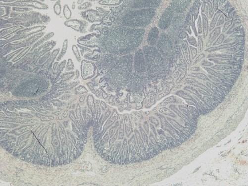
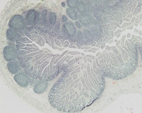
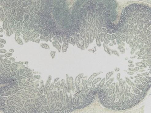

204 (B) Remnant ileal histology Experimental Groups Sham-Saline 0% EN 40% EN Sham-GLP-2 JI - Saline 183


205 JI GLP-2 Representative cross-sections of ileum from all sham and JI surgical groups 7 days after initial surgery are presented. The administration of EN is associated with villus lengthening. The JI groups have longer villi than corresponding sham groups. GLP-2 treatment lengthened villi in the sham and JI group at 0% EN and the sham group only at 40% EN. At 0% EN, GLP- 2 had no effect on crypt depth but with 40% EN administration, GLP-2 reduced crypt depth in the sham group and increased crypt depth in the JI group. All images are at 20X magnification. 184
206 Figure 5-7. Fat absorption The total fat absorption amongst 40% EN SBS pigs following GLP-2 and saline treatment. Mean ± SEM; two-way ANOVA. 185
207 Figure 5-8. Weight gain and gross intestinal morphology comparing 0% EN versus 40% EN piglets. The comparison of (A) total body weight gain, (B) change in remnant intestinal length, (C) intestinal weight and (D) intestinal weight per length in SBS piglets receiving 0% EN versus 40% EN following GLP-2 and saline treatment. Mean ± SEM; three-way ANOVA. * p <.05, versus 0% EN (A) (B) 186
208 (C) (D) 187
209 Figure 5-9. Remnant intestinal histology comparing 0% EN versus 40% EN piglets. The comparison of (A) jejunal villus height, (B) jejunal crypt depth, (C) ileal villus height and (D) ileal crypt depth in SBS piglets receiving 0% EN versus 40% EN following GLP-2 and saline treatment. Mean ± SEM; three-way ANOVA. * p <.05, versus 0% EN (A) (B) 188
210 (C) (D) 189
211 Figure Summary Tables The summary of effects of (A) exogenous GLP-2 administration and (B) intrinsic intestinal adaptation in JI and JC SBS piglets following total PN (0% EN) and 40% EN. * versus JC ** versus sham *** abrogates difference between JI-saline and JC-saline No asterisk denotes comparison to both other models. (A) 190
212 (B) 191
213 Table 5-1: List of target and housekeeping genes for chapter 5. Target Gene Forward primer Reverse primer Nutrient Transporters SGLT1 SLC5A1 AGGTTGGCTGTTCCAACATT CAATCCATTGGGCATGAGTT GLUT5 SLC2A5 CTGGGCTGTGAATCCAGAA AAGCCCTTCAGACAGTCCAG CD36 CD36 TGAAGCAGAACTATATCGTGCCTA CATTTCTGCCTTCTCATCACC FATP4 SLC27A4 AGAAGGAGCTGCCCCTGTA TGAAAGTCCCTGTTTTATGCAG Tight junctional proteins zona TJP1 GAGTTTGATAGTGGCGTT GTGGGAGGATGCTGTTGT occludens-1 occludin OCLN CAT GGC CTA CTC GTC CAA C GAC GCC TCC AAG TTA CCA C claudin-3 CLDN3 CGC GCC CTC ATC GTC GTC ACG TAG TCC TTG CGG TCG TA claudin-7 CLDN7 TGA GCT GCA AAA CGT ACG ACT CAC AAA GAC CTG CCA CGA TG claudin-15 CLDN15 ACAGCAGCTGGCGAGTGT GGCGCAGCTGTACCAGAG Housekeeping β-actin ACTB CACGCCATCCTGCGTCTGGA AGC ACC GTG TTG GCG TAG AG 192
214 Table 5-2. Jejunal and ileal mrna expression of nutrient transporters and tight junctional proteins in piglets on total PN (0% EN). Jejunum Sham - Sham JI - Saline JI GLP-2 JC Saline JC GLP-2 p-value Expression Saline GLP-2 Nutrient Transporters SGLT ± ± ± ± ± ± GLUT ± ± ± ± ± ± * CD ± ± ± ± ± ± FATP ± ± ± ± ± ± Tight junctional proteins occludin 1.00 ± ± ± ± ± ± claudin ± ± ± ± ± ± claudin ± ± ± ± ± ± claudin ± ± ± ± ± ± Ileum Expression Sham - Saline Sham GLP-2 JI - Saline JI GLP-2 p-value Nutrient Transporters SGLT ± ± ± ± GLUT ± ± ± ± CD ± ± ± ± FATP ± ± ± ± Tight junctional proteins occludin 1.00 ± ± ± ± claudin ± ± ± ± claudin ± ± ± ± claudin ± ± ± ± The values are normalized ratios ± SEM. Ratios of target gene expression to housekeeping (β-actin) gene expression are normalized to the sham-saline group. Two-way ANOVA with post-hoc comparisons using either student s t-test or one-way ANOVA with post-hoc Bonferroni s comparisons were used, as appropriate, to detect differences between groups. SGLT1, sodium-glucose linked transporter 1; GLUT5, facilitated glucose/fructose transporter, member 5; CD36, cluster of differentiation 36/fatty acid translocase; FATP4, fatty acid transport protein 4. Note: qrt-pcr of ZO-1, tight junctional protein zona occludens-1, was not performed for this group. *JI anatomy increased GLUT5 expression over sham control. 193
215 Table 5-3. Jejunal and ileal mrna expression of nutrient transporters and tight junctional proteins in piglets receiving 40% EN. Jejunum Sham - Sham JI - Saline JI GLP-2 JC Saline JC GLP-2 p-value Expression Saline GLP-2 Nutrient Transporters SGLT ± ± ± ± ± ± GLUT ± ± ± ± ± ± CD ± ± ± ± ± ± FATP ± ± ± ± ± ± Tight junctional proteins zona occludens ± ± ± ± ± ± occludin 1.00 ± ± ± ± ± ± claudin ± ± ± ± ± ± claudin ± ± ± ± ± ± Ileum Expression Sham - Saline Sham GLP-2 JI - Saline JI GLP-2 p-value Nutrient Transporters SGLT ± ± ± ± GLUT ± ± ± ± CD ± ± 0.04 a 1.11 ± ± 0.06 a 0.05 FATP ± ± ± ± Tight junctional proteins ZO ± ± ± ± occludin 1.00 ± ± ± ± * claudin ± ± ± ± claudin ± ± ± ± claudin ± ± ± ± The values are normalized ratios ± SEM. Ratios of target gene expression to housekeeping (β-actin) gene expression are normalized to the sham-saline group. Two-way ANOVA with post-hoc comparisons using either student s t-test or one-way ANOVA with post-hoc Bonferroni s comparisons were used, as appropriate, to detect differences between groups. Superscripts denote significant differences. SGLT1, sodium-glucose linked transporter 1; GLUT5, facilitated glucose/fructose transporter, member 5; CD36, cluster of differentiation 36/fatty acid translocase; FATP4, fatty acid transport protein 4; ZO-1, tight junctional protein zona occludens-1. Note: qrt-pcr of claudin-15 was not performed in jejunum for this group. *GLP-2 treatment decreased occludin expression over saline control. 194
216 References 1. Vanderhoof JA, Langnas AN. Short-bowel syndrome in children and adults. Gastroenterology. 1997;113(5): Quiros-Tejeira RE, Ament ME, Reyen L, et al. Long-term parenteral nutritional support and intestinal adaptation in children with short bowel syndrome: A 25-year experience. J Pediatr. 2004;145(2): Goulet O, Ruemmele F. Causes and management of intestinal failure in children. Gastroenterology. 2006;130(2 Suppl 1):S Amin SC, Pappas C, Iyengar H, Maheshwari A. Short bowel syndrome in the NICU. Clin Perinatol. 2013;40(1): Nightingale JM. Management of patients with a short bowel. Nutrition. 1999;15(7-8): Gutierrez IM, Kang KH, Jaksic T. Neonatal short bowel syndrome. Semin Fetal Neonatal Med. 2011;16(3): Carlson SJ, Chang MI, Nandivada P, Cowan E, Puder M. Neonatal intestinal physiology and failure. Semin Pediatr Surg. 2013;22(4): Batra A, Beattie RM. Management of short bowel syndrome in infancy. Early Hum Dev. 2013;89(11): Jeppesen PB. Spectrum of short bowel syndrome in adults: Intestinal insufficiency to intestinal failure. JPEN J Parenter Enteral Nutr. 2014;38(1 Suppl):8S-13S. 10. Tappenden KA. Pathophysiology of short bowel syndrome: Considerations of resected and residual anatomy. JPEN J Parenter Enteral Nutr. 2014;38(1 Suppl):14S-22S. 195
217 11. Cisler JJ, Buchman AL. Intestinal adaptation in short bowel syndrome. J Investig Med. 2005;53(8): Dowling RH. Glucagon-like peptide-2 and intestinal adaptation: An historical and clinical perspective. J Nutr. 2003;133(11): Tappenden KA. Intestinal adaptation following resection. JPEN J Parenter Enteral Nutr. 2014;38(1 Suppl):23S-31S. 14. Sondheimer JM, Cadnapaphornchai M, Sontag M, Zerbe GO. Predicting the duration of dependence on parenteral nutrition after neonatal intestinal resection. J Pediatr. 1998;132(1): Buchman AL, Scolapio J, Fryer J. AGA technical review on short bowel syndrome and intestinal transplantation. Gastroenterology. 2003;124(4): Spencer AU, Neaga A, West B, et al. Pediatric short bowel syndrome: Redefining predictors of success. Ann Surg. 2005;242(3):403-9; discussion Thompson JS, Rochling FA, Weseman RA, Mercer DF. Current management of short bowel syndrome. Curr Probl Surg. 2012;49(2): Kelly DA. Liver complications of pediatric parenteral nutrition--epidemiology. Nutrition. 1998;14(1): Guglielmi FW, Boggio-Bertinet D, Federico A, et al. Total parenteral nutrition-related gastroenterological complications. Dig Liver Dis. 2006;38(9): Wales PW, Christison-Lagay ER. Short bowel syndrome: Epidemiology and etiology. Semin Pediatr Surg. 2010;19(1):
218 21. Kelly DG, Tappenden KA, Winkler MF. Short bowel syndrome: Highlights of patient management, quality of life, and survival. JPEN J Parenter Enteral Nutr. 2014;38(4): Wales PW, de Silva N, Kim J, Lecce L, To T, Moore A. Neonatal short bowel syndrome: Population-based estimates of incidence and mortality rates. J Pediatr Surg. 2004;39(5): Squires RH, Duggan C, Teitelbaum DH, et al. Natural history of pediatric intestinal failure: Initial report from the pediatric intestinal failure consortium. J Pediatr. 2012;161(4):723-8.e Javid PJ, Malone FR, Reyes J, Healey PJ, Horslen SP. The experience of a regional pediatric intestinal failure program: Successful outcomes from intestinal rehabilitation. Am J Surg. 2010;199(5): Modi BP, Langer M, Ching YA, et al. Improved survival in a multidisciplinary short bowel syndrome program. J Pediatr Surg. 2008;43(1): Kosar C, Steinberg K, de Silva N, Avitzur Y, Wales PW. Cost of ambulatory care for the pediatric intestinal failure: One-year follow-up after primary discharge. Journal of Pediatric Surgery. 2016;In press. 27. Drucker DJ, Erlich P, Asa SL, Brubaker PL. Induction of intestinal epithelial proliferation by glucagon-like peptide 2. Proc Natl Acad Sci U S A. 1996;93(15): Tsai CH, Hill M, Asa SL, Brubaker PL, Drucker DJ. Intestinal growth-promoting properties of glucagon-like peptide-2 in mice. Am J Physiol. 1997;273(1 Pt 1):E Hartmann B, Johnsen AH, Orskov C, Adelhorst K, Thim L, Holst JJ. Structure, measurement, and secretion of human glucagon-like peptide-2. Peptides. 2000;21(1):
219 30. Drucker DJ, Yusta B. Physiology and pharmacology of the enteroendocrine hormone glucagon-like peptide-2. Annu Rev Physiol. 2014;76: Jeppesen PB. Teduglutide for the treatment of short bowel syndrome. Drugs Today (Barc). 2013;49(10): Sangild PT, Ney DM, Sigalet DL, Vegge A, Burrin D. Animal models of gastrointestinal and liver diseases. animal models of infant short bowel syndrome: Translational relevance and challenges. Am J Physiol Gastrointest Liver Physiol. 2014;307(12):G Vegge A, Thymann T, Lund P, et al. Glucagon-like peptide-2 induces rapid digestive adaptation following intestinal resection in preterm neonates. Am J Physiol Gastrointest Liver Physiol. 2013;305(4):G Suri M, Turner JM, Sigalet DL, et al. Exogenous glucagon-like peptide-2 improves outcomes of intestinal adaptation in a distal-intestinal resection neonatal piglet model of short bowel syndrome. Pediatr Res. 2014;76(4): Thymann T, Stoll B, Mecklenburg L, et al. Acute effects of the glucagon-like peptide 2 analogue, teduglutide, on intestinal adaptation in short bowel syndrome. J Pediatr Gastroenterol Nutr. 2014;58(6): Naberhuis JK, Deutsch AS, Tappenden KA. Teduglutide-stimulated intestinal adaptation is complemented and synergistically enhanced by partial enteral nutrition in a neonatal piglet model of short bowel syndrome. JPEN J Parenter Enteral Nutr Brinkman AS, Murali SG, Hitt S, Solverson PM, Holst JJ, Ney DM. Enteral nutrients potentiate glucagon-like peptide-2 action and reduce dependence on parenteral nutrition in a rat 198
220 model of human intestinal failure. Am J Physiol Gastrointest Liver Physiol. 2012;303(5):G Burrin DG, Stoll B, Jiang R, et al. Minimal enteral nutrient requirements for intestinal growth in neonatal piglets: How much is enough? Am J Clin Nutr. 2000;71(6): Tyson JE, Kennedy KA. Minimal enteral nutrition for promoting feeding tolerance and preventing morbidity in parenterally fed infants. Cochrane Database Syst Rev. 2000;(2)(2):CD Turner JM, Wales PW, Nation PN, et al. Novel neonatal piglet models of surgical short bowel syndrome with intestinal failure. J Pediatr Gastroenterol Nutr. 2011;52(1): Hua Z, Turner JM, Sigalet DL, et al. Role of glucagon-like peptide-2 deficiency in neonatal short-bowel syndrome using neonatal piglets. Pediatr Res. 2013;73(6): Wykes LJ, Ball RO, Pencharz PB. Development and validation of a total parenteral nutrition model in the neonatal piglet. J Nutr. 1993;123(7): Burrin DG, Stoll B, Jiang R, et al. GLP-2 stimulates intestinal growth in premature TPN-fed pigs by suppressing proteolysis and apoptosis. Am J Physiol Gastrointest Liver Physiol. 2000;279(6):G Sigalet DL, de Heuvel E, Wallace L, et al. Effects of chronic glucagon-like peptide-2 therapy during weaning in neonatal pigs. Regul Pept. 2014;188: Horwitz W AI. Official methods of analysis of AOAC international
221 46. Blencowe H, Cousens S, Oestergaard MZ, et al. National, regional, and worldwide estimates of preterm birth rates in the year 2010 with time trends since 1990 for selected countries: A systematic analysis and implications. Lancet. 2012;379(9832): Lim DW, Turner JM, Wales PW. Emerging piglet models of neonatal short bowel syndrome. JPEN J Parenter Enteral Nutr. 2015;39(6): Xiao Q, Boushey RP, Drucker DJ, Brubaker PL. Secretion of the intestinotropic hormone glucagon-like peptide 2 is differentially regulated by nutrients in humans. Gastroenterology. 1999;117(1): Hansen CF, Vrang N, Sangild PT, Jelsing J. Novel insight into the distribution of L-cells in the rat intestinal tract. Am J Transl Res. 2013;5(3): Wales PW, de Silva N, Kim JH, Lecce L, Sandhu A, Moore AM. Neonatal short bowel syndrome: A cohort study. J Pediatr Surg. 2005;40(5): Rocca AS, Brubaker PL. Stereospecific effects of fatty acids on proglucagon-derived peptide secretion in fetal rat intestinal cultures. Endocrinology. 1995;136(12): Brubaker PL, Anini Y. Direct and indirect mechanisms regulating secretion of glucagon-like peptide-1 and glucagon-like peptide-2. Can J Physiol Pharmacol. 2003;81(11): Shin ED, Estall JL, Izzo A, Drucker DJ, Brubaker PL. Mucosal adaptation to enteral nutrients is dependent on the physiologic actions of glucagon-like peptide-2 in mice. Gastroenterology. 2005;128(5): Brubaker PL, Izzo A, Hill M, Drucker DJ. Intestinal function in mice with small bowel growth induced by glucagon-like peptide-2. Am J Physiol. 1997;272(6 Pt 1):E
222 55. Cheeseman CI. Upregulation of SGLT-1 transport activity in rat jejunum induced by GLP-2 infusion in vivo. Am J Physiol. 1997;273(6 Pt 2):R Cheeseman CI, O'Neill D. Basolateral D-glucose transport activity along the crypt-villus axis in rat jejunum and upregulation induced by gastric inhibitory peptide and glucagon-like peptide- 2. Exp Physiol. 1998;83(5): Kato Y, Yu D, Schwartz MZ. Glucagonlike peptide-2 enhances small intestinal absorptive function and mucosal mass in vivo. J Pediatr Surg. 1999;34(1):18-20; discussion Benjamin MA, McKay DM, Yang PC, Cameron H, Perdue MH. Glucagon-like peptide-2 enhances intestinal epithelial barrier function of both transcellular and paracellular pathways in the mouse. Gut. 2000;47(1): Kitchen PA, Fitzgerald AJ, Goodlad RA, et al. Glucagon-like peptide-2 increases sucrase- isomaltase but not caudal-related homeobox protein-2 gene expression. Am J Physiol Gastrointest Liver Physiol. 2000;278(3):G Au A, Gupta A, Schembri P, Cheeseman CI. Rapid insertion of GLUT2 into the rat jejunal brush-border membrane promoted by glucagon-like peptide 2. Biochem J. 2002;367(Pt 1): Ramsanahie AP, Berger UV, Zinner MJ, Whang EE, Rhoads DB, Ashley SW. Effect of glucagon-like peptide-2 (GLP-2) on diurnal SGLT1 expression. Dig Dis Sci. 2004;49(11-12): Cani PD, Possemiers S, Van de Wiele T, et al. Changes in gut microbiota control inflammation in obese mice through a mechanism involving GLP-2-driven improvement of gut permeability. Gut. 2009;58(8):
223 63. Hsieh J, Longuet C, Maida A, et al. Glucagon-like peptide-2 increases intestinal lipid absorption and chylomicron production via CD36. Gastroenterology. 2009;137(3): , 1005.e Yu C, Jia G, Jiang Y, et al. Effect of glucagon-like peptide 2 on tight junction in jejunal epithelium of weaned pigs though MAPK signaling pathway. Asian-Australas J Anim Sci. 2014;27(5): Dong CX, Zhao W, Solomon C, et al. The intestinal epithelial insulin-like growth factor-1 receptor links glucagon-like peptide-2 action to gut barrier function. Endocrinology. 2014;155(2): Burrin DG, Petersen Y, Stoll B, Sangild P. Glucagon-like peptide 2: A nutrient-responsive gut growth factor. J Nutr. 2001;131(3): Petersen YM, Burrin DG, Sangild PT. GLP-2 has differential effects on small intestine growth and function in fetal and neonatal pigs. Am J Physiol Regul Integr Comp Physiol. 2001;281(6):R Petersen YM, Elnif J, Schmidt M, Sangild PT. Glucagon-like peptide 2 enhances maltase- glucoamylase and sucrase-isomaltase gene expression and activity in parenterally fed premature neonatal piglets. Pediatr Res. 2002;52(4): Sangild PT, Tappenden KA, Malo C, et al. Glucagon-like peptide 2 stimulates intestinal nutrient absorption in parenterally fed newborn pigs. J Pediatr Gastroenterol Nutr. 2006;43(2):
224 70. Jeppesen PB, Hartmann B, Thulesen J, et al. Glucagon-like peptide 2 improves nutrient absorption and nutritional status in short-bowel patients with no colon. Gastroenterology. 2001;120(4): Jeppesen PB, Sanguinetti EL, Buchman A, et al. Teduglutide (ALX-0600), a dipeptidyl peptidase IV resistant glucagon-like peptide 2 analogue, improves intestinal function in short bowel syndrome patients. Gut. 2005;54(9): Jeppesen PB, Gilroy R, Pertkiewicz M, Allard JP, Messing B, O'Keefe SJ. Randomised placebo-controlled trial of teduglutide in reducing parenteral nutrition and/or intravenous fluid requirements in patients with short bowel syndrome. Gut. 2011;60(7): Jeppesen PB, Pertkiewicz M, Messing B, et al. Teduglutide reduces need for parenteral support among patients with short bowel syndrome with intestinal failure. Gastroenterology. 2012;143(6): e Koopmann MC, Chen X, Holst JJ, Ney DM. Sustained glucagon-like peptide-2 infusion is required for intestinal adaptation, and cessation reverses increased cellularity in rats with intestinal failure. Am J Physiol Gastrointest Liver Physiol. 2010;299(6):G Dube PE, Forse CL, Bahrami J, Brubaker PL. The essential role of insulin-like growth factor- 1 in the intestinal tropic effects of glucagon-like peptide-2 in mice. Gastroenterology. 2006;131(2): Yusta B, Holland D, Koehler JA, et al. ErbB signaling is required for the proliferative actions of GLP-2 in the murine gut. Gastroenterology. 2009;137(3): Rowland KJ, Brubaker PL. The "cryptic" mechanism of action of glucagon-like peptide-2. Am J Physiol Gastrointest Liver Physiol. 2011;301(1):G
225 204
226 Chapter 6 Synergy of Glucagon-like Peptide-2 and Epidermal Growth Factor Co-administration on Intestinal Adaptation in Neonatal Piglets with Short Bowel Syndrome. Adapted from: Lim DW, Levesque CL, Vine DF, Muto M, Koepke JR, Nation PN, Wizzard PR, Li J, Bigam DL, Brubaker PL, Turner JM, Wales PW. Synergy of glucagon-like peptide-2 and epidermal growth factor co-administration on intestinal adaptation in neonatal piglets with short bowel syndrome. American Journal of Physiology Gastrointestinal & Liver [Revisions Requested August 2016] 205
227 Abstract Background: Glucagon-like peptide-2 (GLP-2) and epidermal growth factor (EGF) treatment enhance intestinal adaptation. To determine whether these growth factors exert synergistic effects on intestinal growth and function, GLP-2 and EGF were administered, alone and in combination, in neonatal piglet models of short bowel syndrome (SBS). Methods: Neonatal Landrace-Large White piglets were block-randomized to 75% midintestinal (JI group) or distal-intestinal (JC group) resection or sham control, with 7-d infusion of saline (control), intravenous human GLP-2 (11 nmol/kg/day) alone, enteral EGF (80 μg/kg/day) alone, or GLP-2 and EGF in combination. Adaptation was assessed by intestinal length, histopathology, Üssing chamber analysis and RT-qPCR of intestinal growth factors. Results: Combined EGF and GLP-2 treatment increased intestinal length in all three surgical models (p<0.01). EGF alone selectively increased bowel weight per length and jejunal villus height in the JI group only. The JC group demonstrated increased intestinal weight and villus height (p<0.01) when given either GLP-2 alone or in combination with EGF, with no effect of EGF alone. Jejunal permeability of mannitol and polyethylene glycol decreased with combination therapy in both SBS groups (p<0.05). No difference was observed in fat absorption or body weight gain. IGF-1 mrna was differentially expressed in JI versus JC piglets with treatment. 206
228 Conclusions: Combined EGF and GLP-2 treatment induced intestinal lengthening and decreased permeability, in addition to the trophic effects of GLP-2 alone. Our findings illustrate the benefits of novel combination GLP-2 and EGF treatment for neonatal SBS, especially in the JC model representing most human infants with SBS. 207
229 INTRODUCTION The treatment and management of infants and children with short bowel syndrome (SBS) remains an ongoing challenge to healthcare practitioners. The diseases that lead to major intestinal resection and SBS in children include congenital anomalies (e.g. intestinal atresia, gastroschisis), intestinal volvulus and most commonly, necrotizing enterocolitis (NEC), which premature infants are especially at risk of developing. 1 Infants with SBS are dependent on parenteral nutrition (PN) therapy to sustain health and normal growth and development. However, many infants with SBS succumb to PNassociated complications, such as liver disease, infection and sepsis, accounting for 1.4% of all deaths in children less than 4 years of age. 2 Intestinal adaptation to massive resection refers to the gradual anatomical and physiological changes that occur in the remnant intestine to restore nutrient absorptive function. Failure of the intestine to adapt results in irreversible intestinal failure, with these infants requiring long-term PN therapy and, potentially, liver and/or intestinal transplantation. 1 Therapies that augment intestinal adaptation are therefore desired to promote enteral function, weaning from PN and improve long-term health outcomes. At the present time, no such therapies are approved for treatment of children with SBS. Glucagon-like peptide-2 (GLP-2) is a distal intestine-derived hormone that mediates the endogenous intestinal adaptive response to feeding. 3 In normal rodents and rodent models of SBS, exogenous GLP-2 administration stimulates intestinal mucosal hyperplasia, up-regulates the expression of nutrient transporters and decreases intestinal permeability to fluorescein isothiocyanate-dextran. 4-7 In adult humans with SBS, treatment with teduglutide, a long-acting GLP-2 analog, for 24 weeks reduces PN volume 208
230 requirements and enhances morphological adaptation, including intestinal villus height. 8,9 While teduglutide has been approved by the FDA for adult SBS, preclinical data on the efficacy of growth factors such as GLP-2 in neonates, where SBS is most frequently encountered, is limited. The rodent models that have been used in preclinical SBS studies have limited clinical relevance to the human neonate due to differences in ontogeny and physiology. 10 Neonates and infants have an innate gut growth potential that may be augmented with growth factor therapies, in comparison to adults whose adaptive capacity is more limited. 11 Hence, we developed models of SBS using the neonatal piglet, a validated model for the human neonatal intestine with similarities in ontogeny and physiology. 10,12 Diseases that lead to SBS in human neonates frequently affect and require removal of the ileum, a site of physiological significance given that GLP-2-producting L-cells are largely found in the distal intestine. Most preclinical models utilize a mid-intestinal resection with retained ileum, due to the feasibility of maintaining this model, but the mid-intestinal resection model has less translational relevance for human neonates. 12,13 Given that remnant anatomy is a significant predictor of pathophysiology and outcome in SBS, we therefore developed two neonatal piglet SBS models, one with mid-intestinal resection and another with distal-intestinal resection. 12 GLP-2 or teduglutide administration for 7 or 14 days in neonatal piglet SBS models stimulates structural adaptation, including increases in remnant villus height and crypt depth, but has limited or transient functional effects on digestive enzyme activity or nutrient transport Because the GLP-2 receptor (GLP-2R) is not expressed by intestinal epithelial cells (IECs), the intestinotropic effects of GLP-2 are believed to be indirect. 18,19 Several 209
231 downstream mediators have been implicated in the growth effects of GLP-2 including the ErbB ligand, epidermal growth factor (EGF). 20,21 In mice, GLP-2 administration upregulates expression of ErbB ligands, an effect that is lost in GLP-2R knockout mice and in mice administered a pan-erbb inhibitor, and significantly diminished in waved-2 mice that harbor a mutated EGF receptor (ErbB1). 21 The ErbB pathway also plays a role in the GLP-2-mediated intestinal adaptive response to re-feeding, which is lost in GLP-2R knockout mice but rescued with EGF administration. 22 Similiar to GLP-2, exogenous EGF administration induces structural and functional adaptation in rodent models of SBS, including increased villus and crypt lengths and decreased permeability to macromolecules. 23,24 In unresected piglets, EGF administration stimulates body weight gain and reverses the changes in intestinal structure and inflammatory indices associated with weaning. 25 Furthermore, in a pilot study, enteral EGF (100 μg/kg/day) administration for 6 weeks in five children with SBS improved 3-0 methylglucose absorption and enteral tolerance, although weight gain and intestinal permeability were not affected. 26 Given the utility of teduglutide in adult SBS, and the demonstrated relationship between GLP-2 and EGF in the regulation of intestinal growth, the aim of our study was to assess the effects of administering GLP-2 and EGF, alone and in combination, on intestinal structure and function in two translational piglet models of neonatal SBS. 27 METHODS Animals and Surgery Animal studies were conducted in compliance with the Canadian Council on Animal Care guidelines and approved by the Animal Policy & Welfare Committee at the 210
232 University of Alberta. Neonatal Landrace-Large White cross F1 male piglets (4 ± 2 days old, 2.3 ± 0.54 kg) obtained from the University of Alberta Swine Research and Technology Center underwent general anesthesia for jugular venous catheterization, laparotomy with measurement of intestinal length followed by assigned surgical procedure and insertion of a Stamm gastrostomy, as previously described. 28 Piglets were block-randomized to 75% mid-intestinal resection (leaving equal lengths of remnant jejunum and ileum) with jejunoileal anastomosis (JI group), 75% distal-intestinal resection (including all of the ileum and proximal 5 cm of colon) with jejunocolic anastomosis (JC group) or sham control (exteriorization of the intestine, measurement of intestinal length and return to the abdominal domain without transection) (Figure 6-1). Animal Care Post-operatively, piglets were secured to a swivel-tether system (Lomir Biomedical Inc., Notre-Dame-de-l Île-Perrot, QC, Canada) and maintained in metabolic cages lined with Plexiglas at 25 C with a 12-hour light/dark cycle. For the first three study days, buprenorphine hydrochloride (Buprinex; Rekitt and Colman Pharmaceutical, Richmond, VA, USA) and oral meloxicam (Metacam; Boehringer Ingelheim, Burlington, ON, Canada) were given for analgesic support and ampicillin sodium (Sandoz, Boucherville, QC, Canada) and trimethoprim-sulfadoxine (Borgal; Merck Animal Health, Kirkland, QC, Canada) were given for prevention of venous catheter sepsis, as described. 15 Piglet activity, body weight, urine output and fluid balance were assessed daily. Piglets with clinical evidence of dehydration, including significant diarrhea and inadequate (<400 ml/day) fluid balance, were given intravenous (IV) normal saline 211
233 (0.9% sodium chlorine; Baxter, Mississauga, ON, Canada) boluses. If piglets developed fever, vomiting or lethargy suggestive of sepsis, blood cultures were taken and antibiotics were resumed. IV enrofloxacin (Baytril; 5 mg/kg; Bayer Inc., Animal Health, Toronto, ON, Canada) and clindamycin (3 mg/kg; Sandoz Canada Inc., Boucherville, QC, Canada) were added if piglets did not improve after 24 and 48 hours, respectively. Piglets were included in the study analysis if they improved on antibiotics and were blood culturenegative. Nutrition Immediately after surgery, all piglets received PN via the venous catheter to meet 100% of daily caloric intake. On post-operative day 2, piglets commenced enteral nutrition (EN) at 20% of daily caloric intake via the gastrostomy tube. In this study, we elected to administer EN at 20% of the total caloric intake because in the previous chapter, we observed that JC piglets do not tolerate EN administered at 40% of total caloric intake, the minimal amount of EN delivery that Burrin et al. demonstrated was required to induce intestinal adaptation in unresected neonatal piglets. 57 Furthermore, Naberhuis et al. demonstrated a synergistic effect between exogenous teduglutide treatment and EN delivered at 20% of total caloric intake in a neonatal piglet SBS model with 80% proximal intestinal resection. 17 Given the diarrhea and malabsorption expected with introducing EN to piglets with SBS, the PN delivery rate was not correspondingly decreased to 80% of the total nutritional fluid rate. Both PN and EN were delivered by pressure-sensitive Alaris infusion pumps (CareFusion Corporation, San Diego, CA, USA). As previously described, the PN and EN solutions were prepared in our laboratory based on a commercially-available formula (Vaminolact; Fresenius Kabi, Bad 212
234 Hömburg, Germany). 15,28 Target nutrient intakes were derived from proof-of-concept studies, as follows: 1100 kj/kg/day, 27% of energy from amino acids, 37% from carbohydrate and 36% from fat. 29 Peptides Piglets were block-randomized to receive either intravenous saline (control), intravenous human GLP-2 alone, enteral EGF or GLP-2 and EGF in combination (Figure 6-1). Normal saline and human GLP-2 (1-33) (11 nmol/kg/day or 42 μg/kg/day; Catalog #CS9065; Lot I074 with 96.83% purity; CS Bio, Menlo Park, CA, USA) 30 were delivered continuously through the venous catheter by a syringe pump (NE-300 Just Infusion Syringe Pump; New Era Pump Systems, Farmingdale, NY, USA) at 0.42 ml/kg/hour beginning immediately post-operatively. EGF (80 μg/kg/day; 5.5 ml/kg) was administered via the gastrostomy tube beginning on post-operative day 2, delivered in the form of EGF-secreting Lactococcus lactis culture supernatant, the generation of which has been previously described. 31 Enteral Fat Absorption Fecal effluent was collected for 48 hours, beginning on study day 5, into drainable ostomy appliances (Two-Piece Pouch System; Hollister, Aurora, ON, Canada). Samples were freeze-dried and fat was extracted by petroleum ether distillation for 6 hours. 32 Enteral fat absorption was calculated by subtracting the average fecal fat content per pig from the total amount of lipid infused and adjusted for the total duration of fecal collection (expressed as g/kg/day). Tissue Collection and Morphology 213
235 On study day 7, piglets were anesthetized and underwent terminal laparotomy, where final intestinal lengths were measured, followed by euthanasia. The entire small intestine from ligament of Treitz to ileocecal value or jejunocolic anastomosis was removed, emptied of fecal matter and weight measured. Mucosal scrapings from 20-cm segments of jejunum and ileum also were weighed. A 20-cm proximal jejunal segment was used to assess intestinal permeability and electrical activity using Üssing chamber analysis under physiological conditions. 33 Cross-sectional jejunal and ileal (in JI and sham piglets) segments were preserved in 10% buffered formaldehyde for histology and immunohistochemistry, while adjacent segments were preserved in RNAlater Stabilization Solution (ThermoFisher Scientific, Waltham, MA, USA) or flash-frozen in liquid nitrogen and stored at -80 C for gene expression analyses (Figure 6-2). Villus height and crypt depth were measured on H&E-stained jejunal and ileal cross-sections (Nikon Eclipse 80i; Nikon, Tokyo, Japan) by a certified veterinary pathologist blinded to treatment. Ten well-oriented villi and crypts were measured on 2-3 different cross sections per piglet. Mucosal crypt cell proliferation was determined using Ki-67 immunohistochemistry staining, as previously described 15, on formalin-fixed, paraffin-embedded, and sectioned (~5 µm) distal intestinal segments taken 5 cm proximal to either the ileocecal valve (in sham and JI piglets) or jejunocolic anastomosis (in JC piglets) (Figure 6-2). Sections were incubated for 1 h at 60 C, deparaffinized, and rehydrated (xylenes, 100% ethanol, and 95% ethanol). Sections were treated with EDTA antigen retrieval for 25 min in a steamer. After incubation with 3% hydrogen peroxide in methanol for 10 min to block endogenous peroxidase, the sections were covered with 2% normal mouse-2% normal goat serum in PBS-Tween for 20 min at room temperature to 214
236 block nonspecific binding sites. After 30 min of incubation with the primary antibody (1:1,000 dilution; catalog no. VP-k451, Vector Laboratories, Burlingame, CA, USA), the sections were incubated with goat anti-rabbit secondary antibody for 30 min and Vector Elite ABC kit for 45 min. Detection was carried out using the chromagen 3-amino-9- ethylcarbazole. The sections were counterstained with hematoxylin and dehydrated, and coverslips were applied in an aqueous mounting medium. The proportion of proliferating crypt cells in 3-5 well-oriented crypts was quantified by a blinded observer counting the number of Ki-67-positive cells (Ki-67-stained nuclei). Intestinal alkaline phosphatase activity Frozen distal intestinal segments (weighing 1.3 g) taken either 5 cm proximal to the ileocecal valve (in sham and JI piglets) or jejunocolic anastomosis (in JC piglets) (Figure 6-2) were thawed in ice-cold homogenization buffer (50 mmol/l D-mannitol, 0.20 mmol/l phenylmethane sulfonyl fluoride, and protease inhibitors at ph 7.4) at a ratio of 20 ml homogenization buffer/g frozen tissue and homogenized using a polytron homogenizer. Tissue homogenate protein content was determined according to the Lowry procedure. Intestinal alkaline phosphatase (IAP) activity was carried out at 37 C for 10 minutes in a final volume of 1 ml suspension containing sample homogenate (1 mg protein/ml), 5 mmol/l MgCl2, 50 mmol/l NaHCO3 and Na2CO3 and 20 mmol/l p- nitrophenol phosphate. 34 Potassium fluoride (2.0 mmol/l) was used to inhibit acid phosphatase activity 35 The IAP activity is expressed as nmol p-nitrophenol liberated/mg protein/min. Intestinal Permeability and Electrical Activity 215
237 Intestinal paracellular transport of radiolabelled mannitol and polyethylene glycol (PEG) (molecular weights: 180 and , respectively) was determined in jejunal segments (taken 20 cm distal to the ligament of Treitz, Figure 6-2) using a modified Üssing chamber (Harvard Apparatus Inc., Holliston, MA, USA) procedure, as previously described 33, by a team blinded to surgical group and treatment. The apparent permeability coefficients (Papp, cm/s) were calculated at steady state as follows: Papp = dq/dt x (1/(A X C0)), where dq/dt is the appearance rate of radiolabelled marker in the receiver chamber, A is the exposed surface area of intestine and C0 is the initial concentration in the donor chamber. The spontaneous transepithelial potential difference (PD) and the short-circuit current (Isc) required to reduce the PD across the tissue were used to calculate transepithelial electrical resistance (TEER), as described. 33 Real-Time RT-PCR Expression of genes related to tissue repair (TFF3), cell proliferation (Ki-67), differentiation (cdx2), apoptosis (caspase-3, or c3), digestion (IAP) and tight junction proteins (claudins-7 and -15) were assessed using total RNA isolated (UltraClean Tissue & Cells RNA Isolation kit; MoBio Laboratories Inc., Carlsbad, CA, USA) from distal intestinal segments, taken 5 cm proximal to the ileocecal valve in sham and JI piglets or jejunocolic anastomosis in JC piglets, and subjected to reverse-transcription (High Capacity cdna Reverse Transcription kit; Applied Biosystems, Foster City, CA, USA). Real-time semi-quantitative PCR was performed in triplicate on an Agilent Technologies Stratagene Mx3005P thermocycler with RT SYBR Green ROX qpcr Mastermix (QIAGEN Inc., Germantown, MD, USA) using primers (Integrated DNA Technologies, Inc., Coralville, IA, USA) listed in Table 6-1. Relative mrna expression was quantified 216
238 using the 2 -ΔCT method with beta-2-microgobulin (B2M) and glyceraldehyde-3-phosphate dehydrogenase (GAPDH) as controls, as previously validated by Ryan et al. for selection of stable porcine intestinal tract reference genes. 36 Expression of intestinal growth factor and receptor genes was assessed using total RNA isolated from whole jejunum and ileum (RNeasy Plus mini kit with Qiashredder; QIAGEN Inc., Germantown, MD, USA) and subjected to reverse-transcription (5X Allin-One RT MasterMix; Applied Biological Materials Inc., Richmond, BC, Canada). Real-time semi-quantitative PCR was performed in duplicate on an Applied Biosystems thermocycler with TaqMan Gene Expression Assays (Life Technologies, Carlsbad, CA, USA) as listed in Table 6-2. Relative messenger RNA (mrna) expression was quantified using the 2 -ΔCT method using 18S ribosomal RNA as the internal control, as validated. 20 Statistical Analyses Sample size was determined based on the outcomes of intestinal and crypt lengthening, with 6-8 piglets per group generating 80% and 87% power (2-sample t-test; α=0.05), respectively. Results are expressed as mean ± SEM per experimental group. Data were analyzed by 2-way ANOVA followed by Bonferroni post-hoc analysis. Some data were transformed to normalize variance. Significance was set at P <.05. Gross morphological data was further analyzed by multiple regression, with the adjusted R 2 value representing the variance in the dependent variable attributable to both surgical model and treatment. For histology and permeability data, with multiple repeated observations per piglet, a linear mixed-model analysis of the relationship between outcome measures and surgical anatomy and treatment was performed. As fixed effects, 217
239 surgery type and treatment were entered (with the interaction term) into the model whereas intercepts for subjects were entered as random effects. Jejunal electrical activity was analyzed by Kruskal-Wallis ANOVA at the 0-minute time-point for each surgical model because data transformation could not satisfy parametric test assumptions. SPSS software, version 23 (IBM Corporation, Armonk, NY, USA) was used for statistical analyses. Ki-67 immunohistochemical staining, IAP activity assays, and gene expression studies on TFF3, Ki-67, c3, cdx2, IAP, and claudins-7 and -15 were analyzed separately using the CONTRAST statement in a one-way ANOVA with SAS software, version 9.2 (SAS Institute, Cary, NC, USA), comparing treatment groups to the saline control within each of the JI and JC models, as these specific outcomes were not studied in the sham group. RESULTS Sixty-four piglets completed the study, while twelve were excluded for complications (Figure 6-1). Baseline characteristics are presented in Table 6-3. Piglets gained kg in body weight during the study period, with no difference between groups (P =.38; Figure 6-3A). There was no difference between groups regarding the amount of PN (over 80% of expected, P =.6) and EN (over 84% of expected, P =.2) delivered. Gross Morphology There was no significant interaction between surgery and treatment on the change in remnant intestinal length between groups (F(6,50)=1.54, P =.2); therefore, both main effects were analyzed separately. Remnant anatomy influenced the change in intestinal length (F(2,50)=11.4, P <.001), with the JI anatomy demonstrating 17.4% (95%CI:
240 26.7) and 11.6% (95%CI: ) greater change in length than the JC and sham groups, respectively (Figure 6-3B). Although no effects of GLP-2 or EGF alone were detected, combination therapy resulted in a 15.4% (95%CI: ) and 13.2% (95%CI: ) greater change in length over EGF alone and saline control, respectively, regardless of anatomy (F(3,50)=4.52, P <.01; Figure 6-3B). Surgical anatomy and treatment independently predicted 33.6% of the variance in the change in intestinal length (P <.001). In the JI group, the remnant ileum demonstrated greater lengthening compared to the remnant jejunum, with combination therapy increasing length over EGF alone in both segments (jejunum: P =.03; ileum: P =.01; Figure 6-3F). There was a significant interaction between remnant anatomy and treatment on bowel weight per length (F(6,50)=3.6, P <.01). While no treatment differences were observed in the sham piglets, EGF increased bowel weight per length by 29.5% compared to saline (P <.01) in JI piglets and GLP-2 increased mean bowel weight per length by 26.8% compared to EGF alone (P <.05) in JC piglets (Figure 6-3C). In addition, in piglets given EGF alone, bowel weight per length was 58.5% and 105.2% greater in the JI group compared to the JC group (P <.01) and sham groups (P <.01), respectively. In piglets given combination therapy, bowel weight per length was 30.0% greater in the JC group (P =.01) and 44.3% greater in the JI group (P <.01), as compared to sham. Surgical anatomy and treatment independently predicted 59.4% of the variance in intestinal weight per length (P <.001). There was a significant interaction between remnant anatomy and treatment on normalized intestinal weight (F(6,50)=2.78, P =.02. In the sham group, GLP-2 alone and combination therapy increased normalized intestinal weight over saline control (P < 219
241 .01) while in the JC group, GLP-2 alone and in combination with EGF increased normalized intestinal weight versus EGF alone and saline control (P <.01); no treatment differences were observed in the JI model (Figure 6-3D). Furthermore, for each treatment, all pairwise comparisons between the three surgical anatomies differed significantly (P <.01). Surgical anatomy and treatment independently predicted 93.6% of the variance in normalized intestinal weight (P <.001). There was no significant interaction between surgery and treatment on jejunal mucosal weight (P =.9); therefore, main effects were analyzed separately. Regarding surgical anatomy, both JI and JC anatomy demonstrated increased jejunal mucosal weight compared to the sham group (F(2,50)=31.7, P <.01; Figure 6-3E). Regarding treatment, GLP-2, alone or in combination with EGF, increased jejunal mucosal weight over both EGF alone and saline control (F(3,50)=14.9, P <.01), with no effect of EGF alone and no difference between GLP-2 alone and combination therapy. Histopathology There was a significant interaction between surgical anatomy and treatment on remnant jejunal villus height (F(6,50)=2.3, P <.05; Figure 6-4A). In the sham group, combination therapy demonstrated a 59.9%-greater increase in jejunal villus height versus saline control (P <.01), while EGF and GLP-2 alone had no effect. In the JI group, GLP-2 and combination therapy increased jejunal villus height by 31.0% (P <.05) and 34.1% (P <.05), respectively, over saline control. In the JC group, GLP-2 alone increased jejunal villus height by 56.5%, 31.3% and 60.7%, respectively, over saline, combination therapy and EGF alone (P <.05). Regarding differences based on anatomy, JC-GLP-2 pigs demonstrated 30.8% (P <.05) greater jejunal villus height compared to 220
242 the sham-glp-2 group, and JI-EGF pigs had 35.2% (P <.05) greater jejunal villus height than the JC-EGF group. Jejunal crypt depth differed as a function of surgical anatomy but not by treatment (P <.01). Thus, the JC and JI groups respectively demonstrated 19.0% (P <.01) and 13.1% (P <.05) greater jejunal crypt depth than the sham group (Figure 6-4B). In sham and JI piglets, there was no interaction between surgical anatomy and treatment on remnant ileal villus height (P =.97); therefore, main effects were analyzed separately. The JI anatomy demonstrated 58.1% (P <.001) greater ileal villus height than the sham group. GLP-2 alone and combination therapy increased ileal villus height in these animals by 29.6% (P <.05) and 26.5% (P <.05) over saline control, respectively (Figure 6-4C). Ileal crypt depth did not differ between groups as a function of either remnant anatomy or treatment (Figure 6-4D). Intestinal Permeability and Electrical Activity In the sham group, EGF alone increased jejunal mucosal-to-serosal (M-to-S) paracellular flux of mannitol compared to saline control by 5.52-fold (P <.05), while GLP-2 had no effect. In contrast, in both JI and JC models, combination therapy decreased M-to-S paracellular flux to mannitol by >70% (P <.05 and =.01, respectively), compared to saline, while monotherapy with either GLP-2 or EGF had no effect on permeability (Figure 6-5A). A similar pattern was observed with the jejunal serosal-to-mucosal (S-to-M) paracellular flux of mannitol. Results for the jejunal permeability of PEG were consistent with those found for mannitol. Hence, EGF increased M-to-S paracellular flux of PEG approximately 6-fold (P <.05) compared to saline in sham piglets, whereas combination therapy in the JI and 221
243 JC groups decreased M-to-S paracellular flux of PEG by approximately 60% (P <.05) and 80% (P =.01), respectively, versus saline (Figure 6-5B); GLP-2 and EGF alone had no effect. In piglets receiving combination therapy, the JI and JC groups also demonstrated decreased M-to-S paracellular flux of PEG compared to sham animals by >75% (P <.01) and 85% (P <.05), respectively. A similar pattern was observed for the jejunal S-to-M paracellular flux of PEG. All intestinal segments established and maintained a transepithelial potential difference (PD) greater than 2 mv, indicating that intestinal integrity was maintained throughout the Üssing experiment (Figure 6-6A). There was no treatment-related difference in PD at T=0 between the sham and JC groups. In the JI group, combination therapy increased the PD across the intestine 2.6-fold compared to EGF alone at T=0 (P <.01) and increased over time (Figure 6-6A). Isc is a summation of all ionic currents across the epithelium and a measure of active transport processes. 37 In the sham group, Isc at T=0 was increased 3.1- and 17.1-fold in response to EGF alone compared to GLP-2 alone (P <.001) and saline (P <.001), respectively, and maintained over time (Figure 6-6B). There were no treatment-related differences in the JI model. In the JC model, combination therapy decreased Isc 2.8-fold at T=0 compared to GLP-2 alone (P <.01). TEER is a measure of tissue integrity and barrier function. 37 In the sham model, EGF alone and combination therapy decreased TEER 9-fold (P <.001) and 7.6-fold (P =.001), respectively, compared to saline, while GLP-2 alone had no effect. In the JI model, there was no effect of treatment at T=0 but over time, there was a gradual increase in TEER with combination therapy, with no effect of either GLP-2 or EGF alone. In the 222
244 JC model, combination therapy increased TEER at T=0 9.8-fold (P <.01) compared to GLP-2 and over time, which likely reflects the increased PD (Figure 6-6C). Fat Absorption Total fat absorption was affected by the SBS resection model, but not by treatment. Thus, the JC anatomy demonstrated 19.7% and 26.0% lower fat absorption compared to both the JI and sham groups, respectively (P <.01, Figure 6-7). Intestinal Gene Expression, IAP activity, and Proliferation For these outcomes, sham animals were not studied. Administration of GLP-2 and EGF, alone or in combination, increased trefoil factor-3 (TFF3) expression by 150% over saline (P <.01) in the JI group (Figure 6-8A), with a similar trend in the JC group (Figure 6-8B). While treatment did not affect ki-67 expression in either JI or JC piglets, ki-67 staining of distal intestine in JI piglets demonstrated a trend towards increased ki- 67-positive staining cells with combination therapy versus saline control (P =.08) (Figure 6-8C). Treatment did not affect relative cdx2, caspase-3 (c3) or IAP expression in JI or JC piglets (Figure 6-8A-B). However, there was a 50%-decrease in IAP activity in the JI group for all treatments as compared to saline control (P <.01) (Figure 6-8D). In the JI piglets, GLP-2 alone and EGF alone increased claudin-7 expression by 40% (P <.05) while GLP-2 alone and combination therapy increased claudin-15 expression by 150% (P <.05) versus saline (Figure 6-8A). A similar trend in claudin-15 expression was seen with combination therapy versus saline in JC piglets (Figure 6-8B). Growth Factor and Receptor Gene Expression Jejunal Glp2r expression differed as a function of surgery (P <.01) but not treatment, such that the JI and JC anatomies demonstrated a 48% (P <.01) and 43% (P < 223
245 .05) decrease in jejunal Glp2r expression versus sham, respectively (Figure 6-9A). Ileal Glp2r expression also differed as a function of surgery (P <.01) but not treatment, with 22% greater ileal Glp2r expression in the JI versus sham animals (Figure 6-9B). There was a significant interaction between surgery and treatment on jejunal Igf1 expression (P <.05). There was no difference in Igf1 expression as compared to saline in all surgical models. However, in the JC anatomy, Igf1 expression was 76% greater with EGF as compared to GLP-2 treatment (Figure 6-9C). Furthermore, differential jejunal Igf1 expression between anatomies was evident in the JI-GLP-2 group demonstrating 76%-greater expression compared to the JC-GLP-2 group (P <.01). There was no difference in ileal Igf1 (Figure 6-9D) or jejunal Igf1r expression (Figure 6-9E). However, ileal Igf1r expression differed as a function of surgical anatomy (P <.02) but not treatment, with the JI group demonstrating 50% increased expression over sham animals (Figure 6-9F). There was no difference in jejunal or ileal relative expression of Gcg (Figures 6-9G and 6-9H) or jejunal expression of ErbB1, the main EGF receptor (Egfr) (Figure 6-9I). EGF treatment decreased ileal Egfr expression compared to saline control in JI piglets (P <.01; Figure 6-9J). Finally, there was no difference in the jejunal or ileal expression of fibroblast growth factors-2, -7, -9, and -10 (Figures 6-9K-R) DISCUSSION There is mounting evidence that growth factors such as GLP-2 and EGF augment intestinal adaptation in SBS. GLP-2 studies in rodent models of SBS and human clinical trials have led to its approval for use in adult SBS Clinical trials of GLP-2 therapy in pediatric SBS are ongoing but the limited preclinical studies using piglet SBS models suggest a benefit. In our previous study, 14 days of GLP-2 therapy increased jejunal 224
246 villus height, and intestinal length to a degree, and JC-piglets given GLP-2 were able to wean off PN sooner than JC piglets given saline. 15 In a neonatal piglet JI model with 80% intestinal resection, Naberhuis et al. demonstrated that teduglutide treatment for 7 days increased mucosal surface area and transiently improved absorption of glucose and glutamine. 17 Finally, in a preterm piglet model with 50% distal intestinal resection and jejunostomy, Vegge et al. showed that GLP-2 treatment for 7 days resulted in intestinal hyperplasia, increases in sucrase and maltase activities and the relative absorption of fluid, energy and all macronutrients. 14 Teduglutide treatment for 7 days in neonatal piglets with 50% distal intestinal resection and jejunostomy demonstrated increases in intestinal weight per length but not functional adaptation (digestive enzyme activity, enteral nutrient absorption or IHC). 16 Regarding EGF, EGF administration has been shown to increase villus height and crypt depth at 50 μg/kg/day in a rat JI model of SBS with 50% resection. 23 In a rodent JC SBS model leaving only a 20 cm jejunal remnant, Sham et al. also demonstrated that treatment with recombinant human EGF prevented weight loss, improved 3-0 methylglucose absorption and reduced permeability (lactulose/mannitol ratio) compared to placebo. 24 In newly weaned 3-week-old piglets without resection, enteral EGF (in the form as administered in the present study) increased weight gain, jejunal villus height, expression of the anti-inflammatory cytokine, IL-13, and reversed the decrease in SGLT-1 and GLP-2R expression and protein levels induced by weaning. 25 A 2005 pilot study of enteral EGF treatment (100 μg/kg/day) for 6 weeks in five children with SBS (defined as having a bowel length less than 25% expected for age) demonstrated improved 3-0 methylglucose absorption and enteral tolerance but did not affect weight gain or intestinal permeability
247 In the present study, we investigated the novel administration of combination GLP-2 and EGF therapy compared to each treatment alone and saline in the setting of neonatal SBS. We used two relevant translational animal models, the JI and JC anatomies, with the latter lacking ileum and representing most human infants with SBS. Preclinical mechanistic studies using the piglet as an established model of the neonatal intestine are timely, as clinical trials with GLP-2 analogues in pediatric SBS are currently in progress. Furthermore, emerging literature has identified common downstream pathways between GLP-2R and ErbB1 signaling, suggesting synergy between GLP-2 and EGF, a natural ErbB1 ligand, in stimulating intestinal growth. Combination treatment with GLP-2 and EGF was associated with tropic intestinal effects and structural adaptation in both the JI and JC models. In parameters such as normalized intestinal weight, mucosal weight and villus height, there was no difference between combination therapy and GLP-2 alone but both treatments were superior to saline control and/or EGF alone. This suggests that GLP-2 is the main factor stimulating increases in these parameters, consistent with prior studies demonstrating that GLP-2 administration in rodents and piglets expands the intestinal mucosal epithelium. 3,4,6,14-17 However, EGF alone appeared to be selectively beneficial in increasing bowel weight per length and jejunal villus height in the JI group only. This finding may relate to the fact that the JI anatomy retains GLP-2-producing L-cells that have been shown to increase their GLP-2 production post-resection to mediate intestinal adaptation. 38,39 We observed evidence of this intrinsic adaptation in our JI model, with intestinal (especially ileal) lengthening, and increased intestinal weight and ileal villus height. Thus, while additional exogenous GLP-2 administration did not augment structural adaptation in the 226
248 JI model, administration of EGF alone may have acted synergistically with endogenous circulating GLP-2. In contrast, the JC model, with markedly reduced numbers of L-cells and thus of endogenous GLP-2, did not exhibit intrinsic adaptation and demonstrated tropic effects only when given either GLP-2, either alone or in combination with EGF. Aside from mucosal expansion, combined GLP-2 and EGF therapy increased intestinal length in all three surgical models. The length of remnant intestine is a predictor of clinical outcomes in SBS 1, but preclinical studies to date have been inconsistent in demonstrating a GLP-2 effect on intestinal lengthening. However, Martin et al. have suggested that the EGFR plays a role in intestinal smooth muscle adaptation after resection, as the EGFR mutant waved-2 mice fail to demonstrate normal smooth muscle proliferation and intestinal lengthening as seen in control mice. 40 Interestingly, our findings show that only co-administration of EGF and GLP-2 increased intestinal length, suggesting the requirement for both factors. Unexpectedly, ki-67 and caspase-3 expression, which reflect cellular proliferation and apoptotic pathways, respectively, did not parallel the morphological or histological findings in the combination treatment. It may be that caspase gene expression may not reflect true rates of apoptosis and that protein expression may be a more representative outcome. Furthermore, cellular hypertrophy, as opposed to cellular hyperplasia, cannot be discounted as another possible underlying adaptive mechanism with administration of trophic peptides. In this study, we measured the jejunal permeability of mannitol (a small sugar alcohol molecule) and PEG (a larger molecular weight molecule representative of the size of bacterial toxin or peptide), which use paracellular pathways of transport. In contrast to Üssing chamber studies using rodent intestine, greater variability was observed when 227
249 using piglet jejunum, both between different segments of the same intestinal specimen and between animals of the same surgical and treatment group. This variability in Papp values may be inherent to performing Üssing chamber studies in intestine from a large animal model such as the piglet. Thus, we performed a linear mixed-model regression analysis of Papp values in order include this variability in our analyses. Studies in healthy rodents have previously demonstrated the GLP-2 decreases permeability through an IGF-1R-dependent mechanism. 41 However, our data showed no difference in intestinal permeability following GLP-2 treatment alone, and this may be due to the degree of variability observed. In contrast, we observed that combination therapy with GLP-2 and EGF decreased jejunal permeability of both mannitol and PEG in JI and JC models compared to the sham and saline treatments, suggesting a benefit in both resection models. These findings may be associated with the tropic effects observed in jejunal mucosal weight and villus height, reflecting an increase in overall epithelial biomass and thickness of the intestinal wall, which may have resulted in a decrease in permeability. This proposed mechanism might also underlie differences in TEER, with combination therapy leading to the greatest intestinal transepithelial resistance in the JI and JC models but not the sham. Furthermore, modulation of tight junctional complexes may underlie the improvements in intestinal permeability. Claudins-7 and -15 are expressed all along the mammalian intestine 42 but have contrasting roles, with claudin-7 weakening the intestinal barrier 43 and claudin-15 maintaining barrier function. 44 We observed that both combination therapy and GLP-2 monotherapy increased claudin-15 expression while GLP-2 or EGF monotherapy increased claudin-7 mrna expression in the JI model while in the JC model, combination therapy increased claudin
250 expression, although not significant. These findings that suggest that modulation of claudin expression may underlie some of the effects observed with barrier function following growth factor treatment, which adds to previous knowledge gained from studies in mice 41, and that these effects may vary according to the SBS resection model. In addition, we observed an increase in mannitol and PEG permeability in response to EGF alone in the sham group but not the JI or JC resection groups. Consistent with this decreased permeability was a decrease in TEER. The sham group also had increased permeability with combination treatment, which was likely to be due to the EGF effect rather than the combination with GLP-2. The mechanisms underlying these effects remain speculative but EGFR signaling has been implicated in the setting of oxidant-induced intestinal hyperpermeability relevant to IBD. 45 This may be translatable to human neonates with SBS that demonstrate impaired intestinal barrier function often associated with NEC and the need for intestinal resection 46 and PN-associated mucosal atrophy 1,47. The consequence of increased paracellular permeability or loss of epithelialtransmembrane barrier function is the potential for bacterial translocation and/or toxin exposure and increased risk of infection and sepsis. These are a significant concern in SBS infants that are at high-risk of developing small bowel bacterial overgrowth. 1 Thus, decreased intestinal permeability is clinically desirable. Interestingly, we did not detect a difference in permeability between surgical anatomies, suggesting that permeability was unaltered as a result of resection, although this lack of statistical significance may be due to the inherent variability of the model. Furthermore, we performed surgery on previously healthy term piglets (with presumably normal baseline permeability) and not 229
251 piglets with NEC that might otherwise demonstrate perturbed intestinal permeability at baseline. While we observed expansion of the mucosal epithelium and intestinal lengthening with combination therapy, we did not observe parallel improvement in nutrient absorptive functional adaptation. We did observe an up-regulation in TFF3 expression, involved in mucosal repair, with GLP-2 and EGF in the JI and JC models, which may also underlie the improvement in permeability with combination treatment. In contrast, we observed no difference in cdx2 expression, a critical factor in early intestinal differentiation, and decreased IAP activity with all treatments compared to saline in the JI model. There were also no treatment-related differences in fat absorption. The Isc, an indicator of active transport, was not affected by growth factor treatment in both JI and JC models. Furthermore, there were no treatment-related differences in weight gain, an important parameter used clinically in the monitoring of infants with SBS. Collectively, these results suggest that while combination therapy may augment the intestinal absorptive surface area, functional adaptation related to nutrient absorption and active transport processes may remain unaffected or immature due to proliferating epithelial mucosal cells that are not yet differentiated. The nutrient absorptive effects of GLP-2 administration in healthy animals and SBS animal models are inconsistent and appear to depend on gestational age 48, with some authors reporting increased nutrient transporter expression 49,50, digestive enzyme activity 14,51, and relative macronutrient absorption 14 while others suggest that such changes are acute and transient. 17 It is important to highlight that our research utilizes a neonatal swine SBS model, with three factors that differ significantly from normal mature rodents but may be relevant for 230
252 translation to human infants with SBS. In our model, it is conceivable that improved intestinal function resulting from growth factor treatment occurs mainly due to increased absorptive surface area, as we have previously shown that JC piglets, following 14 days of GLP-2 treatment, demonstrated increased villus height and a modest increase in intestinal length, tolerated more EN and were able to wean off PN sooner than JC piglets given saline. 15 From a mechanistic perspective, GLP-2R downstream signaling is incompletely understood and complex because the receptor is not found on enterocytes or the intestinal epithelium, where growth and permeability effects are observed. 53 Consequently, intestinal growth factors such as IGF-1 and EGF have been implicated as downstream mediators of GLP-2, although the relative importance of each remains unclear. 20,21 In the present study, we observed decreased jejunal Glp2r expression with either JI or JC resection compared to sham but no effect of treatment, which is in contrast to our previous study demonstrating a12-fold increase in expression in JI-GLP-2 piglets compared to JI-saline piglets after 14 days of treatment. 15 This discrepancy is likely related to differences in EN administration, as piglets in this study were pair-fed while EN delivery in our prior study was increased based on tolerance and piglets given GLP-2 in that study tolerated more EN. In the JI piglets, we did observe increased ileal Glp2r and Igf1r expression compared to sham, potentially an adaptive response to resection. Expression of jejunal Igf1 differed according to surgical model: expression increased in JC piglets given EGF alone compared to GLP-2 while JI piglets given GLP-2 alone had significantly greater Igf1 expression compared to JC piglets given GLP-2 alone. This finding suggests that GLP-2 may differentially upregulate Igf1 expression to a greater 231
253 extent in the JI anatomy compared to the JC anatomy, although this expression was not different than saline. Interestingly, the rodent studies linking GLP-2 signaling and IGF-1 were performed in unresected rodents that still had ileum. 54 Finally, our finding of decreased ileal Egfr expression in JI piglets given EGF alone compared to saline may represent a ligand-activated negative feedback loop, a hypothesis which will require further study. Finally, although fibroblast growth factors are hypothesized to play a role in mediating the intestinal adaptive process, our transcriptomic analyses did not detect any difference in the expression of intestinal fibroblast growth factors. The notion of administering growth factor therapies in combination, in order to elicit a synergistic intestinotropic effect, was first explored in healthy rodents by Drucker et al., where they showed that human recombinant GLP-2, given with either growth hormone (GH) or IGF-1, stimulated histological intestinal growth better than GLP-2 alone, and that villus and crypt lengths were most stimulated when all five growth factors (GLP-2, IGF-1, EGF, IGF-II, and GH) were administered together. In 2005, Kitchen et al. demonstrated that GLP-2 and EGF, given in combination for 6 days to parenterallyfed rats, was superior to either agents given alone in terms of increasing intestinal weight, cell proliferation, and proximal intestinal villus and crypt area. 55 We have now shown that combined administration of EGF and GLP-2, in two neonatal piglet models of SBS representing the spectrum of disease seen in human infants, has beneficial intestinal adaptive effects. While EGF alone and GLP-2 alone had differential effects on morphology and histology, combined therapy specifically increased intestinal length, as well as increasing intestinal weight and villus height compared to saline or EGF alone. Combination therapy also improved intestinal barrier function, which is significant given 232
254 the risk for altered permeability and intestinal bacterial overgrowth and translocation in neonates with SBS. Importantly, both growth and permeability effects were observed in the JC model, which may relate to an intrinsic absence of ileum and endogenous GLP-2 and represents the anatomy most frequently encountered in human neonates. Our study does have limitations. It is possible that greater treatment-related differences in the functional outcomes of gene expression, fat absorption and/or weight gain might be realized with a longer study period. However, a major limitation of study extension is significantly increased mortality in SBS piglets beyond two weeks due to line infection and sepsis, as also often found in infants with SBS. Furthermore, the lack of intestinal structural benefit with EGF alone may relate to the fact that the EGFR is restricted to the basolateral epithelial membrane and is thus normally exposed only in the setting of epithelial injury. 56 Effects of EGF may therefore be better appreciated in using a preclinical model that combines both epithelial injury, such as NEC, and resection but such animal models carry significant morbidity and mortality. Finally, our transcriptomic analyses ultimately require functional validation through characterization of changes in protein levels. In summary, our study demonstrates beneficial effects of combined GLP-2 and EGF administration on intestinal morphology, histology and barrier function in a preclinical model of neonatal SBS. Given these findings, this novel treatment combination has the potential to improve clinical outcomes by decreasing infection risk and improving nutrient absorption and weaning of PN. Importantly, these growth and permeability effects were observed in the SBS model lacking ileum, representing the remnant anatomy most seen in human infants, thus illustrating the potential clinical utility 233
255 of combined GLP-2 and EGF treatment for human infants with SBS. 234
256 Figure 6-1: Study flow chart. The number of piglets per group and excluded from the study are illustrated. 235

257 Figure 6-2: Tissue Collection. Intestinal tissue segments were collected, as illustrated. Note: sham piglets do not have an intestinal anastomosis. 236
258 Table 6-1: Intestinal Growth and Function. List of target and housekeeping primers used to evaluate genes related to intestinal growth and function by RT SYBR Green qpcr. All primers were purchased from Integrated DNA Technologies, Inc., Coralville, IA, USA Target Gene Forward primer (5-3 ) Reverse primer (5-3 ) Trefoil factor-3 TFF3 GGGAGTATGTGGGCCTGTC AGGTGCATTCTGTTTCCTGC (tff3) Antigen Ki-67 MKI67 TGGAGGGAAAGGCTTTTTTAAGT GCAGCCCTGCATCTGTGTAA Homoebox CDX2 CTAAAACAGACACGAGCCTTTCG GCAACCAGTCGATGCATCCT protein cdx-2 Caspase-3 (c3) CASP3 TGCATATTCTACAGCACCTGGTTA CT CTGCACAAAGTGACTGGATGA AC Intestinal ALPI AGCCATATACCTCCATCCTTTATG GTACATGCGGTCGCTAATCT alkaline phosphatase (IAP) Claudin-7 CLDN7 GGGAGACGACAAAGTGAAGAA CATACCAGGAGCAAGCTATC AA Claudin-15 CLDN15 GCGCTGCACGAACATTG GTTGAAGGCATACCAGGAG ATAG Housekeeping beta-2- microgobulin glyceraldehyde -3-phosphate dehydrogenase (GAPDH) B2M CGGAAAGCCAAATTACCTGAAC TCTCCCCGTTTTTCAGCAAAT GAPDH CAGCAATGCCTCCTGTACCA ACGATGCCGAAGTTGTCATG 237
259 Table 6-2: Intestinal Growth Factors and their Receptors. TaqMan gene expression assays using porcine-specific primers purchased from LifeTechnologies for RT-qPCR analysis. Peptide Gene NCBI gene names NCBI mrna Life Technologies accession No. TaqMan Assay ID GLP-2R Glp2r Glucagon-like peptide 2 receptor XM_ * IGF-1 Igf1 Insulin-like growth factor 1 AF , Ss m1 DQ IGF-1R Igf1r Insulin-like growth factor 1 XM_ * receptor Proglucagon Gcg Glucagon NM_ Ss u1 ErbB1 (main Egfr Epidermal growth factor receptor NM_ Ss u1 EGFR) FGF-2 Fgf2 Fibroblast growth factor-2 AJ Ss _u1 FGF-9 Fgf7 Fibroblast growth factor-7 FGF-9 Fgf9 Fibroblast growth factor-9 FGF-10 Fgf10 Fibroblast growth factor-10 XM_ * 18S RN18S 18S ribosomal RNA NM_ Ss u1 Assay ID s denoted by * were custom-made using the Custom TaqMan Assay Design Tool from Life Technologies (Carlsbad, CA) available at: 238
260 Table 6-3: Baseline data. Baseline characteristics of piglets treated with GLP-2, EGF and combined treatment in sham, JI, and JC models of SBS. Data are mean ± SEM, analyzed by two-way ANOVA. Groups Initial Age (days old) Initial weight (kg) Pre-surgery intestinal length (cm) Post-surgery intestinal length (cm) Sham-saline 3.8 ± ± ± ± 34.3 Sham-GLP ± ± ± ± 39.6 Sham-EGF 3.7 ± ± ± ± 27.4 Sham-EGF+GLP ± ± ± ± 60.7 JI-saline 4.6 ± ± ± ± 4.3 JI-GLP ± ± ± ± 4.8 JI-EGF 4.0 ± ± ± ± 6.3 JI-EGF+GLP ± ± ± ± 5.4 JC-saline 3.8 ± ± ± ± 7.2 JC-GLP ± ± ± ± 5.7 JC-EGF 4.8 ± ± ± ± 5.0 JC-EGF+GLP ± ± ± ± 2.3 P value (between JI and JC) 239
261 Figure 6-3A-E: Weight Gain and Gross Morphology. Body weight gain (A), change in remnant intestinal length (B), bowel weight per length (C), normalized intestinal weight (D) and jejunal mucosal weight (E) following GLP-2, EGF and combined treatment in sham, JI and JC piglet SBS models. Mean +/- SEM; twoway ANOVA. Note: For normalized intestinal weight, all pairwise comparisons between the three surgical anatomies differed significantly (sham>ji>jc) for each treatment (not depicted). * P <.05 (as denoted or vs. saline), a P <.05 vs. EGF, b P <.01 vs. sham-egf and JC- EGF, c P <.01 vs. sham-egf+glp-2, d P <.05 vs. JC-EGF, e P <.05 vs. JC-saline and JC-EGF 240
262 Figure 6-3F: Change in length of remnant jejunum and ileum in JI piglets. Unlike JC piglets, JI piglets have both remnant jejunum and ileum, and their respective changes in length are depicted. Mean +/- SEM; one-way ANOVA. * p<0.05 vs. EGF alone 241
263 Figure 6-4: Histopathology. Jejunal villus height (A) and crypt depth (B) and ileal villus height (C) and crypt depth (D) following GLP-2, EGF and combined treatment in sham, JI and JC piglet SBS models. Mean +/- SEM; linear mixed-model analysis. N/A: Not applicable. * P <.05 vs. saline or sham, ** P <.05 vs. JC-saline, JC-GLP-2 and JC-EGF+GLP-2, a P <.05 vs. sham-glp-2, b P <.05 vs. JC-EGF Jejunal villus height mm) 12 A * * b * ** a Jejunal crypt depth mm) 2 * * B Ileal villus height (x 0.1 mm) * * * * * N/A N/A N/A N/A C 242
264 Figure 6-5: Jejunal permeability. Jejunal mucosal-to-serosal (M-to-S) and serosal-to-mucosal (S-to-M) permeability of (A) mannitol and (B) PEG in sham, JI and JC models of SBS. Apparent Permeability (Papp). Mean +/- SEM; linear mixed-model analysis. * P <.05 vs. saline M-to-S Mannitol (Papp, 60 * A * * 60 * B M-to-S PEG (Papp, x10 7 ) * 10 * - Figure 6-6: 243
265 Electrical Parameters of Jejunum. (A) Potential difference (PD), (B) short-circuit current (Isc) and (C) transepithelial electrical resistance (TEER) in GLP-2, EGF and combination treatment groups in sham, JI and JC models of SBS. Mean +/- SEM; Kruskal-Wallis ANOVA at T=0. * P <.05 A TEER (ohms/cm 2 ) I short-circuit (ua/cm 2 ) Potenital Difference (mv) B 250 Sham Anatomy JI Anatomy * JC Anatomy Time (mins) Time (mins) Time (mins) C * Time (mins) Time (mins) Time (mins) Time (mins) * Time (mins) * JC-Saline JC-GLP-2 JC-EGF JC-EGF+GLP-2 JC-Saline JC-GLP-2 JC-EGF JC-EGF+GLP-2 JC-Saline JC-GLP-2 JC-EGF JC-EGF+GLP-2 244
266 Figure 6-7: Fat absorption. Total fat absorption in sham, JI and JC models of SBS. Mean +/- SEM; two-way ANOVA. * P <.05 Fat absorption (g/kg/day) * 2.5 * Sham JI JC 245
, cell proliferation (ki-67), differentiation (cdx2), apoptosis (caspase-3, c3), function (intestinal")
267 Figure 6-8: Intestinal growth and function. Distal intestinal expression of genes involved in intestinal repair (trefoil factor-3, tff3), cell proliferation (ki-67), differentiation (cdx2), apoptosis (caspase-3, c3), function (intestinal alkaline phosphatase, IAP), and permeability (claudins-7 and -15) in (A) JI and (B) JC piglet SBS models, and (C) ki-67 immunohistochemistry and (D) IAP activity in neonatal piglet SBS models following GLP-2, EGF and combination treatment. Mean +/- SEM. * P <.05 vs saline (one-way ANOVA and post-hoc CONTRAST statement) 246
Short Bowel Syndrome: Medical management
 Short Bowel Syndrome: Medical management La Sindrome dell'intestino Corto in età pediatrica Brescia 18 marzo 2011 Jon A.Vanderhoof, M.D. Division of Pediatric GI Harvard Medical School Children s Hospital,
Intestinal Rehabilitation and Transplantation
 Intestinal Rehabilitation and Transplantation Joel Lim, MD Associate Professor of Pediatrics Children s Mercy Hospital University of Missouri in Kansas City Objective: Intestinal Failure/Short Bowel Syndrome
Parenteral Nutrition. Outline. Potential Biomarkers for Use in Intestinal Adaptation. Jejunum is primary site of digestion and absorption
 Outline Potential Biomarkers for Use in Intestinal Adaptation Kelly A. Tappenden, Ph.D., R.D. Professor of Nutrition and GI Physiology 1. Intestinal Adaptation potential regulators 2. Intestinal mucosal
On the horizon: trophic peptide growth factors as therapy for neonatal short bowel syndrome.
 ISSN: 1472-8222 (Print) 1744-7631 (Online) Journal homepage: http://www.tandfonline.com/loi/iett20 On the horizon: trophic peptide growth factors as therapy for neonatal short bowel syndrome. Patricia
Megha Suri. A thesis submitted in conformity with the requirements. for the degree of Master of Science. Institute of Medical Sciences
 EXOGENOUS GLUCAGON-LIKE PEPTIDE-2 IN NEONATAL PIGLET MODELS OF SHORT BOWEL SYNDROME: DOES THE INTESTINAL ADAPTIVE RESPONSE VARY WITH REMNANT INTESTINAL ANATOMY? By Megha Suri A thesis submitted in conformity
Nutrition Management in GI Diseases
 Nutrition Management in GI Diseases Aryono Hendarto MD Nutrition & Metabolic Diseases Division Department of Child Health Cipto Mangunkusumo Hospital University of Indonesia 1 Patient s Care 1. Drugs 2.
Nutritional Requirements in Intestinal Failure
 Nutritional Requirements in Intestinal Failure Christopher Duggan, MD, MPH Center for Nutrition Center for Advanced Intestinal Rehabilitation (CAIR) Division of Gastroenterology, Hepatology and Nutrition
Minimal Enteral Nutrition
 Abstract Minimal Enteral Nutrition Although parenteral nutrition has been used widely in the management of sick very low birth weight infants, a smooth transition to the enteral route is most desirable.
Management of Short Bowel Syndrome in the Era of Teduglutide. Charlene Compher, PhD, RD University of Pennsylvania
 Management of Short Bowel Syndrome in the Era of Teduglutide Charlene Compher, PhD, RD University of Pennsylvania compherc@nursing.upenn.edu Disclosures Research funding for clinical trials by NPS Pharmaceuticals
SHORT GUT SYNDROME (SGS) : A MANAGEMENT CHALLENGE!
 : A MANAGEMENT CHALLENGE!") SHORT GUT SYNDROME (SGS) : A MANAGEMENT CHALLENGE! Muhammad Saaiq DEPARTMENT OF SURGERY,PIMS, ISLAMABAD. Surgical Grand Round, Pakistan Institute of Medical Sciences (PIMS), Islamabad. September 23, 2005.
SHORT GUT SYNDROME (SGS) : A MANAGEMENT CHALLENGE! Muhammad Saaiq DEPARTMENT OF SURGERY,PIMS, ISLAMABAD. Surgical Grand Round, Pakistan Institute of Medical Sciences (PIMS), Islamabad. September 23, 2005.
Gastroschisis Sequelae and Management
 Gastroschisis Sequelae and Management Mary Finn Gillian Lieberman, MD Primary Care Radiology Beth Israel Deaconess Medical Center Harvard Medical School April 2014 Outline I. Definition and Epidemiology
Section Coordinator: Jerome W. Breslin, PhD, Assistant Professor of Physiology, MEB 7208, ,
 IDP Biological Systems Gastrointestinal System Section Coordinator: Jerome W. Breslin, PhD, Assistant Professor of Physiology, MEB 7208, 504-568-2669, jbresl@lsuhsc.edu Overall Learning Objectives 1. Characterize
Home Total Parenteral Nutrition for Adults
 Home Total Parenteral Nutrition for Adults Policy Number: Original Effective Date: MM.08.007 05/21/1999 Line(s) of Business: Current Effective Date: PPO, HMO, QUEST Integration 05/27/2016 Section: Home
PARENTERAL NUTRITION- ASSOCIATED LIVER DISEASE IN CHILDREN
 PARENTERAL NUTRITION- ASSOCIATED LIVER DISEASE IN CHILDREN Praveen Goday MBBS CNSC Associate Professor Pediatric Gastroenterology Medical College of Wisconsin Milwaukee, WI Parenteral Nutrition-Associated
University of Buea. Faculty of Health Sciences. Programme in Medicine
 Faculty of Health Sciences University of Buea Wednesday, 28 th January 2009 Time: 8 00-10 00 Programme in Medicine MED 303 (Gastrointestinal Physiology) EXAMS (2008-2009) Identify the letter of the choice
Small Bowel Obstruction after operation in a severely malnourished man. By: Ms Bounmark Phoumesy
 Small Bowel Obstruction after operation in a severely malnourished man By: Ms Bounmark Phoumesy Normal length of GI tract Normal length(achieved by age 9) Small bowel 600cm (Men: 630 cm; Women: 592 cm)
Series Editors: Daniel Kamin, MD and Christine Waasdorp Hurtado, MD
 NASPGHAN Physiology Lecture Series GI Physiology Module: Absorption of Water and Ions Jason Soden, MD Reviewers: George Fuchs MD: UAMS College of Medicine / Arkansas Children s Hospital Wayne Lencer MD:
Digestion and Absorption
 Digestion and Absorption Digestion and Absorption Digestion is a process essential for the conversion of food into a small and simple form. Mechanical digestion by mastication and swallowing Chemical digestion
Despicable Diarrhea. Darlene G. Kelly, MD, PhD Associate Professor of Medicine Medical Director HPN Program Mayo Clinic Rochester, Minnesota
 Despicable Diarrhea Darlene G. Kelly, MD, PhD Associate Professor of Medicine Medical Director HPN Program Mayo Clinic Rochester, Minnesota Conflict of Interest Statement Commercial Interests None Off
Physiology Unit 4 DIGESTIVE PHYSIOLOGY
 Physiology Unit 4 DIGESTIVE PHYSIOLOGY In Physiology Today Functions Motility Ingestion Mastication Deglutition Peristalsis Secretion 7 liters/day! Exocrine/endocrine Digestion Absorption Digestion of
Enteral and parenteral nutrition in GI failure and short bowel syndrome
 Enteral and parenteral nutrition in GI failure and short bowel syndrome Alastair Forbes University College London Intestinal failure Inadequate functional intestine to allow health to be maintained by
Effect of changing lipid formulation in Parenteral Nutrition in the Newborn Experimental Pathology BSc
 Effect of changing lipid formulation in Parenteral Nutrition in the Newborn Experimental Pathology BSc Word count: 6939 0 CONTENTS Abstract...2 Acknowledgements...3 Introduction...4 Materials and Methods...11
Malabsorption: etiology, pathogenesis and evaluation
 Malabsorption: etiology, pathogenesis and evaluation Peter HR Green NORMAL ABSORPTION Coordination of gastric, small intestinal, pancreatic and biliary function Multiple mechanisms Fat protein carbohydrate
Soft palate elevates, closing off the nasopharynx. Hard palate Tongue Bolus Epiglottis. Glottis Larynx moves up and forward.
 The Cephalic Phase Chemical and mechanical digestion begins in the mouth Saliva is an exocrine secretion Salivary secretion is under autonomic control Softens and lubricates food Chemical digestion: salivary
Nutritional Management in Enterocutaneous fistula Dr Deepak Govil
 Nutritional Management in Enterocutaneous fistula Dr Deepak Govil MS, PhD (GI Surgery) Senior Consultant Surgical Gastroenterology Indraprastha Apollo Hospital New Delhi What is enterocutaneous fistula
PARENTERAL NUTRITION THERAPY
 UnitedHealthcare Benefits of Texas, Inc. 1. UnitedHealthcare of Oklahoma, Inc. 2. UnitedHealthcare of Oregon, Inc. UnitedHealthcare of Washington, Inc. UnitedHealthcare West BENEFIT INTERPRETATION POLICY
Short bowel syndrome: pathophysiological and clinical aspects
 Pathophysiology 6 (1999) 1 19 Review article Short bowel syndrome: pathophysiological and clinical aspects Michael E. Höllwarth * Department of Pediatric Surgery, Uni ersity Klinik für Kinderchirurgie-LKH
The Digestive System. Chapter 25
 The Digestive System Chapter 25 Introduction Structure of the digestive system A tube that extends from mouth to anus Accessory organs are attached Functions include Ingestion Movement Digestion Absorption
Chapter 20 The Digestive System Exam Study Questions
 Chapter 20 The Digestive System Exam Study Questions 20.1 Overview of GI Processes 1. Describe the functions of digestive system. 2. List and define the four GI Processes: 20.2 Functional Anatomy of the
Morabito A UK. -Royal Manchester Children s Hospital (PABRRU) -Intestinal Failure Unit Salford Royal Hospital
 -Intestinal Failure Unit Salford Royal Hospital") NON-TRANSPLANT SURGERY IMPROVES OUTCOME IN SHORT BOWEL PATIENTS Morabito A UK -Royal Manchester Children s Hospital (PABRRU) -Intestinal Failure Unit Salford Royal Hospital antonino.morabito@cmft.nhs.uk
NON-TRANSPLANT SURGERY IMPROVES OUTCOME IN SHORT BOWEL PATIENTS Morabito A UK -Royal Manchester Children s Hospital (PABRRU) -Intestinal Failure Unit Salford Royal Hospital antonino.morabito@cmft.nhs.uk
Physiological processes in the GI tract:
 Gastrointestinal physiology for medical students General principal of gastrointestinal function Motility, nervous control and blood circulation Physiological processes in the GI tract: Motility Secretion
NOTES: The Digestive System (Ch 14, part 2)
") NOTES: The Digestive System (Ch 14, part 2) PANCREAS Structure of the pancreas: The pancreas produces PANCREATIC JUICE that is then secreted into a pancreatic duct. The PANCREATIC DUCT leads to the The
NOTES: The Digestive System (Ch 14, part 2) PANCREAS Structure of the pancreas: The pancreas produces PANCREATIC JUICE that is then secreted into a pancreatic duct. The PANCREATIC DUCT leads to the The
Necrotizing Enterocolitis: The Role of the Immune System
 Necrotizing Enterocolitis: The Role of the Immune System Patricia Denning, M.D. Associate Professor in Pediatrics Division of Neonatology Emory University School of Medicine What is NEC? What is NEC? Necrotizing
SUSTAIN BASELINE DATA COLLECTION FORM. Revised 2/4/2014 (both pediatric and adult data elements) 1. Gestational age at birth ( weeks gestation)
 1. Gestational age at birth ( weeks gestation)") SUSTAIN BASELINE DATA COLLECTION FORM Patient Demographics Revised 2/4/2014 (both pediatric and adult data elements) Did the patient begin Home PN over 90 days ago? Yes No Date began Home PN Patient Number
SUSTAIN BASELINE DATA COLLECTION FORM Patient Demographics Revised 2/4/2014 (both pediatric and adult data elements) Did the patient begin Home PN over 90 days ago? Yes No Date began Home PN Patient Number
Diagnosis of chronic Pancreatitis. Christoph Beglinger, University Hospital Basel, Switzerland
 Diagnosis of chronic Pancreatitis Christoph Beglinger, University Hospital Basel, Switzerland Pancreatitis Pancreas Pancreas - an organ that makes bicarbonate to neutralize gastric acid, enzymes to digest
INTESTINAL FAILURE, REHABILITATION & TRANSPLANTATION: Indications, Techniques and Outcomes
 INTESTINAL FAILURE, REHABILITATION & TRANSPLANTATION: Indications, Techniques and Outcomes Douglas G. Farmer, MD. Professor of Surgery Director, Intestinal Transplant Program Dumont-UCLA Transplant Center
Chapter 14: The Digestive System
 Chapter 14: The Digestive System Digestive system consists of Muscular tube (digestive tract) alimentary canal Accessory organs teeth, tongue, glandular organs 6 essential activities 1. 2. 3. 4. 5. 6.
ESPEN Congress Geneva 2014 NUTRITION IN PAEDIATRIC PATIENTS 2. Nutrition treatment in children with intestinal failure S.
 ESPEN Congress Geneva 2014 NUTRITION IN PAEDIATRIC PATIENTS 2 Nutrition treatment in children with intestinal failure S. Kolaček (HR) Nutrition treatment in children with intestinal failure - IF special
Improved survival in a multidisciplinary short bowel syndrome program
 Journal of Pediatric Surgery (2008) 43, 20 24 www.elsevier.com/locate/jpedsurg Improved survival in a multidisciplinary short bowel syndrome program Biren P. Modi a,b, Monica Langer a,b, Y. Avery Ching
PARENTERAL NUTRITION
 PARENTERAL NUTRITION DEFINITION Parenteral nutrition [(PN) or total parenteral nutrition (TPN)] is the intravenous infusion of some or all nutrients for tissue maintenance, metabolic requirements and growth
NUTRIENT DIGESTION & ABSORPTION
 NUTRIENT DIGESTION & ABSORPTION NUTR 2050: Nutrition for Nursing Professionals Mrs. Deborah A. Hutcheon, MS, RD, LD Lesson Objectives At the end of the lesson, the student will be able to: 1. Differentiate
PHYSIOLOGY OF THE DIGESTIVE SYSTEM
 Student Name CHAPTER 26 PHYSIOLOGY OF THE DIGESTIVE SYSTEM D igestion is the process of breaking down complex nutrients into simpler units suitable for absorption. It involves two major processes: mechanical
10/23/2013 ANIMAL NUTRITION ANIMAL NUTRITION ESSENTIAL NUTRIENTS AN ANIMAL S DIET MUST STUPPLY: AMINO ACIDS
 ANIMAL NUTRITION Food is taken in, taken apart, and taken up in the process of animal nutrition In general, animals fall into three categories: Herbivores Carnivores Omnivores ANIMAL NUTRITION Chapter
Chapter 20 The Digestive System Exam Study Questions
 Chapter 20 The Digestive System Exam Study Questions 20.1 Overview of GI Processes 1. Describe the functions of digestive system. 2. List and define the four GI Processes: 20.2 Functional Anatomy of the
Gastrointestinal Anatomy and Physiology. Bio 219 Napa Valley College Dr. Adam Ross
 Gastrointestinal Anatomy and Physiology Bio 219 Napa Valley College Dr. Adam Ross Functions of digestive system Digestion Breakdown of food (chemically) using enzymes, acid, and water Absorption Nutrients,
Postgastrectomy Syndromes
 Postgastrectomy Syndromes Postgastrectomy syndromes are iatrogenic conditions that may arise from partial gastrectomies, independent of whether the gastric surgery was initially performed for peptic ulcer
The Digestive System. What is the advantage of a one-way gut? If you swallow something, is it really inside you?
 The Digestive System What is the advantage of a one-way gut?! If you swallow something, is it really inside you? Functions and Processes of the Digestive System: Move nutrients, water, electrolytes from
Digestive System 7/15/2015. Outline Digestive System. Digestive System
 Digestive System Biology 105 Lecture 18 Chapter 15 Outline Digestive System I. Functions II. Layers of the GI tract III. Major parts: mouth, pharynx, esophagus, stomach, small intestine, large intestine,
Nutritional assessments and diagnosis of digestive disorders
 Nutritional assessments and diagnosis of digestive disorders AASER ABDELAZIM Assistant professor of Medical Biochemistry Zagazig University, Egypt University of Bisha, KSA aaserabdelazim@yahoo.com 7 Mal
Lower Gastrointestinal Tract KNH 406
 Lower Gastrointestinal Tract KNH 406 Lower GI Tract A&P Small Intestine Anatomy Duodenum, jejunum, ileum Maximum surface area for digestion and absorption Specialized enterocytes from stem cells of crypts
Index. Note: Page numbers of article titles are in boldface type.
 Index Note: Page numbers of article titles are in boldface type. A Acetate, in pediatric surgical patients, 525 526 Acute respiratory distress syndrome (ARDS), immune-modulating nutrition in, 584 585 Aerobic
ESPEN Congress The Hague 2017
 ESPEN Congress The Hague 2017 Meeting nutritional needs of acute care patients Feeding acute pancreatitis patients J. Luttikhold (NL) FEEDING ACUTE PANCREATITIS PATIENTS Joanna Luttikhold, MD PhD Registrar
Strategies for preventing and treating IFALD
 Strategies for preventing and treating IFALD Dr Sue Beath The Liver Unit (including small bowel transplantation) sue.beath@nhs.net Birmingham Children s Hospital Intestinal failure associated liver disease
Understandings, Applications & Skills
 D.2 Digestion Understandings, Applications & Skills Statement D.2.U1 Nervous and hormonal mechanisms control the secretion of digestive juices. D.2.U2 Exocrine glands secrete to the surface of the body
Includes mouth, pharynx, esophagus, stomach, small intestine, large intestine, rectum, anus. Salivary glands, liver, gallbladder, pancreas
 Chapter 14 The Digestive System and Nutrition Digestive System Brings Nutrients Into the Body The digestive system includes Gastrointestinal (GI) tract (hollow tube) Lumen: space within this tube Includes
Our Journey Toward Elimination of. Necrotizing Enterocolitis 4/16/2018. Disclosure. Presentation Outline. Clinical Presentation of NEC
 Our Journey Toward Elimination of Necrotizing Enterocolitis RAY SATO, M.D. TACOMA GENERAL HOSPITAL NICU APRIL 2018 Disclosure Ray Sato, MD has no financial relationship to disclose or conflicts of interest
Digestive Lecture Test Questions Set 4
 Digestive Lecture Test Questions Set 4 1. Which of the following is not associated directly with the small intestine: a. villi b. circular folds c. microvilli d. haustrae e. secretin 2. The largest (longest)
BPK 312 Nutrition for Fitness & Sport. Lecture 2. Digestion & Absorption of Food Nutrients
 BPK 312 Nutrition for Fitness & Sport Lecture 2 Digestion & Absorption of Food Nutrients 1. Overview of digestion & absorption of nutrients 2. Functional anatomy of the gastrointestinal (GI) tract 3. Digestion
Digestive Care Advisor Training #1. Digestion 101 & H.O.P.E.
 Digestive Care Advisor Training #1 & H.O.P.E. The Digestive System in Brief The Process of Digestion The human digestive system is a complex series of organs and glands that process food and excrete waste.
Why would fatty foods aggravate the patient s RUQ pain? What effect does cholecystokinin (CCK) have on gastric emptying?
 have on gastric emptying?") CASE 28 A 43-year-old woman presents to the emergency department with the acute onset of abdominal pain. Her pain is located to the right upper quadrant (RUQ) and radiates to the right shoulder. She reports
CASE 28 A 43-year-old woman presents to the emergency department with the acute onset of abdominal pain. Her pain is located to the right upper quadrant (RUQ) and radiates to the right shoulder. She reports
What location in the gastrointestinal (GI) tract has tight, or impermeable, junctions between the epithelial cells?
 tract has tight, or impermeable, junctions between the epithelial cells?") CASE 32 A 17-year-old boy presents to his primary care physician with complaints of diarrhea for the last 2 days. The patient states that he just returned to the United States after visiting relatives
CASE 32 A 17-year-old boy presents to his primary care physician with complaints of diarrhea for the last 2 days. The patient states that he just returned to the United States after visiting relatives
Intestinal Microbiota in Health and Disease
 Intestinal Microbiota in Health and Disease February 27, 2015 Master s Course in Gastroenterology Prof. Kathy McCoy 1 Overview Overview of Gut Microbiota Microbiota in Health Microbiota in Disease 2 Gut
Fecal incontinence causes 196 epidemiology 8 treatment 196
 Subject Index Achalasia course 93 differential diagnosis 93 esophageal dysphagia 92 95 etiology 92, 93 treatment 93 95 work-up 93 Aminosalicylates, pharmacokinetics and aging effects 36 Antibiotics diarrhea
RADIATION INDUCED SMALL BOWEL DISEASE. Dr Mnguni Supervisor: Dr Lohlun Radiation Oncology
 RADIATION INDUCED SMALL BOWEL DISEASE Dr Mnguni Supervisor: Dr Lohlun Radiation Oncology INTRODUCTION Radiation therapy is not regularly indicated in the treatment of small bowel disease. Reasons are complex
A. Incorrect! The esophagus connects the pharynx and the stomach.
 Human Physiology - Problem Drill 19: Digestive Physiology and Nutrition Question No. 1 of 10 Instructions: (1) Read the problem and answer choices carefully, (2) Work the problems on paper as 1. This organ
Pathology of Intestinal Obstruction. Dr. M. Madhavan, MBBS., MD., MIAC, Professor of Pathology Saveetha Medical College
 Pathology of Intestinal Obstruction Dr. M. Madhavan, MBBS., MD., MIAC, Professor of Pathology Saveetha Medical College Pathology of Intestinal Obstruction Objectives list the causes of intestinal obstruction
Hepatoprotective Therapies for TPN-Associated Cholestasis
 Hepatoprotective Therapies for TPN-Associated Cholestasis Robert A. Cowles, M.D. Department of Surgery Columbia University Medical Center and Morgan Stanley Children s Hospital New York, NY USA For the
Principles of Anatomy and Physiology
 Principles of Anatomy and Physiology 14 th Edition CHAPTER 24 The Digestive System Introduction The purpose of this chapter is to Identify the anatomical components of the digestive system as well as their
Small-Bowel and colon Transit. Mahsa Sh.Nezami October 2016
 Small-Bowel and colon Transit Mahsa Sh.Nezami October 2016 Dyspeptic symptoms related to dysmotility originating from the small bowel or colon usually include : Abdominal pain Diarrhea Constipation However,
SIBO
 SIBO What is it? Small Intestinal Bowel Overgrowth A chronic bacterial infection of the small intestine Caused by bad bacteria such as E Coli and Clostridium migrating to the small intestine There is not
Intestinal Obstruction Clinical Presentation & Causes
 Intestinal Obstruction Clinical Presentation & Causes V Chidambaram-Nathan Consultant Transplant and General Surgeon Sheffield Kidney Institute Northern General Hospital Intestinal Obstruction One of the
MCAT Biology Problem Drill 20: The Digestive System
 MCAT Biology Problem Drill 20: The Digestive System Question No. 1 of 10 Question 1. During the oral phase of swallowing,. Question #01 A. Initially, the food bolus is moved to the back of the tongue and
Digestive System Module 4: The Stomach *
 OpenStax-CNX module: m49286 1 Digestive System Module 4: The * Donna Browne Based on The by OpenStax This work is produced by OpenStax-CNX and licensed under the Creative Commons Attribution License 4.0
 SUMMARY Coeliac disease is a common food intolerance in Western populations, in which it has a prevalence of about 1%. In early infancy, when the transition is made to a gluten-containing diet (particularly
The Small Intestine. The pyloric sphincter at the bottom of the stomach opens, squirting small amounts of food into your small intestine.
 The Small Intestine The pyloric sphincter at the bottom of the stomach opens, squirting small amounts of food into your small intestine. approximately six metres (the longest section of your digestive
Dietary management. Dr Alison Culkin Lead Intestinal Failure Dietitian St Mark s Hospital RSM December 2017
 Dietary management Dr Alison Culkin Lead Intestinal Failure Dietitian St Mark s Hospital RSM December 2017 Overview Effect of resection on absorption Evidence for dietary management in short bowel Practicalities
Emerging Strategies for the Management of Intestinal Failure
 Emerging Strategies for the Management of Intestinal Failure Kelly Tappenden, PhD, RD, FASPEN Human Nutrition Endowed Professor University Distinguished Teacher-Scholar Provost Fellow University of Illinois
7/18/13. Outline of Talk. Neonatal bowel injury: Diverting energy to avoid a nutritional crisis. The Developing Gut. Types of Bowel Injury
 Outline of Talk Neonatal bowel injury: Diverting energy to avoid a nutritional crisis Jae Kim MD PhD Neonatology & Pediatric Gastroenterology Hepatology and Nutrition May 13, 2013 SCANN meeting UCSD Auditorium
A review on enteral nutrition guidelines for traumatic brain injury
 A review on enteral nutrition guidelines for traumatic brain injury According to the Centers for Disease Control and Prevention, at least 1.7 million people suffer from traumatic brain injury (TBI) every
Chapter 15 Gastrointestinal System
 Chapter 15 Gastrointestinal System Dr. LL Wang E-mail: wanglinlin@zju.edu.cn Rm 608, Block B, Research Building, School of Medicine, Zijingang Campus Pancreatic Secretion The exocrine cells in the pancreas
Chapter 26 The Digestive System
 Chapter 26 The Digestive System Digestive System Gastroenterology is the study of the stomach and intestine. Digestion Catabolism Absorption Anabolism The actions of the digestive system are controlled
2.08 Understand the functions and disorders of the digestive system Essential questions
 2.08 Understand the functions and disorders of the digestive system Essential questions What are the functions of the digestive system? How do the functions of chemical and physical digestion interrelate?
Gastric Residuals in Preterm Infants
 Neonatal Nursing Education Brief: Gastric Residuals in the Preterm Infant https://www.seattlechildrens.org/healthcareprofessionals/education/continuing-medical-nursing-education/neonatalnursing-education-briefs/
Monosaccharides: Little amounts Don t need any digestion
 Slide 8 Digestion result in mono and disaccharides & alpha-dextrins (oligosaccharides) Alpha1-4 in sequences / alpha1-6 at branches Dietary carbohydrates: Polysaccharides: 1) Containing α(1,4)/ α(1,6)
Physiology 12. Overview. The Gastrointestinal Tract. Germann Ch 19
 Physiology 12 The Gastrointestinal Tract Germann Ch 19 Overview 1 Basic functions of the GI tract Digestion Secretion Absorption Motility Basic functions of the GI tract Digestion: : Dissolving and breaking
Corporate Medical Policy
 Corporate Medical Policy Small Bowel, Small Bowel with Liver, or Multivisceral Transplant File Name: Origination: Last CAP Review: Next CAP Last Review: small_bowel_liver_and_multivisceral_transplant 2/1996
DIGESTIVE. CHAPTER 17 Lecture: Part 1 Part 2 BIO 212: ANATOMY & PHYSIOLOGY II
 BIO 212: ANATOMY & PHYSIOLOGY II CHAPTER 17 Lecture: DIGESTIVE Part 1 Part 2 Dr. Lawrence G. Altman www.lawrencegaltman.com Some illustrations are courtesy of McGraw-Hill. SMALL INTESTINE DUODENUM > JEJUNUM
Total Parenteral Nutrition and Enteral Nutrition in the Home. Original Policy Date 12:2013
 MP 1.02.01 Total Parenteral Nutrition and Enteral Nutrition in the Home Medical Policy Section Durable Medical Equipment Issue Original Policy Date Last Review Status/Date Return to Medical Policy Index
Safety and Efficacy of Teduglutide After 52 Weeks of Treatment in Patients With Short Bowel Syndrome Intestinal Failure
 Safety and Efficacy of Teduglutide After 52 Weeks of Treatment in Patients With Short Bowel Syndrome Intestinal Failure O keefe, S.J.D et al, Clinical Gastroenterology and Hepatology Accepted date: 21
BILE FORMATION, ENTEROHEPATIC CIRCULATION & BILE SALTS
 1 BILE FORMATION, ENTEROHEPATIC CIRCULATION & BILE SALTS Color index Important Further explanation 2 Mind map...3 Functions of bile & stages of bile secretion... 4 Characteristics & composition of bile...5
New Directions in Lactose Intolerance: Moving from Science to Solutions
 New Directions in Lactose Intolerance: Moving from Science to Solutions PAPANDREOU DIMITRIOS, PhD, MS., RD. Ass. Professor of Nutrition, University of Nicosia A Barrier to Dairy Consumption Lactose intolerance
Gastrointestinal Surgery for Severe Obesity 2.0 Contact Hours Presented by: CEU Professor
 Gastrointestinal Surgery for Severe Obesity 2.0 Contact Hours Presented by: CEU Professor 7 www.ceuprofessoronline.com Copyright 8 2007 The Magellan Group, LLC All Rights Reserved. Reproduction and distribution
WHEN To Initiate Parenteral Nutrition A Frequent Question With New Answers
 WHEN To Initiate Parenteral Nutrition A Frequent Question With New Answers Ainsley Malone, MS, RD, LD, CNSC, FAND, FASPEN Dubai International Nutrition Conference 2018 Disclosures No commercial relationship
Ever wonder what s really happening on the inside?
 For Practitioners Ever wonder what s really happening on the inside? Are your patients suffering from diarrhea, constipation, bloating, gas or indigestion? Rocky Mountain Analytical is now offering Gut-Well
ESPEN Congress Florence 2008
 ESPEN Congress Florence 2008 PN Guidelines presentation PN Guidelines in gastroenterology A. van Gossum (Belgium) ESPEN-Parenteral Guidelines in Gastroenterology André Van Gossum, Eduard Cabre, Xavier
Index. Note: Page numbers of article titles are in boldface type.
 Note: Page numbers of article titles are in boldface type. A Abdominal pain, enteral therapy in acute pancreatitis and, 812 Abscess(es), pancreatic, nutritional support for, 814 815 Acute Physiology and
16 April 2010 Resident Teaching Conference. Pancreatitis. W. H. Nealon, M.D., F.A.C.S. J.J. Smith, M.D., D.W.D.
 16 April 2010 Resident Teaching Conference Pancreatitis W. H. Nealon, M.D., F.A.C.S. J.J. Smith, M.D., D.W.D. Santorini Wirsung anatomy.med.umich.edu/.../ duodenum_ans.html Bud and ductology Ventral pancreatic
MCT AND THE ROLES NUTRITION
 MCT AND THE ROLES NUTRITION Nguyen Hoang Nhut Hoa Department of Nutrition Children's Hospital 2 OBJECTIVES Structure Absorption and metabolic Effects of MCT in the treatment of certain diseases Demand