WNT2 and WNT7B Cooperative Signaling in Lung Development
|
|
|
- Juniper Norris
- 5 years ago
- Views:
Transcription

1 University of Pennsylvania ScholarlyCommons Publicly Accessible Penn Dissertations WNT2 and WNT7B Cooperative Signaling in Lung Development Mayumi Miller University of Pennsylvania, Follow this and additional works at: Part of the Developmental Biology Commons, and the Molecular Biology Commons Recommended Citation Miller, Mayumi, "WNT2 and WNT7B Cooperative Signaling in Lung Development" (2012). Publicly Accessible Penn Dissertations This paper is posted at ScholarlyCommons. For more information, please contact
2 WNT2 and WNT7B Cooperative Signaling in Lung Development Abstract The development of a complex organ, such as the lung, relies upon precisely controlled temporal and spatial expression patterns of signaling pathways for proper specification and differentiation of the cell types required to build a lung. While progress has been made in dissecting the network of signaling pathways and the integration of their positive and negative feedback mechanisms, there is still much to discover. For example, the Wnt signaling pathway is required for lung specification and growth, but a combinatorial role for Wnt ligands has not been investigated. In this dissertation, I combine mouse genetic models and in vitro and ex vivo lung culture assays, to determine a cooperative role for Wnt2 and Wnt7b in the developing lung. This body of work reveals the requirement of cooperative signaling between Wnt2 and Wnt7b for smooth muscle development and proximal to distal patterning of the lung. Additional findings reveal a role for the Pdgf pathway and homeobox genes in potentiating this cooperation. In total, these findings elucidate how strong bursts of Wnt signaling activity are spatially and temporally controlled to affect specific cell populations of the developing lung. Degree Type Dissertation Degree Name Doctor of Philosophy (PhD) Graduate Group Cell & Molecular Biology First Advisor Edward E. Morrisey Keywords Development, Lung, Wnt Subject Categories Developmental Biology Molecular Biology This dissertation is available at ScholarlyCommons:
3 WNT2 AND WNT7B COOPERATIVE SIGNALING IN LUNG DEVELOPMENT Mayumi F. Miller A DISSERTATION in Cell and Molecular Biology Presented to the Faculties of the University of Pennsylvania In Partial Fulfillment of the Requirements for the Degree of Doctor of Philosophy 2012 Supervisor of Dissertation Edward E. Morrisey, Ph.D., Professor of Medicine Chair, Cell and Molecular Biology Graduate Group Dr. Daniel S. Kessler, Ph.D., Associate Professor of Cell and Developmental Biology Dissertation Committee Catherine Lee May, Ph.D., Assistant Professor of Pathology and Laboratory Medicine Peter S. Klein, M.D., Ph.D., Associate Professor of Medicine Sarah E. Millar, Ph.D., Professor of Dermatology Michael A. Pack, M.D., Associate Professor of Medicine
4 ABSTRACT WNT2 AND WNT7B COOPERATIVE SIGNALING IN LUNG DEVELOPMENT Mayumi F. Miller Edward E. Morrisey, Ph.D. The development of a complex organ, such as the lung, relies upon precisely controlled temporal and spatial expression patterns of signaling pathways for proper specification and differentiation of the cell types required to build a lung. While progress has been made in dissecting the network of signaling pathways and the integration of their positive and negative feedback mechanisms, there is still much to discover. For example, the Wnt signaling pathway is required for lung specification and growth, but a combinatorial role for Wnt ligands has not been investigated. In this dissertation, I combine mouse genetic models and in vitro and ex vivo lung culture assays, to determine a cooperative role for Wnt2 and Wnt7b in the developing lung. This body of work reveals the requirement of cooperative signaling between Wnt2 and Wnt7b for smooth muscle development and proximal to distal patterning of the lung. Additional findings reveal a role for the Pdgf pathway and homeobox genes in potentiating this cooperation. In total, these findings elucidate how strong bursts of Wnt signaling activity are spatially and temporally controlled to affect specific cell populations of the developing lung. ii
5 Table of Contents ABSTRACT. ii TABLE OF CONTENTS... iii LIST OF FIGURES. v CHAPTER 1: INTRODUCTION... 1 Summary... 1 Foregut Development. 1 Lung Development. 3 Lung endoderm development 3 Lung mesenchyme development 4 Epithelial mesenchymal interactions Signaling pathways involved in lung development branching... 6 Signaling pathways involved in lung development proximal and distal patterning... 7 Signaling pathways involved in lung development smooth muscle... 9 Discovery of Wnt pathway.. 10 Canonical and non-canonical Wnt signaling Wnt signaling in the lung. 12 Wnt signaling lung endoderm Wnt signaling lung mesenchyme.. 15 Expression of Wnt ligands in the lung. 15 The PDGF pathway.. 17 Wnt interactions with the PDGF pathway CHAPTER 2: SUBSETS OF WNT LIGANDS SIGNAL IN A COOPERATIVE MANNER THROUGH THE PDGF PATHWAY TO PROMOTE FOREGUT ORGANOGENESIS.. 27 Summary.. 27 Introduction.. 27 RESULTS Wnt2 and Wnt7b are expressed in a complementary fashion in the lung Specific subfamilies of Wnt ligands drive cooperative signaling activation Wnt2/Wnt7b cooperative signaling is essential for early lung development.. 31 Wnt2/Wnt7b cooperation is required for development of distal lung endoderm progenitors Wnt2/Wnt7b cooperation is required for smooth muscle development.. 32 Wnt2/Wnt7b cooperative signaling occurs in mesenchymal but not epithelial lineages 33 Frizzleds 5 and 8 promote Wnt2/Wnt7b cooperative signaling.. 34 Wnt2/Wnt7b cooperative signaling does not lead to increased nuclear β-catenin levels 36 iii
6 High-throughput small molecule screen reveals the importance of PDGF signaling on Wnt2/Wnt7b cooperative signaling 36 PDGF signaling is necessary for smooth muscle development and Wnt2 signaling in the lung 38 DISCUSSION. 39 MATERIALS AND METHODS.. 42 CHAPTER 3: HIGH THROUGHPUT GENOMIC SCREEN IDENTIFIES MULTIPLE FACTORS THAT PROMOTE COOPERATIVE WNT SIGNALING 74 Summary.. 74 Introduction.. 74 RESULTS Optimization of Wnt2/Wnt7b transfection for high throughput genomic screen 76 Validation of HTS results 77 Msx1 and Msx2 function as repressors in the cooperation between Wnt2 and Wnt7b Microarray to determine repressors of Wnt signaling that are down regulated in Wnt2/Wnt7b cooperation. 79 Does sumoylation affect the activity of Msx1? Can the cooperation between Wnt2 and Wnt7b occur in vitro or in vivo without Msx1 or Msx2? 80 DISCUSSION. 82 MATERIALS AND METHODS.. 84 CHAPTER 4: CONCLUSIONS AND FUTURE DIRECTIONS 104 Summary 104 PRELIMINARY DATA. 105 Do Wnt7b and Wnt8a cooperate during heart development? 105 FUTURE DIRECTIONS. 106 A role for msh containing genes in the cooperation between Wnt2 and Wnt7b? 106 Is Wnt7b acting non-canonically in this cooperation? What is the role of the PDGF pathway in the cooperation between Wnt2 and Wnt7b? Are Lef/Tcf factors being modified?. 108 Is β-catenin modified? Possible reactivation of Wnt2/Wnt7b in pulmonary disease? Endodermal/mesenchymal lung stem cells 112 Concluding remarks REFERENCES. 116 iv
7 List of Figures Figure 1.1 Mouse lung development Figure 1.2 Illustration of BMP4, FGF, and SHH interactions for lung bud branching Figure 1.3 Canonical Wnt signaling pathway.. 25 Figure 2.1 Wnt2/Wnt7b expression patterns in early lung development. 48 Figure 2.2 Wnt2 and Wnt7b cooperate and show ligand specificity Figure 2.3 Wnt2-/-;Wnt7b-/- double mutant lungs are hypoplastic and have severe branching defects. 52 Figure 2.4 Proximal to distal patterning is disrupted in Wnt2-/-;Wnt7b-/- double mutant lungs. 54 Figure 2.5 Smooth muscle formation is disrupted in Wnt2-/-;Wnt7b-/- double mutant lungs. 56 Figure 2.6 Cooperation between Wnt2/Wnt7b is specific to mesenchymal cells 58 Figure 2.7 Cell type specificity may be a result of Fzd5 or Fzd8 availability.. 60 Figure 2.8 β-catenin is necessary but not solely responsible for the cooperation between Wnt2/Wnt7b. 62 Figure 2.9 PDGF and EGF pathway inhibitors decrease the cooperation between Wnt2 and Wnt7b 64 Figure 2.10 The PDGF pathway specifically affects the cooperation between Wnt2 and Wnt7b Figure 2.11 Knockdown of PDGFRs results in decreased smooth muscle formation and Wnt signaling Figure 2.12 Overexpression of rwnt2 and rwnt7a results in increased smooth muscle formation.. 70 Figure 2.13 Model of cooperation between Wnt2/Wnt7b/PDGF 72 Figure 3.1 Optimization of HTS.. 88 Figure 3.2 Results of HTS highlight homeodomain genes and sumoylation gene s capacity to increase Wnt signaling.. 90 Figure 3.3 Msx1 and Msx2 specifically enhance Wnt2 and Wnt7b signaling 92 Figure 3.4 Msx HB acts as a repressor of a repressor. 94 Figure 3.5 Msx expression in combination with Wnt signaling does not affect ten repressors of Wnt signaling. 96 Figure 3.6 Dbf4, RacGAP1, and RRM2 do not repress the cooperation between Wnt2/Wnt7b 98 Figure 3.7 Single point mutations of Msx1 affect Wnt cooperation, while dual point mutations reverse the effect Figure 3.8 Msx1/2 are not required for the cooperation between Wnt2 and Wnt7b. 102 Figure 4.1 Wnt7b and Wnt8a cooperate in vitro and in vivo for heart development 114 v
8 CHAPTER 1: Introduction Summary The development of foregut derived organs relies upon a network of epithelialmesenchymal signaling interactions. The lung, a foregut derived organ, highlights the complexity and necessity of this network of signaling pathway interactions. This introductory chapter will discuss lung development, and offer a general overview of signaling pathways involved. The focus will then shift to a specific signaling pathway critical to lung development, the Wnt pathway, and the affects global loss of Wnt signaling has on specific compartments of the lung. Next, the roles of specific Wnt ligands will be addressed, followed by an introduction to the platelet derived growth factor (PDGF) pathway and its role in smooth muscle development, which is affected by loss of specific Wnt ligands from the lung. The goal of this chapter is to introduce the reader to the importance of signaling pathway interactions during lung development, and the vital role Wnt signaling plays in this process. Foregut development During the blastula stage of development, the early embryo is composed of three distinct germ layers mesoderm, endoderm, and ectoderm. Following the blastula stage is gastrulation, when the endoderm and mesoderm involute, with the endoderm forming the gut tube and the mesoderm generating several lineages including bone and skeletal, cardiac, and smooth muscle. The third layer, the ectoderm, encloses the mesoderm and will go on to form structures such as the skin and neural organs. 1
9 Post-gastrulation, the primitive gut endoderm exhibits anterior-posterior (AP) patterning, and is highly responsive to paracrine signals. By E7.5, the gut tube is patterned into three distinct regions - the foregut, which forms organs including the thyroid, lung, liver, and pancreas, the midgut, which forms the small intestine, and the hindgut, which forms the large intestine (1). A gradient of Wnt, fibroblast growth factor (FGF), and bone morphogenetic protein (BMP) signaling develops along the AP axis, with highest expression of these factors at the posterior hindgut and low to no expression at the anterior foregut. For the foregut derived organs to initiate development, interaction between the endodermal and mesodermal compartments is necessary. For example, the cardiac mesoderm is required to specify the endoderm to form the liver. Cardiac cell specification begins at E7.5 in the cardiac crescent of the splanchnic mesoderm (2). It is this cardiac mesoderm that is vitally important to the specification of the liver in the ventral foregut endoderm. FGF signals from the cardiac mesoderm are transmitted to the juxtaposed ventral endoderm and without this FGF signal, the liver will not develop (3). The capacity of the foregut endoderm to give rise to such a diverse array of organs also depends upon tightly coordinated spatial and temporal gene expression. The specification of the lung, at the anterior portion of the ventral foregut endoderm, is dependent upon β-catenin mediated Wnt signaling. Lung agenesis occurs with loss of β- catenin in the foregut endoderm or loss of Wnt2/2b in the splanchnic mesoderm (4, 5). However, at E8.0 Wnt signaling inhibits anterior foregut specification, highlighting the dynamic nature of endoderm responsiveness (1). Therefore, the development of the foregut organs involves very tightly controlled spatial and temporal actions of multiple signaling pathways. 2
10 Lung development Mouse lung development initiates at embryonic day 9 (E9) with the evagination of the anterior portion of the ventral foregut endoderm into the surrounding splanchnic mesoderm to form the primitive lung buds. The dorsal portion of this evagination will become the esophagus, while the ventral portion will become the lung. As development proceeds, the lung buds extend to form the two main bronchi of the lung and progress through a stereotypic branching process to form the arborized blueprint of the adult lung. The terminal portions of the branches then narrow, and begin to form the alveolar saccules that will allow for the gas exchange function of the lower airways (6). Lung endoderm development The earliest marker of specified lung endoderm is Nkx2.1, which is expressed in the ventral foregut beginning at E9.0 (7). These Nkx2.1 positive cells are the foundation of a diverse array of specialized cell types. The earliest demarcation of differing cell types in the lung begins with the specification of a proximal to distal (P-D) axis, noted by the expression of Sox2 in the proximal, non-branching epithelium and Sox9 in the distal branching epithelium (8-10). Of note, Sox2 is also expressed in the esophagus and is expressed in a reciprocal pattern to Nkx2.1 (9, 11). The epithelial cells of the lung differentiate in the proximal lung first, with the first differentiated cells being the Clara cells and ciliated cells, which are specified between E14 E15.5, and function to protect the airways by secreting glycoproteins to capture and remove inhaled particulates (6, 12). In the distal epithelium, the alveolar type II cells (AECII) and alveolar type I cells 3
11 (AECI) begin developing at E16.5 and continue to develop through the perinatal period (12). Interestingly, all epithelial cell types of the lung can be derived from a distal endoderm pool of inhibitor of DNA binding 2 (ID2) positive cells until at least E12.5, highlighting the plasticity and paracrine influences on cell differentiation during lung endoderm development (13). Lung mesenchyme development The precise mechanisms involved in the development of airway and vascular lung smooth muscle are as yet unclear. However, previous studies have shown that the mesenchymal cells of the developing lung will form the airway smooth muscle, supporting the large airways (parabronchial smooth muscle) and alveoli, and the vascular smooth muscle, supporting the endothelium of the developing vascular system. The vascular smooth muscle begins to form in the proximal lung at approximately E12, with the mesenchyme condensing to surround endothelial vessels and subsequently differentiating into smooth muscle cells (14). Airway smooth muscle is published to form in a similar manner, with distal mesenchymal cells migrating proximally and condensing along the epithelial tubes of the developing airways (6). However, recent unpublished work from our lab suggests that airway smooth muscle is derived from the cardiogenic second heart field mesenchyme, which migrates to surround the airways. Defects in the specification, proliferation, or differentiation of lung smooth muscle can lead to multiple pathophysiological conditions, including pulmonary hemorrhage, chronic asthma, and pulmonary hypertension. 4
12 Epithelial mesenchymal interactions Epithelial-mesenchymal interactions are essential for the specification and development of the primitive lung. Tissue recombination studies using explanted E11 mouse lungs first illustrated the importance of these interactions. If the lung mesenchyme is separated from the epithelium prior to culturing, the isolated epithelium cannot branch, nor can the cells differentiate. However, addition of increasing amounts lung mesenchyme results in a graded recovery of branching and cell differentiation (15). The mesenchyme is not a static tissue, however, and localized signaling interactions occur in separate compartments of the developing lung. When the distal lung mesenchyme is attached to the trachea, the normally unbranched tracheal epithelium begins to bud, branch, and differentiate into distal lung epithelial cell lineages (16, 17). Conversely, when tracheal mesenchyme is grafted to distal lung epithelium, branching is inhibited, indicating not only the sufficiency of lung mesenchyme to induce differentiation of lung epithelium, but also a proximal-distal spatial specificity of the mesenchymal signal (17). Several factors involved in lung epithelial-mesenchymal interactions have been identified, including FGFs, sonic hedgehog (SHH), and Wnts. Several FGF family members are expressed in the developing lung mesenchyme, while their receptors (FGFRs) are expressed in the epithelium (18, 19). Mice expressing a dominant negative form of FGFR2IIIb, the predominant FGFR in the lung, die in the perinatal period and have elongated airway tubes that fail branch (19). Distal lung formation is absent in FGF10 null mutants, but tracheal development proceeds normally, demonstrating a requirement of FGFs and their receptors for lung branching (20). Shh is expressed throughout the lung endoderm, but most highly expressed at the distal lung bud tips, 5
13 while the surrounding mesenchyme is enriched for the Shh receptor, Patched (Ptc) (21). The Shh-/- mutant has incomplete septation of the trachea and esophagus, decreased lobe number, and defective branching morphogenesis (22). Multiple Wnt ligands are expressed during lung development, and later in this chapter, I will discuss the necessity of Wnt signaling for lung development. Wnt2 is expressed in the developing mesenchyme, Wnt7b expressed in the developing epithelium, and Wnt5a and Wnt11 are expressed in both compartments (4, 23, 24). The mutants for Wnt2, Wnt5a, and Wnt7b have severe lung defects, and will also be discussed later in this chapter. Signaling pathways involved in lung development - branching Nkx2.1, which is the earliest known marker of lung specification, is a homeodomain transcription factor expressed throughout the endoderm in early lung development. However, as development proceeds, expression becomes confined to the distal epithelium. The Nkx2.1-/- lung consists of two unbranched, sac-like cysts distal to the main stem bronchi, with defective specification of distal epithelial cells. A possible component of the mechanism for the severe branching defect may be the near absence of Bmp4 expression in Nkx2.1-/- lungs, as Bmp4 is involved in lung bud branching and outgrowth (11, 25). Branching of the lung airways requires epithelial-mesenchymal interactions. Loss of endodermally expressed Shh leads to defective branching, increased endodermal Bmp4 expression and expanded mesodermal Fgf10 expression (22). Meanwhile, overexpression of Shh in the lungs results in decreased Fgf10 expression (18). Bmp4 expression in the developing endoderm is normally restricted to the distal bud tips and a 6
14 cleft forms in the area of low Bmp4 expression to allow for the bud to bifurcate (26). Fgf10 is expressed in the mesenchyme surrounding the distal endoderm buds, with highest expression where the lung bud will branch and elongate, and low expression at the cleft (18). Lung explant cultures show that Fgf10 is a chemoattractant for endodermal cells and that Bmp4 expression is initiated when Fgf10 is in proximity to the endoderm (26). These feedback mechanisms can be put together to provide a node of branching morphogenesis (Figure 1.2). Shh in the distal endoderm inhibits mesenchymal Fgf10 expression at the distal point of the bud, which will become the cleft of the branching bud. Fgf10 in the mesenchyme on either side of the nascent bifurcating bud upregulates Bmp4 expression on the lateral sides of the bud, causing it to bifurcate. This Bmp4 expression causes outgrowth of the branch points towards the two points of highest Fgf10 expression (22, 26). Therefore, the growth and branching defects in the Shh-/- lung can be partially explained by the loss of distinct Bmp4 and Fgf10 expression patterns, mediated by the initial Shh signal, necessary to specify the branch point by negatively regulating Fgf10. The signaling pathways involved in branching, though, do not necessarily affect patterning of the lung epithelium, as even with the severe branching defects in the Shh-/- lungs, proximal to distal patterning of the endoderm is normal (22). Signaling pathways involved in lung development proximal and distal patterning The precise mechanisms of P-D patterning are still under study, and the feedback loops that exist can occasionally obscure the initiating signal, but multiple knockout and transgenic overexpression models provide an approximate understanding of P-D lung patterning. In addition to Bmp s role in branching morphogenesis, Bmp signaling is also 7
15 important for the development of the P-D axis of the lung. When the Bmp receptors, Bmpr1a and Bmpr1b, are conditionally deleted from the lung endoderm, the trachea fails to form, and the two main bronchi of the lung grow out from the esophagus. In these mutant lungs, Nkx2.1 is initially expressed in the ventral foregut endoderm, but Nkx2.1 expression is not maintained, and Sox2 expression, in the dorsal endoderm, which denotes the esophagus, expands. Bmp has a role in repressing esophageal fate in the foregut by repressing the Sox2 promoter, and with the loss of Bmpr1a and Bmpr1b, Sox2 is not repressed, and the trachea does not form (27). Interestingly, the bronchi of the lungs do form, and branching, though defective, does occur. The proximal to distal patterning of the lung is abnormal, as there is a significant reduction in the proportion of distal cell types (27). One hypothesis for the ability of the lung to develop directly from the esophagus is that the bronchi of the lungs elongate into the lung mesenchyme, and the mesenchyme is able to maintain lung identity in the bronchi, which further supports the importance of epithelial mesenchymal interactions (27). Foxa1 and Foxa2, transcription factors containing winged helix DNA binding domains, are expressed in the epithelial cells of the developing lung. While deletion of Foxa1 does not have a reported lung phenotype, loss of Foxa2 in epithelial cells leads to increased goblet cells in the proximal airways, and defects in the formation of the alveoli of the lung postnatally (28-30). Combined loss of Foxa1 and Foxa2 results in a much more severe phenotype than either single mutant, as Foxa1-/-;Foxa2-/- lungs have smooth muscle and epithelial cell differentiation defects. The lungs lack expression of markers for the ciliated and Clara cells of the upper airways and the AECII cells of the lower airways (31). Foxa1 and Foxa2 function upstream of Shh, and Foxa2 binds to and 8
16 activates the Wnt7b promoter (32). Foxa1/2 regulation of Shh and Wnt7b may explain the smooth muscle phenotype in the Foxa1-/-;Foxa2-/- lungs, as both Shh and Wnt7b signal to the mesenchyme, and mutants of both have smooth muscle defects (22, 23, 31, 33). GATA6, a zinc finger transcription factor, is expressed in the epithelium of the developing lung and is also important for development of the distal lung epithelium. Deletion of GATA6 in the lung epithelium results in defective epithelial cell development, and also results in ectopic expression of Clara cells in the distal airways (34). An interesting phenotype of the GATA6 deletion is an increase in bronchioalveolar stem cells (BASC), which are thought to function as a progenitor cell in the lung. The increased abundance of BASCs, at the loss of differentiated epithelial cells, demonstrates the importance of GATA6 in cell differentiation. GATA6 functions by increasing noncanonical Wnt signaling in the lung to decrease canonical signaling, as the two Wnt pathways can antagonize each other. Therefore, this work also highlights a role for Wnt signaling in cell differentiation (34, 35). Signaling pathways involved in lung development smooth muscle The Wnt pathway is important for lung endoderm development, but also plays an important role in lung smooth muscle development. Though the precise mechanisms are as yet unknown, there is strong evidence in the literature that Fgf and Wnt signaling work in concert to regulate mesenchymal proliferation and subsequent differentiation. Fgf9 is expressed in a dynamic pattern during early lung development, with expression in both the mesothelium (pleural lining surrounding the lung) and lung endoderm until E12.5, 9
17 when expression becomes restricted to the mesothelium (36). The Fgf9-/- lungs are hypoplastic in both the epithelial and mesenchymal compartments and have reduced branching with dilated distal tips, but P-D epithelial development is comparable to control lungs (37). Conditional deletion of Fgf9 in either the mesothelium or the endoderm reveals a dual role for Fgf9. Mesothelial expression serves to maintain mesenchymal cell proliferation, while endodermal expression maintains endodermal cell proliferation (38). Explant studies have shown that Fgf9 prevents mesenchymal cell differentiation into smooth muscle, which may explain why loss of Fgf9 results in hypoplastic mesenchyme, as it may have prematurely differentiated without proliferating (39). Fgf9 also plays an important role in potentiating mesenchymal Wnt2 expression, as loss of Fgf9 results in absence of Wnt2 by E12.5, but overexpression results in increased Wnt2 expression (40). Wnt2 is required for proper smooth muscle development and has a regulatory role over Fgf10. As mentioned previously, Fgf10 works in concert with Bmp4 and Shh to regulate epithelial branching, which may be the cause of the Fgf9-/- lung branching defects (4, 22, 26). The early events of lung development highlight the cross-regulation that occurs among multiple signaling pathways to coordinate lung development. The next section of this chapter will discuss the Wnt pathway, which has multiple functions in the lung including specification, P-D cell differentiation, and smooth muscle development. Discovery of Wnt pathway In 1980, Christiane Nusslein-Volhard and Eric Wieschaus discovered the Wingless (wg) gene in drosophila, which controls segment polarity, while in 1982 Roel 10
18 Nusse and Harold Varmus discovered a mutation in the int-1 gene that results in mouse mammary tumors (41). In 1987, the drosophila wg gene was found to be identical to the murine int-1 gene, and by 1990, the nomenclature of wg/int signaling combined to become Wnt signaling (42, 43). Canonical and non-canonical Wnt signaling Wnt proteins are characterized by twenty-two highly conserved cysteine residues and are post-translationally modified by glycosylation and acetylation prior to cell secretion. When Wnt signaling is inactive, cytoplasmic β-catenin interacts with a complex of proteins including adenomatous polyposis coli (APC), glycogen synthase kinase 3β (GSK3β), Axin, and casein kinase 1α (Ck1α). In this complex, Ck1α phosphorylates β-catenin on Ser-45, followed by GSK3 phosphorylation on Ser33, Ser37, and Thr41, resulting in recognition of β-catenin by β-transducin repeat containing protein (βtrcp), which then mediates ubiquitin mediated proteolysis of β-catenin (44-46). Wnt signaling is commonly delineated into two categories: canonical (β-catenin dependent) and non-canonical (non-β-catenin dependent). In the canonical pathway, secreted Wnt proteins bind to the Fzd/LRP co-receptor, resulting in Disheveled (Dsh) binding to Fzd and causing dissociation of GSK-3β, Axin, and APC. β-catenin is then free to accumulate in the cytoplasm and translocate to the nucleus. Once in the nucleus, β-catenin interacts with the LEF/TCF DNA binding proteins to activate gene expression, as β-catenin does not have a DNA binding domain (Fig 1.3) (47, 48). Non-canonical signaling is more divergent and not as clearly understood. It can be divided into at least 11
19 two additional signaling pathways: the Wnt/Ca 2+ pathway and the Wnt/Rho/JNK pathway. In the Wnt/Ca 2+ pathway, Wnt signaling modulates the activity of Ca 2+ /Calmodulin-dependent protein kinase II (CamKII) and PKC, through Fzd proteins in a heterotrimeric G-protein dependent manner (49, 50). In the Wnt/Rho/JNK pathway, Wnt signaling affects cell polarity and movement through the activity of Rho/Rhoassociated Kinase (ROK) and Jun-N-terminal kinase (JNK) (51, 52). Non-canonical Wnt signaling inhibits the canonical pathway in many contexts, suggesting a possible regulatory function between different Wnt proteins and their downstream signaling components (35). Of note, whether a Wnt ligand will activate the canonical or non-canonical pathway cannot be determined by protein sequence or structure. Also, depending upon receptor availability and context, Wnt ligands can activate both the canonical and noncanonical pathways, (53). For example, Wnt5a activates the canonical pathway by binding to the Fzd4 receptor, whereas signaling through the alternate Ror2 receptor activates the non-canonical pathway (54). In Xenopus, Wnt11 signals canonically in the developing embryo to regulate axis specification, but signals non-canonically in heart development to inhibit β-catenin signaling (55, 56). Wnt signaling in the lung To determine if Wnt signaling plays a role in lung development, Wnt signaling was visualized using three transgenic mouse lines BATGAL, TOPGAL, and Axin2 LacZ (57-59). The BATGAL transgenic reporter mouse has seven TCF/LEF binding sites upstream of the Xenopus siamois minimal promoter, which drives the expression of β- 12
20 galactosidase in the nucleus (58). The TOPGAL transgenic reporter mouse has three TCF/LEF sites upstream of the c-fos minimal promoter, driving expression of β- galactosidase in the cytoplasm (57). The Axin2 LacZ knock-in reporter mouse utilizes a different approach, as Axin2 is a target of canonical Wnt signaling, and therefore the Axin2 LacZ mouse is a reporter of endogenous Wnt signaling activity (59). The visualization of these reporter lines in the developing lung show that canonical Wnt signaling is active from the point of lung specification at E9.5 in the ventral foregut (60). Because each reporter line is unique in its construction, there are differences in the Wnt signaling expression patterns in the lung. At E11.5 the BATGAL reporter strain shows Wnt signaling in punctate locations throughout the lung epithelium, and by E13.5, there is reporter activity in the mesenchyme adjacent to the proximal non-branching airways. As development proceeds through E18.5, expression is reduced, but punctate regions of BATGAL activity are maintained in the distal airways and upper airway smooth muscle (58, 59, 61). The TOPGAL reporter strain shows β-catenin signaling in the lung endoderm beginning at E9.5, and then from E11.5-E12.5, expression becomes more concentrated in the distal epithelium. From E15.5 to birth, TOPGAL expression becomes progressively restricted to the proximal airways (60). The Axin2 LacZ reporter shows Wnt activity in the ventral foregut endoderm at E9.25, but not in the dorsal endoderm that will form the esophagus (5). By E11.5 Axin2 LacZ is expressed throughout the lung endoderm and at lower levels in the mesenchyme. By E13.5, expression is in the distal airways and proximal airway smooth muscle, and from E16.5 until birth, there is signaling throughout the airways and adjacent mesenchyme (59). While each of these reporter lines has their 13
21 strengths and weaknesses, all three clearly show the dynamic expression of Wnt signaling throughout lung development. Wnt signaling - lung endoderm Since we know Wnt signaling is active in the developing lung, the next question to address is the functional role of Wnt signaling during lung development. Genetic deletion of β-catenin allows for the dissection of temporally and spatially specific roles of β-catenin in the lung. When β-catenin is deleted from the lung endoderm early in development, at approximately E10.5, there are profound defects in the specification of the distal epithelium of the lung, which result in a proximalized lung phenotype. However, when β-catenin is deleted later in lung development (E14.5), the lung develops normally (62). Since β-catenin also has a role in cytoskeletal stability, Shu et al. employed a different method to decrease canonical Wnt signaling in the developing lung endoderm, without structurally affecting β-catenin (61, 63). Dikkopf-1 (Dkk1) is a negative regulator of Wnt signaling, binding to LRP5/6 and preventing the formation of a Fzd/Wnt/LRP signaling complex (64, 65). Overexpression of Dkk1 in the lung endoderm beginning at E10.5 results in a similar phenotype as the early β-catenin deletion, confirming the early loss of β-catenin phenotype as Wnt specific (61). When β-catenin is deleted from the foregut endoderm prior to lung specification, Nkx2.1 positive lung endoderm progenitors are not specified, but other endodermally derived organs are specified (4, 5). Interestingly, if β-catenin is overexpressed in the 14
22 foregut endoderm, prior to lung formation, the proximal stomach expresses the lung restricted marker Nkx2.1, illustrating the ability of β-catenin to ectopically specify lung fate (4, 5, 66). Wnt signaling - lung mesenchyme Two approaches have been taken to delete Wnt signaling in the lung mesenchyme. The first approach is a global deletion of β-catenin from the lung mesenchyme, resulting in hypoplastic lungs, with decreased epithelial branching and reduced mesenchyme (40, 66, 67). By E14.5, lung growth is halted and the trachea begins to degenerate. By E18.5, the terminal airways are not developed, and mice harboring this mutation are perinatal lethal (40, 66). The second approach specifically deletes β-catenin in the vascular and airway smooth muscle (33, 68). The results of this experiment clearly demonstrate that smooth muscle development in the lung is dependent upon Wnt signaling, as there are fewer smooth muscle precursor cells, and subsequently a thinner smooth muscle layer surrounding the airways and vasculature when compared to control (33). Lithium chloride injections can increase β-catenin signaling by inhibiting GSK3β, thereby increasing the available β-catenin in the cytoplasm (69). If the Wnt pathway is exogenously activated during embryogenesis, through LiCl injections to pregnant dams, the smooth muscle progenitor cells in the lung increase in number, resulting in a thicker smooth muscle layer surrounding the airways and vasculature, compared to controls (33). Expression of Wnt ligands in the lung 15
23 There are at least four Wnt ligand families expressed in the developing lung, Wnt2, Wnt5, Wnt7, and Wnt11 (4, 23, 24, 32, 70). Wnt11 is broadly expressed in both the mesenchymal and epithelial compartments of the lung, and no lung phenotype has been reported in Wnt11 mutants (32). Wnt5a is expressed in both the mesenchymal and epithelial compartments of the lung, with higher expression in the distal branching tips beginning at E16.5 (24). In Wnt5a-/- mutants, the lungs are larger than control lungs, and have increased branching and overgrowth of the distal compartments (24). Shh, Fgf10, and Bmp4 expression is increased in these mutants, which may partly explain the branching defects, as previously described in this chapter. Overexpression of Wnt5a in the endoderm results in hypoplastic lungs with decreased branching and dilated distal airways (71). Shh expression is decreased, and Bmp4 expression, while not quantitatively different, becomes more broadly expressed (71). Wnt2 expression is restricted to the lung mesenchyme, with higher expression in the distal lung mesenchyme (32, 72). Wnt2-/- mutants are 80% perinatal lethal and have hypoplastic lungs (4, 73). Upon closer inspection, the Wnt2-/- lungs have thinned airway smooth muscle, but normal vascular smooth muscle development (74). Further work has shown that loss of Wnt2 results in decreased expression of the smooth muscle progenitor marker, PDGFR-β, as well as smooth muscle transcription factors myocardin and Mrtf-B (74). Wnt7b is expressed throughout the developing lung epithelium. There are two reported Wnt7b mutant alleles, the Wnt7b LacZ, which has the coding sequence of the first exon of Wnt7b replaced with the LacZ gene, and the Wnt7b C3, which has loxp sites flanking the third exon of Wnt7b (23, 75). The Wnt7b gene has two isoforms, with two 16
24 distinct first exons and ATG start sites (75). As such, the Wnt7b LacZ mouse strain most likely functions as a potent hypomorph, as the alternate first exon is not disrupted by the insertion of the LacZ allele (23). Of note, the Wnt7b LacZ has robust knockdown of the Wnt7b transcript, with a 90% decrease in Wnt7b transcript levels compared to control lungs (33). The Wnt7b mutant alleles result in perinatal lethality and have severely hypoplastic lungs with defects in AECI development (23, 75). The Wnt7b LacZ mutant also has airway and vascular smooth muscle defects (23). The smooth muscle phenotype in the Wnt7b LacZ mutant lungs is attributed to an indirect effect on PDGF signaling (33). Expression of Wnt7b in Pac1 cells, a vascular smooth muscle cell line, results in increased Tenascin C (Tnc) expression. Expression of Tnc protein in Pac1 cells results in increased PDGFR-β expression and the use of Tnc blocking antibodies in lung explants results in decreased PDGFR-β expression, suggesting Wnt7b has an indirect role in PDGFR-β expression (33). Of note, the Wnt2 and Wnt7b mutant lungs have very similar phenotypes, and the ligands are expressed in complementary patterns during lung development. Knowing the importance of epithelial and mesenchymal interactions during development, the question arises as to whether Wnt2 and Wnt7b interact. The PDGF pathway The PDGF signaling pathway utilizes receptor tyrosine kinases to activate downstream transcriptional events. There are four PDGF ligands (PDGF-A, PDGF-B, PDGF-C, and PDGF-D) and two receptors (PDGFR-α and PDGFR-β). The focus of my work will be on PDGF-A and PDGF-B. The PDGF ligands bind to the PDGFR s as homo- and heterodimers. Only the PDGF-B ligands can bind to PDGFR-β, but PDGF- 17
25 AA, PDGF-AB, and PDGF-BB can bind to PDGFR-α. Because signaling occurs through ligand dimers, when the ligands bind, two receptors are brought into close apposition and autophosophorylation events occur between intracellular tyrosines on the two receptors, activating multiple signaling pathways, including Ras-MAPK, PI3K, and PLCγ (76, 77). Of note, unique kinase sites on the cytosolic side of the receptor activate different downstream pathways (78, 79). PDGF signaling plays an important role in smooth muscle formation during embryogenesis. PDGF-A is expressed in the developing lung epithelium, with expression decreasing by E18.5, while PDGFR-α is expressed in the developing mesenchyme directly adjacent to the epithelium (80). PDGF-A-/- mice are viable and have an emphysema like phenotype. They have dilated lower airways with poor alveolar air sac formation and loss of supporting myofibroblasts, leading to atelectasis. However, only the smooth muscle formation in the lower airways is defective, as vascular and parabronchial smooth muscle development is normal (80). Overexpression of PDGF-A in the lung endoderm results in overgrowth of the lung, with mesenchymal thickening to such an extent that sacculation of the lower airways is inhibited (81). These two mouse models highlight the potent affect PDGF-A has on mesenchymal proliferation and distal airway and smooth muscle development. PDGF-B is expressed in vascular endothelial cells, while PDGFR-β is expressed on pericytes and developing smooth muscle cells (82). Mouse mutants for both of these genes exist, and have similar phenotypes. The PDGF-B-/- mice are born with obvious subcutaneous hemorrhaging, heart and kidney defects, and hematological disorders including anemia and immature blood cell development (83). The PDGFR-β-/- mice are 18
26 born with subcutaneous hemorrhaging, kidney defects, and hematological disorders (84). Further work in the field has lead to an understanding of PDGF s role in angiogenesis. PDGF-B expressed in endothelial cells of developing vessels attract, and cause the proliferation of, PDGFR-β expressing smooth muscle cells to increase the integrity of the growing blood vessels. The hemorrhagic phenotype of the PDGF-B-/- and PDGFR-β-/- mice may result from rupture of weakened vessels that do not have the increased integrity smooth muscle support confers (85). Wnt interactions with the PDGF pathway Roles for the PDGF pathway and the Wnt signaling pathway in vascular smooth muscle development have been established (76, 86, 87). However, a more recent paradigm illustrates an interaction between the Wnt and PDGF pathways that affects smooth muscle cell proliferation and differentiation. As mentioned previously, there is a decrease in the expression of PDGFR-β in the Wnt2-/- lung and the Wnt7b-/- lung, however a direct role for Wnt signaling in PDGFR-β expression levels was not addressed in either study (33, 74). Recent evidence suggests that the Wnt pathway can directly regulate the expression of PDGF ligands and that downstream components of the PDGF pathway mediate the effects of Wnt signaling (88, 89). Work in glioblastoma and endothelial cell lines show that Wnt1 and Wnt3a can regulate the expression of PDGF-B directly, as β-catenin activation domain-lef1 DNA binding domain fusion constructs increase both the transcription and translation of PDGF-B (88). Following injury, VSMC proliferation and vessel wall thickening occurs in conjunction with increased Wnt signaling. To illustrate a requirement for Wnt 19
27 signaling in VSMC proliferation, explant cultures of dissected aortas were treated with PDGF-BB to increase VSMC proliferation. However, if the explants are also treated with Dkk1 or sirna against Wnt4a, there is a significant decrease in PDGF-BB mediated VSMC proliferation (64, 89). In human mesenchymal stem cells and preosteoblastic cell lines, which are of mesenchymal origin, Wnt3a increases cell proliferation through the transient activation of PDGFRs, as measured by receptor phosphorylation (90). In this context, Wnt3a signals non-canonically, and decreasing PDGF signaling through the use of PDGFR inhibitors or PDGFR-β sirna, blocks the affect of Wnt3a on cell proliferation (90). These studies provide evidence that Wnt signaling and the PDGF pathway cooperate to regulate smooth muscle proliferation, and by extension, smooth muscle development. 20
28 21
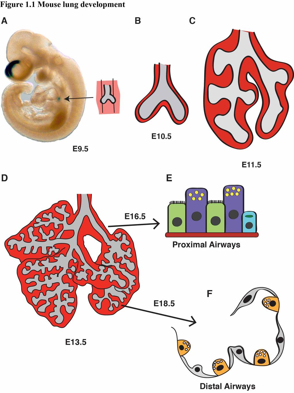
29 Figure 1.1 Mouse lung development (A) At E9.5 the ventral foregut endoderm evaginates into the surrounding mesenchyme to form the primitive lung bug. The LacZ staining in the lung and hindbrain show the expression of Wnt7b at this time point. (B) At E10.5, the bronchi of the lungs extend into the mesenchyme, and by (C) E11.5, the stereotyped branching of the lung has initiated. (D) At E13.5, the arborized blueprint of the lung has formed, and as development proceeds through E16.5, (E) epithelial cells in the proximal airways such as ciliated cells, Clara cells, and goblet cells, will become specified. (F) By E18.5, the terminal airways have begun to form alveoli, and AECII and AECI become specified and continue to develop throughout the perinatal period. 22
30 23
31 Figure 1.2 Illustration of BMP4, FGF, and SHH interactions for lung bud branching (A) The branching distal endoderm expresses SHH and BMP4, while the distal mesenchyme adjacent to the branching endoderm expresses FGF. (B) SHH down regulates FGF signaling at the tip of the branching bud, creating a zone of decreased FGF signaling. BMP4 expression is induced by the two FGF signaling centers, and the endoderm extends toward the FGF source, causing a cleft at the SHH high, FGF low zone. 24
32 25
33 Figure 1.3 Canonical Wnt signaling pathway (A) When Wnt signaling is not active, β-catenin is bound to a complex of proteins including APC, Axin, and GSK3 and phosphorylated to mark β-catenin for ubiquitin mediated proteolysis. Lef/Tcf signaling does not occur. (B) When a Wnt ligand binds to the Fzd/LRP5/6 receptor complex, Dsh causes the disruption of the APC, Axin, GSK3 complex, and β-catenin accumulates in the cytoplasm and then translocates to the nucleus to bind Lef/Tcf factors and activate target gene transcription. 26
34 CHAPTER 2: Subsets of Wnt ligands signal in a cooperative manner through the PDGF pathway to promote foregut organogenesis A portion of this chapter is published in: Proc Natl Acad Sci (Miller et al. 2012) Summary In Chapter 1, I highlighted the importance of epithelial-mesenchymal signaling in the lung, described some of the signaling pathways involved in lung development, and offered a brief overview of the signaling crosstalk that occurs. I also introduced Wnt2 and Wnt7b as complementarily expressed Wnt ligands in the lung, which have very similar mutant phenotypes. In this chapter, I am going to address an outstanding question in the Wnt and lung fields concerning Wnt ligand crosstalk, and also address the combinatorial role of Wnt2 and Wnt7b during lung development. I show that Wnt2 and Wnt7b cooperate in mesenchymal cells to potentiate high levels of canonical signaling, important for P-D endoderm patterning and smooth muscle development. I also show that the level of signaling cannot be explained by increased β-catenin accumulation, and therefore utilize a high throughput chemical screen to identify the PDGF pathway s capacity to influence Wnt signaling. Introduction As mentioned in Chapter 1, distinct regions of the foregut endoderm are specified to become the lung, liver, stomach, pancreas, and thyroid. The capacity of the foregut endoderm to develop into these organs is mediated in part through reciprocal signaling interactions with the adjacent mesenchyme (15, 17). One signaling pathway of note is the Wnt pathway, with Wnt ligands expressed in spatially specific patterns along the 27
35 anterior-posterior axis of the developing gut tube in both the developing endodermal and mesenchymal components. One goal of this chapter is to add to the understanding of how Wnt expression functions to promote regionally specific foregut development. The anterior aspect of the foregut endoderm is surrounded by the splanchnic mesoderm, which expresses Wnt2, and Wnt2-/- mutant mice have hypoplastic lungs with defective airway smooth muscle development. Of note, combined loss with Wnt2b results in lung agenesis (4, 5). Wnt7b is expressed in the developing lung endoderm, and Wnt7b- /- mice also have hypoplastic lungs with defective airway smooth muscle development, along with defective vascular smooth muscle development (23, 75). The importance of epithelial-mesenchymal interactions was highlighted in Chapter 1, and one goal of this chapter is to determine if two families of Wnt ligands, Wnt2 and Wnt7, cooperate to promote lung development. A recent study has shown two Wnt ligands, Wnt5a and Wnt11, can interact and cooperate to promote Xenopus axis formation, supporting our hypothesis that specific Wnt ligand interactions may promote important aspects of vertebrate development (91). Given the complementary expression patterns of Wnt2 and Wnt7b and the previous findings that genetic inactivation of either Wnt2 or Wnt7b leads to a similar hypoplastic lung phenotype with defects in smooth muscle development (4, 23, 33, 74, 75), we sought to determine whether context dependent Wnt autocrine and paracrine signals active in the developing anterior foregut cooperate to promote development of the lung. In this Chapter, I show that Wnt2 and Wnt7b cooperate to drive high levels of Wnt signaling activity specifically in mesenchymal cells, with cell type specificity mediated through Fzd receptor availability. Through the use of a high throughput small molecule 28
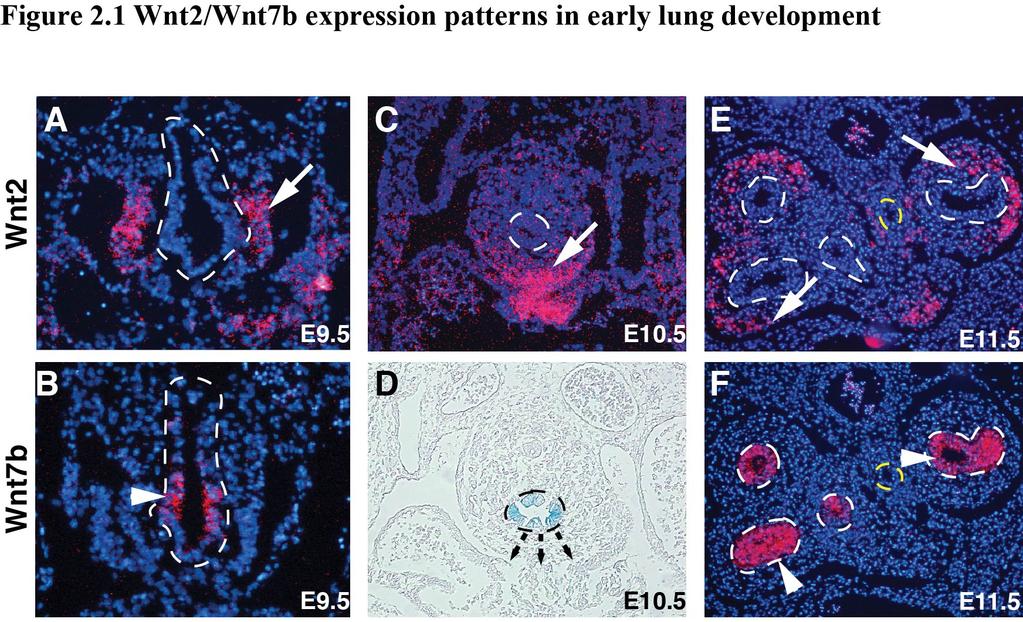
36 screens, I then identify the PDGF pathway as necessary and sufficient to promote the cooperation between Wnt2 and Wnt7b. Taken together, these studies define a novel mechanism whereby specific Wnt ligands cooperate to promote high levels of signaling in a cell lineage specific manner, in part through PDGF signaling. This mechanism illustrates how combinations of Wnt ligands can confer spatial and temporal canonical Wnt regulation of foregut organogenesis. RESULTS Wnt2 and Wnt7b are expressed in a complementary fashion in the lung Previous work has shown Wnt2 and Wnt7b are expressed in mutually exclusive domains in the developing anterior foregut and lung (4, 23). We performed in situ hybridization and utilized the LacZ knock-in to the Wnt7b allele to visualize the expression patterns of Wnt2 and Wnt7b in the developing lung from E9.5-E11.5. Wnt2 expression is observed in the mesenchyme on the ventral side of the anterior foregut at E9.5 (Fig. 2.1A). At this same time point, Wnt7b is expressed exclusively in the ventral anterior foregut endoderm where the lung endoderm progenitors are specified (Fig. 2.1B). This complementary expression pattern with Wnt2 expressed in the developing mesenchyme and Wnt7b expression restricted to the developing lung endoderm continues at E10.5 and E11.5 (Fig. 2.1C-F). In Chapter 1, I introduced that signaling pathways in close proximity work to cross regulate specific developmental programs, and the expression patterns of Wnt2 And Wnt7b suggest that they may also interact. Specific subfamilies of Wnt ligands drive cooperative signaling activation 29
37 To test whether Wnt2 and Wnt7b can cooperate to promote Wnt signaling, we transfected the rat lung mesenchymal cell line, RFL6, with expression plasmids for Wnt2 and Wnt7b, alone or in combination, along with the SuperTopFlash (STF) reporter, to detect canonical Wnt signaling activity. The STF reporter consists of 7 repeated Lef/Tcf binding sites preceding a firefly luciferase gene (92). While either Wnt2 or Wnt7b expression alone results in reproducible but low-level STF activation, the combination of Wnt2 and Wnt7b results in a dramatic cooperative activation of STF (Fig. 2.2A). To determine whether Wnt2 and Wnt7b can also activate an endogenous target of Wnt signaling in a cooperative manner, we assessed expression of endogenous axin2, a well described direct target of Wnt signaling, using quantitative PCR (Q-PCR) (93). These data show that Wnt2 and Wnt7b can activate endogenous axin2 in a cooperative manner in RFL6 cells (Fig. 2.2B). To determine whether the cooperative Wnt activation by Wnt2 and Wnt7b is specific to these ligands, we tested whether Wnt1, a traditionally canonical Wnt ligand, or Wnt5a, a traditionally non-canonical Wnt ligand, could cooperate with Wnt2 or Wnt7b (94, 95). Our studies show that neither Wnt1 nor Wnt5a cause cooperative activation of Wnt signaling with Wnt2 or Wnt7b (Fig. 2.2C,D). However, Wnt2b and Wnt7a are able to cooperatively activate STF, indicating that members of the Wnt2 and Wnt7 families are capable of cooperation (Figure 2.2E,F). To determine if either Wnt2 or Wnt7b could cooperate in the presence of a constitutively active β-catenin, we expressed a stable form of activated β-catenin (ABC) containing point mutations that prevent its degradation, along with either Wnt2 or Wnt7b (96). Expression of ABC along with either Wnt2 or Wnt7b did not result in the 30
38 cooperative effect observed between Wnt2 and Wnt7b (Fig. 2.2G). However, ABC did potentiate Wnt7b signaling, suggesting that Wnt7b may act to enhance canonical Wnt pathway activation. But, this potentiation was multiple folds lower than the Wnt2 and Wnt7b cooperation (Fig. 2.2H). These data suggest that Wnt2/Wnt7b signaling cooperation involves a mechanism distinct from simple activation of the β-catenin dependent canonical pathway. Wnt2/Wnt7b cooperative signaling is essential for early lung development To determine if the in vitro cooperation observed between Wnt2 and Wnt7b plays a significant role in vivo, we determined the consequences of Wnt2 and Wnt7b deletion during early lung development. As described in Chapter 1, loss of either Wnt2 or Wnt7b alone results in a similar lung hypoplasia and decreased smooth muscle phenotype (4, 23, 74, 75). Wnt2-/-;Wnt7b-/- double mutant lungs show severe defects in branching morphogenesis with an almost complete loss of secondary branching after trachea separation from the esophagus (Fig. 2.3A-J). Because the Wnt2-/-;Wnt7b-/- mutants die by E13.5, our analysis is restricted to E12.5 and earlier (data not shown). Although trachea-esophagus separation is apparent in the histological sections of Wnt2-/-;Wnt7b-/- double mutants at all time points tested, we assessed expression of Nkx2.1 and p63, markers of lung and esophagus endoderm respectively, to determine whether there were defects in early specification of lung endoderm. Expression of Nkx2.1 is restricted to the developing trachea while expression of p63 is restricted to the developing esophagus in both control and Wnt2-/-;Wnt7b-/- mutants (Fig. 2.3K-N) (7, 97). Thus, while loss of Wnt2 or Wnt7b alone leads to a minor lung hypoplasia phenotype, the combined loss of 31
39 both Wnt2 and Wnt7b leads to a dramatic loss of lung branching morphogenesis but normal lung endoderm specification (23, 74). Wnt2/Wnt7b cooperation is required for development of distal lung endoderm progenitors Given the significant defects in early branching of the lung, we examined whether there were alterations in proximal and distal progenitor development in the Wnt2-/- ;Wnt7b-/- mutants. As stated in Chapter 1, deletion of β-catenin in the developing lung mesenchyme or endoderm results in defective distal epithelial cell development, leading us to hypothesize that we may also observe defects in P-D epithelial cell development (40, 62). We assessed expression of Sox2, a marker of proximal endoderm progenitors, and Sox9, a marker of distal endoderm progenitors, in E11.5 lungs using immunostaining (98, 99). While Sox2 is confined to the proximal regions of the developing airway branches as well as the esophagus in both Wnt2+/-:Wnt7b+/- controls and Wnt2-/- ;Wnt7b-/- double mutants (Fig. 2.4A-D), Sox9 expression is expressed at low levels and diffusely throughout the defective airways in the Wnt2-/-;Wnt7b-/- mutants and does not exhibit the polarized expression pattern apparent in the control lungs (Fig. 2.4E-H). These data suggest that P-D patterning of the lung endoderm progenitors is disrupted in Wnt2-/-;Wnt7b-/- mutants. Wnt2/Wnt7b cooperation is required for smooth muscle development As described in Chapter 1, epithelial-mesenchymal signaling is essential for development of lung mesodermal derivatives such as airway smooth muscle (6). Given 32
40 the defects in P-D endoderm patterning in the Wnt2-/-;Wnt7b-/- mutants, as noted by diffuse expression of Sox9, we examined expression of endothelial and smooth muscle markers in Wnt2-/-;Wnt7b-/- mutant lung buds. Combined loss of Wnt2 and Wnt7b leads to a significant decrease in smooth muscle gene expression. Immunostaining for SM22α, a smooth muscle marker, shows a dramatic loss, and in some cases absence, of smooth muscle development in Wnt2-/-;Wnt7b-/- mutant lungs (Fig. 2.5A,B). Interestingly, while loss of Wnt7b leads to decreased expression of endothelial cell markers CD31 and Flk-1, QPCR results show that additional loss of Wnt2 did not further decrease expression of these genes, while SM22α and SMA show an additional decrease in expression, suggesting that smooth muscle differentiation is particularly sensitive to the cooperative affects of Wnt2 and Wnt7b signaling (Fig. 2.5C). Together, these data indicate that cooperative signaling by Wnt2/Wnt7b is required for proper P-D patterning of early lung endoderm progenitors and development of airway smooth muscle from the multipotent lung mesenchyme. The affects on Sox9 patterning could be due to the inability of the defective mesenchyme to signal back to the developing endoderm to promote P-D patterning of Sox9+ progenitors. Wnt2/Wnt7b cooperative signaling occurs in mesenchymal but not epithelial cell lineages Since Wnt2 is expressed in the developing mesenchyme and Wnt7b is expressed in the developing epithelium, we wanted to determine whether the cooperation between Wnt2 and Wnt7b functions in both cell lineages. Therefore, we expressed Wnt2, Wnt7b or both in two additional mesenchymal cells lines Pac1 and 10T1/2 cells, which represent rat vascular smooth muscle and mouse embryonic fibroblasts, respectively. We also 33
41 performed the same experiments in two epithelial cell lines, HEK-293 and MLE15 cells, which represent human kidney and mouse lung epithelium, respectively. These studies showed that co-expression of Wnt2 and Wnt7b results in cooperative Wnt signaling in both Pac1 and NIH-3T3 cells while no cooperative signaling is observed in HEK-293 or MLE15 cells (Fig. 2.6A-D). To further assess whether mesenchymal cell lineages are the responsive cell type for Wnt2/Wnt7b cooperative signaling and whether this occurs in a paracrine manner, we expressed Wnt2 and Wnt7b in RFL6 and HEK-293 cells, respectively, while transfecting the STF reporter in only one of the cell types to assess cell specific Wnt signaling activity. When the STF reporter is transfected into HEK-293 cells, Wnt2/Wnt7b cooperative signaling was not observed (Fig. 2.6E). However, when the STF reporter is transfected into RFL6 cells, we observe high levels of Wnt2/Wnt7b cooperative signaling (Fig. 2.6E). Taken together, these data indicate that mesenchymal cell lineages respond to Wnt2/Wnt7b cooperative signaling in a paracrine manner which helps to explain the dramatic loss of airway smooth muscle development in Wnt2-/- ;Wnt7b-/- mutants and also suggests that the defects in patterning of Sox9+ progenitors is secondary to defects in the adjacent mesenchyme. Frizzleds 5 and 8 promote Wnt2/Wnt7b cooperative signaling To assess Wnt signaling components that may promote or inhibit Wnt2/Wnt7b cooperative signaling, we performed a Q-PCR screen of 84 genes known to be involved in the Wnt pathway. From this screen, Fzd5 was identified as a receptor that is strongly induced by Wnt2/Wnt7b cooperative signaling (Fig. 2.7A). Over-expression of Fzd5 in RFL6 cells enhances Wnt2/Wnt7b cooperation (Fig. 2.7B). Moreover, sirna 34
42 knockdown of Fzd5 inhibits Wnt2/Wnt7b cooperative signaling in RFL6 cells (Fig. 2.7C). Thus, the finding that Fzd5 can promote Wnt2/Wnt7b cooperative signaling suggests that receptor availability may partly explain the ability of Wnt2/Wnt7b to display cooperative signaling in mesenchymal but not epithelial cell lineages. To test whether expression of Fzd5 could confer Wnt2/Wnt7b cooperation in a non-responsive cell type, we transfected Fzd1, Fzd2, Fzd10, and Fzd5 along with Wnt2 and/or Wnt7b into HEK-293 cells, a cell type normally non-responsive to Wnt2/Wnt7b cooperation. Of note, previous studies have shown that Wnt7b displays strong specificity for Fzd/LRP receptor combinations and can signal through Fzd1 and Fzd10, but not Fzd4 or Fzd7 in HEK-293 cells (100). In the current assay, Fzd5 expression was able to confer Wnt2/Wnt7b cooperative signaling to HEK-293 cells (Fig. 2.7D). Interestingly, Fzd5 increased Wnt2, but not Wnt7b signaling suggesting that Fzd5 promotes the Wnt2/Wnt7b cooperative signaling by enhancing Wnt2 and not Wnt7b activity, which is in line with previous work showing that Wnt2 synergizes with the Fzd5 receptor in Xenopus axis development (101). Although Fzd5 is expressed in the developing lung, the Fzd5 conditional mutants are viable, suggesting that additional Fzd receptors work redundantly with Fzd5 in lung development (101, 102). Fzd8 is highly homologous to Fzd5 and is also expressed in the lung (103, 104). Expression of Fzd8 is also sufficient to confer Wnt2/Wnt7b cooperative signaling to HEK-293 cells and also increased Wnt2 signaling (Fig. 2.7D). These data suggest that specific Fzd receptors including Fzd5 and Fzd8 transmit Wnt2/Wnt7b cooperative signaling and do so, at least in part, by enhancing Wnt2 activity. 35
43 Wnt2/Wnt7b cooperative signaling does not lead to increased nuclear β-catenin levels The dramatic induction in canonical Wnt signaling observed by Wnt2/Wnt7b signaling suggests that this cooperation may lead to a concordant increase in nuclear β- catenin levels. Therefore, we performed Western blots for β-catenin on both cytoplasmic and nuclear fractions of RFL6 cells expressing Wnt2, Wnt7b, or Wnt2 and Wnt7b (Figure 2.8A). These experiments revealed a lack of increased nuclear or cytoplasmic β- catenin levels by co-expression of Wnt2 and Wnt7b, although a slight increase was observed by expression of Wnt2 alone (Fig. 2.8A). These data suggest that the Wnt2/Wnt7b cooperative signaling involves additional pathways and factors beyond accumulation of nuclear β-catenin. To determine whether β-catenin expression is necessary for Wnt2/Wnt7b cooperative signaling, we performed sirna knockdown of β-catenin in RFL6 cells, along with expression of Wnt2 and Wnt7b (Figure 2.8B). Loss of β-catenin resulted in a significant decrease in Wnt2/Wnt7b cooperative signaling indicating that while this cooperation did not lead to increased levels of β-catenin, steady state expression of β- catenin is necessary for signaling to occur. Together, these data suggest that β-catenin expression is required but additional factors or pathways are necessary to promote Wnt2/Wnt7b cooperative signaling. High-throughput small molecule screen reveals the importance of PDGF signaling on Wnt2/Wnt7b cooperative signaling 36
44 To identify potential pathways that promote Wnt2/Wnt7b signaling independent of β-catenin signaling, we performed a high-throughput small molecule screen utilizing 1280 pharmacologically active compounds (LOPAC-Library of Pharmacologically Active Compounds) that selectively inhibit components of most major signaling pathways to detect pathways important for Wnt2/Wnt7b cooperation. We excluded hits that promoted cell cycle arrest, cell death, or proliferation based on software accompanying the library and focused on multiple hits towards common signaling pathways that inhibited signaling by at least 35% (Figure 2.9A). This screen identified four compounds that target the epidermal growth factor (EGF)/PDGF signaling pathways: 1) Tyrphostin AG1478, a selective inhibitor of EGFR activity (105), 2) U0126, a MEK1/2 inhibitor (106), 3) GW2974, a dual EGFR and ErbB-2 receptor tyrosine kinase inhibitor (107), and 4) Tyrphostin A9, a selective PDGFR tyrosine kinase receptor inhibitor (108). Interestingly, the EGF and PDGF pathways have been implicated in regulating Wnt signaling activity (33, 109). All four of these compounds inhibited Wnt2/Wnt7b cooperative signaling in a dose dependent manner (Figure 2.9B-E). Based on our findings that expression of β-catenin is necessary for Wnt2/Wnt7b cooperative signaling, but steady state levels of β-catenin do not change significantly in this cooperative signaling, we assessed whether inhibition of EGF or PDGF signaling inhibited Wnt/β-catenin signaling in general, or specifically inhibited the Wnt2/Wnt7b cooperative signaling. A pathway inhibitor that affects Wnt2/Wnt7b cooperative signaling without affecting activated β-catenin signaling would implicate this pathway in the specific regulation of Wnt2/Wnt7b signaling. These studies show that only the PDGF signaling inhibitor Tyrphostin A9 inhibits Wnt2/Wnt7b cooperative signaling without 37
45 effecting activated β-catenin signaling (Fig. 2.10A,B). To further support the PDGFR chemical inhibitor data, we expressed a dominant negative truncated form of PDGFR-α (PDGFR-α Truncated) in RFL6 cells, along with Wnt2, Wnt7b, and STF. We observed a similar decrease in the cooperation between Wnt2 and Wnt7b with the expression of a truncated form of PDGFR-α as we observed with Tyrphostin A9 (Fig. 2.10C). To test whether activation of PDGF signaling could enhance Wnt2/Wnt7b cooperative signaling, RFL6 cells expressing Wnt2 and Wnt7b were treated with recombinant PDGF-BB ligand. These studies show that PDGF-BB causes a significant increase in Wnt2/Wnt7b cooperative signaling (Fig. 2.10D). PDGF signaling is necessary for smooth muscle development and Wnt signaling in the lung To determine if this potent role for PDGF signaling in the Wnt2/Wnt7b cooperation is necessary for smooth muscle development in the lung, lung explants were treated with splice blocking morpholinos against PDGFR-α and PDGFR-β, which resulted in significantly decreased expression of these two receptors (Figure 2.11A). Knockdown of PDGFR-α and PDGFR-β resulted in a significant decrease in smooth muscle gene expression as well as a decreased smooth muscle development surrounding the lung airways (Fig. 2.11B,C). Moreover, decreased PDGFR-α/β expression in the lung leads to decreased axin2 gene expression, suggesting that PDGFR-α/β are required for endogenous levels of Wnt activity in the lung (Fig. 2.11D). To further assess whether the combined activity of the Wnt2 and Wnt7 ligand families could cooperatively increase 38
46 smooth muscle development in the lung, we treated lung explants with recombinant Wnt2, Wnt7a, or Wnt2 plus Wnt7a. Recombinant Wnt7a was used in these studies because recombinant Wnt7b is not available and Wnt7a cooperates with Wnt2 in vitro. These data show that Wnt2 and Wnt7a can cooperatively increase both smooth muscle gene expression and development in the lung (Fig. 2.12A,B). Together, these data reveal a novel Wnt signaling mechanism whereby PDGF signaling promotes a specific Wnt2/Wnt7b cooperative signaling mechanism required for mesenchymal cell differentiation in lung morphogenesis (Fig. 2.13A). DISCUSSION In this study, we show that specific Wnt ligands can cooperatively induce high levels of Wnt signaling to regulate P-D patterning and mesenchyme development in the lung. We found that the combinatorial actions of Wnt2 and Wnt7b promote mesenchymal cell specific Wnt activity that is mediated, in part, through the PDGF pathway. While this cooperative effect of Wnt2/Wnt7b signaling requires β-catenin activity, it does not result in an increase in nuclear steady state expression levels of β- catenin. The necessity of this Wnt2/Wnt7b cooperation is demonstrated by the dramatic lung phenotype in Wnt2-/-;Wnt7b-/- mutant lungs, which display a severe truncation of branching morphogenesis along with, disrupted distal endoderm progenitor patterning. Our results highlight the importance of reciprocal epithelial/mesenchymal signaling for lung development and show the necessity of specific combinations of Wnt ligands to promote foregut derived organ development. 39
47 Epithelial-mesenchymal interactions are a recurrent theme throughout embryonic development. Though the foregut endoderm begins as an undifferentiated sheet of epithelium, the ventral aspect of the foregut endoderm will eventually give rise to complex, multicellular organs such as the thyroid, lung, liver, and ventral pancreas. The specification and the subsequent differentiation of this endoderm into a myriad of organ specific epithelial lineages are dependent upon epithelial-mesenchymal interactions. Recent work in the pluripotent stem cell reprogramming field has shown the requirement of temporally specific expression of Wnt signaling to differentiate foregut endoderm cells into lung endoderm (110). If Wnt signaling is activated prior to the expression of anterior foregut endoderm markers, the reprogrammed cells express posterior endoderm markers. Following endoderm reprogramming to anterior foregut endoderm, Wnt3a, in combination with additional growth factors, is required in a precise time window to allow for the ventralization, and not dorsalization, of the anterior foregut endoderm. Then, to promote the expression of distal lung endoderm markers, continued Wnt3a expression, in addition to retinoic acid and Fgf ligands, is required (110). The exogenously applied growth factors exemplify an in vitro system modeling the in vivo requirement for epithelial to mesenchymal signaling for epithelial lung cell differentiation. An abundance of literature highlights the in vivo importance of epithelialmesenchymal interactions for lung specification and development. During lung endoderm specification, Wnt2/Wnt2b ligands function in a paracrine manner to specify Nkx2.1+ lung endoderm progenitors within the ventral anterior foregut endoderm. Conditional deletion of β-catenin from the foregut endoderm phenocopies the Wnt2/Wnt2b lung agenesis phenotype indicating that Wnt2/2b act in a canonical fashion to specify Nkx
48 lung progenitors. These studies also showed that Wnt2/2b expression is required for Wnt7b expression in the anterior foregut endoderm. Based on the present and previous studies, we propose a model in which Wnt2 activates the canonical Wnt pathway in the anterior foregut endoderm including Wnt7b expression. Expression of Wnt7b, along with PDGFR activity, promotes maximal Wnt signaling activity in the lung mesenchyme that promotes proper differentiation of specific cell lineages such as smooth muscle. How Wnt7b promotes this maximal signaling in cooperation with PDGF signaling is unclear but could involve several different mechanisms including post-translational modifications of β-catenin that enhance its activity by releasing the repressive activity of a repressor, or enhancing the activity of a β-catenin co-activator. We have shown that while β-catenin is required for the cooperation between Wnt2 and Wnt7b, the accumulation of β-catenin is not solely sufficient. Our work has shown that the combination of Wnt2 and Wnt7b does not result in a concordant increase in β-catenin accumulation that could explain the high levels of reporter activity observed. Examples of increased β-catenin signaling, with modest to no effects on β-catenin accumulation, have been seen in previous studies where modifications to β-catenin or the binding of co-activators to promoter regions enhances canonical signaling. The transcriptional co-activator p300 has been shown to affect the activity of β-catenin through acetylation. This acetylation results in an increased β-catenin affinity for Tcf4 and higher levels of signaling, without affecting nuclear β-catenin accumulation (111). Protein kinase A (PKA) has been shown to phosphorylate β-catenin, which results in β- catenin binding to the transcriptional coactivator CREB- binding protein, leading to 41
49 increased signaling without affecting β-catenin accumulation (112). A similar mechanism could occur in the cooperation between Wnt2 and Wnt7b, in which PDGF signaling results in co-activator activation leading to high levels of canonical signaling, without a reciprocal increase in nuclear β-catenin accumulation. Our finding that Wnt2 and Wnt7b specifically cooperate during lung development to uniquely enhance transcriptional activation may allow for refinements in current reprogramming techniques. We show that simply activating the canonical Wnt pathway is not sufficient to replicate the cooperative activity of the Wnt2 and Wnt7b ligands. Although Wnt3a has been used to promote reprogramming of ips cells into lung endoderm, our work suggests that there exist distinct functions for specific Wnt ligands that should to be taken into account for promotion of lung progenitor cell fates. These combinatorial actions of specific Wnt ligands may explain how broad domains of canonical Wnt activity during embryonic development affect only specific cell lineages to allow for organ specification and development. MATERIALS AND METHODS Animals Generation and genotyping of Wnt7b LacZ and Wnt2 mouse lines have been previously described (4, 23). Embryos were collected from E10.5-E12.5 as noted. The University of Pennsylvania Institutional Animal Care and Use Committee approved all animal protocols. Histology 42
50 Embryos were collected and fixed in 4% paraformaldehyde (PFA) for immunohistochemistry and in situ hybridization. Following fixation, embryos were dehydrated in an increasing gradient of ethanol washes, imbedded in paraffin, and sectioned. Tissue sections were immunostained with the following antibodies: anti- Nkx2.1 (Santa Cruz), anti-p63 (Santa Cruz), anti-sox2 (Seven Hills Bioreagents), anti- Sox9 (Santa Cruz), and anti-sm22alpha (Abcam). In situ hybridization was performed as previously described (4, 23). For all histology, at least 3 control embryos and at least 3 Wnt2-/-;Wnt7b-/- embryos were stained and visualized. Cell Culture and Transient Transfection Assays RFL6, Pac1, and HEK293 were cultured in DMEM supplemented with 10% FBS and 1% antibiotic/antimycotic. 10T1/2 cells were cultured in Eagle s Basal medium supplemented with 10% FBS, 2mM L-Glutamine, and 1% antibiotic/antimycotic. MLE15 cells were cultured in HITES medium supplemented with 10% FBS and 1% penicillin/streptomycin as previously described (113). Cells were transfected with the indicated expression plasmids using Fugene 6 (Roche). Forty-eight hours following transfection, luciferase assays were performed using the Dual-Luciferase Reporter Assay System (Promega). For Wnt1 and ABC transfections, Wnt1 and ABC levels were titrated for sub-maximal STF activation. For cell mixture experiments, RFL6 and HEK293 cells were transfected with the indicated plasmids for 24 hours after which they were mixed in equal numbers. Forty-eight hours following mixture, luciferase assays were performed. For sirna knockdown experiments, 24 hours following transfection with expression vectors, cells were transfected with sirna pools against b-catenin or Fzd5 (Dharmacon) 43
51 using Lipofectamine 2000 (Invitrogen). Luciferase assays were performed 24 hours following transfection of sirna. For chemical inhibitor assays, 24 hours following transfection with expression vectors, chemical inhibitors were added at the indicated concentrations for 24 additional hours, followed by luciferase assays. All of the indicated chemical inhibitors were obtained from Sigma and solubilized in DMSO. For cell culture assays, a pcmv b-gal plasmid was used as a control plasmid to equalize the amount of plasmid being transfected per condition. 50,000 cells were plated in each well of a 24-well plate on Day 1. On Day 2, the wells were transfected in triplicate, with a half well excess calculated, as shown below: pcmv β- gal Wnt2 Wnt7b STF Renilla Control µg µg µg Wnt µg µg µg µg Wnt7b µg µg µg µg Wnt2+Wnt7b µg µg µg µg µg Fugene 6 was used as the transfection reagent, and was added at a ratio of 3uL per 1ug of plasmid. On Day 4, we used the Dual-Luciferase Assay System to perform luciferase assays. To normalize transfection efficiency, the firefly luciferase values were divided by the Renilla luciferase values, and the ratio was used to compare the control, Wnt2, Wnt7b, and Wnt2+Wnt7b conditions. Rat On-Target Plus SMARTpool sirnas were purchased from Dharmacon (Ctnnb1 L , Fzd5 L , Non-Targeting Pool D ). 44
52 Quantitative PCR and Western blotting Total RNA was isolated from cells and tissues using Trizol reagent, reverse transcribed using SuperScript II First-Strand Synthesis Kit (Invitrogen), and used in quantitative realtime PCR analysis utilizing SYBR green (Applied Biosystems). Lung explant mrna was isolated using the Qiagen RNeasy Mini Kit. For Western blotting, cells were collected 48 hours following transfection, lysed, and nuclear and cytoplasmic fractions were generated using a standard protocol. Briefly, 500mL of Buffer A (10mM HEPES, 1.5mM MgCl2, 10mM KCl, 0.5mM DTT, 0.05% NP40) was added to transfected cells, cells were scraped, and lysates were incubated for 10 minutes on ice. Lysates were centrifuged at 4 0 C at 3000rpm for 10 minutes, and the supernatant was collected as the cytoplasmic fraction. The pellet was resuspended in Buffer B (300mM NaCl, 5mM HEPES, 1.4mM MgCl2, 0.2mM EDTA, 0.5mM DTT, 26% glycerol, and protease inhibitor cocktail), homogenized, and incubated on ice for 30 minutes. Nuclear lysate was centrifuged at 21,130 g s at 4 0 C for 30 minutes. The supernatant was collected as the nuclear fraction. Protein concentration was quantified using the Bio-Rad Protein Assay and 15mg of protein were resolved on SDS-PAGE gels and transferred to PVDF membranes. Antibodies used include in Western blotting include: b-catenin (BD Transduction Laboratories), b-tubulin (Abcam), HN-RNPA1 (Santa Cruz Biotech). LOPAC high-throughput small molecule screen RFL6 cells were transfected with CMV-bgal or pcdna3.1-wnt2 and Wnt7b expression plasmids, along with STF. Twenty-four hours following transfection, cells were replated 45
53 into 384-well tissue culture plates at 3X10 4 cells per well and compounds from the Library of Pharmacologically Active Compounds (LOPAC) library (Sigma-Aldrich) were added at a final concentration of 10mM. Twenty-four hours following plating and addition of the LOPAC library, luciferase assays were performed using BriteLite reagent (Perkin-Elmer). The assay was repeated and data was normalized to the median of each 384 well plate. Lung Explant Culture Lungs were isolated from E11.5 CD1 embryos and cultured in phenol free DMEM/F12 (Cellgro) on 0.4mm transwells (Falcon) for either 48 or 72 hours at 37 degrees C and 5% CO2. Lungs for Q-PCR were cultured for 48 hours, and lungs for immunohistochemistry were cultured for 72 hours. PDGFRa and PDGFRb morpholinos (GeneTools) were added into the culture medium at a concentration of 15uM. The PDGFR-α morpholino (ATGTGTGGATACATACCTGTGAGG) targeted the splice donor site of exon 2, and the PDGFR-β morpholino (ATCTGTCAAGAGCAGAGCCAAGGAA) targeted the splice acceptor site of exon 4. A standard control morpholino (CCTCTTACCTCAGTTACAATTTATA) designed by Gene-Tools was used as the control morpholino. Morpholino knockdown was assessed by QPCR using primers (TGAGGGAGAGAAACAAACGGAGGA) and (AGCTCCTGAGACCTTCTCCTTCTA) for PDGFR-α and (ACCAGCGAGGTTTCACTGGTACTT) and (ATCATTGCCCATCACAATGCACCG) for PDGFR-β. Recombinant Wnt2 (Novus Biologicals) and recombinant Wnt7a (R&D) were added to the culture medium at a concentration of 0.25ug/mL. For QPCR, at least 3 46
54 lungs were pooled for RNA for each experimental condition, with the number held constant between compared conditions, and for histology, at least 3 lungs were visualized for each condition. 47
55 48
56 Figure 2.1 Wnt2/Wnt7b expression patterns in early lung development In situ hybridizations show that Wnt2 is expressed in the developing lung mesenchyme from E9.5-E11.5 (A, C, E, arrows), while Wnt7b is expressed in the developing lung endoderm from E9.5-E11.5 (B, F, arrowheads). (D) The Wnt7b LacZ allele can be used to visualize Wnt7b expression in the lung endoderm, which signals to the surrounding Wnt2 positive mesenchyme (arrows). The endoderm of the lung is outlined. 49
57 50
58 Figure 2.2 Wnt2 and Wnt7b cooperate and show ligand specificity Expression of Wnt2 and Wnt7b promote high levels of cooperative signaling activity as noted by the activation levels of the STF reporter (A) and by measuring endogenous Axin2 expression by Q-PCR (B) as readouts of Wnt signaling. Wnt2 does not cooperate with Wnt1 or Wnt5a to promote STF activity (C). Wnt7b does not cooperate with Wnt1 or Wnt5a to promote STF activity (J). Wnt2b can cooperate with Wnt7b (E) and Wnt7a can cooperate with Wnt2 (F) to drive high levels of Wnt signaling. Wnt2 does not cooperate with activated β-catenin to promote STF activity and Wnt7b plus activated β- catenin results in only an additive increase in STF activity (G). 51

59 52
60 Figure 2.3 Wnt2-/-;Wnt7b-/- double mutant lungs are hypoplastic and have severe branching defects While Wnt2-/-;Wnt7b-/- mutants at E10.5 and E11.5 do form a separate trachea/lung and esophagus (A and B, E and F, red versus black arrows), but the lung fails to undergo extensive subsequent branching after the first airway bifurcation (H, black arrow). By E12.5, Wnt2-/-;Wnt7b-/- mutant lungs have not progressed while control (Wnt2+/- :Wnt7b+/-) lungs have branched extensively (I and J). The expression patterns of Nkx2.1 and p63 at E10.5, which mark the primitive trachea (T) and esophagus (E) respectively, are normal in Wnt2-/-;Wnt7b-/- mutants compared to double heterozygous controls (K- N). E= esophagus, T=trachea. 53
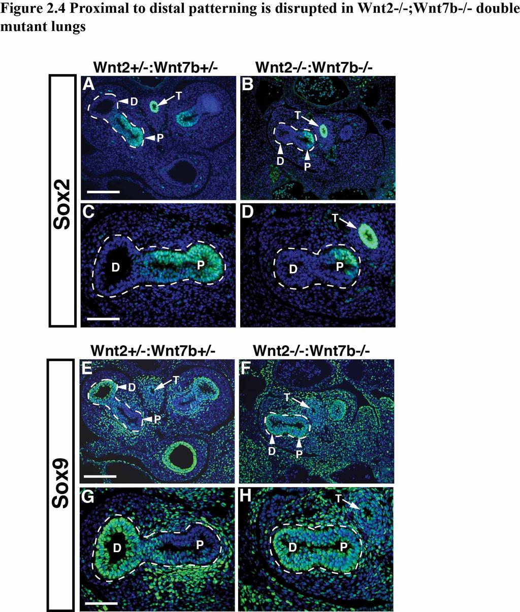
61 54
62 Figure 2.4 Proximal to distal patterning is disrupted in Wnt2-/-;Wnt7b-/- double mutant lungs The Sox2 expression pattern, which marks the proximal endoderm progenitor population, is normal in Wnt2-/-:Wnt7b-/- mutants compared to controls (A-D). However, the Sox9 expression pattern is more diffuse indicating defective distal progenitor development in Wnt2-/-:Wnt7b-/- mutants (E-H). D=distal lung endoderm, P=proximal lung endoderm, T=trachea. The lung endoderm is outlined. 55
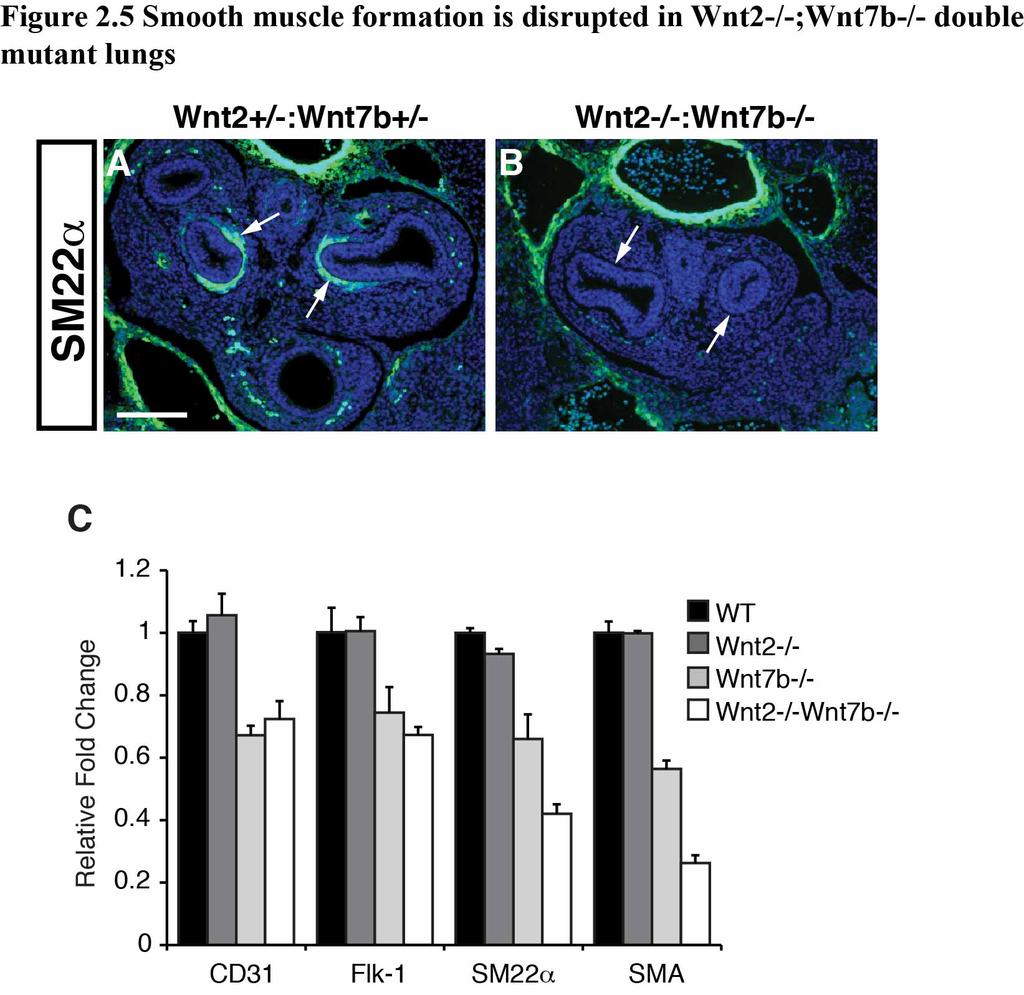
63 56
64 Figure 2.5 Smooth muscle formation is disrupted in Wnt2-/-;Wnt7b-/- double mutant lungs SM22α expression reveals a significant loss in smooth muscle development in Wnt2-/- ;Wnt7b-/- mutants (A,B, arrows). Q-PCR analysis shows that smooth muscle markers SM22α and smooth muscle α-actin (SMA) are decreased in a cooperative manner by loss of both Wnt2 and Wnt7b expression while endothelial markers are decreased in Wnt7b-/- mutants but loss of Wnt2 does not lead to a further decrease (C). 57
65 58
66 Figure 2.6 Cooperation between Wnt2/Wnt7b is specific to mesenchymal cells Wnt2-Wnt7b cooperative signaling occurs in Pac1 and 10T1/2 cells while no cooperation is observed in HEK293 and MLE15 cells (A-D). Wnt2-Wnt7b cooperative signaling occurs robustly in a paracrine fashion in mesenchymal cells (RFL6) but less so in epithelial cells (HEK293) in a cell mixture assay (E). 59
67 60
68 Figure 2.7 Cell type specificity may be a result of Fzd5 and Fzd8 availability Analysis of a Q-PCR Superarray assay shows that Fzd5 is induced by Wnt2/Wnt7b cooperative signaling (A). Over-expression of Fzd5 in RFL6 cells can strongly enhance Wnt2/Wnt7b cooperative signaling (B) while sirna knockdown of Fzd5 expression inhibits this cooperation (C). Amongst Fzd s tested including Fzd1, Fzd4, and Fzd10 only Fzd5 and the highly related Fzd8, can confer Wnt2/Wnt7b cooperative signaling to normally non-responsive HEK293 cells (D). 61
69 62
70 Figure 2.8 β-catenin is necessary but not solely responsible for the cooperation between Wnt2/Wnt7b RFL6 cells transfected with Wnt2, Wnt7b, or Wnt2+Wnt7b expression plasmids were fractionated as described in the Materials and Methods section and Western blots were performed on cytoplasmic and nuclear protein fractions. While a slight increase in β- catenin levels were observed for Wnt2 expressing cells, no further increase was observed in Wnt2+Wnt7b expressing cells (A). The nuclear and cytoplasmic fractions were pure as noted by separate expression of β-tubulin and HNRNPA-1, respectively. sirna knockdown of β-catenin in RFL6 cells inhibits the Wnt2-Wnt7b cooperation (B). 63
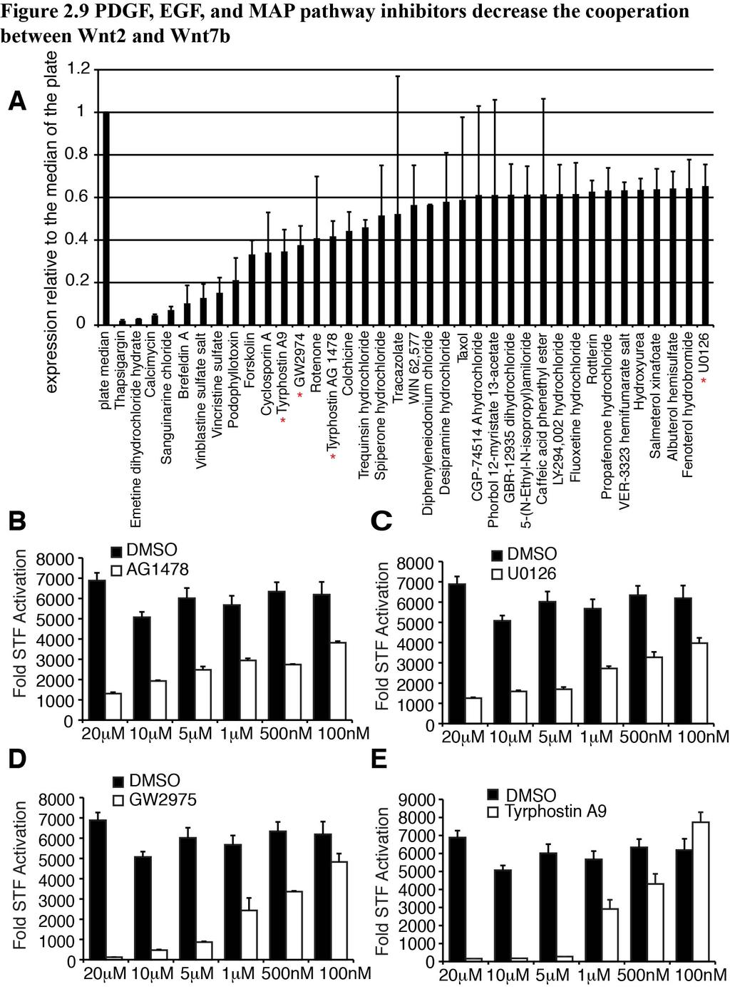
71 64
72 Figure 2.9 PDGF and EGF pathway inhibitors decrease the cooperation between Wnt2 and Wnt7b The LOPAC library was screened for compounds that inhibit the Wnt2-Wnt7b cooperative signaling. From this screen, 37 compounds were identified that inhibited the Wnt2/Wnt7b signal by at least 35% (A). Of these, four compounds, AG1478, U0126, GW2974, and Tyrphostin A9 targeted the EGFR or PDGFR pathways (denoted by *). All four compounds inhibited Wnt2-Wnt7b cooperative signaling in a dose dependent manner (B-E). Of note, cell survival was not affected in all doses less than 10mM (data not shown). 65
73 66
74 Figure 2.10 The PDGF pathway specifically affects the cooperation between Wnt2 and Wnt7b The effects of AG1478, U0126, GW2974, and Tyrphostin A9 on Wnt2/Wnt7b signaling and activated β-catenin signaling were compared using the 1mM dose for each compound. While AG1478, U0126, and GW2974 all inhibited β-catenin signaling (A), only Tyrphostin A9 affected Wnt2/Wnt7b signaling specifically, without affecting β- catenin signaling (B). A dominant negative truncated PDGFR-a decreases the cooperation between Wnt2/Wnt7b, adding support to the Tyrphostin A9 chemical inhibitor data (C). The addition of PDGF-BB ligand to Wnt2+Wnt7b transfected RFL6 cells results in increased STF activation at a concentration of 25ng/mL (D). 67

75 68
76 Figure 2.11 Knockdown of PDGFRs results in decreased smooth muscle formation and Wnt signaling The PDGFR-α and PDGFR-β morpholinos knock down the expression of PDGFR-α by approximately 80% and PDGFR-β by approximately 90% in cultured lung explants (A). The knockdown of PDGFR-α and PDGFR-β results in decreased smooth muscle markers SMA and SM22α by Q-PCR (B) and expression of SM22α is decreased by immunohistochemistry, as denoted by arrows (C). The knockdown of PDGFR-α and PDGFR-β also decreases the Wnt downstream target, Axin2, demonstrating decreased Wnt signaling with the loss of PDGF in the lung explants. 69
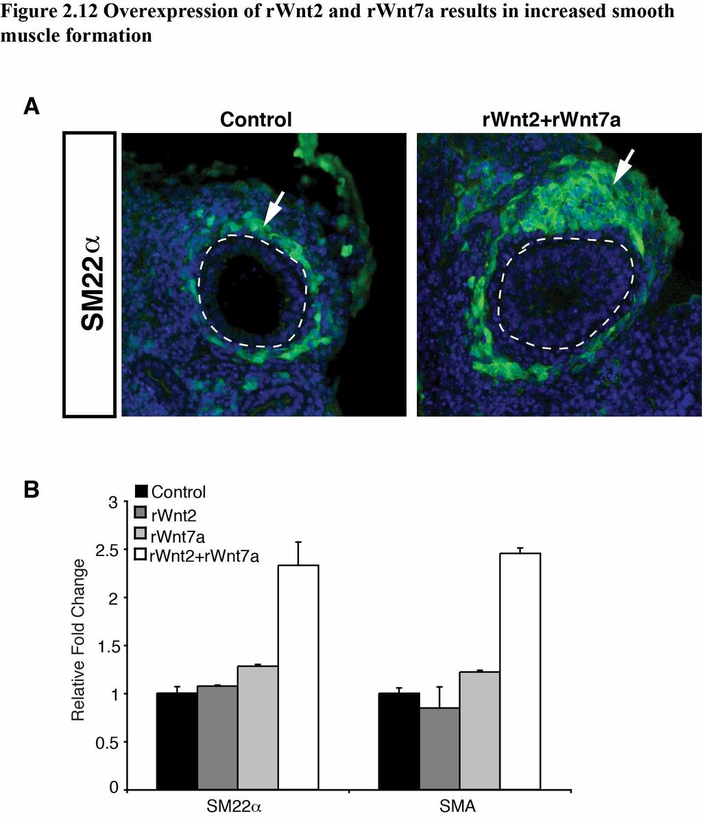
77 70
78 Figure 2.12 Overexpression of rwnt2 and rwnt7a results in increased smooth muscle formation Addition of rwnt2 and rwnt7a to lung explant cultures results in increased SM22α expression, compared to lung explants cultured without recombinant Wnt, arrows point to SM22α expression, dashed line denotes lung endoderm outline (A). QPCR for SM22α and SMA on lung explants treated with rwnt2, rwnt7a, or rwnt2+rwnt7a show increased smooth gene transcription only with the combination of rwnt2 and rwnt7a (B). 71
79 72
80 Figure 2.13 Model of cooperation between Wnt2/Wnt7b/PDGF Epithelial Wnt7b signals in a paracrine manner to Wnt2 expressing mesenchymal cells. The combined action of Wnt2 and Wnt7b, in combination with PDGF signaling, results in increased canonical Wnt signaling, which allows for proper smooth muscle development and proximal-distal patterning of the lung epithelium. 73
81 CHAPTER 3: High throughput genomic screen identifies multiple factors that promote cooperative Wnt signaling A portion of the data presented in this chapter is included in a forthcoming manuscript. Summary In Chapter 2, I demonstrated that Wnt2 and Wnt7b can promote high levels of cooperative signaling in a cell type specific manner. To explore the underlying mechanism of this cooperative Wnt signaling, we performed a high-throughput screen of more than 14,000 cdnas to identify genes that promote Wnt signaling in the context of a single Wnt ligand, Wnt2. This screen identified several homeobox containing genes including Msx2, Nkx5.2, and Esx1, in addition to other factors known to promote Wnt signaling, including Pias4. Generation of dominant-active or dominant-negative forms of the Msx homeodomain indicate that the mechanism by which this homeobox factor cooperatively promotes Wnt signaling is through its ability to repress gene transcription. These data identify a broad homeobox code, which acts to increase Wnt signaling through transcriptional repression. Introduction As shown in Chapter 2, cooperative signaling by Wnt2 and Wnt7b is necessary and sufficient to promote smooth muscle and distal endoderm progenitor cell development in the lung. Moreover, this cooperative activation of Wnt signaling, while requiring β-catenin expression, is not the result of increased β-catenin stabilization. Precedent for increased Wnt signaling without a concomitant increase in β-catenin protein levels has been demonstrated by the activity of β-catenin co-activators, including 74
82 p300 and pontin52, both of which cause a significant increase in Wnt reporter activation without increasing β-catenin stabilization (114, 115). Homeotic (Hox) transcription factors are a large group of transcriptional regulators that are critical for embryonic development. Hox genes were first discovered in Drosophila as random gene duplication and deletion events resulting in structure duplication i.e. a fly with two pairs of wings. The genes causing these segment duplication and deletion mutations were found to contain highly homologous DNA binding domains of 180 base pairs with extremely high evolutionary conservation (116, 117). Interestingly, Hox genes are distributed along chromosomes in the same anterior to posterior position that they are expressed in the body. In the mouse, there are 39 Hox genes located on four chromosomes. They are thought to have arisen from gene and chromosomal duplication events of the original eight Drosophila Hox genes, which are located on a single chromosome (118). Paralogous Hox genes have the same number designation, and are differentiated by letter e.g. hoxa-3, hoxb-3, hoxc-3, and hoxd-3. Paralogues are homologous enough that they can be substituted for each other (118). Hox genes in the same family are all related by a highly homologous homeobox DNA binding domain and can function as transcriptional activators or repressors. Several homeobox genes have been implicated in enhancing Wnt signaling. For example, Pitx2, a paired-homeodomain containing gene (Prd), can directly bind to β- catenin and synergistically activate the promoter of Lef-1 (119). The homeobox factor Hhex binds to a Wnt signaling repressor, Sox13, displacing it from Tcf3/4 to relieve transcriptional repression and allow for Wnt mediated transcriptional activation (120). 75
83 Such studies indicate the importance of homeobox factor interactions in promoting Wnt signaling activity. In this Chapter, I perform a gain of function high throughput cdna overexpression screen to further interrogate the mechanism of the cooperation between Wnt2 and Wnt7b. I identify multiple homeobox factors that promote high levels of signaling in the presence of Wnt2. These results indicate that a homeobox code may be important for Wnt signaling activity in lung development. The work in this chapter may have broader implications for development along the entire embryonic axis, where areas of Hox gene and Wnt expression overlap define specific transcriptional responses. RESULTS Optimization of Wnt2/Wnt7b transfection for high throughput genomic screen The work described in Chapter 2 demonstrates the importance of the cooperation between Wnt2 and Wnt7b in the development of the early mouse lung (121). To determine factors that could act upstream or downstream of Wnt2, we performed a high throughput screen (HTS), in collaboration with John Hogenesch s lab at the University of Pennsylvania, utilizing the Mammalian Genome Collection (Open Biosystems), which includes 9,017 mouse and 5,445 human full length cdnas driven by the CMV promoter arrayed in 46 x 384-well plates. We performed the screen in RFL6 cells, the same cell line that most of the work in Chapter 2 was performed in (121). To carry out this screen, we first scaled down our STF Wnt cooperation assay to a 384-well plate format, from a 6-well plate format. To do this, we scaled down into a 96-well plate format, and then scaled down to a 384-well plate format, optimizing plasmid and transfection reagent 76
84 ratios as well as cell number (Fig. 3.1A). Additional steps were taken to ensure that the signal to noise ratio between control transfected cells and cells transfected with Wnt2+Wnt7b plasmids was significant enough that the screen produced consistent and reproducible results. To perform the screen, a transfection mix containing control expression plasmids as well as the STF reporter, along with the transfection reagent Fugene HD, was added to 384-well plates containing the Mammalian Genome Collection in expression vectors (Fig. 3.1B). RFL6 cells were then added to the wells and after a 48-hour incubation period, the STF activity was quantified utilizing the BriteLite assay and the Perkin Elmer Envision luminometer. The screen was performed twice and the STF measurements were normalized and averaged. Validation of HTS results A cut-off point of 35-fold or higher than the median value of each 384-well plate was chosen to identify cdnas from the screen for further investigation. This resulted in 23 unique genes to validate (Fig. 3.1B). From the 23 genes, we tested whether they could activate the STF reporter by themselves or if they would activate the SuperFOPFLASH (SFF) reporter, which has mutated LEF/TCF sites and thus should not be activated by Wnt signaling activity. Of the 23 genes, five genes specifically activated the STF reporter in combination with Wnt2 (Fig. 3.2A). Of these five, three were homeobox containing genes (Esx1, Msx2, and Nkx5.2) and one was the sumoylation factor Pias4, which is known to regulate and alter Wnt/β-catenin signaling ( ). All of these 77
85 factors cooperatively enhanced Wnt signaling when co-expressed with either Wnt2 or Wnt7b (Fig. 3.2B-F). Msx1 and Msx2 function as repressors of repressors in the cooperation between Wnt2 and Wnt7b Of the three homeobox genes, we chose to focus on Msx2, and its highly related family member Msx1, since previous reports have demonstrated that these factors regulate Wnt signaling, particularly in mesenchymal cells ( ). To determine whether the Msx1/2 genes enhance the activity of other Wnt ligands, we expressed Msx1/2 along with either Wnt1 or Wnt5a in RFL6 cells. When co-expressed with Wnt2 and Wnt7b, the fold activation Msx1/2 were able to confer over the Wnt alone was 18 and 20 fold, respectively (Fig. 3.3A and B). However, with Wnt1 and Wnt5a, Msx only enhanced the signal by 5-fold for Wnt1, and had no positive effect on Wnt5a signaling (Fig. 3.3C and D). These data suggest that Msx1/2 enhance canonical Wnt signaling and that their effects are Wnt ligand specific. To determine whether Wnt2 or Wnt7b activity can affect the transcription of Msx1 or Msx2, we performed qualitative Q-PCR on RFL6 cells expressing Wnt2 and/or Wnt7b. These studies show that increased Wnt2 and Wnt7b expression induce both Msx1 and Msx2 (Fig. 3.4A). Next, we wanted to determine how Msx1/2 affect the cooperation between Wnt2 and Wnt7b. To determine the activity of Msx1/2 in the context of Wnt2 and Wnt7b, we produced fusion constructs containing the Msx1 homeodomain (MsxHB) to construct MsxHB-Engrailed and MsxHB-VP16 expression plasmids to repress and activate Msx target genes, respectively (Fig. 3.4B). The MsxHB-VP16 construct, which 78
86 we initially hypothesized would increase the cooperation between Wnt2 and Wnt7b, decreased the Wnt2/Wnt7b cooperative signaling. In contrast, the MsxHB-Engrailed construct increased Wnt2/Wnt7b cooperative Wnt signaling (Fig. 3.4C,D). Thus, our findings suggests that Msx1/2, and possibly other homeobox factors, promote cooperative Wnt2/Wnt7b signaling by repressing gene expression. Microarray to determine repressors of Wnt signaling that are down regulated in Wnt2/Wnt7b cooperation The results of the VP16 and engrailed fusion constructs led to the hypothesis that the role of Msx, or a gene containing the conserved homeodomain, is acting as a repressor of a Wnt repressor. A list of known Wnt repressors was compiled and QPCR was performed to determine if any known Wnt repressors show decreased transcriptional activity when Wnt2 and Wnt7b are transiently overexpressed. From our candidate list, none showed a decrease in transcription levels (Fig. 3.5A-C). Therefore, to expand our inquiry of possible candidate repressors, we performed a microarray comparing the transcriptional profiles of control transfected RFL6 cells, Wnt2 transfected cells, Wnt7b transfected cells, and Wnt2+Wnt7b transfected cells. From the microarray, we found 247 genes are down regulated two-fold or greater (p-value </= 0.05) when comparing the control condition to the Wnt2+Wnt7b condition. Three of the genes, RRM2, Racgap1, and Dbf4, stood out as being previously implicated to be repressors of Wnt signaling ( ). All three genes showed a decrease in transcript levels with the addition of Wnt2+Wnt7b, and also with the addition of Msx1 (Fig. 3.6A). When the three genes were expressed in the cooperation assay, however, 79
87 they did not result in a decrease in STF reporter levels, which is what we hypothesized would occur if either of these repressors was acting as the repressor of Wnt signaling that the Msx genes were affecting (Fig. 3.6B-D). Does sumoylation affect the activity of Msx1? One of the genes identified to cooperatively activate the STF reporter with Wnt2 was PIAS4, which can function as an E3-SUMO ligase and sumoylate target proteins on lysines located in conserved sequence motifs. PIAS mediated sumoylation is thought to enhance the stability of proteins or regulate protein-protein interactions (135). We found that Msx1 has two lysines located in predicted sumoylation motifs, designated by ψkxd/e, where ψ is a hydrophobic residue, x is any amino acid, and K is the sumoylated lysine (Fig. 3.7A). By using site directed mutagenesis, we changed lysine 15 (K15) and lysine 122 (K133) to arginines singly, and in combination. We found that when Msx1 has a K15R or K133R mutation, there is a decrease in the cooperation between Wnt2 and Wnt7b. However, when both K15R and K133R are mutated the cooperation between Wnt2 and Wnt7b is restored (Fig. 3.7B). These data implicate a possible role for sumoylation of Msx1 in the cooperation between Wnt2 and Wnt7b, but the results of the dual point mutations complicate any interpretation made from the single point mutations. Can the cooperation between Wnt2 and Wnt7b occur in vitro or in vivo without Msx1 or Msx2? 80
88 We next wanted to determine the necessity of Msx1 and Msx2 in the cooperation between Wnt2 and Wnt7b. Commercially available sirna against Msx1 was not able to decrease Msx1 transcript expression below approximately 60% of normal expression levels, and at this level of knockdown, there were no significant effects on the cooperation between Wnt2 and Wnt7b (Fig. 3.8A,B). Therefore, we harvested mouse embryonic fibroblasts (MEFs) from Msx1 fl/fl ;Msx2 fl/fl embryos and treated the MEFs with an adeno-cre virus to cause the recombination of the floxed alleles and produce MEFs deficient in functional Msx1 or Msx2. The recombination was highly efficient, resulting in a greater than 80% decrease in Msx1 and Msx2 expression (Fig. 3.8C). However, the cooperation between Wnt2 and Wnt7b still occurred in the Msx1/Msx2 conditional mutant MEFs, which can mean 1) there is a redundant gene, such as Msx3, acting in place of Msx1 and Msx2, 2) only very low expression levels of Msx1 and Msx2 are required for the cooperation, or 3) Msx1 and Msx2 are not necessary for the in vitro cooperation between Wnt2 and Wnt7b (Fig. 3.8D). We wanted to determine whether or not the in vivo loss of Msx1 and Msx2 would result in a lung phenotype similar to the Wnt2/Wnt7b double null lungs. Because there were no available lung mesenchymal Cre lines available at the time this experiment was performed, we bred the Msx1 fl/fl ;Msx2 fl/fl females to CMV-cre males to produce Msx1 +/- ; Msx2 +/- ;CMV-cre + females. The CMV-cre transgene is X-linked, and therefore, no males produced from these breedings will have either Msx gene recombined. We bred Msx1 +/- ;Msx2 +/- males and females to produce Msx1 -/- ;Msx2 -/- double null embryos and collected at E14.5 (136). We found that the Msx1-/-;Msx2-/- double null lungs did not display a phenotype similar to the Wnt2-/-;Wnt7b-/- double null lungs. 81
89 DISCUSSION Wnt signaling is a critical regulator of early development, as well as stem cell differentiation and self-renewal. How the Wnt signaling activity is regulated by interactions with transcriptional modulators is still poorly understood. By performing a HTS screen with the majority of the coding genes in the mammalian genome, we have shown that several homeobox factors cooperate with Wnt2 and Wnt7b to promote high levels of Wnt signaling activity in mesenchymal cells. These homeobox factors encompass several subfamilies including the Msh, Prd-like, and Nkx subfamilies. Our data also suggest that this cooperative increase in Wnt signaling activity is driven by the ability of homeobox factors to repress gene transcription, possibly through repression of inhibitors of Wnt signaling activity. However, when we tested the necessity of Msx1 and Msx2 for the cooperation between Wnt2 and Wnt7b, we found that while sufficient to confer cooperation, they are not necessary. One possible explanation for this is that the Msx gene was found through the use of a gain of function screen, and therefore overexpression resulted in an off target, non-endogenous effect. Gain of function screens are useful for finding genes that are sufficient to cause a specific outcome, whereas loss of function screens are useful for finding genes that are necessary for a specific outcome. Our rationale for performing a gain of function screen was that we wanted to spread the widest net to find genes capable of cooperating with Wnt2. We believe that while we did not find the specific gene responsible for the cooperation between Wnt2 and Wnt7b, we did discover a broader cooperation between homeobox factors and Wnt signaling. Homeobox factors may both promote and fine tune Wnt signaling in specific developmental contexts through inhibition of negative inputs of the Wnt pathway. 82
90 Wnt signaling has been described as a network rather than a single linear pathway (137). Our data showing a broad role for homeobox factors in promoting Wnt signaling correlates with such a network assessment for Wnt signaling. Several previous reports have demonstrated roles for homeobox factors in the Wnt pathway. Prop1, which contains a Prd-like homeodomain, works in conjunction with Wnt signaling by interacting with β-catenin to both activate and repress transcription in a tissue specific manner to allow for proper cell differentiation in the pituitary gland (138). Alx4, another Prd-domain containing gene, binds to and physically interacts with Lef-1 in vitro, and has been shown to genetically interact with Lef-1 in heart, head, and dorsal vessel development in vivo (139, 140). Despite these examples, how homeobox factors regulate Wnt signaling output is still unclear. Our studies suggest that transcriptional repression may underlie at least part of the mechanism. Such repression could be through a simple inhibition of repressors of Wnt signaling allowing for maximal signaling output in certain contexts. Alternatively, homeobox factors may inhibit portions of the Wnt pathway that provide important negative feedback on signaling. Future studies will be needed to assess how homeodomain containing genes promote Wnt signaling through transcriptional repression. The HTS screen performed in these studies was done in the context of a specific ligand, Wnt2. However, most of the positive hits we obtained also showed significant cooperative increases with the Wnt7b ligand but not other ligands including Wnt1 or Wnt5a. These results further indicate that the large number of Wnt ligands evolved to perform specific functions in specific contexts. This is an important concept given the wide use of some recombinant Wnt proteins i.e. Wnt3a to demonstrate active Wnt 83
91 signaling in multiple different contexts ( ). Thus, our data suggest that the use of a Wnt ligand to study a given biological system in vitro should match with the expression pattern of the ligands used in vivo where possible. MATERIALS AND METHODS High throughput genomic screen of the Mammalian Gene Collection 50ng of each cdna from the Open Biosystems Mammalian Gene Collection was arrayed on 46 x 384-well plates. The final plasmid amount in each well was 155ng (50ng MGC cdna + 40ng Wnt2 expression plasmid + 75ng SuperTopFlash). FugeneHD (Roche) was dispensed at a DNA:Fugene HD ratio of 1µg:3.5µL. 10,000 RFL6 cells were dispensed to each well. The transfected cells were grown in DMEM (Gibco), 10%FBS (Gibco), and 1%Antibiotic/Antimycotic (Gibco). Transfection master mixes containing plasmid/dmem/fugenehd were dispensed to each well using the Matrix Wellmate. 20µl of RFL6 cells at 5x10 5 cells/ml were then dispensed to each well using the Matrix Wellmate. For transfection controls, we utilized both a Wnt7b plasmid and a constitutively active b-catenin mutant plasmid. Plates were incubated at 37 o C and 5% CO 2 for 48 hours. 35µl of BriteLite (PerkinElmer) was dispensed to each well using a multi-channel pipette and allowed to incubate for 5 minutes. Luciferase values were then recorded using the PerkinElmer Envision plate reader. The screen was repeated, and the luciferase values for each well averaged. The median value of each plate was used to normalize the values between the 46 plates. VP16 and engrailed fusions with the Msx homeodomain 84
92 The VP16 activation domain (amino acids ) was cloned into pcdna3.1 using NheI and KpnI. The Xenopus engrailed repression domain (amino acids 1-297) was cloned into pcdna3.1 using NheI and KpnI. MsxHB (amino acids of Msx1) was cloned into pcdna3.1 and downstream of the activation and repression domains of the VP16 and Engrailed pcdna3.1 constructs using KpnI and XhoI. Cell Transfections RFL6 cells were plated into 24-well plates at a concentration of 1x10 5 cells/ml. One day later, cells were transfected using Fugene 6 at a ratio of 3µL Fugene 6:1µg plasmid. A pcmv-βgal plasmid was used as a control plasmid and to equalize total plasmid amounts per transfection. 48 hours following transfection, we performed a Dual- Luciferase Assay (Promega) to determine activation of the STF reporter and Renilla transfection efficiency. QPCR Cells were lysed and RNA collected following the Trizol Reagent (Invitrogen) protocol. Following DNase treatment (Roche), cdna was synthesized using the First-Strand cdna synthesis kit (Invitrogen). QPCR was then performed using the below primers and SYBR green (Applied Biosystems). Msx1: Forward 5 TCCTCCTGGCCATCGCATCTTAAA, Reverse 5 ATATTGGGAAGAGGTGGACAGGCA Msx2: Forward 5 ATTGAAGCCATGTGTTGGGCTTGG, Reverse 5 ATATTGGGAAGAGGTGGACAGGCA 85
93 Microarray 3x10cm plates of RFL6 cells were transfected with each of the following: control expression plasmid, the Wnt2 expression plasmid, the Wnt7b expression plasmid, or the combination of the Wnt2 and Wnt7b expression plasmids. 48 hours post-transfection, the cells were PBS washed, and RNA was isolated using Trizol Reagent. Following isolation, RNA was DNase treated following the Roche DNase I protocol. Ammonium acetate and 2.5 volumes of 100% ethanol were used to precipitate the RNA from the DNase reaction. The RNA was then submitted to the University of Pennsylvania Microarray Core and the samples were processed and run on the Affymetrix Rat Gene 1.0st Array. Site directed mutagenesis of Msx1 expression plasmid We used SUMOsp2.0 to analyze the Msx1 amino acid sequence to determine if there were putative sumoylation sites that Pias4 could target. Two sites were found at K15 and K133. To mutate K to R, primers were made to anneal to the sequence of Msx1 and change the sequence to code for an R, instead of a K. A PCR reaction was set up with 5ul 10X Takara PCR Buffer, 4µL dntps, 1.25µL 100µM primer, 1µL 5-50ng template, 1µL pfu Turbo Taq, and 33.75µL water. The PCR machine was set so that for every 1kb, there would be 2 minutes extension time. The PCR program was: step 1-95 C 1 minute, step minute, step 3 60 C 1 minute, step C 11 minutes, step 5 68 C 7 minutes, repeat steps 2 4 x 18 before progressing to step 5. Then add 1µL DpnI restriction enzyme (10units/µL) directly to PCR product, and incubate for 1 hour. 86
94 Precipitate DNA with 100% ethanol and resuspend in water. Transform cells, and sequence plasmids for point mutants. MEF preparation Dissect embryos at E13.5 and remove head, limbs, and all organs. Using a razor blade, finely chop the body wall. Add trypsin and incubate at 37 C until cells disperse. To help cells disperse, pipette trypsin/cell mix vigorously. When cells are dispersed, add media with FBS to inhibit trypsin. Strain media with cells to ensure single cell suspension (100 micron pore size), and centrifuge. Remove media and bring up cells in fresh media. Transducing and transfecting MEFs Day 1: Plate Msx1 fl/fl ;Msx2 fl/fl MEFs at a density of 1x10 6 cells/ml on a 10cm plate. Day 2: Transduce with 1000 MOI of either Adeno-Cre virus (H5:040.CMV.PI.CRE, 4.74x10 11 infectious units/ml) or Adeno-LacZ control virus. Day 3: Change media on cells to remove virus. Days 4 and 5: Passage MEFs when needed. Day 6: Plate transduced MEFs into 12-well plates. Day 7: Transfect MEFs with control, Wnt2, Wnt7b, STF, and Renilla plasmids using 6.5µL Invitrogen s Lipofectamine LTX + 1µL Invitrogen s PLUS Reagent per 1µg plasmid. Day 9: Perform Promega s Dual- Luciferase Assay to determine STF activation values. 87
95 88
96 Figure 3.1 Optimization of high throughput screen (A) The standard 6-well assay was first optimized for 96-well format and then for the high throughput 384-well format. (B) Diagram of how the 384-well HTS screen was performed. Our cutoff was at 35-fold higher than the median of each plate. Each 384-well plate contained internal positive and negative controls. Red-dashed line signifies cut-off point. 89
97 90
98 Figure 3.2 Results of HTS highlight homeodomain genes and a sumoylation gene s capacity to increase Wnt signaling (A) The 23 hits that were obtained in the HTS screen were repeated in a 96-well format. Two of the hits, Wnt3 and HSPA12B, activated the STF reporter in the absence of Wnt2 and did not cooperatively promote signaling in the presence of Wnt2. The five genes specifically acting with Wnt2 are denoted by (*). Wnt2 plus Wnt7b cooperatively activates STF (B). Expression of Esx1 (C), Msx2 (D), Nkx5.2 (E), and Pias4 (F) further promotes this cooperative signaling in the presence of Wnt2 or Wnt7b. 91
99 92
100 Figure 3.3 Msx1 and Msx2 specifically enhance Wnt2 and Wnt7b signaling Wnt cooperative signaling assays were performed in RFL6 cells in the presence or absence of Wnt1 (A), Wnt7b (B), Wnt5 (C), or Wnt2 (D). Msx1 and Msx2 cooperate more robustly with Wnt2 and Wnt7b than Wnt5 or Wnt1. 93
101 94
102 Figure 3.4 Msx HB acts as a repressor of a repressor Wnt2 and Wnt7b increase Msx1/2 gene expression in RFL6 cells (A). Diagram of Msx1 HB fusion proteins made with the VP16 activation domain and the engrailed repression domain (B). Expression of the MsxHB-VP16 fusion represses Wnt2-Wnt7b cooperative signaling (C), while expression of the MsxHB-engrailed fusion enhances Wnt2-Wnt7b cooperative signaling (D). 95
103 96
104 Figure 3.5 Msx expression in combination with Wnt signaling does not affect ten repressors of Wnt signaling The transcriptional changes of ten genes implicated in the negative regulation of Wnt signaling were determined in the context of Wnt2, Wnt7b, and Wnt2+Wnt7b transfected RFL6 cells (A). The changes in expression seen in (A) were compared to the transcriptional changes observed when RFL6 were also transfected with Msx1 (B) or Msx2 (B). None of the tested genes show a significant decrease in transcriptional activation following addition of Msx with either Wnt2 or Wnt7b. 97
105 98
106 Figure 3.5 Dbf4, RacGAP1, and RRM2 do not repress the cooperation between Wnt2/Wnt7b Results from a microarray performed on RFL6 cells transfected with Wnt2 and Wnt7b revealed a down regulation of three genes that have been shown to negatively regulate Wnt signaling - Dbf4, RacGAP1, and RRM2 (A). The down regulation of the three genes is maintained with the additional transfection of Msx1 into RFL6 cells (A). However, there is no change in the activation of STF in either the Wnt2, Wnt7b, or Wnt2+Wnt7b conditions with the addition of Dbf4, RacGAP1, or RRM2 (B-D). 99
107 100
108 Figure 3.7 Single point mutations of Msx1 affect Wnt cooperation, while dual point mutations reverse the effect (A) The amino acid sequence for Msx1 contains two predicted sumoylation sites at K15 and K133 (highlighted in red). The grey box denotes the homeodomain region of Msx1. (B) Mutation of either sumoylation site in Msx1 results in decreased STF activation with Wnt2 and Wnt7b. However, mutation of both sumoylation sites rescues the decreased cooperative activity of Msx1 with Wnt2 and Wnt7b. 101
109 102
110 Figure 3.8 Msx1/2 are not required for the cooperation between Wnt2 and Wnt7b (A) sirna against Msx1 in RFL6 cells results in an approximately 60% decrease in Msx1 transcripts, but this decreased expression does not affect the cooperation between Wnt2 and Wnt7b (B). Msx1 fl/fl ;Msx2 fl/fl MEFs transduced with either one dose of 1000 MOI of Adeno-Cre or two sequential doses of Adeno-Cre at 1000 MOI and then 500 MOI 48-hours later, results in a robust deletion of Msx1 and Msx2 expression of approximately 80% (C). MEFs transduced with either a control Adeno-LacZ virus or the Adeno-Cre virus, which deletes Msx1 and Msx2, show very similar STF activity levels with the transfection of Wnt2, Wnt7, or Wnt2+Wnt7b. 103
111 CHAPTER 4: Conclusions and Future Directions Summary Lung development relies upon the precise temporal and spatial interactions of multiple signaling pathways. Both gain and loss of function studies have highlighted the importance of Wnt signaling in every aspect of lung development - from specification, to branching, to cell differentiation. However, these studies have focused on single, or combinations of redundant, Wnt ligands, or global loss or gain of β-catenin. The studies in this dissertation were focused instead on a cooperative role for Wnt ligands during lung development. The goals of this dissertation were to determine 1) if the complementarily expressed Wnt2 and Wnt7b cooperate, 2) what role their cooperation has in lung development, and 3) through what mechanism the cooperation occurs. In Chapter 2, I demonstrated that Wnt2 and Wnt7b cooperate both in vitro and in vivo to promote high levels of Wnt signaling. This cooperative activation to spatially and temporally fine-tune high levels of canonical Wnt signaling is novel. Previous studies have shown the negative regulation non-canonical Wnt signaling can have on canonical Wnt signaling, but positive effects on canonical signaling, to the levels I identified, have not been reported. My studies also show that the PDGF pathway can interact with Wnt2 and Wnt7b to potentiate β-catenin signaling without increased β-catenin accumulation. The precise mechanisms of this interaction are unclear, however there are several posttranslational modifications that may be involved, which I will discuss in my future directions. In Chapter 3, I extend my understanding of the cooperation between Wnt2 and Wnt7b by performing a high throughput genomic screen to find genes that may function 104
112 downstream of Wnt2 and Wnt7b. From this screen, three homeodomain containing genes were found to cooperate very strongly with both Wnt2 and Wnt7b. Of note, a BLAST search of one of these homeodomain genes results in the alignment of the homeodomain region of the other two genes, suggesting that these genes may have similar transcriptional targets. While we were not able to determine the precise gene involved in the cooperation between Wnt2 and Wnt7b, we have found that homeodomain containing genes can cooperate with Wnt ligands to activate high levels of canonical Wnt signaling. This work suggests the possibility of a homeodomain-wnt code that could be used throughout development to allow for high levels of Wnt signaling in precise expression domains. PRELIMINARY DATA Do Wnt7b and Wnt8a cooperate during heart development? Preliminary work not included in the main chapters of this dissertation support the findings in Chapter 2 that Wnt ligands cooperate to allow for tightly controlled spatial and temporal bursts of transcriptional activity. In vitro work shows that Wnt7b and Wnt8a cooperatively activate the STF reporter in vitro (Figure 4.1A). Wnt7b is expressed in the developing lung endoderm, limb buds, and brain, while Wnt8a is expressed in the developing heart and hindbrain (23, 145, 146). Surprisingly, preliminary work on Wnt7b+/-;Wnt8a+/- double heterozygous crosses have produced compound mutants with cardiac defects, which is especially interesting, as a cardiac phenotype has not been seen in either the Wnt7b-/- mutant heart or the Wnt8a-/- mutant heart. The Wnt7b/Wnt8a compound mutants have ventricular septation defects and thin 105
113 myocardiums (Figure 4.1B-E). A further point of interest is that while Wnt8a is expressed in the developing heart, Wnt7b is not, though a thorough analysis of Wnt7b expression prior to E9.0 has not yet been undertaken. The close proximity of the heart and lung during early embryonic development may highlight another instance in which cooperative Wnt signaling is utilized. These results lead to the question: How and in what cell types do Wnt7b and Wnt8a cooperate during heart development? FUTURE DIRECTIONS A role for msh containing genes in the cooperation between Wnt2 and Wnt7b? The VP16 and engrailed fusion constructs in Chapter 3 of this dissertation suggest that a gene containing the same, or highly similar, homeodomain as Msx1 and Msx2 is involved in the cooperation between Wnt2 and Wnt7b. A potential method to determine a candidate list of genes would be to perform a BLAST search of the Msx1 homeodomain sequence used for the fusion constructs. This search would result in a list of genes with sequence similarity to the Msx homeodomain, which would suggest that they may bind to and transcriptionally affect the same targets. To first determine if the candidate gene is expressed in the lung, QPCR primers could be designed and transcript levels measured on E10.5 E11.5 lung cdna. If the gene is expressed, the expression pattern of the candidate could be determined by in situ hybridization, and candidates that are expressed in the mesenchymal compartment of the developing lung could be further investigated. By determining the relevant homeodomain containing gene involved in the cooperation between Wnt2 and Wnt7b, the mechanism through which the Wnt/homeodomain interaction occurs could be further investigated. 106
114 Is Wnt7b acting non-canonically in this cooperation? An outstanding question from this dissertation is what role each of the Wnt ligands has in the cooperation. There is strong evidence from previous work that Wnt2 is a canonical ligand in the lung, as loss of Wnt2/2b phenocopies the loss of β-catenin from the foregut endoderm (4, 5). However, the Wnt7 class of ligands is known to work both canonically and non-canonically (20, 33, 147, 148). Preliminary data with chemical inhibitors and phorbol 12-myristate 13-acetate (PMA) indicate that the protein kinase C (PKC) pathway is involved in the cooperation between Wnt2 and Wnt7b, which would support a role for the non-canonical pathway in this cooperation (149). However, the PDGF pathway also utilizes PKC signaling, which could indicate either a junction point between Wnt2/Wnt7b and PDGF, a downstream component of Wnt7b signaling, or a downstream component of PDGF signaling (150). If Wnt7b is signaling through the non-canonical pathway, this would support a cooperative role between canonical and non-canonical signaling, which would add to the current knowledge of canonical/noncanonical pathway interactions. What is the role of the PDGF pathway in the cooperation between Wnt2 and Wnt7b? In Chapter 2 of this dissertation, the PDGF pathway is shown to play a role in the cooperation between Wnt2 and Wnt7b, and knockdown of the pathway in lung explants results in decreased smooth muscle formation and canonical Wnt signaling. However, the mechanism by which the PDGF pathway is potentiating the cooperation is not known. The cytoplasmic domain of PDGFRs are autophosphorylated following PDGF ligand 107
115 dimer binding, and the phosphorylated tyrosines serve as docking sites for individual downstream signaling proteins. Specific tyrosine residues have been mutated and characterized in mice to determine the effects of blocking specific downstream pathways of PDGF signaling (78, 79, 151, 152). By using the information known about which tyrosines act as docking sites for effectors of the downstream pathways, we can construct PDGFR plasmids with the same point mutations and see which downstream pathway(s) play a role in the cooperation between Wnt2 and Wnt7b. However, there are definite caveats to this proposed experiment. Both Wnt2-/- and Wnt7b-/- single mutant mice have decreased PDGFR expression, which could mean that both Wnt2 and Wnt7b affect PDGFR transcription (33, 74). As such, by transfecting cells with either Wnt2 or Wnt7b, wild type PDGFR may be transcribed, and may obscure the ability to measure any affect a PDGFR kinase mutant would have. However, epithelial cells do not normally express PDGFRs and therefore an epithelial cell line will have less PDGFR present on the cell surface following transfection, and perhaps allow a greater ability to measure any effect a PDGFR kinase mutant may have. If the specific PDGF downstream signaling pathway, or combination of pathways, supporting Wnt2/Wnt7b cooperative signaling is determined, we will be one step closer to determining how the PDGF pathway works in concert with Wnt signaling. Are Lef/Tcf factors being modified? The work in Chapter 2 of this dissertations illustrates that β-catenin is necessary for the cooperation between Wnt2 and Wnt7b, but that there is not a concomitant increase in β-catenin nuclear accumulation. How Wnt2 and Wnt7b cooperation results in such 108
116 high reporter activation is not known. Recent work on the DNA binding partners Lef/Tcf have revealed a method of transcriptional activation by post-translational modifications. In Chapter 3 of this dissertation, I discussed a high throughput genomic screen that identified Pias4 as having a positive effect on Wnt2 signaling. In data not presented in this dissertation, the combination of Wnt2 and Wnt7b increases the transcription of Pias4, as measured by QPCR. Published work shows that Pias4 sumoylates Tcf4 to positively affect the TOP-fos-Luc reporter, which is very similar to the STF reporter used in Chapters 2 and 3 of this dissertation, except that it only contains three Lef/Tcf binding sites (153). By transfecting 293 cells and keeping constant the amount of β-catenin in the cell, the authors found that increasing amounts of Pias4 caused stepwise increases of the TOP-fos-Luc reporter activity from approximately 20 to 100 fold (123). Perhaps a similar mechanism is occurring in the Wnt2/Wnt7b cooperation, whereby Pias4 is transcriptionally upregulated by Wnt2 and Wnt7b, resulting in the sumoylation of Lef or Tcf, allowing for increased signaling without affecting β-catenin accumulation. An additional modification of Lef/Tcf factors that affects the strength of transcriptional activation, or repression, is phosphorylation. Homeodomain-interacting protein kinase 2 (Hipk2) functions as an on/off switch for the Vent2 gene, a Wnt target gene aiding in dorsal-ventral patterning of the Xenopus embryo. Hipk2 phosphorylates Tcf3, which in the context of the Vent2 promoter, has a repressive function on gene transcription. However, upon phosphorylation, Tcf3 dissociates from the promoter, allowing Lef1 to bind, and upon Wnt8a mediated nuclear β-catenin accumulation, activates transcription (154). Wnt signaling also plays an important role in maintaining progenitor cell identity in intestinal crypts. Traf2 and Nck-interacting kinase (Tnik) was 109
117 discovered proteomically to directly interact with Tcf4, and through its kinase domain, phosphorylates Tcf4 upon Wnt stimulation to activate β-catenin mediated transcription (155). These examples illustrate possible mechanisms through which post-translational modifications to Lef/Tcf factors could account for the cooperation between Wnt2 and Wnt7b. By performing immunoprecipitations and Western blots for Lef/Tcf modifications in the context of the Wnt2/Wnt7b cooperation assay, we can determine if there are post-translational modifications in response to expression of both ligands. Is β-catenin being modified? In Chapter 2 of this dissertation, a Western blot experiment illustrates that the accumulation of nuclear β-catenin does not correlate to the level of STF reporter activation. In addition to Lef/Tcf post-translational modifications, modifications to β- catenin have shown increases in signal output without an increase in protein accumulation. As mentioned previously, acetylation of β-catenin by p300 increases the affinity of β-catenin to Tcf4, and leads to increased TOPFlash reporter activation, without appearing to increase β-catenin accumulation (111). Also mentioned previously, β- catenin is phosphorylated by PKA, which can be activated in many contexts, including by PDGF signaling (156). This phosphorylation leads to increased transcriptional activity, without affecting nuclear β-catenin accumulation (112). Perhaps a similar mechanism is occurring in the cooperation between Wnt2 and Wnt7b. To determine if β-catenin is being acetylated or phosphorylated in the cooperation between Wnt2 and Wnt7b, we 110
118 could immunoprecipitate β-catenin and perform a Western blot using antibodies against acetylation and phosphorylation. Possible reactivation of Wnt2/Wnt7 in pulmonary disease? In Chapter 2, I described mesenchymal cell specificity for the cooperation between Wnt2 and Wnt7b, and also demonstrated that exogenous Wnt2 andwnt7b proteins in lung explants increase smooth muscle formation. There are multiple pathologic conditions in the human lung resulting from increased smooth muscle deposition, including idiopathic pulmonary fibrosis (IPF) and pulmonary hypertension. In IPF, fibroblasts are recruited to sites of lung microinjuries and in an aberrantly coordinated repair process, cause fibrosis of the airways, resulting in a rigid network of fibrotic scars which restrict lung function (157). Without a lung transplant, the lifespan post diagnosis is 2 to 3 years. Of note, Wnt7b expression is increased in areas of fibrosis and high fibroblast proliferation, and down regulation of canonical Wnt signaling reduces fibrosis following airway injury ( ). In pulmonary hypertension, the smooth muscle and endothelial cell layer of the pulmonary artery thickens, resulting in decreased vessel diameter, requiring increased right ventricular force to maintain circulation through the lungs. This increased ventricular force results in dilated cardiomyopathy (161). In both published and preliminary work from our lab, there is increased canonical Wnt signaling and PDGFR expression in patient samples of pulmonary hypertension and in mouse models of pulmonary hypertension (33). The underlying cause of both IPF and pulmonary hypertension is increased smooth muscle recruitment and proliferation, which can both result from Wnt2/Wnt7b cooperative signaling. Therefore, perhaps in these 111
119 pathologic conditions, Wnt2 and Wnt7b are aberrantly expressed, and reinitiate uncontrolled smooth muscle growth and development. Endodermal/mesenchymal lung stem cells The body of this dissertation describes a unique role for Wnt2 and Wnt7b in the developing lung. The combination of Wnt2 and Wnt7b results in high levels of canonical Wnt signaling, in a ligand specific manner. These data can be informative to work in ES and ips cell fields studying differentiation towards specific cell lineages. For example, current techniques utilize the readily available recombinant Wnt3a to serve as a canonical signal, thereby generalizing all activities of the canonical pathway. However, this dissertation highlights specific and unique roles for Wnt ligands, and their interacting pathways. The data from Chapter 2 highlights that the combined inputs from Wnt2 and Wnt7b act through either additional or separate pathways than simply stabilized β- catenin. This work suggests that future studies on progenitor cell differentiation should consider focusing on the use of ligands expressed in the target tissue of interest to facilitate timely and effective stem cell differentiation. Concluding remarks The work contained in this dissertation highlights a novel aspect of Wnt signaling, and a new role for Wnts in development. Previous work utilizes Wnt ligands interchangeably for canonical activation, especially with the difficulty in isolating recombinant Wnt proteins. However, the work in this dissertation demonstrates that not all canonical signaling, or Wnt ligands, are equal - that there are specific downstream effectors that are 112
120 utilized in a ligand specific manner. Wnt ligands are broadly expressed throughout development, and additional Wnt ligands pairs may cooperate in a manner similar to Wnt2 and Wnt7b, to allow for short bursts of intense activity, tightly controlled by the spatial and temporal expression of the Wnt ligands. 113

121 114
CHAPTER 6 SUMMARIZING DISCUSSION
 CHAPTER 6 SUMMARIZING DISCUSSION More than 20 years ago the founding member of the Wnt gene family, Wnt-1/Int1, was discovered as a proto-oncogene activated in mammary gland tumors by the mouse mammary
Wnt signaling. Ramray Bhat.
 Wnt signaling Ramray Bhat ramray@mrdg.iisc.ernet.in Starting with animal biology and viral infections The discovery of certain laboratory murine strains that were highly susceptible to mammary gland cancer.
Branching morphogenesis of the lung: new molecular insights into an old problem
 86 Review TRENDS in Cell Biology Vol.13 No.2 February 2003 Branching morphogenesis of the lung: new molecular insights into an old problem Pao-Tien Chuang 1 and Andrew P. McMahon 2 1 Cardiovascular Research
Lung Physiology. Jamie Havrilak, PhD Postdoctoral Research Associate Layden Lab October 26th, 2018
 Lung Physiology Jamie Havrilak, PhD Postdoctoral Research Associate Layden Lab October 26th, 2018 Nkx2.1- Lung epithelium Endomucin- Vasculature Alveoli/Capillaries: Site of Gas Exchange in the Lung
Cancer. The fundamental defect is. unregulated cell division. Properties of Cancerous Cells. Causes of Cancer. Altered growth and proliferation
 Cancer The fundamental defect is unregulated cell division. Properties of Cancerous Cells Altered growth and proliferation Loss of growth factor dependence Loss of contact inhibition Immortalization Alterated
Tetrapod Limb Development
 Biology 4361 Developmental Biology Tetrapod Limb Development July 29, 2009 Tetrapod Limbs Merlin D. Tuttle Vicki Lockard and Paul Barry Father Alejandro Sanchez Anne Fischer Limb Development - Overview
Signaling Vascular Morphogenesis and Maintenance
 Signaling Vascular Morphogenesis and Maintenance Douglas Hanahan Science 277: 48-50, in Perspectives (1997) Blood vessels are constructed by two processes: vasculogenesis, whereby a primitive vascular
Tetrapod Limb Development
 IBS 8102 Cell, Molecular and Developmental Biology Tetrapod Limb Development February 11, 2008 Tetrapod Limbs Merlin D. Tuttle Vicki Lockard and Paul Barry Father Alejandro Sanchez Anne Fischer Limb Patterning
Development of Respiratory System. Dr. Sanaa Alshaarawy& Dr. Saeed Vohra
 Development of Respiratory System Dr. Sanaa Alshaarawy& Dr. Saeed Vohra OBJECTIVES At the end of the lecture the students should be able to: Identify the development of the laryngeotracheal (respiratory)
Axis Formation and Mesoderm Induction
 Developmental Biology Biology 4361 Axis Formation and Mesoderm Induction October 27, 2005 Amphibian anteroposterior specification polarized eggs animal/vegetal pigment yolk v. clear cytoplasm mitochondrial
Tetrapod Limb Development
 Biology 4361 Developmental Biology Tetrapod Limb Development July 29, 2009 Tetrapod Limbs Merlin D. Tuttle Vicki Lockard and Paul Barry Father Alejandro Sanchez Anne Fischer Limb Development - Overview
Polarity and Segmentation. Chapter Two
 Polarity and Segmentation Chapter Two Polarization Entire body plan is polarized One end is different than the other Head vs. Tail Anterior vs. Posterior Front vs. Back Ventral vs. Dorsal Majority of neural
A Genetic Program for Embryonic Development
 Concept 18.4: A program of differential gene expression leads to the different cell types in a multicellular organism During embryonic development, a fertilized egg gives rise to many different cell types
ODONTOGENESIS- A HIGHLY COMPLEX CELL-CELL INTERACTION PROCESS
 ODONTOGENESIS- A HIGHLY COMPLEX CELL-CELL INTERACTION PROCESS AMBRISH KAUSHAL, MALA KAMBOJ Department of Oral and Maxillofacial Pathology Career Post Graduate Institute of Dental Sciences and Hospital
Hepatogenesis I Liver development
 Hepatogenesis I Liver development HB 308 George Yeoh Room 2.59 MCS Building yeoh@cyllene.uwa.edu.au Topics Early liver development Tissue interaction - role of morphogens and cytokines Liver enriched transcription
Vertebrate Limb Patterning
 Vertebrate Limb Patterning What makes limb patterning an interesting/useful developmental system How limbs develop Key events in limb development positioning and specification initiation of outgrowth establishment
The Evolution and Development of the Gut. Dr Mike Wride School of Natural Sciences Zoology Department
 The Evolution and Development of the Gut Dr Mike Wride School of Natural Sciences Zoology Department email: wridem@tcd.ie The gut? Gut Function and Regulation (Dr. Alan Tuffery) Absorption of nutrients
CHAPTER VII CONCLUDING REMARKS AND FUTURE DIRECTION. Androgen deprivation therapy is the most used treatment of de novo or recurrent
 CHAPTER VII CONCLUDING REMARKS AND FUTURE DIRECTION Stathmin in Prostate Cancer Development and Progression Androgen deprivation therapy is the most used treatment of de novo or recurrent metastatic PCa.
Histology and development of the respiratory system
 Histology and development of the respiratory system Árpád Dobolyi Semmelweis University, Department of Anatomy, Histology and Embryology Outline of the lecture 1. Structure of the trachea 2. Histology
Bronchioles. Alveoli. Type I alveolar cells are very thin simple squamous epithelial cells and form most of the lining of an alveolus.
 276 Bronchioles Bronchioles continue on to form bronchi. The primary identifying feature is the loss of hyaline cartilage. The epithelium has become simple ciliated columnar, and there is a complete ring
Cell Cell Communication
 IBS 8102 Cell, Molecular, and Developmental Biology Cell Cell Communication January 29, 2008 Communicate What? Why do cells communicate? To govern or modify each other for the benefit of the organism differentiate
Urogenital Development
 2-5-03 Urogenital Development Greg Dressler Assoc. Professor Dept. of Pathology x46490 Dressler@umich.edu The Origin of the Kidney In the vertebrate embryo, the first stage of kidney development occurs
C. elegans Embryonic Development
 Autonomous Specification in Tunicate development Autonomous & Conditional Specification in C. elegans Embryonic Development Figure 8.36 Bilateral Symmetry in the Egg of the Tunicate Styela partita Fig.
Cooperative and independent functions of FGF and Wnt signaling during early inner ear development
 Wright et al. BMC Developmental Biology (2015) 15:33 DOI 10.1186/s12861-015-0083-8 RESEARCH ARTICLE Open Access Cooperative and independent functions of FGF and Wnt signaling during early inner ear development
Developing Molecularly Targeted Therapies for Basal Cell Carcinoma. Ivor Caro, MD, FAAD
 Developing Molecularly Targeted Therapies for Basal Cell Carcinoma Ivor Caro, MD, FAAD Disclosures Genentech, Inc Medical Director, Dermatology (employee) Stock holder Hedgehog Signaling Pathway Fundamental
BIOL2005 WORKSHEET 2008
 BIOL2005 WORKSHEET 2008 Answer all 6 questions in the space provided using additional sheets where necessary. Hand your completed answers in to the Biology office by 3 p.m. Friday 8th February. 1. Your
Cell Cell Communication
 IBS 8102 Cell, Molecular, and Developmental Biology Cell Cell Communication January 29, 2008 Communicate What? Why do cells communicate? To govern or modify each other for the benefit of the organism differentiate
The Multiple Phases and Faces of Wnt Signaling During Cardiac Differentiation and Development
 This Review is the introduction of a new thematic series on Wnts in Cardiovascular Development and Disease, which includes the following articles: An Updated Overview on Wnt Signaling Pathways: A Prelude
Regionalization of the nervous system. Paul Garrity 7.68J/9.013J February 25, 2004
 Regionalization of the nervous system Paul Garrity 7.68J/9.013J February 25, 2004 Patterning along: Rostral/Caudal (AP) axis Dorsal/Ventral (DV) axis Start with DV axial patterning in Spinal Cord Dorsal/Ventral
Overview: Conducting the Genetic Orchestra Prokaryotes and eukaryotes alter gene expression in response to their changing environment
 Overview: Conducting the Genetic Orchestra Prokaryotes and eukaryotes alter gene expression in response to their changing environment In multicellular eukaryotes, gene expression regulates development
The Building Blocks of Mammalian Lung Development
 a DEVELOPMENTAL DYNAMICS 240:463 476, 2011 SPECIAL ISSUE REVIEWS-A PEER REVIEWED FORUM The Building Blocks of Mammalian Lung Development Emma L. Rawlins* Progress has recently been made in identifying
Lecture 21Development of respiratory system Dr. Rehan Asad At the end of session students should able to Describe formation of lung buds Describe
 Lecture 21Development of respiratory system Dr. Rehan Asad At the end of session students should able to Describe formation of lung buds Describe development of larynx, trachea and bronchi. Describe the
Function of Breathing. Jeanine D Armiento, M.D., Ph.D. Respiratory Portion. Conducting Portion. Critical to the Development of the Lung
 Function of Breathing Jeanine D Armiento, M.D., Ph.D. Associate Professor Department of Medicine P&S 9-449 5-3745 jmd12@columbia.edu Air Sacs (alveoli) Ventilation-air conduction Moving gas in and out
Midgut. Over its entire length the midgut is supplied by the superior mesenteric artery
 Gi Embryology 3 Midgut the midgut is suspended from the dorsal abdominal wall by a short mesentery and communicates with the yolk sac by way of the vitelline duct or yolk stalk Over its entire length the
CYTOKINE RECEPTORS AND SIGNAL TRANSDUCTION
 CYTOKINE RECEPTORS AND SIGNAL TRANSDUCTION What is Cytokine? Secreted popypeptide (protein) involved in cell-to-cell signaling. Acts in paracrine or autocrine fashion through specific cellular receptors.
Cell Birth and Death. Chapter Three
 Cell Birth and Death Chapter Three Neurogenesis All neurons and glial cells begin in the neural tube Differentiated into neurons rather than ectoderm based on factors we have already discussed If these
MBios 401/501: Lecture 12.1 Signaling IV. Slide 1
 MBios 401/501: Lecture 12.1 Signaling IV Slide 1 Pathways that require regulated proteolysis 1. Notch and Delta 2. Wnt/ b-catenin 3. Hedgehog 4. NFk-B Our last topic on cell signaling are pathways that
Biol403 MAP kinase signalling
 Biol403 MAP kinase signalling The mitogen activated protein kinase (MAPK) pathway is a signalling cascade activated by a diverse range of effectors. The cascade regulates many cellular activities including
Genetics and Cancer Ch 20
 Genetics and Cancer Ch 20 Cancer is genetic Hereditary cancers Predisposition genes Ex. some forms of colon cancer Sporadic cancers ~90% of cancers Descendants of cancerous cells all cancerous (clonal)
Lecture IV. Mechanisms of Neural. Neural Development
 Lecture IV. Mechanisms of Neural Bio 3411 Monday 1 Readings NEUROSCIENCE: 5 th ed, pp 477-506 (sorta) 4 th ed, pp 545-575 (sorta) References : Fainsod, A., Steinbeisser, H., & De Robertis, E. M. (1994).
Biology Developmental Biology Spring Quarter Midterm 1 Version A
 Biology 411 - Developmental Biology Spring Quarter 2013 Midterm 1 Version A 75 Total Points Open Book Choose 15 out the 20 questions to answer (5 pts each). Only the first 15 questions that are answered
Emx2 patterns the neocortex by regulating FGF positional signaling
 Emx2 patterns the neocortex by regulating FGF positional signaling Tomomi Fukuchi-Shimogori and Elizabeth A Grove Presented by Sally Kwok Background Cerebral cortex has anatomically and functionally distinct
VIII Curso Internacional del PIRRECV. Some molecular mechanisms of cancer
 VIII Curso Internacional del PIRRECV Some molecular mechanisms of cancer Laboratorio de Comunicaciones Celulares, Centro FONDAP Estudios Moleculares de la Celula (CEMC), ICBM, Facultad de Medicina, Universidad
Supplementary Figure 1. A microarray screen of organizers compared to non-organizer tissue reveals a putative organizer gene set.
 Supplementary Figure 1. A microarray screen of organizers compared to non-organizer tissue reveals a putative organizer gene set. (a, b) Venn diagrams of 31 enriched (a) and 17 depleted (b) genes significantly
Development of the Heart
 Development of the Heart Thomas A. Marino, Ph.D. Temple University School of Medicine Stages of Development of the Heart 1. The horseshoe-shaped pericardial cavity. 2. The formation of the single heart
Development of the Urinary System. 3 Distinct Embryonic Kidney Structures
 Development of the Urinary System Excretory portion of urinary system derived from intermediate mesoderm Week 4: 1 st nephrons/renal corpuscles form Nephrotomes form and develop hollow lumens to form nephric
Organogenesis Part 2. V. Lateral Plate Mesoderm VI. Endoderm VII. Development of the Tetrapod Limb VIII. Sex Determination. V. Lateral Plate Mesoderm
 Organogenesis Part 2 V. Lateral Plate Mesoderm VI. Endoderm VII. Development of the Tetrapod Limb VIII. Sex Determination V. Lateral Plate Mesoderm chordamesoderm paraxial mesoderm intermediate mesoderm
RAS Genes. The ras superfamily of genes encodes small GTP binding proteins that are responsible for the regulation of many cellular processes.
 ۱ RAS Genes The ras superfamily of genes encodes small GTP binding proteins that are responsible for the regulation of many cellular processes. Oncogenic ras genes in human cells include H ras, N ras,
Subject Index. AXIN2, cleft defects 24, 26
 Subject Index ADAMTS, mouse mutants and palate development 37, 38 Africa, cleft lip and palate prevalence 6, 7 Alcohol dependence, pregnancy risks for cleft 25, 61 Altitude, pregnancy risks for cleft 25,
Summary and Concluding Remarks
 Summary and Concluding Remarks Chapter 6 The intestinal epithelium provides an excellent model system for investigating molecular mechanisms regulating cell lineage establishment, stem cell proliferation,
Organs Histology D. Sahar AL-Sharqi. Respiratory system
 Respiratory system The respiratory system provides for exchange of O2 and CO2 to and from the blood. Respiratory organs include the lungs and a branching system of bronchial tubes that link the sites of
Patterning the Embryo
 Patterning the Embryo Anteroposterior axis Regional Identity in the Vertebrate Neural Tube Fig. 2.2 1 Brain and Segmental Ganglia in Drosophila Fig. 2.1 Genes that create positional and segment identity
Heart Development. Origins of congenital heart defects Properties of cardiac progenitor cells. Robert G. Kelly
 ESC CBCS Summer School on Cardiovascular Sciences Heart Development 19th June 2013 Origins of congenital heart defects Properties of cardiac progenitor cells Robert G. Kelly Animal models of heart development
Problem Set 5 KEY
 2006 7.012 Problem Set 5 KEY ** Due before 5 PM on THURSDAY, November 9, 2006. ** Turn answers in to the box outside of 68-120. PLEASE WRITE YOUR ANSWERS ON THIS PRINTOUT. 1. You are studying the development
CHAPTER 3. Expression pattern of Wnt signaling components in the adult intestine. Gastroenterology 23: (2005)
") CHAPTER 3 Expression pattern of Wnt signaling components in the adult intestine Gastroenterology 23:1825-1833 (2005) Chapter 3 Expression pattern of Wnt signaling components in the adult intestine Alex
CHAPTER 3 Expression pattern of Wnt signaling components in the adult intestine Gastroenterology 23:1825-1833 (2005) Chapter 3 Expression pattern of Wnt signaling components in the adult intestine Alex
SUPPLEMENTARY INFORMATION
 DOI: 10.1038/ncb2610 Figure S1 FSMCs derived from MSLN CLN transgenic mice express smooth muscle-specific proteins. Beta-galactosidase is ubiquitously expressed within cultured FSMCs derived from MSLN
Regulation of Gene Expression
 Chapter 18 Regulation of Gene Expression PowerPoint Lecture Presentations for Biology Eighth Edition Neil Campbell and Jane Reece Lectures by Chris Romero, updated by Erin Barley with contributions from
a) They are the most common cause of pediatric kidney failure. b) They are always symptomatic. c) They can be asymmetric.
 They are the most common cause of pediatric kidney failure. b) They are always symptomatic. c) They can be asymmetric.") Practice questions: 1. The paraxial mesoderm gives rise to somites. The structure of the somite a) is a loose mesenchymal sheet that will migrate toward the notochord. b) is an epithelial rosette with
Practice questions: 1. The paraxial mesoderm gives rise to somites. The structure of the somite a) is a loose mesenchymal sheet that will migrate toward the notochord. b) is an epithelial rosette with
Molecular biology :- Cancer genetics lecture 11
 Molecular biology :- Cancer genetics lecture 11 -We have talked about 2 group of genes that is involved in cellular transformation : proto-oncogenes and tumour suppressor genes, and it isn t enough to
Chapter 15: Signal transduction
 Chapter 15: Signal transduction Know the terminology: Enzyme-linked receptor, G-protein linked receptor, nuclear hormone receptor, G-protein, adaptor protein, scaffolding protein, SH2 domain, MAPK, Ras,
Endocrine Pancreas Development and Regeneration: Noncanonical Ideas From Neural Stem Cell Biology
 314 Diabetes Volume 65, February 2016 Jimmy Masjkur, 1 Steven W. Poser, 1 Polyxeni Nikolakopoulou, 1 George Chrousos, 2 Ronald D. McKay, 3 Stefan R. Bornstein, 1 Peter M. Jones, 4 and Andreas Androutsellis-Theotokis
The embryonic endoderm initially is widely connected with the yolk sac. As a consequence of cephalocaudal and lateral folding, a portion of the
 DIGESTIVE SYSTEM The embryonic endoderm initially is widely connected with the yolk sac. As a consequence of cephalocaudal and lateral folding, a portion of the endoderm-lined yolk sac cavity is incorporated
Receptor mediated Signal Transduction
 Receptor mediated Signal Transduction G-protein-linked receptors adenylyl cyclase camp PKA Organization of receptor protein-tyrosine kinases From G.M. Cooper, The Cell. A molecular approach, 2004, third
Heart Development. Robert G. Kelly Developmental Biology Institute of Marseilles - Luminy
 ESC CBCS Summer School on Cardiovascular Sciences 15th June 2011 Heart Development Robert G. Kelly Developmental Biology Institute of Marseilles - Luminy Animal models of heart development Tinman/Nkx2.5
Angiogenesis in Human Development. Vascular Development
 Angiogenesis in Human Development Jan Kitajewski ICRC 217B, ph 851-4688, email: jkk9 BACKGROUND READING: Vascular Development Signaling Vascular Morphogenesis and Maintenance Douglas Hanahan. Science 277:
Platelet derived growth factor receptor alpha (PDGFRα) signaling in liver biology: Promises and Perils. Prince Kwaku Awuah
 signaling in liver biology: Promises and Perils. Prince Kwaku Awuah") Platelet derived growth factor receptor alpha (PDGFRα) signaling in liver biology: Promises and Perils by Prince Kwaku Awuah [B.S in Biology, University of Maryland Eastern Shore, 2008] Submitted to the
Platelet derived growth factor receptor alpha (PDGFRα) signaling in liver biology: Promises and Perils by Prince Kwaku Awuah [B.S in Biology, University of Maryland Eastern Shore, 2008] Submitted to the
Cellular Signaling Pathways. Signaling Overview
 Cellular Signaling Pathways Signaling Overview Signaling steps Synthesis and release of signaling molecules (ligands) by the signaling cell. Transport of the signal to the target cell Detection of the
Fig. S1. RT-PCR analyses of the expression and distribution of Xdscr6 transcripts during early development.
 Fig. S1. RT-PCR analyses of the expression and distribution of Xdscr6 transcripts during early development. (A) Temporal expression of Xdscr6 at various stages (numbers on the top) and its distribution
Regulation of Gene Expression in Eukaryotes
 Ch. 19 Regulation of Gene Expression in Eukaryotes BIOL 222 Differential Gene Expression in Eukaryotes Signal Cells in a multicellular eukaryotic organism genetically identical differential gene expression
Wnt/β-catenin s regulatory role in embryonic stem cell pluripotency and differentiation
 Wnt/β-catenin s regulatory role in embryonic stem cell pluripotency and differentiation Wim de Jonge 3361039 Supervisor: Prof. Dr. S.J.L. van den Heuvel Group: Developmental Biology, Faculty of Science,
Principles of Genetics and Molecular Biology
 Cell signaling Dr. Diala Abu-Hassan, DDS, PhD School of Medicine Dr.abuhassand@gmail.com Principles of Genetics and Molecular Biology www.cs.montana.edu Modes of cell signaling Direct interaction of a
Signaling. Dr. Sujata Persad Katz Group Centre for Pharmacy & Health research
 Signaling Dr. Sujata Persad 3-020 Katz Group Centre for Pharmacy & Health research E-mail:sujata.persad@ualberta.ca 1 Growth Factor Receptors and Other Signaling Pathways What we will cover today: How
REGULATORY MECHANISMS OF TRANSCRIPTION FACTOR FUNCTION
 Transcription Regulation And Gene Expression in Eukaryotes Cycle G2 (lecture 13709) RG Clerc 04.04.2012 REGULATORY MECHANISMS OF TRANSCRIPTION FACTOR FUNCTION Protein synthesized Protein phosphorylated
Development of pancreas and Small Intestine. ANATOMY DEPARTMENT DR.SANAA AL-AlSHAARAWY DR.ESSAM Eldin Salama
 Development of pancreas and Small Intestine ANATOMY DEPARTMENT DR.SANAA AL-AlSHAARAWY DR.ESSAM Eldin Salama OBJECTIVES At the end of the lecture, the students should be able to : Describe the development
HLA and antigen presentation. Department of Immunology Charles University, 2nd Medical School University Hospital Motol
 HLA and antigen presentation Department of Immunology Charles University, 2nd Medical School University Hospital Motol MHC in adaptive immunity Characteristics Specificity Innate For structures shared
Cell Type Nervous System I. Developmental Readout. Foundations. Stem cells. Organ formation. Human issues.
 7.72 10.11.06 Cell Type Nervous System I Human issues Organ formation Stem cells Developmental Readout Axes Cell type Axon guidance 3D structure Analysis Model + + organisms Foundations Principles 1 What
Chapter 2. Investigation into mir-346 Regulation of the nachr α5 Subunit
 15 Chapter 2 Investigation into mir-346 Regulation of the nachr α5 Subunit MicroRNA s (mirnas) are small (< 25 base pairs), single stranded, non-coding RNAs that regulate gene expression at the post transcriptional
Molecular Mechanisms of Early Lung Specification and Branching Morphogenesis
 0031-3998/05/5705-0026R PEDIATRIC RESEARCH Vol. 57, No. 5, Pt 2, 2005 Copyright 2005 International Pediatric Research Foundation, Inc. Printed in U.S.A. Molecular Mechanisms of Early Lung Specification
G-Protein Signaling. Introduction to intracellular signaling. Dr. SARRAY Sameh, Ph.D
 G-Protein Signaling Introduction to intracellular signaling Dr. SARRAY Sameh, Ph.D Cell signaling Cells communicate via extracellular signaling molecules (Hormones, growth factors and neurotransmitters
5 Dr. Heba Kalbouneh
 5 Dr. Heba Kalbouneh Glandular epithelium Gland: Is a collection of epithelial cells the secrets a certain product, like: proteins, lipids and carbohydrates. Secretion : A certain material that is produced
Neuroepithelial Cells and Neural Differentiation
 Neuroepithelial Cells and Neural Differentiation Neurulation The cells of the neural tube are NEUROEPITHELIAL CELLS Neural crest cells migrate out of neural tube Neuroepithelial cells are embryonic stem
Biochemistry of Cancer
 4/6/15 Biochemistry of Cancer Tumor Suppressor Genes 1 Tumor Suppressor Genes Recessive phenotype Oncogenes (viral or cellular) dominate a phenotype i.e. acfvafon of cancer like behaviors. Ras, Myc, Erb
Week 14. Development of the Musculoskeletal System
 Week 14 Development of the Musculoskeletal System Skeletal System Derived from: paraxial mesoderm somites and somitomeres sclerotome sclerotome differentiation induced by SHH from notochord and floor plate
Cell Signaling part 2
 15 Cell Signaling part 2 Functions of Cell Surface Receptors Other cell surface receptors are directly linked to intracellular enzymes. The largest family of these is the receptor protein tyrosine kinases,
THE ROLE OF CONNECTIVE TISSUE GROWTH FACTOR IN ISLET. Michelle Guney. Dissertation. Submitted to the Faculty of the
 THE ROLE OF CONNECTIVE TISSUE GROWTH FACTOR IN ISLET MORPHOGENESIS AND β CELL PROLIFERATION By Michelle Guney Dissertation Submitted to the Faculty of the Graduate School of Vanderbilt University In partial
Cell Biology Lecture 9 Notes Basic Principles of cell signaling and GPCR system
 Cell Biology Lecture 9 Notes Basic Principles of cell signaling and GPCR system Basic Elements of cell signaling: Signal or signaling molecule (ligand, first messenger) o Small molecules (epinephrine,
CNS Developmental. Anke van Eekelen, PhD. Telethon Institute for Child Health Research
 CNS Developmental Anke van Eekelen, PhD Telethon Institute for Child Health Research (Some slides are modified versions of Prof. Alan Harvey s Neuroscience lecture at ANHB and Dr. Joanne Britto s Dev Neuroscience
Supplementary Figure 1.TRIM33 binds β-catenin in the nucleus. a & b, Co-IP of endogenous TRIM33 with β-catenin in HT-29 cells (a) and HEK 293T cells
 and HEK 293T cells") Supplementary Figure 1.TRIM33 binds β-catenin in the nucleus. a & b, Co-IP of endogenous TRIM33 with β-catenin in HT-29 cells (a) and HEK 293T cells (b). TRIM33 was immunoprecipitated, and the amount of
Supplementary Figure 1.TRIM33 binds β-catenin in the nucleus. a & b, Co-IP of endogenous TRIM33 with β-catenin in HT-29 cells (a) and HEK 293T cells (b). TRIM33 was immunoprecipitated, and the amount of
number Done by Corrected by Doctor Maha Shomaf
 number 19 Done by Waseem Abo-Obeida Corrected by Abdullah Zreiqat Doctor Maha Shomaf Carcinogenesis: the molecular basis of cancer. Non-lethal genetic damage lies at the heart of carcinogenesis and leads
4/18/2011. Physiology 67 Lecture on Neural Development
 Physiology 67 Lecture on Neural Development 1 2 3 4 5 6 Neural cell categories After the ectodermal tissue has folded into the neural tube, another series of signaling interactions determine the type of
Analysis on the mechanism of reduced nephron number and the pathological progression of chronic renal failure in Astrin deficient rats
 Analysis on the mechanism of reduced nephron number and the pathological progression of chronic renal failure in Astrin deficient rats Summary of Doctoral Thesis Hidenori Yasuda Graduate School of Veterinary
WNT signalling pathways as therapeutic targets in cancer
 WNT signalling pathways as therapeutic targets in cancer Jamie N. Anastas 1,2,3,4 and Randall T. Moon 1,2,4 Abstract Since the initial discovery of the oncogenic activity of WNT1 in mouse mammary glands,
Journal Club. 03/04/2012 Lama Nazzal
 Journal Club 03/04/2012 Lama Nazzal NOTCH and the kidneys Is an evolutionarily conserved cell cell communication mechanism. Is a regulator of cell specification, differentiation, and tissue patterning.
Review From fruitflies to mammals: mechanisms of signalling via the Sonic hedgehog pathway in lung development Minke van Tuyl* and Martin Post*
 Review From fruitflies to mammals: mechanisms of signalling via the Sonic hedgehog pathway in lung development Minke van Tuyl* and Martin Post* *Hospital for Sick Children Research Institute, Toronto,
Meeting Report. From December 8 to 11, 2012 at Atlanta, GA, U.S.A
 Meeting Report Affiliation Department of Transfusion Medicine and Cell Therapy Name Hisayuki Yao Name of the meeting Period and venue Type of your presentation Title of your presentation The 54 th Annual
Development of the nasal cavity :
 Development of the nasal cavity : several processes contribute to the development of the nose, the nose consists of 2 cavities separated by a septum, and the nasal cavity is separated from the oral cavity
Development of the Digestive System. W.S. O The University of Hong Kong
 Development of the Digestive System W.S. O The University of Hong Kong Plan for the GI system Then GI system in the abdomen first develops as a tube suspended by dorsal and ventral mesenteries. Blood
Protein Kinase C Modulates Wnt Signaling In Colon Tumoral Cell Lines
 Protein Kinase C Modulates Wnt Signaling In Colon Tumoral Cell Lines Dra Martha Robles Flores Department of Biochemistry Facultad de Medicina Universidad Nacional Autónoma de México Tissue anatomy of the
The functional investigation of the interaction between TATA-associated factor 3 (TAF3) and p53 protein
 and p53 protein") THESIS BOOK The functional investigation of the interaction between TATA-associated factor 3 (TAF3) and p53 protein Orsolya Buzás-Bereczki Supervisors: Dr. Éva Bálint Dr. Imre Miklós Boros University of
THESIS BOOK The functional investigation of the interaction between TATA-associated factor 3 (TAF3) and p53 protein Orsolya Buzás-Bereczki Supervisors: Dr. Éva Bálint Dr. Imre Miklós Boros University of