NBME Anatomy Review. Sylvia Nelsen, Ph.D. March 19, 2015
|
|
|
- Damon Atkinson
- 5 years ago
- Views:
Transcription
1 NBME Anatomy Review Sylvia Nelsen, Ph.D. March 19, 2015
2 UPPER & LOWER LIMBS
3 1. What is the most likely diagnosis in this case? A. Rotator cuff tendinitis: pain w/o weakness B. Adhesive capsulitis: absolute loss of ROM C. Subcapsular bursitis: pain localized to upper back D. Rotator cuff tear: pain & weakness


4 2. Which of the rotator cuff muscles does NOT rotate the humerus? A. Supraspinatus m. B. Infraspinatus m. C. Teres minor m. D. Subscapularis m. Attachments? Innervation? Theime
5


6 1. What is the most likely explanation for the patient s presentation? A. Upper brachial plexus palsy B. Ulnar nerve mononeuropathy C. Lower brachial plexus palsy D. T1 radiculopathy E. Radial nerve mononeuropathy 452N5 If mononeuropathy, which nerve? H9.4.2


7 2. Which of the following BEST describes the region of sensory deficit? A. Medial arm, forearm, and hand B. Anterior and posterior arm and forearm C. Anterior forearm and palmar surface of hand D. Lateral arm, forearm, and hand E. Posterior forearm and dorsal surface of hand 401N5
8

9 1. Which nerve is most likely affected? A. Fibular n. B. Obturator n. C. Genitofemoral n. D. Femoral n. E. Tibial n. 526N5

10 2. Which of the following BEST describes the region of sensory deficit? A. Anterior and posterior thigh B. Lateral thigh, leg, and foot C. Anteromedial thigh, medial leg and foot D. Posterior leg and plantar surface of foot E. Anterior leg and dorsal surface of foot 526N5


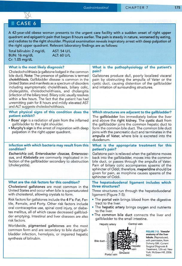
11 3. The patient s lower limb hematoma is located in the region of the femoral triangle. Which of the following structures would NOT be found in the femoral triangle? A. Inguinal lymph nodes B. Lateral circumflex femoral artery C. Femoral vein D. Femoral branch of the genitofemoral n. E. Pectineus m. 492N5 526N5
12
13 THORAX

14 1. What is the most likely cause of the patient s chest pain? A. Myocardial infarction B. Plueritis C. Pneumothorax D. Costochondritis E. Pericarditis

15 2. Which type of nerve innervates the intercostal space? A. Dorsal ramus B. Dorsal root C. Spinal nerve D. Ventral ramus E. Ventral root 174N5


16 3. Which of the following arteries is the LEAST likely to supply blood to the intercostal space? A. Subclavian a. B. Internal thoracic a. C. Inferior epigastric a. D. Musculophrenic a. E. Axillary a. 415N5 184N5
17

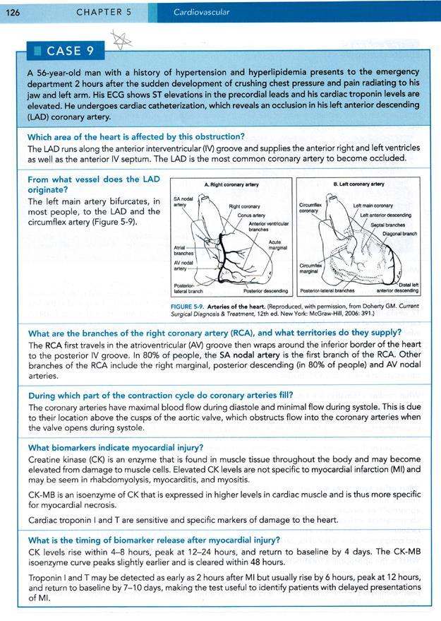
18 1. Which area of the heart is affected by this obstruction? A. Posterior interventricular septum B. SA node C. Anterior right ventricle D. Left atrium From which vessel does the LAD originate? 211N5

19 2. Which of the following is NOT typically a branch of the RCA? A. Circumflex branch B. SA nodal branch C. Right marginal branch D. Posterior interventricular branch (PDA)

20 3. During which part of the contraction cycle do coronary arteries fill? A. Late systole B. Isovolumetric ventricular contraction C. Diastole D. Early systole 218N5
21


22 1. Which of the following nerves would be LEAST at risk for injury by a stab wound to the thorax? A. Cardiac and pulmonary plexuses B. Recurrent laryngeal n. C. Phrenic n. D. Vagus n. E. Lesser splanchnic n. 220N5 225N5

23 2. Which of the following is NOT a major vein of the thorax? A. Azygos v. B. Cephalic v. C. Posterior intercostal vv. D. Internal thoracic v. E. Brachiocephalic v. 186N5

24 3. Lymph from which of the following regions drains to the thoracic duct? A. Right upper limb B. Right side of the head C. Right lower limb D. Right breast E. Right side of the neck
25
26 ABDOMEN

27 1. The bullet likely damaged retroperitoneal structures. Which of the following structures is NOT retroperitoneal? A. Suprarenal glands B. Ascending colon C. Rectum D. Sigmoid colon E. Aorta/IVC 2665

28 2. Which of the following organs does NOT supply the splenic vein? A. Stomach B. Cecum C. Rectum D. Pancreas E. Spleen 289N5
29 3. The bullet penetrated the anterolateral abdominal wall. Which series of layers is correctly listed, superficial to deep (not necessarily adjacent)? A. Scarpa s fascia, transversalis fascia, internal oblique m., parietal peritoneum B. Transversalis fascia, camper s fascia, transversus abdominis m., parietal peritoneum C. Camper's fascia, external abdominal oblique m., internal abdominal oblique m., transversalis fascia D. Camper s fascia, transversus abdominis m., internal abdominal oblique m., extraperitoneal fat E. Scarpa s fascia, internal abdominal oblique m., extraperitoneal fat, transversalis fascia


30 4. The bullet penetrated the skin. Which of the following is NOT a histological layer of the epidermis of thin skin? A. Stratum lucidum B. Stratum corneum C. Stratum basale D. Stratum spinosum E. Stratum granulosum
31
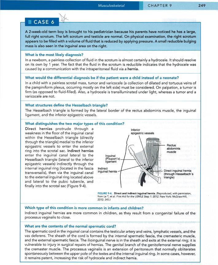
32 1. What is the most likely diagnosis? A. Tumor firm on palpation B. Varicocele dilated veins of the pampiniform plexus (left side) C. Hydrocele transilluminated under light (others are not)
33 2. Which of the following structures is NOT a boundary of the Hesselbach triangle? A. Rectus abdominis muscle B. Internal abdominal oblique muscle C. Inguinal ligament D. Inferior epigastric vessels

34 3. What is the primary distinguishing characteristic of the two major types of this condition? A. Indirect hernias are typically reducible, direct are not B. Indirect hernias are found only in children, direct in adults C. Indirect hernias do not involve intestines, direct do D. Indirect hernias pass through the inguinal canal, direct do not
 D. Genital branch of genitofemoral nerve E.")
35 4. Which of the following is NOT found within the normal spermatic cord? A. Artery to ductus deferens B. Pampiniform plexus of veins C. EAO aponeurosis (renamed) D. Genital branch of genitofemoral nerve E. Transversus abdominis muscle (renamed) 4.47GAS
36

37 Diagnosis? 1. Which of the following arteries is NOT a major branch of the aorta below the diaphragm? 2 1 A. Superior phrenic arteries B. Middle suprarenal arteries C. Gonadal arteries D. Inferior mesenteric artery Renal arteries , , GAS

38 Diagnosis? 2. In which of the following layers of a muscular artery would you find smooth muscle? A. Tunica intima B. Tunica media C. Tunica adventitia
39

40 1. Which organ is most likely injured in this case? A. Spleen B. Liver C. Pancreas D. Stomach E. Left kidney 4.166GAS2


41 2. What surface anatomy landmark corresponds to the location of the spleen? A. Posterior ribs 9-11 B. Inferior to costal margin C. Anterior ribs D. Transumbilical plane Spleen 4.168GAS2

42 3. Where is the most common site of referred pain in this injury? A. C1-C3 dermatomes B. C3-C5 dermatomes C. T2-T4 dermatomes D. T9-T11 dermatomes 159N5
43
44 HEAD & NECK

45 1. Care must be taken during thyroidectomy not to remove all functioning parathyroid gland tissue. How many parathyroid glands are there? A. One B. Two C. Three D. Four E. Five Embryologic origin? 76N5

46 2. Which one of the following muscles would you be LEAST likely to encounter during a routine thyroidectomy? A. Platysma B. Sternothyroid C. Thyrohyoid D. Sternyhyoid 28N5
47 3. Which nerve in this region is particularly at risk during thyroidectomy? A. Recurrent laryngeal n. B. Vagus n. C. Sympathetic trunk D. Superior laryngeal n. 76N5
48

49 1. Which nerve relays the painful sensation in this patient? A. Mandibular n. B. Ophthalmic n. C. Facial n. D. Maxillary n. What infectious agent? 2N5


50 2. What is the characteristic distribution pattern of this condition? A. Follows territory of cutaneous nerve B. Follows territory of myotome C. Follows territory of dermatome 76N5
51
52 MORE CASES: ANATOMY & EMBRYOLOGY
53
54
55
56
57
58
59
HUMAN HEART. Learn the following structures on the heart models.
 HUMAN HEART Learn the following structures on the heart models. The human heart has four chambers that consist of the right atrium, left atrium, right ventricle, and left ventricle. The atria are smaller
Day 5 Respiratory & Cardiovascular: Respiratory System
 Day 5 Respiratory & Cardiovascular: Respiratory System Be very careful not to damage the heart and lungs while separating the ribs! Analysis Questions-Respiratory & Cardiovascular Log into QUIA using your
The Thoracic wall including the diaphragm. Prof Oluwadiya KS
 The Thoracic wall including the diaphragm Prof Oluwadiya KS www.oluwadiya.com Components of the thoracic wall Skin Superficial fascia Chest wall muscles (see upper limb slides) Skeletal framework Intercostal
The posterior abdominal wall. Prof. Oluwadiya KS
 The posterior abdominal wall Prof. Oluwadiya KS www.oluwadiya.sitesled.com Posterior Abdominal Wall Lumbar vertebrae and discs. Muscles opsoas, quadratus lumborum, iliacus, transverse, abdominal wall
CARDIOVASCULAR DANIL HAMMOUDI.MD
 CARDIOVASCULAR DANIL HAMMOUDI.MD 18 Systemic Circulation Figure 19.19 Pulmonary Circulation Figure 19.18b 1. Thyroid gland 2. Trachea 3. Brachiocephalic 4. Common carotid 5. Internal jugular 6. Superior
VESSELS: GROSS ANATOMY
 ACTIVITY 10: VESSELS AND CIRCULATION OBJECTIVES: 1) How to get ready: Read Chapter 23, McKinley et al., Human Anatomy, 4e. All text references are for this textbook. 2) Observe and sketch histology slide
Artery 1 Head and Thoracic Arteries. Arrange the parts in the order blood flows through them.
 Artery 1 Head and Thoracic Arteries 1. Given the following parts of the aorta: 1. abdominal aorta 2. aortic arch 3. ascending aorta 4. thoracic aorta Arrange the parts in the order blood flows through
YOU MUST BRING GLOVES FOR THIS ACTIVITY
 ACTIVITY 10: VESSELS AND CIRCULATION OBJECTIVES: 1) How to get ready: Read Chapter 23, McKinley et al., Human Anatomy, 5e. All text references are for this textbook. 2) Observe and sketch histology slide
3 Circulatory Pathways
 40 Chapter 3 Circulatory Pathways Systemic Arteries -Arteries carry blood away from the heart to the various organs of the body. -The aorta is the longest artery in the body; it branches to give rise to
Misc Anatomy. Upper Limb! 2. Lower Limb! 5. Venous Drainage! Head & neck! 8
 Misc Anatomy Upper Limb! 2 Arteries!... 2 Veins!... 2 Spaces!... 4 Lower Limb! 5 Arteries!... 5 Venous Drainage!... 6 Spaces!... 7 Head & neck! 8 Artery!... 8 Ultrasound View for IJ CVL!... 8 Arteries
Group of students. - Rawan almujabili د. محمد المحتسب - 1 P a g e
 - 14 - Group of students - Rawan almujabili د. محمد المحتسب - 1 P a g e Nerves of the posterior abdominal wall The spinal cord gives off spinal nerves between the vertebrae. In the abdomen, through the
Dr. Weyrich G07: Superior and Posterior Mediastina. Reading: 1. Gray s Anatomy for Students, chapter 3
 Dr. Weyrich G07: Superior and Posterior Mediastina Reading: 1. Gray s Anatomy for Students, chapter 3 Objectives: 1. Subdivisions of mediastinum 2. Structures in Superior mediastinum 3. Structures in Posterior
Abdomen: Introduction. Prof. Oluwadiya KS
 Abdomen: Introduction Prof. Oluwadiya KS www.oluwadiya.com Abdominopelvic Cavity Abdominal Cavity Pelvic Cavity Extends from the inferior margin of the thorax to the superior margin of the pelvis and the
musculoskeletal system anatomy nerves of the lower limb 1 done by: dina sawadha & mohammad abukabeer
 musculoskeletal system anatomy nerves of the lower limb 1 done by: dina sawadha & mohammad abukabeer What is the importance of plexuses? plexuses provides us the advantage of a phenomenon called convergence
Figure ) The specific chamber of the heart that is indicated by letter A is called the. Diff: 1 Page Ref: 364
 The specific chamber of the heart that is indicated by letter A is called the. Diff: 1 Page Ref: 364") Essentials of Anatomy and Physiology, 9e (Marieb) Chapter 11 The Cardiovascular System Short Answer Figure 11.1 Using Figure 11.1, identify the following: 1) The Purkinje fibers are indicated by label.
Essentials of Anatomy and Physiology, 9e (Marieb) Chapter 11 The Cardiovascular System Short Answer Figure 11.1 Using Figure 11.1, identify the following: 1) The Purkinje fibers are indicated by label.
Gateway to the upper limb. An area of transition between the neck and the arm.
 Gateway to the upper limb An area of transition between the neck and the arm. Pyramidal space inferior to shoulder @ junction of arm & thorax Distribution center for the neurovascular structures that serve
ABDOMINAL WALL & RECTUS SHEATH
 ABDOMINAL WALL & RECTUS SHEATH Learning Objectives Describe the anatomy, innervation and functions of the muscles of the anterior, lateral and posterior abdominal walls. Discuss their functional relations
Abdominal muscles. Subinguinal hiatus and ingiunal canal. Femoral and adductor canals. Neurovascular system of the lower limb. Sándor Katz M.D.,Ph.D.
 Abdominal muscles. Subinguinal hiatus and ingiunal canal. Femoral and adductor canals. Neurovascular system of the lower limb. Sándor Katz M.D.,Ph.D. External oblique muscle Origin: outer surface of the
Academic Year 2017/2018. FALL SEMESTER DDS Program Monday: 5:45pm 7:15pm Thursday: 5:30pm- 7:00pm
 CURRICULUM FOR HUMAN ANATOMY COURSE LAB SESSIONS Academic Year 2017/2018 FALL SEMESTER DDS Program Monday: 5:45pm 7:15pm Thursday: 5:30pm- 7:00pm Category 1: Bones name, type, structure Joints: type, articular
Definition of anatomy 1 Questions 5
 Contents Chapter 1: Introduction 1 5 Definition of anatomy 1 Questions 5 Chapter 2: Skeletal System 6 74 Skeleton 6 Skeletal system 8 Bones of superior extremity 12 Articulated skeleton of hand 17 Clinical
The front of the thigh. Dr.Amjad shatarat
 The front of the thigh Femoral triangle (Scarpa s triangle) Is a triangular depressed area located in the upper part of the medial aspect of the thigh immediately below the inguinal ligament. Superiorly:
Cardiovascular System. Chapter 22
 Cardiovascular System Blood Vessels Chapter 22 Cardiac Contractions Cardiac cycle consists of alternate periods of contraction and relaxation Contraction is the systolic pressure Blood is ejected into
Upper limb Pectoral region & Axilla
 Upper limb Pectoral region & Axilla 黃敏銓 mchuang@ntu.edu.tw 1 Pectoral region Intercostal nerve Anterior branch of lateral cutaneous branch Lateral cutaneous branch Anterior cutaneous branch Anterior cutaneous
cardiac plexus is continuous with the coronary and no named branches pain from the heart and lungs
 Nerves of the Thoracic Region Nerve Source Branches Motor Sensory Notes cardiac plexus cardiac brs. of the vagus n. and cervical ; thoracic l nn. the heart and lungs cardiac, cervical cardiac, vagal vagus
Anatomy of thoracic wall
 Anatomy of thoracic wall Topographic Anatomy of the Thorax 1 Bones of Thoracic wall ribs 1-7"true" ribs -those which attach directly to the sternum true ribs actually attach to the sternum by means of
Anatomy: Know Your Abdomen
 Anatomy: Know Your Abdomen Glossary Abdomen - part of the body below the thorax (chest cavity); separated by the diaphragm. Anterior - towards the front of the body. For example, the umbilicus is anterior
OBJECTIVE: To obtain a fundamental knowledge of the root of the neck with respect to structure and function
 The root of the neck Jeff Dupree, Ph.D. e mail: jldupree@vcu.edu OBJECTIVE: To obtain a fundamental knowledge of the root of the neck with respect to structure and function READING ASSIGNMENT: Moore and
Brachial plexuses and axillary lymph nodes
 Brachial plexuses and axillary lymph nodes Introduction about nervous system nervous system central nervous system periphral nervous system brain spinal cord 31 pairs of spinal nerves 12 paris of cranial
thoracic cage inlet and outlet landmarks of the anterior chest wall muscles of the thoracic wall sternum joints ribs intercostal spaces diaphragm
 Thoracic Wall Lecture Objectives Describe the shape and outline of the thoracic cage including inlet and outlet. Describe the anatomical landmarks of the anterior chest wall. List various structures making
GI module Lecture: 9 د. عصام طارق. Objectives:
 GI module Lecture: 9 د. عصام طارق Objectives: To list structures forming posterior abdominal wall. To follow aorta & its main branches. To describe IVC & its main tributaries. To list nerves of posterior
Identify the lines used in anatomical surface descriptions of the thorax. median line mid-axillary line mid-clavicular line
 L 14 A B O R A T O R Y Thorax THORACIC WALL Identify the lines used in anatomical surface descriptions of the thorax. median line mid-axillary line mid-clavicular line Identify the surface landmarks of
G24: Shoulder and Axilla
 G24: Shoulder and Axilla Syllabus - Pg. 2 ANAT 6010- Medical Gross Anatomy David A. Morton, Ph.D. Objectives Upper limb Systemically: Bones (joints) Muscles Nerves Vessels (arteries/veins) Fascial compartments
Netter's Anatomy Flash Cards Section 4 List 4 th Edition
 Netter's Anatomy Flash Cards Section 4 List 4 th Edition https://www.memrise.com/course/1577335/ Section 4 Abdomen (31 cards) Plate 4-1 Bony Framework of Abdomen 1.1 Costal cartilages 1.2 Iliac crest 1.3
Benha University. Faculty of Medicine. Anatomy Department Course code (MED 0701) Model answer of Anatomy examination. (Abdomen,Pelvis and Thorax)
 Model answer of Anatomy examination. (Abdomen,Pelvis and Thorax)") 1 Benha University Faculty of Medicine Anatomy Department Course code (MED 0701) Model answer of Anatomy examination (Abdomen,Pelvis and Thorax) 1 st year 2 nd term Date :18 /5 /2013 2 I-Short account
1 Benha University Faculty of Medicine Anatomy Department Course code (MED 0701) Model answer of Anatomy examination (Abdomen,Pelvis and Thorax) 1 st year 2 nd term Date :18 /5 /2013 2 I-Short account
3 Mohammad Al-Mohtasib Areej Mosleh
 3 Mohammad Al-Mohtasib Areej Mosleh ***Muscles Connecting the Upper Limb to the Vertebral Column 1.Trapezius Muscle ***The first muscle on the back is trapezius muscle, it s called so according
M. Al-Mohtaseb. Tala Saleh. Faisal Nimri
 4 5 M. Al-Mohtaseb Tala Saleh Faisal Nimri Inguinal Hernia - An abdominal hernia is the protrusion of part of the abdominal content beyond the normal confines of the abdominal wall through weak points
DESCRIPTION: This is the part of the trunk, which is located between the root of the neck and the superior border of the abdominal region.
 1 THE THORACIC REGION DESCRIPTION: This is the part of the trunk, which is located between the root of the neck and the superior border of the abdominal region. SHAPE : T It has the shape of a truncated
ANATOMY. Schedule for 2014/2015 academic school year (2x15 weeks)
") ANATOMY Schedule for 2014/2015 academic school year (2x15 weeks) SEMESTER LECTURES LAB CLASSES SEMINARS TOTAL FIRST 4 hours (2+2) 4 hours (2+2) 1 hour 135 hours SECOND 3 hours 4 hours (2+2) 2 hours 135
ANATOMY Schedule for 2014/2015 academic school year (2x15 weeks) SEMESTER LECTURES LAB CLASSES SEMINARS TOTAL FIRST 4 hours (2+2) 4 hours (2+2) 1 hour 135 hours SECOND 3 hours 4 hours (2+2) 2 hours 135
Anatomy of the renal system. Professor Nawfal K. Al-Hadithi
 Anatomy of the renal system Professor Nawfal K. Al-Hadithi Objectives To describe the posterior abdominal wall To identify the main anatomical landmarks of the kidneys & ureters To describe the suprarenal
THE GOOFY ANATOMIST QUIZZES
 THE GOOFY ANATOMIST QUIZZES 5. NERVES Q1. Which of the following classifications of the nervous systems is correct? A. The autonomic nervous system is composed of the brain, cranial nerves and spinal nerves.
General Anatomy p. 1 Organization of the Human Body p. 1 Skeleton of the Human Body p. 4 Ossification of the Bones p. 6 Bone Structure p. 8 Joints p.
 General Anatomy p. 1 Organization of the Human Body p. 1 Skeleton of the Human Body p. 4 Ossification of the Bones p. 6 Bone Structure p. 8 Joints p. 10 Principal Joints (Immovable) p. 12 Synovial Joints
Lab Activity 25. Blood Vessels & Circulation. Portland Community College BI 232
 Lab Activity 25 Blood Vessels & Circulation Portland Community College BI 232 Artery and Vein Histology Walls have 3 layers: Tunica intima Tunica media Tunica externa 2 Tunica Intima Is the innermost layer
Synapse Homework. Back page last question not counted. 4 pts total, each question worth 0.18pts. 26/34 students answered correctly!
 Synapse Homework Back page last question not counted 26/34 students answered correctly! 4 pts total, each question worth 0.18pts Business TASS hours extended! MWF 1-2pm, Willamette 204 T and Th 9:30-10:30am,
Which Artery am I? I am one of two smaller arteries that arise from the brachial. I supply blood to the medial aspect of the forearm.
 I am one of two smaller arteries that arise from the brachial. I supply blood to the medial aspect of the forearm. A. I supply blood to the head and neck. I am large and will branch into two smaller arteries.
Mediastinum It is a thick movable partition between the two pleural sacs & lungs. It contains all the structures which lie
 Dr Jamila EL medany OBJECTIVES At the end of the lecture, students should be able to: Define the Mediastinum. Differentiate between the divisions of the mediastinum. List the boundaries and contents of
Anatomy of the Thorax
 Anatomy of the Thorax A) THE THORACIC WALL Boundaries Posteriorly by the thoracic part of the vertebral column Anteriorly by the sternum and costal cartilages Laterally by the ribs and intercostal spaces
Femoral Triangle and Adductor Canal. Dr. Heba Kalbouneh Associate Professor of Anatomy and Histology
 Femoral Triangle and Adductor Canal Dr. Heba Kalbouneh Associate Professor of Anatomy and Histology Femoral Triangle and Adductor Canal Femoral triangle Is a triangular depressed area located in the upper
Inguinal Canal. It is an oblique passage through the lower part of the anterior abdominal wall. Present in both sexes
 Inguinal canal Inguinal Canal It is an oblique passage through the lower part of the anterior abdominal wall Present in both sexes It allows structures to pass to and from the testis to the abdomen in
GI anatomy Lecture: 2 د. عصام طارق
 GI anatomy Lecture: 2 د. عصام طارق Objectives: To define rectus sheath. To describe anatomy of inguinal canal. To relates types of inguinal hernia to the region. To explore spermatic cord. Rectus Abdominis
1 Right & left Hepatic ducts Gastric Impression of spleen
 Pancreatic Model 1 Right & left Hepatic ducts 14 Gastric Impression of spleen 2 Common hepatic duct 15 Renal Impression of spleen 3 Cystic Duct 16 Colic Impression of spleen 4 Common Bile Duct 17 Splenic
BLOCK IV: OFFICIAL BODY PARTS LIST FOR ANTERIOR ABDOMINAL WALL AND ABDOMINAL CONTENTS
 BLOCK IV: OFFICIAL BODY PARTS LIST FOR ANTERIOR ABDOMINAL WALL AND ABDOMINAL CONTENTS External oblique muscle Muscular portion Aponeurotic portion Superficial inguinal ring Lateral (inferior) crus Medial
Borders of the Abdomen
 Abdominal wall Borders of the Abdomen Abdomen is the region of the trunk that lies between the diaphragm above and the inlet of the pelvis below Borders Superior: Costal cartilages 7-12. Xiphoid process:
REVIEW SHEET Anatomy of Blood Vessels
 REVIEW SHEET Anatomy of Blood Vessels Name LabTime/Date Microscopic Structure of the Blood Vessels 1. Cross-sectional views of an aftery of a vein are shown here. ldentify each; on the lines to the sides,
2. List the 8 pelvic spaces: list one procedure or dissection which involves entering that space.
 Name: Anatomy Quiz: Pre / Post 1. In making a pfannensteil incision you would traverse through the following layers: a) Skin, Camper s fascia, Scarpa s fascia, external oblique aponeurosis, internal oblique
Cardiovascular System
 Cardiovascular System I. Structure of the Heart A. Average adult heart is 14 cm long and 9 cm wide. B. Lies in the mediastinum. C. Enclosed in the pericardium. 1. Fibrous pericardium- Outer, tough connective
BY DR NOMAN ULLAH WAZIR
 BY DR NOMAN ULLAH WAZIR The stomach (from ancient Greek word stomachos, stoma means mouth) is a muscular, hollow and the most dilated part of the GIT. It starts from the point where esophagus ends. It
KARPAGAM FACULTY OF MEDICAL SCIENCES & RESEARCH DEPARTMENT OF ANATOMY TEACHING SCHEDULE GENERAL ANATOMY I - MBBS
 TEACHING SCHEDULE GENERAL ANATOMY I - MBBS S.NO DATE & TIME 1. 3.10.16 Monday 2. 4.10.16 Tuesday 8 10 3. 5. 10.16 Wednesday 4. 11 1pm 5. 6.10.16 Thursday 8 10 6. 6.10.16 Thursday 12 1pm 7. 7.10.16 Friday
First BHMS Anatomy Question Papers Calicut University
 First BHMS Anatomy Question Papers Calicut University 1996-2000 FIRST B.H.M.S. DEGREE EXAMINATION, DECEMBER 1996 Time: Three Hours Maximum: 100 Marks Answer any five questions. Draw diagrams wherever needed.
The Cardiovascular System
 PowerPoint Lecture Slide Presentation by Patty Bostwick-Taylor, Florence-Darlington Technical College The Cardiovascular System 11PART B The Heart: Cardiac Output Cardiac output (CO) Amount of blood pumped
أحمد رواجبة- محمود الحربي- أحمد السالمان-
 -6 أحمد رواجبة- محمود الحربي- أحمد السالمان- 1 P a g e The Male Reproductive System The male genital system structures are divided into: Internal structures: 1- Prostate 3-Ejaculatory ducts External structures:
حسام أبو عوض. -Dr. Mohammad Muhtasib. 1 P a g e
 5 حسام أبو عوض - -Dr. Mohammad Muhtasib 1 P a g e There are two types of inguinal hernia: direct and indirect. Hernia: protrusion of the small intestine or the greater omentum of the intra-abdominal organs
STERNUM. Lies in the midline of the anterior chest wall It is a flat bone Divides into three parts:
 STERNUM Lies in the midline of the anterior chest wall It is a flat bone Divides into three parts: 1-Manubrium sterni 2-Body of the sternum 3- Xiphoid process The body of the sternum articulates above
Note : I put the sheet's info within the slides to easily understand this lecture Done by : Zaid Al-Ghnaneem
 Note : I put the sheet's info within the slides to easily understand this lecture Done by : Zaid Al-Ghnaneem Thoracic Wall Lecture Objectives Describe the shape and outline of the thoracic cage including
[ANATOMY #12] April 28, 2013
![[ANATOMY #12] April 28, 2013](/thumbs/86/93473883.jpg "[ANATOMY #12] April 28, 2013") Sympathetic chain : Sympathetic chain is each of the pair of ganglionated longitudinal cords of the sympathetic nervous system; extend from level of atlas (base of skull) till coccyx. It is paravertebral
Sympathetic chain : Sympathetic chain is each of the pair of ganglionated longitudinal cords of the sympathetic nervous system; extend from level of atlas (base of skull) till coccyx. It is paravertebral
Mediastinum and pericardium
 Mediastinum and pericardium Prof. Abdulameer Al-Nuaimi E-mail: a.al-nuaimi@sheffield.ac.uk E. mail: abdulameerh@yahoo.com The mediastinum: is the central compartment of the thoracic cavity surrounded by
The Heart & Pericardium Dr. Rakesh Kumar Verma Assistant Professor Department of Anatomy KGMU UP Lucknow
 The Heart & Pericardium Dr. Rakesh Kumar Verma Assistant Professor Department of Anatomy KGMU UP Lucknow Fibrous skeleton Dense fibrous connective tissue forms a structural foundation around AV & arterial
Copy Right- Hongqi ZHANG-Department of Anatomy-Fudan University. Systematic Anatomy. Angiology Part 4. Veins. Dr.Hongqi Zhang ( 张红旗 )
") Systematic Anatomy Angiology Part 4 Veins Dr.Hongqi Zhang ( 张红旗 ) Email: zhanghq58@126.com 1 General introduction of the veins Vessel which return the blood back to atrium No pulsation,veneous blood, metabolic
Systematic Anatomy Angiology Part 4 Veins Dr.Hongqi Zhang ( 张红旗 ) Email: zhanghq58@126.com 1 General introduction of the veins Vessel which return the blood back to atrium No pulsation,veneous blood, metabolic
Cat Dissection. Muscular Labs
 Cat Dissection Muscular Labs Tibialis anterior External oblique Pectroalis minor Gastrocnemius Sartorius Pectoralis major Gastrocnemius Semitendinosis Sartorius External oblique Trapezius Latissimus dorsi
Chapter 5: Other mediastinal structures. The Large Arteries. The Aorta. Ascending aorta
 Chapter 5: Other mediastinal structures The Large Arteries The Aorta The aorta is the main arterial trunk of the systemic circulation and in the healthy state its wall contain a large amount of yellow
Large veins of the thorax Brachiocephalic veins
 Large veins of the thorax Brachiocephalic veins Right brachiocephalic vein: formed at the root of the neck by the union of the right subclavian & the right internal jugular veins. Left brachiocephalic
Biology 323 Human Anatomy for Biology Majors Lecture 11 Dr. Stuart S. Sumida. Peripheral Circulation
 Biology 323 Human Anatomy for Biology Majors Lecture 11 Dr. Stuart S. Sumida Peripheral Circulation Structures of the Splanchnopleure: receive unpaired vessels of the abdominal aorta. Structures of the
The abdominal Esophagus, Stomach and the Duodenum. Prof. Oluwadiya KS
 The abdominal Esophagus, Stomach and the Duodenum Prof. Oluwadiya KS www.oluwadiya.com Viscera of the abdomen Abdominal esophagus: Terminal part of the esophagus The stomach Intestines: Small and Large
THE DESCENDING THORACIC AORTA
 Intercostal Arteries and Veins Each intercostal space contains a large single posterior intercostal artery and two small anterior intercostal arteries. The anterior intercostal arteries of the lower spaces
Bio& 242, Unit 3/ Lab 4 Blood Vessels, Lymphatic System and Blood Pressure G. Blevins/ G. Brady Summer 2009
 Bio& 242, Unit 3/ Lab 4 Blood Vessels, Lymphatic System and Blood Pressure G. Blevins/ G. Brady Summer 2009 Major Arteries and for arteries and veins with common names your answer must include either artery
IP UNIVERSITATEA DE STAT DE MEDICINĂ ŞI FARMACIE NICOLAE TESTEMIŢANU FACULTATEA MEDICINĂ nr. 1 CATEDRA DE ANATOMIE A OMULUI
 IP UNIVERSITATEA DE STAT DE MEDICINĂ ŞI FARMACIE NICOLAE TESTEMIŢANU FACULTATEA MEDICINĂ nr. 1 CATEDRA DE ANATOMIE A OMULUI MD 2004, Chişinău, Republica Moldova, bd. Ştefan cel Mare, 192, tel.: +373.22.205349
STRUCTURAL BASIS OF MEDICAL PRACTICE EXAMINATION 3. October 16, 2015
 STRUCTURAL BASIS OF MEDICAL PRACTICE EXAMINATION 3 October 16, 2015 PART l. Answer in the space provided. (12 pts) 1. Identify the structures. (2 pts) A. B. A B C. D. C D 2. Identify the structures. (2
Contents. Pig Dissection. Contents. External Features Sex Determination Mouth and Maxillary Nerve Muscles Index Internal Systems Index
 Pig Dissection External Features Sex Determination Mouth and Maxillary Nerve Muscles Index Internal Systems Index External features Sex determination Male Female Male to External anatomy 1. Pinna 2. External
slide 23 The lobes in the right and left lungs are divided into segments,which called bronchopulmonary segments
 Done By : Rahmeh Alsukkar Date : 26 /10/2017 slide 23 The lobes in the right and left lungs are divided into segments,which called bronchopulmonary segments Each segmental bronchus passes to a structurally
Diaphragm and intercostal muscles. Dr. Heba Kalbouneh Associate Professor of Anatomy and Histology
 Diaphragm and intercostal muscles Dr. Heba Kalbouneh Associate Professor of Anatomy and Histology Skeletal System Adult Human contains 206 Bones 2 parts: Axial skeleton (axis): Skull, Vertebral column,
c) What is the name of RBC (erythrocyte) formation? Where do blood cells form?
 What is the name of RBC (erythrocyte) formation? Where do blood cells form?") UNIT 6: CARDIOVASCULAR SYSTEM 1) List the three general functions of BLOOD. REVIEW QUESTIONS Blood 2) a) What are the three formed elements /cellular elements in blood? b) Describe the composition of the
UNIT 6: CARDIOVASCULAR SYSTEM 1) List the three general functions of BLOOD. REVIEW QUESTIONS Blood 2) a) What are the three formed elements /cellular elements in blood? b) Describe the composition of the
Spinal nerves and cervical plexus Prof. Abdulameer Al Nuaimi. E mail: a.al E. mail:
 Spinal nerves and cervical plexus Prof. Abdulameer Al Nuaimi E mail: a.al nuaimi@sheffield.ac.uk E. mail: abdulameerh@yahoo.com Branches of ophthalmic artery Muscles of face A spinal nerve Spinal
Breathing. Heart Rate
 Breathing Heart Rate Inspiration Expiration (Pressos not Stretched) Heart Rate increases with inspiration (Pressos Stretched) Heart Rate decreases with expiration Upside Down (Pressos Stretched) HR Decreases
Anatomy of the Large Intestine
 Large intestine Anatomy of the Large Intestine 2 Large Intestine Extends from ileocecal valve to anus Length = 1.5-2.5m = 5 feet Regions Cecum = 2.5-3 inch Appendix= 3-5 inch Colon Ascending= 5 inch Transverse=
Anatomy and Physiology, Spring 2015 Exam II: Form A April 9, Name Student Number
 Anatomy and Physiology, Spring 2015 Exam II: Form A April 9, 2015 Name Student Number For Questions 1 2 refer to the following table. 1 Ventricular pressure is greater than aortic 6 AV valve is open 2
Right lung. -fissures:
 -Right lung is shorter and wider because it is compressed by the right copula of the diaphragm by the live.. 2 fissure, 3 lobes.. hilum : 2 bronchi ( ep-arterial, hyp-arterial ), one artery mediastinal
THE THORACIC WALL. Boundaries Posteriorly by the thoracic part of the vertebral column. Anteriorly by the sternum and costal cartilages
 THE THORACIC WALL Boundaries Posteriorly by the thoracic part of the vertebral column Anteriorly by the sternum and costal cartilages Laterally by the ribs and intercostal spaces Superiorly by the suprapleural
Lab 9 Abdomen MUSCLES
 Lab 9 Abdomen MUSCLES External abdominal oblique continuous with the external intercostal muscle; its fibers point in a caudal direction as it moves anteriorly until it inserts on the linea alba via its
CARDIOVASCULAR SYSTEM
 CARDIOVASCULAR SYSTEM CARDIAC SYSTEM TWO TYPES OF CIRCULATION Systemic system delivers blood to ALL body cells and carries away waste. The red blood cells use hemoglobin to carry oxygen to the cells Pulmonary
Nerves on the Posterior Abdominal Wall
 Nerves on the Posterior Abdominal Wall Lumbar Plexus The lumbar plexus, which is one of the main nervous pathways supplying the lower limb, is formed in the psoasmuscle from the anterior ramiof the upper
ANATOMY OF PELVICAYCEAL SYSTEM -DR. RAHUL BEVARA
 1 ANATOMY OF PELVICAYCEAL SYSTEM -DR. RAHUL BEVARA 2 KIDNEY:ANATOMY OVERVIEW Kidneys are retroperitoneal, in posterior abdominal region, extending from T12 L3 Bean-shaped Right kidney is lower than left
Lecturer: Ms DS Pillay ROOM 2P24 25 February 2013
 Lecturer: Ms DS Pillay ROOM 2P24 25 February 2013 Thoracic Wall Consists of thoracic cage Muscle Fascia Thoracic Cavity 3 Compartments of the Thorax (Great Vessels) (Heart) Superior thoracic aperture
PERIPHERAL NERVOUS SYSTEM
 CHAPTER 13 PERIPHERAL NERVOUS SYSTEM Functional division of nervous system = afferent info to the CNS ascending spinal cord = efferent info from CNS descending spinal cord somatic skin, muscles visceral
Internal abdominal wall and inguinal region. Mathew Wedel, 2015
 Internal abdominal wall and inguinal region Mathew Wedel, 2015 gut tube umbilicus gut tube dorsal mesentery visceral peritoneum gut tube FOREGUT dorsal mesentery parietal peritoneum MIDGUT & HINDGUT gut
Chapter 14. The Nervous System. The Spinal Cord and Spinal Nerves. Lecture Presentation by Steven Bassett Southeast Community College
 Chapter 14 The Nervous System The Spinal Cord and Spinal Nerves Lecture Presentation by Steven Bassett Southeast Community College Introduction The Central Nervous System (CNS) consists of: The spinal
ANATYOMY OF The thigh
 ANATYOMY OF The thigh 1- Lateral cutaneous nerve of the thigh Ι) Skin of the thigh Anterior view 2- Femoral branch of the genitofemoral nerve 5- Intermediate cutaneous nerve of the thigh 1, 2 and 3 are
The Human Body. Lesson Goal. Lesson Objectives 9/10/2012. Provide a brief overview of body systems, anatomy, physiology, and topographic anatomy
 The Human Body Lesson Goal Provide a brief overview of body systems, anatomy, physiology, and topographic anatomy Medial Lateral Proximal Distal Superior Inferior Anterior Lesson Objectives Explain the
Adductor canal (Subsartorial) or Hunter s canal
 or Hunter s canal") Adductor canal (Subsartorial) or Hunter s canal John Hunter described the exposure and ligation of the femoral artery in this canal for aneurysm of the popliteal artery; this method has the advantage that
Adductor canal (Subsartorial) or Hunter s canal John Hunter described the exposure and ligation of the femoral artery in this canal for aneurysm of the popliteal artery; this method has the advantage that
Year 2004 Paper one: Questions supplied by Megan
 QUESTION 47 A 58yo man is noted to have a right foot drop three days following a right total hip replacement. On examination there is weakness of right ankle dorsiflexion and toe extension (grade 4/5).
In the Last Three Lectures We Already Discussed the Importance of the Thoracic Cage.
 -This Lecture Will Revise what we took in the last three lectures and will introduce the concept of the chest cavity ( Thoracic Cavity ) In the Last Three Lectures We Already Discussed the Importance of
Lower Limb Nerves. Clinical Anatomy
 Lower Limb Nerves Clinical Anatomy Lumbar Plexus Ventral rami L1 L4 Supplies: Abdominal wall External genitalia Anteromedial thigh Major nerves.. Lumbar Plexus Nerves relation to psoas m. : Obturator n.