Biology 323 Human Anatomy for Biology Majors Lecture 11 Dr. Stuart S. Sumida. Peripheral Circulation
|
|
|
- Corey Miller
- 5 years ago
- Views:
Transcription
1 Biology 323 Human Anatomy for Biology Majors Lecture 11 Dr. Stuart S. Sumida Peripheral Circulation
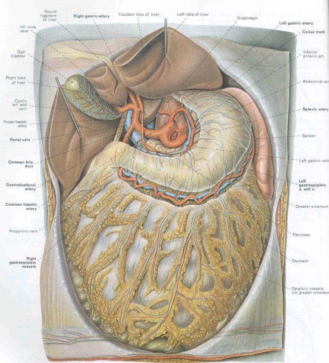
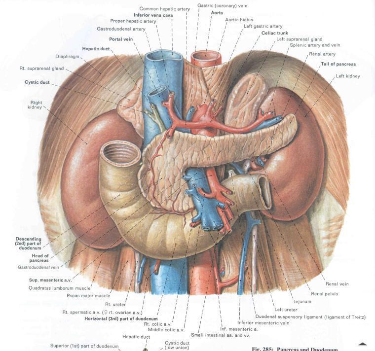
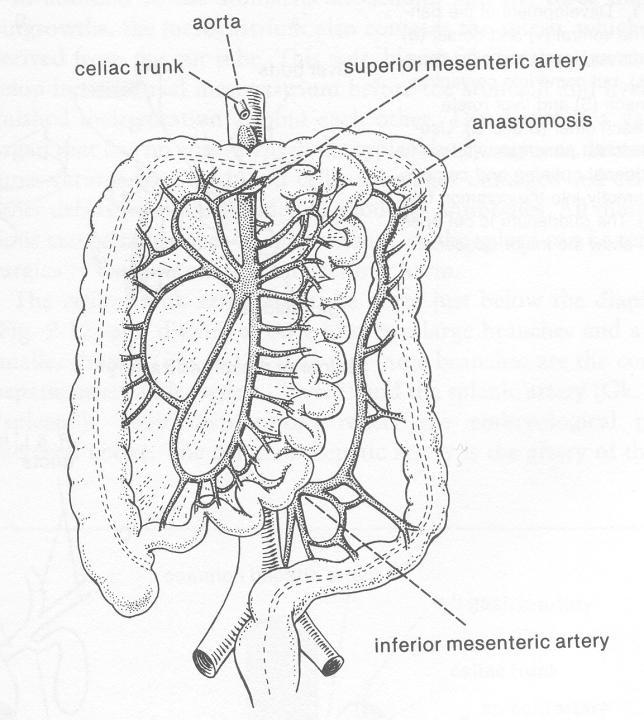
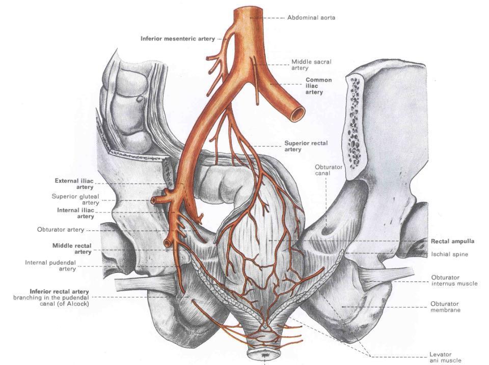
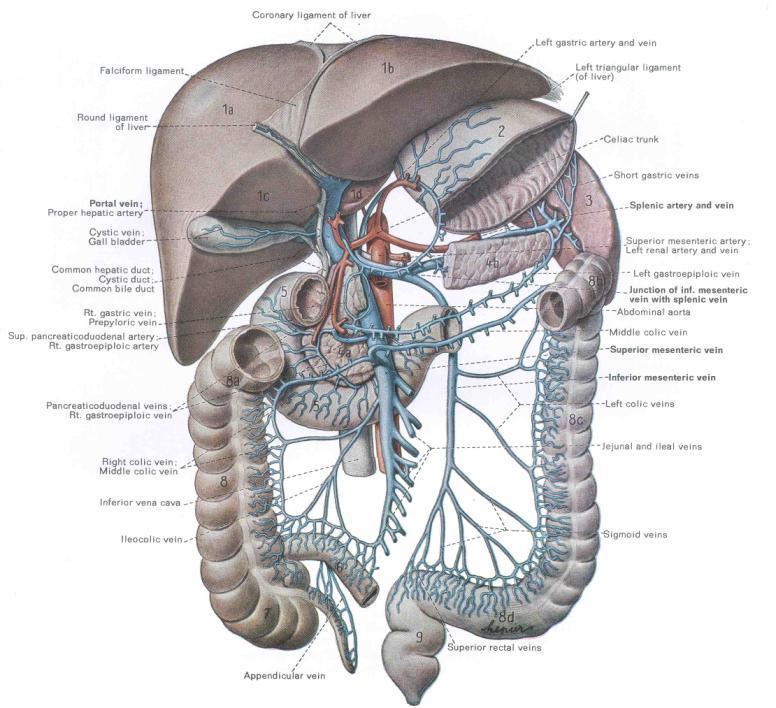
2 Structures of the Splanchnopleure: receive unpaired vessels of the abdominal aorta. Structures of the Somatopleure: receive PAIRED vessels of the abdominal aorta.
3 Structures of the Splanchnopleure: receive unpaired vessels of the abdominal aorta.
4
5
6
7
8
9
10 Structures of the Somatopleure: receive paired vessels of the aorta.
11
12

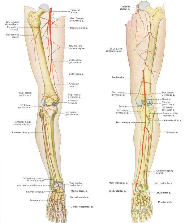
13 Major Arteries of Leg: External Iliac Artery Femoral Artery Profunda femoris Medial femoral circumflex Lateral femoral circumflex Popliteal and Saphenous AA. Descending branch lateral femoral circumflex
 medial circumflex femoral artery. 2) ascending branch of the lateral circumflex femoral artery. 3) inferior gluteal artery.")
14 Arteries & nerves of gluteal region Trochanteric Anastomosis anastomotic ring of arteries found in the trochanteric fossa and around the neck of the femur. Formed by the union of branches from: 1) medial circumflex femoral artery. 2) ascending branch of the lateral circumflex femoral artery. 3) inferior gluteal artery. 4) superior gluteal artery.

15 Femoral triangle / RELATIONS Deep contents Femoral a. & v. surrounded by femoral sheath Profunda femoris a. principal artery of thigh Lat and med. femoral circumflex aa. Deep external pudendal a. Femoral n. Anterior view A few deeper lymph nodes --

16 Femoral triangle / RELATIONS Anterior view

17 Femoral triangle / PRINCIPAL VASCULATURE OF THIGH

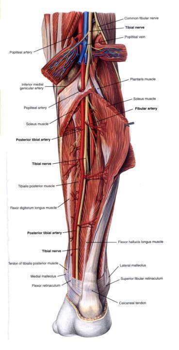
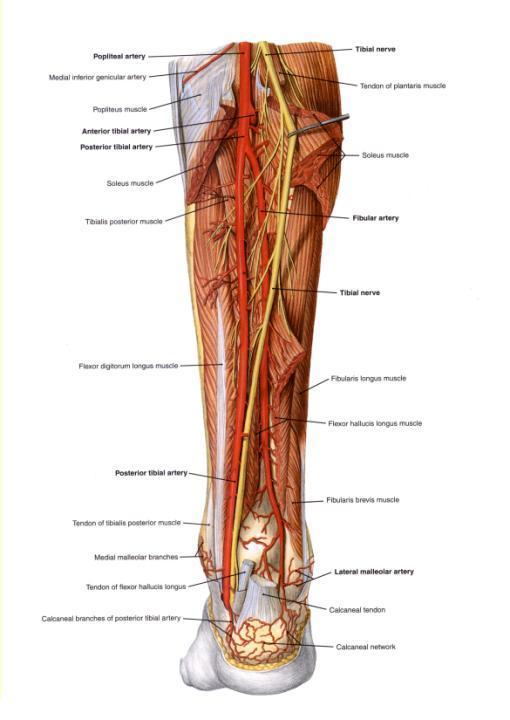
18 Region of Popliteal Fossa Femoral Popliteal Posterior Anterior Tibial A. Tibial A. Peroneal A. Anastomosing branches of Popliteal Artery: Medial and lateral superior geniculate aa. Medial and lateral sural aa. Medial and lateral inferior geniculate aa.
19

20 Tibialis Anterior Artery running in anterior compartment next to tibialis anterior muscle.

21 Anterior Tibial Artery Dorsalis pedis artery Deep plantar artery
22

23 Anastomosis of Internal Thoracic Artery with External Iliac Artery

24 Sublcavian AA. pass into each arm, becomin Axillary A. past clavicle. Subclavian Branches: Vertebral A. Internal Thoracic A. Thyrocervical trunk
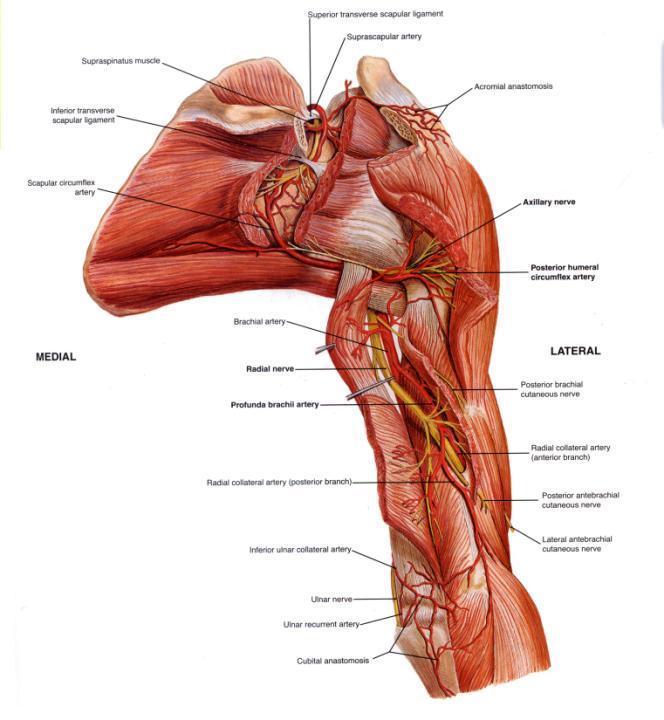
25 What is the Axilla? A region (the axillary space) associated with the armpit. It actually begins around the cervicoaxillary canal, at the edge of the first rib. It continues to the armpit, with the bottom being the axillary fascia. (remember? The lower attachment of the clavipectoral membrane?) It has musculoskeletal boundaries that are lateral, medial, anterior and posterior.

26 AXILLARY SPACE
27 Walls of the axillary space Medial Serratus anterior muscle Lateral Intertubercular sulcus. Anterior Pectoralis major and minor MM. Posterior Scapula with subscapularis M.; in places, latisimus dorsi M. and teres major M. Apex clavicle. Base Axillary fascia.
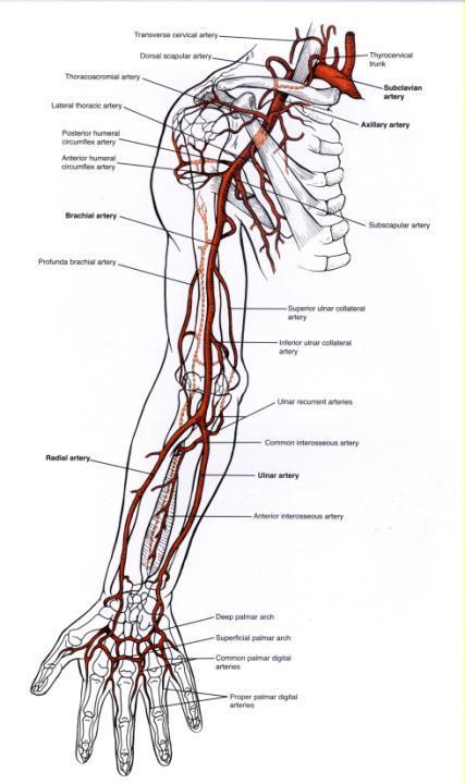
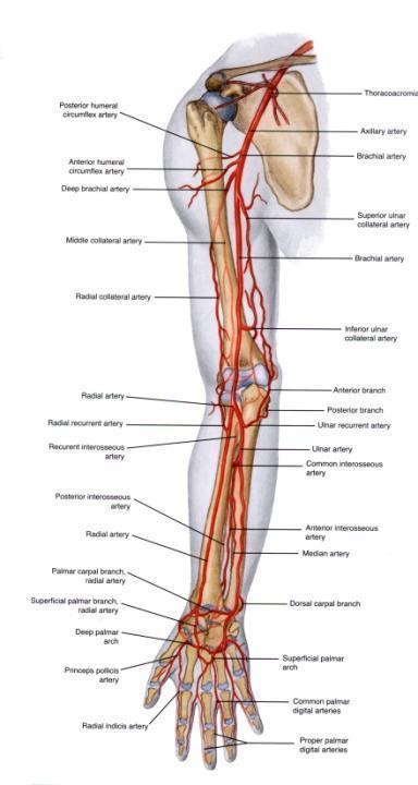
28 MUSCLES Subscapularis M Latissimus dorsi M Major structures inside: Axillary sheath and contents! Teres major M Pectoralis major M Pectoralis minor M Most of the rest of the space is adipose tissue. Serratus anterior M
29 Axillary sheath Derived, at least in part, from anterior and middle scalene muscle fascia. Covers over a series of contents: Axillary artery Axillary vein Brachial plexus and nerves derived from it. The axillary sheath is just the fascia surrounding these structures. You will open it up in lab to see them.
30 Axillary Artery: divided into three parts Subclavian A. Part 1 (proximal) one branch Brachial A. Part 2 (intermediate) two branches. Part 3 (distal) three branches.
31 Axillary Artery: First Part From lateral border of 1 st rib to medial border of Pectoralis Major M. Named Branch: Supreme Thoracic A. (to external thoracic body wall) Supplies blood to first and second intercostal spaces
32 Axillary Artery: Second part Deep to the pectoralis minor M. Thoracoacromial trunk Branches to: Clavicular area Pectoralis region Acromion of Scapula Deltoid Muscle. Lateral Thoracic Artery Bbr. to Serratus Ant. M.
33 Axillary Artery: third part Lateral border of Pectoralis minor M. to lateral border of Teres major M. Posterior circumflex humeral A. Anterior circumflex humeral A. Subscapular A.: Branches: 1. Circumflex scapular A. (to multiple muscles associated with the scapula) 2. Thoracodorsal A. (to Latissimus dorsi M.) How it will look in lab

34 Subscapular A. Thoracoacromial A. Lateral thoracic A. Supreme thoracic A. Post. Circumflex humoral A. Ant. Circumflex humoral A. Note, there is a broad anastamosis of the entire scapular region including circumflex humorals, subscapular, dorsal scapular, and suprascapular AA.
35
36 Arteries of Proximal Arm The arterial pattern has one major vessel, with several important branches, which can supply muscles: Deep brachial A. to posterior compartment (branches to medial collateral and radial collateral AA). Superior ulnar collateral A. Inferior ulnar collateral A. Note, many muscles are supplied directly by unnamed muscular branches. Do not even think of giving all the vessels you see a distinct name.
37 Axillary A. Brachial A. Radial collateral A. (a branch of the deep brachial A.) Not seen, middle collateral A., another branch of the deep brachial A. Deep brachial A. Superior ulnar collateral A. Inferior ulnar collateral A.

38 Collateral anastomosis around elbow.


39 Antebrachium Major Arteries Superficial Deeper Dissection

40 Antebrachium Major Arteries (Deepest Dissection) Brachial Artery Radial Artery Ulnar Artery Common Interosseous A. Anterior Posterior Interosseus Interosseus Artery Artery

41 Manus Major Arteries of Palmar Aspect Ulnar & Radial AA Superficial Palmar Arch Common Palmar Digital AA.
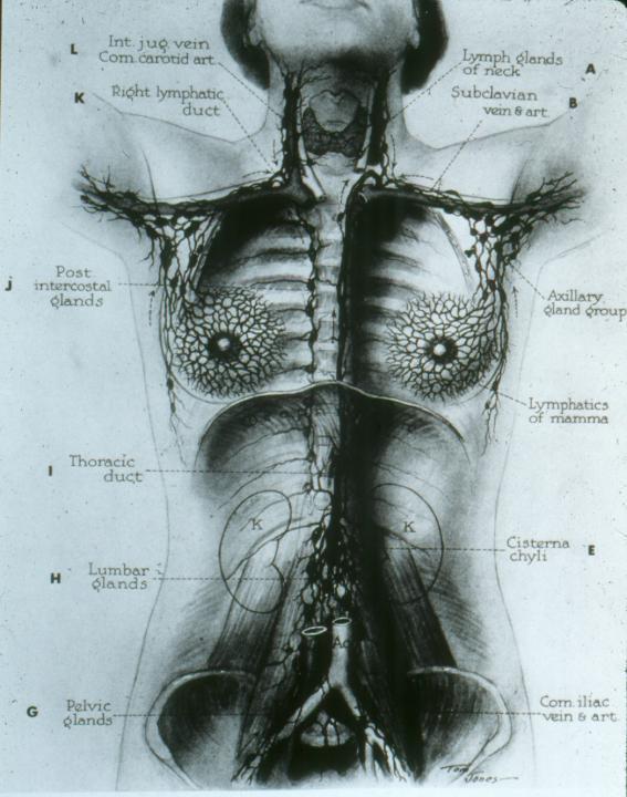
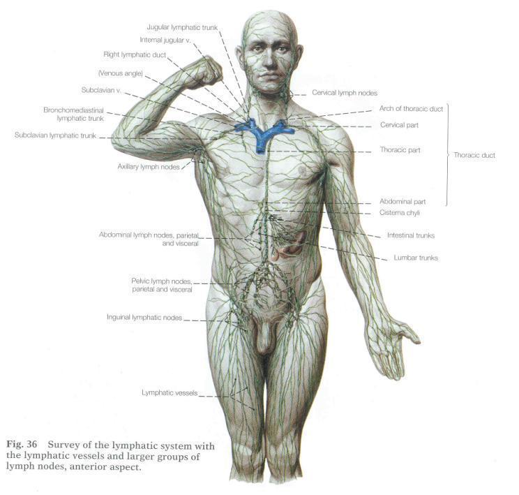
42 Lymphatic System
43
44
45 STRUCTURAL & MORPHOLOGICAL ORGANIZATION Generally run parallel to arteries and/or veins. Over three-fourths of the body dump into the thoracic duct which runs on the inside or the dorsal body wall (retroperitoneal).
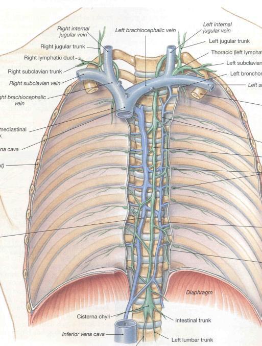
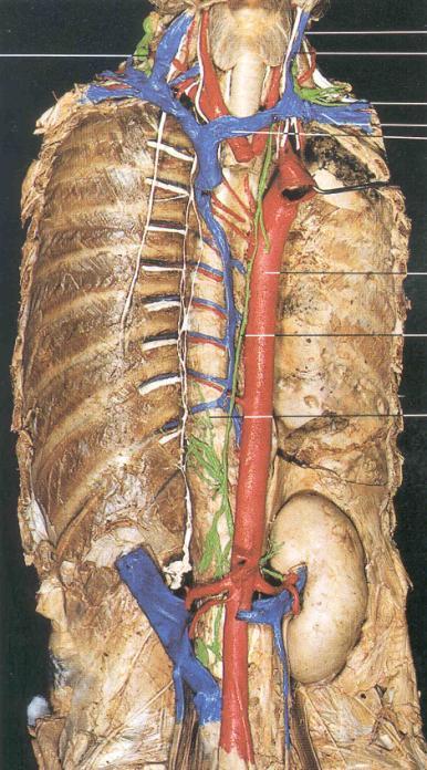
46 THORACIC DUCT Begins as a loosely dilated sac and connections in the abdomen called the CYSTERNA CHYLI. Drains both legs, and left side of body. Goes through thorax, receives tributaries from: LEFT SUBCLAVIAN TRUNK (from left arm) and LEFT JUGULAR TRUNK (left side of head and neck). Dumps into venous circulation at junction between left subclavian vein and left jugular vein. (Technically into left brachiocephalic vein.)
47 THORACIC DUCT Begins as a loosely dialated sac and connections in the abdomen called the CYSTERNA CHYLI. Drains both legs, and left side of body. Goes through thorax, receives tributaries from: LEFT SUBCLAVIAN TRUNK (from left arm) and LEFT JUGULAR TRUNK (left side of head and neck). Dumps into venous circulation at junction between left subclavian vein and left jugular vein. (Technically into left brachiocephalic vein.)
48 RIGHT LYMPHATIC DUCT Upper right quadrant is drained by right lymphatic duct. It dumps into venous circulation at junction between right subclavian vein and right jugular vein. (Technically into right brachiocephalic vein.)

49 RIGHT LYMPHATIC DUCT Upper right quadrant is drained by right lymphatic duct. It dumps into venous circulation at junction between right subclavian vein and right jugular vein. (Technically into right brachiocephalic vein.)
50
51
52 OTHER LYMPHATIC STRUCTURES Lymph Nodes Tonsils Spleen Thymus Gland Pyer s Patches
53 LYMPH NODES Scattered along lymph vessels are concentrated masses of lymph tissue called "lymph nodes." Usually 1-25 mm in length, but they can be larger. Greatest concentration near groin, axilla, neck, thorax, and along gut tube in abdomen. In women, near mammary glands. Macrophages and lymphocytes resident in the outer ("cortex") region of a lymph node. Thus, the nodes can act as filters. Afferent (entering) vessels bring lymph in; lymph is filtered through cortex. "Medulla" is the inner collecting area. Efferent (exiting) vessel leaves at the "hilus."
 vessel leaves at the \"hilus.\" Usually 1-25 mm in length, but they can be larger.")
54 Macrophages and lymphocytes resident in the outer ("cortex") region of a node. Afferent (entering) vessels bring lymph in; lymph is filtered through cortex. "Medulla" is the inner collecting area. Efferent (exiting) vessel leaves at the "hilus." Usually 1-25 mm in length, but they can be larger. Greatest concentration near groin, axilla, neck, thorax, and along gut tube in abdomen.
55 TONSILS (Sort of like large, glorified lymph nodes, but ) They don t act as filters. Only produce lymphocytes for export. Phayrngeal tonsils, palatine tonsils, and lingual tonsils are defensive structures at the mouth, entrance to digestive and respiratory systems.

56 TONSILS They don t act as filters. Only produce lymphocytes for export. Pharyngeal tonsils, palatine tonsils, and lingual tonsils are defensive structures at the mouth, entrance to digestive and respiratory systems.

57 Pharyngeal tonsils, palatine tonsils, and lingual tonsils are defensive structures at the mouth, entrance to digestive and respiratory systems. TONSILS They don t act as filters. Only produce lymphocytes for export.
58 SPLEEN Not part of the gut (just near it). Largest lymphoid organ of body. Highly vascularized (perfect for a filter). In spleen, BLOOD passes resident macrophages and lymphocytes. Not strictly a lymph filter, but its interaction with blood can stimulate production and action of materials normally found in lymph. Macrophages abundant: help to scavenge spent red blood cells and recycle hemoglobin. Antigens (nasty stuff) in blood active lymphocytes in spleen for antibody production. Produces red blood cells in fetus, can be called back into action in adults under stressful conditions.

59 Highly vascularized. In spleen, BLOOD passes resident macrophages and lymphocytes. Not strictly a lymph filter, but its interaction with blood can stimulate production and action of materials normally found in lymph. SPLEEN Macrophages abundant: help to scavenge spent red blood cells and recycle hemoglobin. Antigens (nasty stuff) in blood active lymphocytes in spleen for antibody production.
60 SPLEEN
61
62 THYMUS GLAND Ventral to heart and laryngeal structures. Has outer cortex (containing many lymphocytes) and inner medulla. Fetal thymus: transforms undifferentiated lymphocytes from bone marrow into T- lymphocytes. (More later )

63 Adult THYMUS

64 Adult THYMUS

65 One-year old THYMUS
66 PYER S PATCHES (Also known as aggregated lymph nodes.) Clusters of lymphoid tissue without a fibrous capsule. Common in tonsils, small intestine, and appendix. Secrete antibodies in response to antigens in gut tube, particularly ingested viruses and bacteria.
CIRCULATORY SYSTEMS. Has a pump (heart)
") CIRCULATORY SYSTEMS Cardiovascular Derived from mesoderm Transport System Has a pump (heart) Arteries Veins for return Veins have valves Carries RC, WBC, plasma Lymphatic Derived from mesoderm Transport
CIRCULATORY SYSTEMS Cardiovascular Derived from mesoderm Transport System Has a pump (heart) Arteries Veins for return Veins have valves Carries RC, WBC, plasma Lymphatic Derived from mesoderm Transport
Gateway to the upper limb. An area of transition between the neck and the arm.
 Gateway to the upper limb An area of transition between the neck and the arm. Pyramidal space inferior to shoulder @ junction of arm & thorax Distribution center for the neurovascular structures that serve
3 Mohammad Al-Mohtasib Areej Mosleh
 3 Mohammad Al-Mohtasib Areej Mosleh ***Muscles Connecting the Upper Limb to the Vertebral Column 1.Trapezius Muscle ***The first muscle on the back is trapezius muscle, it s called so according
OBJECTIVE: To obtain a fundamental knowledge of the root of the neck with respect to structure and function
 The root of the neck Jeff Dupree, Ph.D. e mail: jldupree@vcu.edu OBJECTIVE: To obtain a fundamental knowledge of the root of the neck with respect to structure and function READING ASSIGNMENT: Moore and
Upper limb Pectoral region & Axilla
 Upper limb Pectoral region & Axilla 黃敏銓 mchuang@ntu.edu.tw 1 Pectoral region Intercostal nerve Anterior branch of lateral cutaneous branch Lateral cutaneous branch Anterior cutaneous branch Anterior cutaneous
VENOUS DRAINAGE O US F UPPER UPPER LIM B BY dr.fahad Ullah
 VENOUS DRAINAGE OF UPPER LIMB BY dr.fahad Ullah Venous drainage of the supper limb The venous system of the upper limb drains deoxygenated blood from the arm, forearm and hand It can anatomically be divided
Axilla and Brachial Region
 L 4 A B O R A T O R Y Axilla and Brachial Region BRACHIAL PLEXUS 5 Roots/Rami (ventral rami C5 T1) 3 Trunks Superior (C5, C6) Middle (C7) Inferior (C8, T1) 3 Cords Lateral Cord (Anterior Superior and Anterior
Day 5 Respiratory & Cardiovascular: Respiratory System
 Day 5 Respiratory & Cardiovascular: Respiratory System Be very careful not to damage the heart and lungs while separating the ribs! Analysis Questions-Respiratory & Cardiovascular Log into QUIA using your
Artery 1 Head and Thoracic Arteries. Arrange the parts in the order blood flows through them.
 Artery 1 Head and Thoracic Arteries 1. Given the following parts of the aorta: 1. abdominal aorta 2. aortic arch 3. ascending aorta 4. thoracic aorta Arrange the parts in the order blood flows through
CARDIOVASCULAR DANIL HAMMOUDI.MD
 CARDIOVASCULAR DANIL HAMMOUDI.MD 18 Systemic Circulation Figure 19.19 Pulmonary Circulation Figure 19.18b 1. Thyroid gland 2. Trachea 3. Brachiocephalic 4. Common carotid 5. Internal jugular 6. Superior
Misc Anatomy. Upper Limb! 2. Lower Limb! 5. Venous Drainage! Head & neck! 8
 Misc Anatomy Upper Limb! 2 Arteries!... 2 Veins!... 2 Spaces!... 4 Lower Limb! 5 Arteries!... 5 Venous Drainage!... 6 Spaces!... 7 Head & neck! 8 Artery!... 8 Ultrasound View for IJ CVL!... 8 Arteries
BRACHIAL PLEXUS. DORSAL SCAPULAR NERVE (C5) supraclavicular branch innervates rhomboids (major and minor) and levator scapulae
 supraclavicular branch innervates rhomboids (major and minor) and levator scapulae") THE BRACHIAL PLEXUS DORSAL SCAPULAR NERVE (C5) supraclavicular branch innervates rhomboids (major and minor) and levator scapulae SCHEMA OF THE BRACHIAL PLEXUS THE BRACHIAL PLEXUS PHRENIC NERVE supraclavicular
THE BRACHIAL PLEXUS DORSAL SCAPULAR NERVE (C5) supraclavicular branch innervates rhomboids (major and minor) and levator scapulae SCHEMA OF THE BRACHIAL PLEXUS THE BRACHIAL PLEXUS PHRENIC NERVE supraclavicular
Brachial plexuses and axillary lymph nodes
 Brachial plexuses and axillary lymph nodes Introduction about nervous system nervous system central nervous system periphral nervous system brain spinal cord 31 pairs of spinal nerves 12 paris of cranial
Al-Balqa Applied University
 Al-Balqa Applied University Faculty Of Medicine *You can use this checklist as a guide to you for the lab. the items on this checklist represent the main features of the models that you have to know for
Region of upper limb attachment to the trunk Proximal segment of limb overlaps parts of the trunk (thorax and back) and lower lateral neck.
 and lower lateral neck.") Region of upper limb attachment to the trunk Proximal segment of limb overlaps parts of the trunk (thorax and back) and lower lateral neck. includes Pectoral Scapular Deltoid regions of the upper limb
Region of upper limb attachment to the trunk Proximal segment of limb overlaps parts of the trunk (thorax and back) and lower lateral neck. includes Pectoral Scapular Deltoid regions of the upper limb
Key Relationships in the Upper Limb
 Key Relationships in the Upper Limb This list contains some of the key relationships that will help you identify structures in the lab. They are organized by dissection assignment as defined in the syllabus.
Femoral Artery. Its entrance to the thigh Position Midway between ASIS and pubic symphysis
 Lower Limb Vessels Lecture Objectives Describe the major arteries of the lower limb. Describe the deep and superficial veins of the lower limb. Describe the topographical relationships of the arteries
3 Circulatory Pathways
 40 Chapter 3 Circulatory Pathways Systemic Arteries -Arteries carry blood away from the heart to the various organs of the body. -The aorta is the longest artery in the body; it branches to give rise to
The Lymphoid System Pearson Education, Inc.
 23 The Lymphoid System Introduction The lymphoid system consists of: Lymph Lymphatic vessels Lymphoid organs An Overview of the Lymphoid System Lymph consists of: Interstitial fluid Lymphocytes Macrophages
Returns fluids that leaked from blood vessels back to blood Consists of three parts
 Lymphatic System Returns fluids that leaked from blood vessels back to blood Consists of three parts 1. Network of lymphatic vessels (lymphatics) 2. Lymph fluid in vessels 3. Lymph cleanse lymph 1 Lymphoid
Functional anatomy and variability of the blood vessels of the upper and lower limbs. Anastasia Bendelic Human Anatomy Departament
 Functional anatomy and variability of the blood vessels of the upper and lower limbs Anastasia Bendelic Human Anatomy Departament Plan: 1. Variations of the branching pattern of the aortic arch 2. Arterial
HUMAN HEART. Learn the following structures on the heart models.
 HUMAN HEART Learn the following structures on the heart models. The human heart has four chambers that consist of the right atrium, left atrium, right ventricle, and left ventricle. The atria are smaller
BOGOMOLETS NATIONAL MEDICAL UNIVERSITY. Department of Human Anatomy GUIDELINES. The theme of the lesson The vessels of the upper limb.
 BOGOMOLETS NATIONAL MEDICAL UNIVERSITY Department of Human Anatomy GUIDELINES Academic discipline HUMAN ANATOMY Module 2 The theme of the lesson The vessels of the upper limb. Course Faculties І Medical
213: HUMAN FUNCTIONAL ANATOMY: PRACTICAL CLASS 1: Proximal bones, plexuses and patterns
 213: HUMAN FUNCTIONAL ANATOMY: PRACTICAL CLASS 1: Proximal bones, plexuses and patterns CLAVICLE Examine an isolated clavicle and compare it with a clavicle on an articulated skeleton. Viewed from above,
The Arm and Cubital Fossa
 The Arm and Cubital Fossa Dr. Andrew Gallagher School of Anatomical Sciences University of the Witwatersrand Introduction The ARM (BRACHIUM) is the most proximal segment of the upper limb musculoskeletal
The Lymphatic System
 The Lymphatic System The Lymphatic Systems Overview General Functions Organization Components Lymphatic System General Functions Transportation Excess fluid from capillary exchange Fats & fat soluble vitamins
Gross Anatomy: Upper Extremity Arteries
 Gross Anatomy: Upper Extremity Arteries By: Trevor Lohman DPT Illustrated by: Dennis Breese 1 Subclavian and Axillary arteries Hardening of the heart ages people more quickly than hardening of the arteries
Upper limb Arm & Cubital region 黃敏銓
 Upper limb Arm & Cubital region 黃敏銓 1 Arm Lateral intermuscular septum Anterior (flexor) compartment: stronger Medial intermuscular septum Posterior (extensor) compartment 2 Coracobrachialis Origin: coracoid
Human Anatomy and Physiology - Problem Drill 20: Immunity and the Lymphatic System
 Human Anatomy and Physiology - Problem Drill 20: Immunity and the Lymphatic System Question No. 1 of 10 The lymphatic system is formed early during human development. Which of the following statements
MUSCLES. Anconeus Muscle
 LAB 7 UPPER LIMBS MUSCLES Anconeus Muscle anconeus origin: distal end of dorsal surface of humerus insertion: lateral surface of ulna from distal margin of the semilunar notch to proximal end of the olecranon
Pectoral region. Lecture 2
 Pectoral region Lecture 2 Muscle Action Each muscle has: Origin Beginning. Insertion End. Body (belly). Law: When a muscle performs its action, its insertion, moves towards its origin. Spring 2016 Dr.
Which Artery am I? I am one of two smaller arteries that arise from the brachial. I supply blood to the medial aspect of the forearm.
 I am one of two smaller arteries that arise from the brachial. I supply blood to the medial aspect of the forearm. A. I supply blood to the head and neck. I am large and will branch into two smaller arteries.
VESSELS: GROSS ANATOMY
 ACTIVITY 10: VESSELS AND CIRCULATION OBJECTIVES: 1) How to get ready: Read Chapter 23, McKinley et al., Human Anatomy, 4e. All text references are for this textbook. 2) Observe and sketch histology slide
YOU MUST BRING GLOVES FOR THIS ACTIVITY
 ACTIVITY 10: VESSELS AND CIRCULATION OBJECTIVES: 1) How to get ready: Read Chapter 23, McKinley et al., Human Anatomy, 5e. All text references are for this textbook. 2) Observe and sketch histology slide
Chapt 21: The Lymphatic and Immune Systems
 Chapt 21: The Lymphatic and Immune Systems Goals 1. Discuss the organization of the lymphatic system, including the vessels, principal lymph nodes, thymus, and spleen 2. Explain the relationship between
Bio& 242, Unit 3/ Lab 4 Blood Vessels, Lymphatic System and Blood Pressure G. Blevins/ G. Brady Summer 2009
 Bio& 242, Unit 3/ Lab 4 Blood Vessels, Lymphatic System and Blood Pressure G. Blevins/ G. Brady Summer 2009 Major Arteries and for arteries and veins with common names your answer must include either artery
STRUCTURAL BASIS OF MEDICAL PRACTICE EXAMINATION 5 October 6, 2006
 STRUCTURAL BASIS OF MEDICAL PRACTICE EXAMINATION 5 October 6, 2006 PART l. Answer in the space provided. (8 pts) 1. Identify the structures. (2 pts) B C A. _pisiform B. _ulnar artery A C. _flexor carpi
Definition of anatomy 1 Questions 5
 Contents Chapter 1: Introduction 1 5 Definition of anatomy 1 Questions 5 Chapter 2: Skeletal System 6 74 Skeleton 6 Skeletal system 8 Bones of superior extremity 12 Articulated skeleton of hand 17 Clinical
Netter's Anatomy Flash Cards Section 6 List 4 th Edition
 Netter's Anatomy Flash Cards Section 6 List 4 th Edition https://www.memrise.com/course/1577581/ Section 6 Upper Limb (66 cards) Plate 6-1 Humerus and Scapula: Anterior View 1.1 Acromion 1.2 Greater tubercle
This figure (of humerus) is from Dr. Maher's newest slides. -Its added here just for consideration-
 is from Dr. Maher's newest slides. -Its added here just for consideration-") This figure (of humerus) is from Dr. Maher's newest slides. -Its added here just for consideration- Slides of Anatomy Please note : These slides are Dr. Maher Hadidi s slides of spring 2016 and were edited
This figure (of humerus) is from Dr. Maher's newest slides. -Its added here just for consideration- Slides of Anatomy Please note : These slides are Dr. Maher Hadidi s slides of spring 2016 and were edited
BOGOMOLETS NATIONAL MEDICAL UNIVERSITY. Department of Human Anatomy GUIDELINES. The theme of the lesson The vessels of the lower limb.
 BOGOMOLETS NATIONAL MEDICAL UNIVERSITY Department of Human Anatomy GUIDELINES Academic discipline HUMAN ANATOMY Module 2 The theme of the lesson The vessels of the lower limb. Course Faculties І Medical
Posterior Triangle of the Neck By Prof. Dr. Muhammad Imran Qureshi
 Posterior Triangle of the Neck By Prof. Dr. Muhammad Imran Qureshi For the purpose of anatomical description the neck is sub divided into two major triangles, the Anterior and the Posterior by muscle bellies
ANATOMY & PHYSIOLOGY II
 ANATOMY & PHYSIOLOGY II THE BODY SYSTEMS Anatomy & Physiology II The Body Systems Michelle Cochrane 2014 All rights reserved. This material is subject to copyright and may not be reprinted or reproduced
Anatomy of the Musculoskeletal System
 Anatomy of the Musculoskeletal System Kyle E. Rarey, Ph.D. Department of Anatomy & Cell Biology and Otolaryngology University of Florida College of Medicine Outline of Presentation Vertebral Column Upper
DISSECTION SCHEDULE. Session I - Hip (Front) & Thigh (Superficial)
 & Thigh (Superficial)") DISSECTION SCHEDULE Session I - Hip (Front) & Thigh (Superficial) Surface anatomy Inguinal region Gluteal region Thigh Leg Foot bones Hip bone Femur Superficial fascia Great saphenous vein Superficial
DISSECTION SCHEDULE Session I - Hip (Front) & Thigh (Superficial) Surface anatomy Inguinal region Gluteal region Thigh Leg Foot bones Hip bone Femur Superficial fascia Great saphenous vein Superficial
G24: Shoulder and Axilla
 G24: Shoulder and Axilla Syllabus - Pg. 2 ANAT 6010- Medical Gross Anatomy David A. Morton, Ph.D. Objectives Upper limb Systemically: Bones (joints) Muscles Nerves Vessels (arteries/veins) Fascial compartments
Muscle Anatomy Review Chart
 Muscle Anatomy Review Chart BACK Superficial (5) Trapezius Transverse cervical a. Latissimus dorsi Thoracodorsal a. Rhomboideus major Dorsal scapular a. Rhomboideus minor Levator scapulae Intermediate
Mediastinum It is a thick movable partition between the two pleural sacs & lungs. It contains all the structures which lie
 Dr Jamila EL medany OBJECTIVES At the end of the lecture, students should be able to: Define the Mediastinum. Differentiate between the divisions of the mediastinum. List the boundaries and contents of
ANATOMY & PHYSIOLOGY ONLINE COURSE - SESSION 11 THE LYMPHATIC SYSTEM AND IMMUNITY
 ANATOMY & PHYSIOLOGY ONLINE COURSE - SESSION 11 THE LYMPHATIC SYSTEM AND IMMUNITY Functions of the Lymphatic System The lymphatic system has three primary functions. First of all, it returns excess interstitial
Introduction to anatomy
 Introduction to anatomy Dr. Maher Hadidi Fareed Halteh 3 7/2/2013 Subscapularis: It is located on the anterior side of the scapula. It has a triangular shape. It is like Pectoralis major and Teres major
LYMPHATIC ANATOMY LAB. BIO 139 ANATOMY AND PHYSIOLOGY II MARY CATHERINE FLATH, Ph.D.
 LYMPHATIC ANATOMY LAB BIO 139 ANATOMY AND PHYSIOLOGY II MARY CATHERINE FLATH, Ph.D. THE LYMPHATIC SYSTEM ORGANS PRIMARY BONE MARROW THYMUS SECONDARY LYMPH NODES SPLEEN FUNCTIONS CONTROL DISEASE TRANSPORT
MUSCULOSKELETAL LOWER LIMB
 MUSCULOSKELETAL LOWER LIMB Spinal Cord Lumbar and Sacral Regions Spinal cord Dorsal root ganglion Conus medullaris Cauda equina Dorsal root ganglion of the fifth lumbar nerve End of subarachnoid space
Cat Dissection. Muscular Labs
 Cat Dissection Muscular Labs Tibialis anterior External oblique Pectroalis minor Gastrocnemius Sartorius Pectoralis major Gastrocnemius Semitendinosis Sartorius External oblique Trapezius Latissimus dorsi
Pectoral region. Lecture 2
 Pectoral region Lecture 2 Muscle Action Each muscle has: Origin Beginning. Insertion End. Body (belly). Law: When a muscle performs its action, its insertion, moves towards its origin. Spring 2016 Dr.
3/17/2014. The Lymphatic System. Lymphatic System Overview Lymphatic Vessels and Flow of Lymph Lymphoid Cells, Tissues, and Organs
 The Lymphatic System Lymphatic System Overview Lymphatic Vessels and Flow of Lymph Lymphoid Cells, Tissues, and Organs Overview of the Lymphatic System Slide 2 Major Components of the Lymphatic System
2/19/2018. Lymphatic System and Lymphoid Organs and Tissues. What is Lymph?
 Lymphatic System and Lymphoid Organs and Tissues Lymphatic system a transport system for tissue fluids 1. elaborate network of one-way drainage vessels returning lymph to systemic circulation 2. Lymph:
THE VESSELS OF BLOOD CIRCULATION
 THE VESSELS OF BLOOD CIRCULATION scientistcindy.com /the-vessels-of-blood-circulation.html NOTE: You should familiarize yourself with the anatomy of the heart and have a good understanding of the flow
Chapter 21 The Lymphatic System Pearson Education, Inc.
 Chapter 21 The Lymphatic System Overview of the Lymphatic System The Lymphatic System Protects us against disease Lymphatic system cells respond to: Environmental pathogens Toxins Abnormal body cells,
The peripheral (secondary) lymphoid tissues
 lymphoid tissues") The peripheral (secondary) lymphoid tissues The peripheral (secondary) lymphoid tissues : are the lymph nodes, spleen, Mucosal associated lymphoid tissue (MALT). All secondary lymphoid organs have one
The peripheral (secondary) lymphoid tissues The peripheral (secondary) lymphoid tissues : are the lymph nodes, spleen, Mucosal associated lymphoid tissue (MALT). All secondary lymphoid organs have one
Part One Anatomy Practice MeQ's
 Part One natomy Practice MeQ's 1. Regarding the flexor retinaculum, which of the following statements is FLS?: o It is crossed by palmaris longus, the palmar branch of the median nerve, and the superficial
Scapular and Deltoid Regions
 M1 Gross and Developmental Anatomy Scapular and Deltoid Regions Dr. Peters 1 Outline I. Skeleton of the Shoulder and Attachment of the Upper Extremity to Trunk II. Positions and Movements of the Scapula
Large veins of the thorax Brachiocephalic veins
 Large veins of the thorax Brachiocephalic veins Right brachiocephalic vein: formed at the root of the neck by the union of the right subclavian & the right internal jugular veins. Left brachiocephalic
musculoskeletal system <lower limb vessle> <1> done by:renad abu ruman &rama alawamleh
 musculoskeletal system done by:renad abu ruman &rama alawamleh The entrance to the anterior compartment of the leg is through lingual lig. & superior ramus, & to the posterior compartment
Femoral Triangle and Adductor Canal. Dr. Heba Kalbouneh Associate Professor of Anatomy and Histology
 Femoral Triangle and Adductor Canal Dr. Heba Kalbouneh Associate Professor of Anatomy and Histology Femoral Triangle and Adductor Canal Femoral triangle Is a triangular depressed area located in the upper
The Neck the lower margin of the mandible above the suprasternal notch and the upper border of the clavicle
 The Neck is the region of the body that lies between the lower margin of the mandible above and the suprasternal notch and the upper border of the clavicle below Nerves of the neck Cervical Plexus Is formed
Dr. Weyrich G07: Superior and Posterior Mediastina. Reading: 1. Gray s Anatomy for Students, chapter 3
 Dr. Weyrich G07: Superior and Posterior Mediastina Reading: 1. Gray s Anatomy for Students, chapter 3 Objectives: 1. Subdivisions of mediastinum 2. Structures in Superior mediastinum 3. Structures in Posterior
Cardiovascular & lymphatic system both are supply fluid flow in to the body. but bothe are deferent type of fluid..
 Hap unit 6th Introduction:- All body tissues are bathed in tissue fluid, consisting of the diffusible constituent of blood & waste material from cell. Some tissue fluid returnes to capillaries at their
Salvador Dali - Anthropomorphic Chest of Drawers, 1936
 Salvador Dali - Anthropomorphic Chest of Drawers, 1936 Kaan Yücel M.D., Ph.D. 05.March.2014 the part between the neck and the abdomen Chest X-ray 1.1. REGIONS/T ERMS Thoracic cavity cavity between neck
The arterial system of upper limb begins with the
 Kathmandu University Medical Journal (2009), Vol. 7, No. 3, Issue 27 Case Note Multiple arterial anomalies in upper limb Baral P 1, Vijayabhaskar P 2, Roy S 1, Kumar S 2, Ghimire S 3, Shrestha U 3 1 Lecturer,
NBME Anatomy Review. Sylvia Nelsen, Ph.D. March 19, 2015
 NBME Anatomy Review Sylvia Nelsen, Ph.D. March 19, 2015 UPPER & LOWER LIMBS 1. What is the most likely diagnosis in this case? A. Rotator cuff tendinitis: pain w/o weakness B. Adhesive capsulitis: absolute
The arm: *For images refer back to the slides
 The arm: *For images refer back to the slides Muscles of the arm: deltoid, triceps (which is located at the back of the arm), biceps and brachialis (it lies under the biceps), brachioradialis (it lies
Lymphatic and Immune Systems
 Lymphatic and Immune www.vastaccess.com 2 Specialized component of circulatory system Lymphatic system functions: Maintenance of internal fluid balance Immunity Lymph derived from blood and tissue fluid
The Lymphatic and Immune Systems
 PowerPoint Lecture Slides prepared by Leslie Hendon University of Alabama, Birmingham C H A P T E R 21 Part 1 The Lymphatic and Immune Systems The Lymphatic and Immune Systems Lymphatic system Main function
*Our main subject is the brachial plexus but it's important to understand the spinal cord first in order to understand the brachial plexus.
 *Our main subject is the brachial plexus but it's important to understand the spinal cord first in order to understand the brachial plexus. *Vertebral column is formed by the union of 33 sequential vertebrae
Lymphatic System and Immunity. Lymphatic System
 Lymphatic System and Immunity Lymphatic System Lymphatic System High hydrostatic pressure in the arterioles and capillaries at the arterial part of the circulation leads to move plasma fluid from the capillaries
Muscle Action Origin Insertion Nerve Innervation Chapter Page. Deltoid. Trapezius. Latissimus Dorsi
 Muscle Action Origin Insertion Nerve Innervation Chapter Page All Fibers Abduct the shoulder (glenohumeral joint) Deltoid Anterior Fibers Flex the shoulder (G/H joint) Horizontally adduct the shoulder
LIST OF STRUCTURES TO BE IDENTIFIED IN LAB: UPPER EXTREMITY REVIEW 2016
 LIST OF STRUCTURES TO BE IDENTIFIED IN LAB: UPPER EXTREMITY REVIEW 2016 BONES Ribs, sternum, clavicle Humerus: Head, greater tubercle, lesser tubercle, intertubercular sulcus, surgical neck, anatomical
Lab Activity 25. Blood Vessels & Circulation. Portland Community College BI 232
 Lab Activity 25 Blood Vessels & Circulation Portland Community College BI 232 Artery and Vein Histology Walls have 3 layers: Tunica intima Tunica media Tunica externa 2 Tunica Intima Is the innermost layer
Introduction to Lesson 4 - The Lymphatic System
 Introduction to Lesson 4 - The Lymphatic System Your circulatory system is not your body s only vascular transport system. Closely associated with the blood vessels of the circulatory system is the lymphatic
Sinusoids and venous sinuses
 LYMPHOID SYSTEM General aspects Consists of organs that are made of lymphoid tissue; Immune defense Breakdown of red blood cells. 1 Sinusoids In place of capillaries Endothelium; often fenestrated More
DESCRIPTION: This is the part of the trunk, which is located between the root of the neck and the superior border of the abdominal region.
 1 THE THORACIC REGION DESCRIPTION: This is the part of the trunk, which is located between the root of the neck and the superior border of the abdominal region. SHAPE : T It has the shape of a truncated
Anatomy of the Blood Vessels
 Biology 212: Anatomy and Physiology II Anatomy of the Blood Vessels References: Saladin, KS: Anatomy and Physiology, The Unity of Form and Function 8 th (2018). Required reading before beginning this lab:
THE SHOULDER JOINT T H E G L E N O H U M E R A L ( G H ) J O I N T
 J O I N T") THE SHOULDER JOINT T H E G L E N O H U M E R A L ( G H ) J O I N T CLARIFICATION OF TERMS Shoulder girdle = scapula and clavicle Shoulder joint (glenohumeral joint) = scapula and humerus Lippert, p115
THE SHOULDER JOINT T H E G L E N O H U M E R A L ( G H ) J O I N T CLARIFICATION OF TERMS Shoulder girdle = scapula and clavicle Shoulder joint (glenohumeral joint) = scapula and humerus Lippert, p115
ANATOMY OF PELVICAYCEAL SYSTEM -DR. RAHUL BEVARA
 1 ANATOMY OF PELVICAYCEAL SYSTEM -DR. RAHUL BEVARA 2 KIDNEY:ANATOMY OVERVIEW Kidneys are retroperitoneal, in posterior abdominal region, extending from T12 L3 Bean-shaped Right kidney is lower than left
Supplied in part by the musculocutaneous nerve. Forms the axis of rotation in movements of pronation and supination
 Anatomy: Upper limb (15 questions) 1. Latissimus Dorsi: Is innervated by the dorsal scapular nerve Lies above feres major muscle Medially rotates the humerus All of the above 2. Supinator muscle is: Deep
Venous drainage of the lower limb
 Venous drainage of the lower limb INTRODUCTION It is of immense clinical and surgical importance. The venous blood against gravity. FACTORS HELPING THE VENOUS DRAINAGE OF THE LOWER LIMB The contraction
The posterior abdominal wall. Prof. Oluwadiya KS
 The posterior abdominal wall Prof. Oluwadiya KS www.oluwadiya.sitesled.com Posterior Abdominal Wall Lumbar vertebrae and discs. Muscles opsoas, quadratus lumborum, iliacus, transverse, abdominal wall
Upper Limb Muscles Muscles of Axilla & Arm
 Done By : Saleh Salahat Upper Limb Muscles Muscles of Axilla & Arm 1) Muscles around the axilla A- Muscles connecting the upper to thoracic wall (4) 1- pectoralis major Origin:- from the medial half of
7/31/2012 THE SHOULDER JOINT CLARIFICATION OF TERMS OSTEOLOGY OF THE GH JOINT(BONES)
") THE SHOULDER JOINT T H E G L E N O H U M E R AL ( G H ) J O I N T CLARIFICATION OF TERMS Shoulder girdle = scapula and clavicle Shoulder joint (glenohumerual joint) = scapula and Lippert, p115 OSTEOLOGY
THE SHOULDER JOINT T H E G L E N O H U M E R AL ( G H ) J O I N T CLARIFICATION OF TERMS Shoulder girdle = scapula and clavicle Shoulder joint (glenohumerual joint) = scapula and Lippert, p115 OSTEOLOGY
The Thoracic wall including the diaphragm. Prof Oluwadiya KS
 The Thoracic wall including the diaphragm Prof Oluwadiya KS www.oluwadiya.com Components of the thoracic wall Skin Superficial fascia Chest wall muscles (see upper limb slides) Skeletal framework Intercostal
ANATYOMY OF The thigh
 ANATYOMY OF The thigh 1- Lateral cutaneous nerve of the thigh Ι) Skin of the thigh Anterior view 2- Femoral branch of the genitofemoral nerve 5- Intermediate cutaneous nerve of the thigh 1, 2 and 3 are
THYROID & PARATHYROID. By Prof. Saeed Abuel Makarem & Dr. Sanaa Al-Sharawy
 THYROID & PARATHYROID By Prof. Saeed Abuel Makarem & Dr. Sanaa Al-Sharawy 1 OBJECTIVES By the end of the lecture, the student should be able to: Describe the shape, position, relations and structure of
ANATOMY. Schedule for 2014/2015 academic school year (2x15 weeks)
") ANATOMY Schedule for 2014/2015 academic school year (2x15 weeks) SEMESTER LECTURES LAB CLASSES SEMINARS TOTAL FIRST 4 hours (2+2) 4 hours (2+2) 1 hour 135 hours SECOND 3 hours 4 hours (2+2) 2 hours 135
ANATOMY Schedule for 2014/2015 academic school year (2x15 weeks) SEMESTER LECTURES LAB CLASSES SEMINARS TOTAL FIRST 4 hours (2+2) 4 hours (2+2) 1 hour 135 hours SECOND 3 hours 4 hours (2+2) 2 hours 135
KARPAGAM FACULTY OF MEDICAL SCIENCES & RESEARCH DEPARTMENT OF ANATOMY TEACHING SCHEDULE GENERAL ANATOMY I - MBBS
 TEACHING SCHEDULE GENERAL ANATOMY I - MBBS S.NO DATE & TIME 1. 3.10.16 Monday 2. 4.10.16 Tuesday 8 10 3. 5. 10.16 Wednesday 4. 11 1pm 5. 6.10.16 Thursday 8 10 6. 6.10.16 Thursday 12 1pm 7. 7.10.16 Friday
THE AORTA AND IT S MAJOR BRANCHES
 1 THE AORTA AND IT S MAJOR BRANCHES The aorta commences at the aortic valve, above the vestible of the left ventricle and terminates at the level of the fourth lumbar vertebra (L4), where it bifurcates
Lymphoid Organs. Dr. Sami Zaqout. Dr. Sami Zaqout IUG Faculty of Medicine
 Lymphoid Organs Dr. Sami Zaqout Cells of the Immune System Lymphocytes Plasma cells Mast cells Neutrophils Eosinophils Cells of the mononuclear phagocyte system Distribution of cells of the immune system
Bilateral Variations in the Branching Pattern of the Axillary Artery in a Single Cadaver
 Case Report Bilateral Variations in the Branching Pattern of the Axillary Artery in a Single Cadaver Dr. Purnendu Rang 1, Dr. Parijat Mukherjee 2, Dr. Aradhana Sanga 3, Dr. Arunima Nag (Ray) 4, Dr. Champak
THE GOOFY ANATOMIST QUIZZES
 THE GOOFY ANATOMIST QUIZZES 8. BREAST AND LYMPHATICS Q1. Which of the following statements concerning the breast is true? A. The inferomedial region of the breast contains the anterior axillary nodes.
The front of the thigh. Dr.Amjad shatarat
 The front of the thigh Femoral triangle (Scarpa s triangle) Is a triangular depressed area located in the upper part of the medial aspect of the thigh immediately below the inguinal ligament. Superiorly:
First BHMS Anatomy Question Papers Calicut University
 First BHMS Anatomy Question Papers Calicut University 1996-2000 FIRST B.H.M.S. DEGREE EXAMINATION, DECEMBER 1996 Time: Three Hours Maximum: 100 Marks Answer any five questions. Draw diagrams wherever needed.