Ankle and hindfoot Note medial malleolus, lateral malleolus, inferior tibiofibular joint, talocrural joint and subtalar joint form the 3 joint complex
|
|
|
- Kory Snow
- 5 years ago
- Views:
Transcription
1 Session 4 Look at the ankle (talocrural joint) and the subtalar joint (hind foot) Anatomy of the joints Muscles and how the joints move (biomechanics) Structure of tendons and Achilles tendinitis Some common conditions including sprained ankle, involvement of the common peroneal nerve, the importance of retraining balance

2

3 Ankle and hindfoot Note medial malleolus, lateral malleolus, inferior tibiofibular joint, talocrural joint and subtalar joint form the 3 joint complex
 on the medial side of")
4 Looking down on the talus bone Note the circumference of the medial aspect of the trochlear surface The trochlear surface is convex The circumference of the lateral aspect is longer which will influence the movement which occurs at the ankle Moving forwards the talus has a neck and convex head which articulates with the navicular (not shown) on the medial side of the foot

5 The calcaneus A looking down on the calcaneus, 3 areas for articulation with the talus. Note the angulation of the talus on the calcaneus B Looking at the underneath surface note the groove for the tendon of flexor halluces longus which bends the big the toe C side view of the calcaneus showing the areas for articulation with the talus

6 Note the domed surface of the talus which articulates with the tibia The navicular on the medial side of the foot and the cuboid on the lateral side of the foot are also shown

7 Anterior view of the talocrural joint and lateral view of the subtalar joint View to show the relationship of the hindfoot to the midfoot and forefoot
8 Lateral ligaments of the ankle The inferior tibiofibular joint has anterior and posterior ligaments plus an interosseous ligament inside the joint The lateral ligament of the ankle has 3 portions Anterior from the fibula to the talus this is the ligament most frequently injured in a sprained ankle The middle band from the fibula to the calcaneus The posterior band from the fibula to the talus Note the large gap in between the anterior and middle bands of the lateral ligament

9 Medial ligament of the ankle The medial ligament is fan shaped and more substantial The capsule of the ankle is thin and weak particularly anteriorly and posteriorly The stability of the joint is dependent on the ligaments

10 Interosseous ligament of the subtalar joint Interosseous talocalcaneal ligament lies in the tarsal canal It is a very strong ligament and is composed of collagen with little elastin

11 Muscles of the anterior shin Lateral to the tibia tibialis anterior, extensor digitorum longus, extensor hallucis longus, peroneus longus Tibialis anterior pulls the ankle up into dorsiflexion Extensor digitorum and extensor hallucis dorsiflex the ankle and extend the toes These muscles are surrounded by fascia inelastic tissue which can cause problems if there is bleeding into the area or rapid development of the muscle. Anterior compartment syndrome see later Note also the retinacula over the front of the ankle. This is bands of fascia which prevent the tendons from bowstringing outwards when the muscle contracts
 The tendon of peroneus brevis runs along the outside of the foot to insert into the base of the 5 th")
12 Muscles of the lateral aspect of the calf Peroneus longus and brevis are the main muscles to note Their tendons run under the lateral malleolus Peroneus longus passes under the sole of the foot to insert into the base of the first metatarsal and into the medial cuneiform (cf next week) The tendon of peroneus brevis runs along the outside of the foot to insert into the base of the 5 th metatarsal The action of these muscles which evert the foot are very important in preventing an inversion sprain of the ankle

13 Muscles of the posterior calf Gastrocnemius is the most superficial muscle It has a medial and lateral head arising from the medial and lateral femoral condyles The muscle bellies insert into an aponeurosis Underneath gastrocnemius is soleus This originates from the posterior fibula and tibia The muscle fibres unite to form a tendon which blends with the aponeurosis of gastrocnemius to become the tendo achilles (calcaneal tendon) which inserts onto the calcaneus
 The muscle can plantarflex and invert the foot Flexor digitorum longus")
14 Deep posterior muscles of the calf Middle tibialis posterior arises from the interosseous membrane between the tibia and fibula, it also arises from the fibula In the lower calf it forms a tendon which runs around the medial malleolus and under the instep of the foot to insert into several bones of the foot (cf next week) The muscle can plantarflex and invert the foot Flexor digitorum longus arises from the tibia. The tendon passes alongside tibialis posterior going around the medial malleolus into the foot The muscle flexes the toes and plantarflexes the ankle Flexor hallucis longus arises from the fibula and the interosseous membrane The tendon grooves the posterior tibia and talus before passing under the instep

15 Synovial sheaths of the tendons The tendons are held down by bands of fascia the retinaculae They are protected by synovial sheaths shown in blue

16 Movements at the Talocrural joint In dorsiflexion there is some slide and roll of the talus within the mortice of the tibia and fibula The anterior and middle fibres of the ligament slacken whilst the posterior portion becomes tight In plantar flexion the anterior capsule of the joint tenses as does the anterior band of the ligament
17 Combined movement of the talocrural joint Several important features to note the talus is wider anteriorly than posteriorly When the foot is in full dorsiflexion the inferior tibiofibular joint widens slightly, the fibula rotates laterally and glides superiorly Therefore movement at the ankle causes movement in the inferior and superior tibiofibular joints. This is important in to consider in sprains of the ankle (cf later) Because the circumference of the lateral surface of the trochlear of the talus is longer on the lateral side plantarflexion occurs with inversion, dorsiflexion with eversion

18 Movement of the tibia when lunging If you stand with the right foot forwards and perform a lunge movement the tibia moves over the surface of the talus as shown This produces medial tibial rotation If rotation movements of the tibia and fibula are lost after trauma full range of movement at the ankle cannot be achieved

19 Combined movements of the subtalar joint The axis of the subtalar joint is offset in relation to the TC joint by 16 degrees along a line which would fall inside the big toe It is also aligned 42 degrees upward from the horizontal axis Movements at the subtalar joint, as seen below, are eversion and inversion

20 Visualising the subtalar joint as a boat It is easiest to imagine the subtalar joint as a boat under the ankle which can rock from side to side
21 Subtalar joint as a mitred hinge Classically the subtalar joint is considered as a mitred hinge (Inman and Mann 1973) Note for next week that inversion of the subtalar joint (heel) leads to supination of the foot Eversion of the subtalar joint leads to pronation of the foot Note also that the tibia rotates internally with pronation and rotates externally with supination

22 Combined movements in action

23 Any Questions?

24 Course of the sciatic nerve in the posterior thigh Note the close relationship to piriformis muscle in the buttock Tightness in this muscle can irritate the nerve It also runs in close proximity to the hamstring muscles Pain in this area could be related to problems with the muscle or nerve The nerve divides in the middle of the thigh into the tibial branch running down into the calf and the common peroneal nerve

25 Course of the common peroneal nerve The nerve divides into superficial and deep branches Note the that the deep branch winds round the neck of the fibula It is vulnerable to damage from fractures of the upper end of the fibula and trauma to the ankle, due in particular to the movement of the fibula with ankle dorsiflexion Note the proximity of the cutaneous branches to the lateral aspect of the ankle In severe inversion sprains of the ankle the nerve can be subject to traction causing pain and altered sensation Abnormal nerve function may also affect joint proprioception
26 Function of tendon Link between muscle and bone, muscle compliant bone stiff, graduated change in tissue characteristics between these two situations. Minimises areas of concentrated stress Muscle belly bulky, tendon allows application of force at a distance. Tendon works like a lever arm reducing the forces required to produce movement Contrasting roles tendons of fingers low stresses and strains but high precision. Achilles withstands multiples of the body weight. Also acts like a spring to store energy when stretched and releasing the energy at push off Requires degree of stiffness to provide efficient force transfer but also elastic to enable stretching and storage of energy Tendons have slightly different structures depending on specific function

27 Structure Cellular component-tenocytes 10% of dry weight Sensitive to mechanical loading Extracellular matrix 60-90% type I collagen Also contains 0.5-3% elastin, 2-5% proteoglycans, small amounts of other collagens Collagen interspersed with proteoglycans rich matrix Collagen molecules crosslink to build collagen fibrils, aggregated into fibres, fascicles, and then tendon The hierarchical organisation of the tendon gives tensile properties

28 Transition of tendon to bone Bone/tendon junction area of transition between the more flexible tendon and stiffer bone Changes from type I to type II and III fibrocartilage As closer to the bone mineralised fibrocartilage Then gradual transition to bone Called enthesis Often bursae between tendon and bone to protect the tendon

29 Structural differences in tendo achilles


30 Structure of tendo achilles In energy storing tendons, like the achilles, stretching causes sliding of the fascicles With age ability of fascicles to slide may decrease increasing the risk of injury The tendon also has a tendon sheath or paratenon protects and enhances movement Some tendons have an epitenon producing synovial fluid helping to reduce friction

31 Behaviour of the tendon under tension As force is applied to stretch the tendon the crimp of the fibres is pulled out The gradient of the elastic region is individual to tendon and its composition

32 Potential issues with the tendo achilles Midportion Achilles - tendinopathy wide-spread disorder. Prevalence of 2.01 per 1,000 patients Aetiology - multiple factors including overuse, poor vascularity, a lack of flexibility, genetic makeup, gender, endocrine, a high body mass index or metabolic factors Located about 2 6 cm proximal to Achilles tendon insertion The painful region coincides with the tendon area possessing the poorest blood supply tendon pathology continuum model describes a discrepancy between load in relation to intrinsic factors like genetics, adiposity, cholesterol, and diabetes finally leading to degeneration and insufficient regenerative capability of an individual achilles tendon

33 Signs and symptoms Obvious swelling of the affected area Pain on palpation Pain on toe standing Maybe palpable crepitus on plantarflexion

34 Treatment of achilles tendinitis Conservative modalities include load modification, eccentric exercises, orthoses, massage, electrotherapy, cryotherapy, nonsteroidal anti-inflammatory drugs extracorporeal shockwave therapy- application of acoustic shockwaves to the area. Can be high dose applied once or lower dose applied over several visits. Difficult to assess the effectiveness as different study protocols used. NICE recommends further studies Steroid injection is not recommended as it is thought that this can weaken the tendon predisposing it to rupture However, about 25 % of the patients continue to have persistent symptoms
35 Surgical removal of the affected paratendon Minimally invasive techniques have been found to be most successful in removing vascularised painful tissue This is followed by rehabilitation to restore range of movement, muscle strength and tensile properties of the tendon

36 Partial and complete tears of the tendo achilles Can occur suddenly due to increased stress on the tendon May be an audible snap or pop On examination there may be a dip or gap in the tendon Inability to toe stand in complete tear

37 Classic test for complete rupture of the tendon Note in a partial tear the foot may still plantarflex when the calf is squeezed If complete tear go to ED If left for more than 3 weeks cannot be repaired Depending on age and activity of the individual the injury may be treated conservatively Surgical repair is more likely to be successful in preventing recurrent tears After surgery rehabilitation is required to return to full function

38 Lateral ligament injuries The anterior band of the lateral ligament is most frequently damaged in an inversion sprain Typical mechanism is foot down a rabbit hole, foot on the edge of a kerb Swelling, pain and bruising over the lateral aspect of the foot In more severe injuries the calcaneofibular ligament can also be involved The tip of the lateral malleolus can be pulled off The inferior tibiofibular joint can be disrupted The tip of the base of the 5 th metatarsal can be pulled off by contraction of peroneus brevis The lateral malleolus can be fractured The cuboid can be subluxed due to the pull of peroneus longus The proximal tibiofibular joint can be subluxed The proximal fibula can also be fractured


39 Management of mild to moderate sprains RICE Rest, Ice, Compression, Elevation Strapping stirrup pulling the foot into dorsiflexion eversion to reduce the stress on the ligament Figure of eight strapping from the mid foot up to the calf Mobilising with a stick if required Gentle exercises after a few days to increase range of movement Progression of exercises as pain and swelling allows Retraining of balance

40 Severe ankle sprain Extensive bruising affecting the lateral and medial aspects of the foot Bruising over the anterior ankle may suggest damage to the ankle joint Significant swelling Inability to bear weight A feeling of instability of the ankle Suspect complete rupture of one or more ligaments Possible fractures Seek medical assistance ED Depending on the injury may require immobilisation, surgical intervention Rehabilitation to increase range of movement, muscle strength, balance If involvement of the common peroneal nerve specific techniques to restore mobility of the nerve

41 Sprain of the medial ligament This is a less common injury due to the strength of the medial ligament However this can lead to a fracture of the medial malleolus Severe pain, inability to bear weight, a feeling of instability, marked swelling and bruising Seek medical help for optimal management
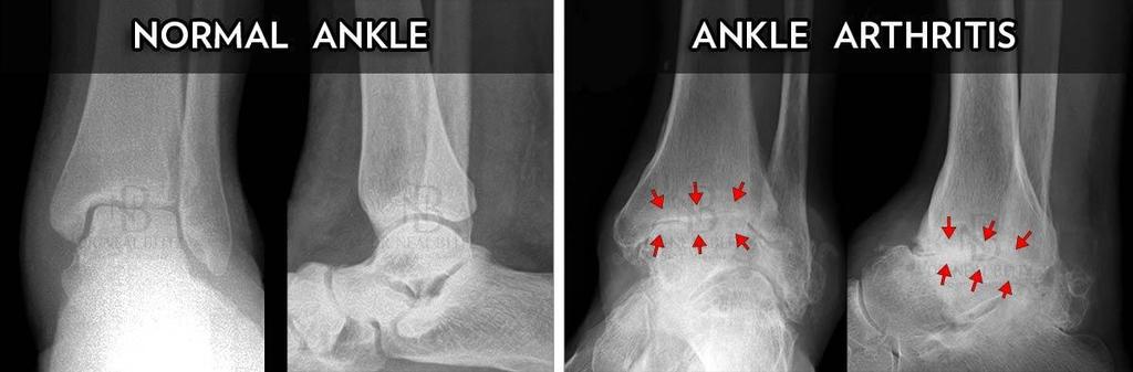
42 Osteoarthritis of the ankle

43 Ankle joint replacement OA can follow severe ankle sprains or previous fractures If conservative management insufficient surgery may be considered Ankle arthrodesis still considered for younger patients More recently joint replacement provided in some areas Of the 30,000 cases of ankle osteoarthritis seen by hospital specialists every year in the UK, only about 1,200 of them will undergo ankle replacement surgery With ankle joint replacement there is a failure rate of up to 19% after 10 years
44 Ankle exercises Early mobilising exercises taking the ankle through full range dorsiflexion, plantarflexion, inversion and eversion


45 Increasing the range of dorsiflexion Using a belt around the foot the ankle can be pulled into more dorsiflexion Standing lunging forwards can push the ankle further into dorsiflexion

46 Using resistance to strengthen the dorsiflexor muscles Theraband is an elastic band which you can buy on the internet from Amazon, Tesco etc. It comes in various levels of resistance. Anchor one end to something stable Place the loop over your foot and pull up into dorsiflexion Repeat 10 times, 4 sets. 8,9 and 10 should be difficult If it becomes easy progress to the next level of resistance

47 Using resistance to strengthen plantar flexion The plantar flexors are generally much stronger than the dorsiflexors so you may need to start with more resistance and or progress to exercises in standing using body weight to give increased resistance

48 Resisted inversion and eversion Using theraband to strengthen the invertors and evertors It is important when doing this exercise that the knee and leg remain still It is easy to cheat by rolling the whole leg in and out
49 Stretching both calf muscles


50 Balance re-education Standing on tip toes with eyes open and closed Standing on one foot eyes open, eyes closed Standing on tip toes one foot eyes open, eyes closed, throwing and catching a ball Standing on an unstable surface eyes open, eyes closed, throwing and catching a ball Standing on a wobble board with two feet, with one foot, eyes open, eyes closed, throwing and catching a ball
51 Further rehabilitation If the persons job or hobbies involve running, hopping, jumping these activities would also be included in the rehabilitation programme Cycling, walking on the treadmill, weights in the gym are also good ways to get back to full fitness

52 Any questions?
53 Next week The foot bony architecture, muscles, how it can be both pliable to walk on rough ground but also act as a solid lever at push off The importance of the arches of the foot The foot in normal gait and posture and its influence in abnormal alignment of the lower limb Common conditions hallux valgus (bunions), heel pain, hammer toes
Leg. Dr. Heba Kalbouneh Associate Professor of Anatomy and Histology
 Leg Dr. Heba Kalbouneh Associate Professor of Anatomy and Histology Skin of the Leg Cutaneous Nerves Medially: The saphenous nerve, a branch of the femoral nerve supplies the skin on the medial surface
Understanding Leg Anatomy and Function THE UPPER LEG
 Understanding Leg Anatomy and Function THE UPPER LEG The long thigh bone is the femur. It connects to the pelvis to form the hip joint and then extends down to meet the tibia (shin bone) at the knee joint.
Main Menu. Ankle and Foot Joints click here. The Power is in Your Hands
 1 The Ankle and Foot Joints click here Main Menu Copyright HandsOn Therapy Schools 2009 K.8 http://www.handsonlineeducation.com/classes/k8/k8entry.htm[3/27/18, 1:40:03 PM] Ankle and Foot Joint 26 bones
Clarification of Terms
 Clarification of Terms The plantar aspect of the foot refers to the role or its bottom The dorsal aspect refers to the top or its superior portion The ankle and foot perform three main functions: 1. shock
The Lower Limb VII: The Ankle & Foot. Anatomy RHS 241 Lecture 7 Dr. Einas Al-Eisa
 The Lower Limb VII: The Ankle & Foot Anatomy RHS 241 Lecture 7 Dr. Einas Al-Eisa Ankle joint Synovial, hinge joint Allow movement of the foot in the sagittal plane only (1 degree of freedom): dorsiflexion:
BLUE SKY SCHOOL OF PROFESSIONAL MASSAGE AND THERAPEUTIC BODYWORK Musculoskeletal Anatomy & Kinesiology KNEE & ANKLE MUSCLES
 BLUE SKY SCHOOL OF PROFESSIONAL MASSAGE AND THERAPEUTIC BODYWORK Musculoskeletal Anatomy & Kinesiology KNEE & ANKLE MUSCLES MSAK201-I Session 3 1) REVIEW a) THIGH, LEG, ANKLE & FOOT i) Tibia Medial Malleolus
Anatomy and evaluation of the ankle.
 Anatomy and evaluation of the ankle www.fisiokinesiterapia.biz Ankle Anatomical Structures Tibia Fibular Talus Tibia This is the strongest largest bone of the lower leg. It bears weight and the bone creates
Copyright 2004, Yoshiyuki Shiratori. All right reserved.
 Ankle and Leg Evaluation 1. History Chief Complaint: A. What happened? B. Is it a sharp or dull pain? C. How long have you had the pain? D. Can you pinpoint the pain? E. Do you have any numbness or tingling?
The Leg. Prof. Oluwadiya KS
 The Leg Prof. Oluwadiya KS www.oluwadiya.sitesled.com Compartments of the leg 4 Four Compartments: 1. Anterior compartment Deep fibular nerve Dorsiflexes the foot and toes 2. Lateral Compartment Superficial
Prevention and Treatment of Injuries. Anatomy. Anatomy. Tibia: the second longest bone in the body
 Prevention and Treatment of Injuries The Ankle and Lower Leg Westfield High School Houston, Texas Anatomy Tibia: the second longest bone in the body Serves as the principle weight-bearing bone of the leg.
The Lower Limb VI: The Leg. Anatomy RHS 241 Lecture 6 Dr. Einas Al-Eisa
 The Lower Limb VI: The Leg Anatomy RHS 241 Lecture 6 Dr. Einas Al-Eisa Muscles of the leg Posterior compartment (superficial & deep): primary plantar flexors of the foot flexors of the toes Anterior compartment:
Anatomy of Foot and Ankle
 Anatomy of Foot and Ankle Surface anatomy of the ankle & foot Surface anatomy of the ankle & foot Medial orientation point medial malleous sustentaculum tali tuberosity of navicular TA muscle TP muscle
Therapeutic Foot Care Certificate Program Part I: Online Home Study Program
 Therapeutic Foot Care Certificate Program Part I: Online Home Study Program 1 Anatomy And Terminology Of The Lower Extremity Joan E. Edelstein, MA, PT, FISPO Associate Professor of Clinical Physical Therapy
ANKLE PLANTAR FLEXION
 ANKLE PLANTAR FLEXION Evaluation and Measurements By Isabelle Devreux 1 Ankle Plantar Flexion: Gastrocnemius and Soleus ROM: 0 to 40-45 A. Soleus: Origin: Posterior of head of fibula and proximal1/3 of
Recognizing common injuries to the lower extremity
 Recognizing common injuries to the lower extremity Bones Femur Patella Tibia Tibial Tuberosity Medial Malleolus Fibula Lateral Malleolus Bones Tarsals Talus Calcaneus Metatarsals Phalanges Joints - Knee
Section Three: The Leg, Ankle, and Foot Lecture: Review of Clinical Anatomy, Patterns of Dysfunction and Injury, and
 Section Three: The Leg, Ankle, and Foot Lecture: Review of Clinical Anatomy, Patterns of Dysfunction and Injury, and Treatment Implications for the Leg, Ankle, and Foot Levels I and II Demonstration and
Review relevant anatomy of the foot and ankle. Learn the approach to examining the foot and ankle
 Objectives Review relevant anatomy of the foot and ankle Learn the approach to examining the foot and ankle Learn the basics of diagnosis and treatment of ankle sprains Overview of other common causes
Managing Tibialis Posterior Tendon Injuries
 Managing Tibialis Posterior Tendon Injuries by Thomas C. Michaud, DC Published April 1, 2015 by Dynamic Chiropractic Magazine Tibialis posterior is the deepest, strongest, and most central muscle of the
Key Points for Success:
 ANKLE & FOOT 1 2 All of the stretches described in this chapter are detailed to stretch the right side. Key Points for Success: Keep your movements slow and precise. Breathe in before you move and breathe
Introduction to Anatomy. Dr. Maher Hadidi. Laith Al-Hawajreh. Mar/25 th /2013
 Introduction to Anatomy Dr. Maher Hadidi Laith Al-Hawajreh 22 Mar/25 th /2013 Lower limb - The leg The skeleton of the leg is formed by two bones: 1) Medial: Tibia 2) Lateral: Fibula The two bones are
بسم هللا الرحمن الرحيم
 بسم هللا الرحمن الرحيم Laboratory RHS 221 Manual Muscle Testing Theory 1 hour practical 2 hours Dr. Ali Aldali, MS, PT Department of Physical Therapy King Saud University Talocrural and Subtalar Joint
Sky Ridge Medical Center, Aspen Building Ridgegate Pkwy., Suite 309 Lone Tree, Colorado Office: Fax:
 ANKLE SPRAIN What is the ATFL? The ankle joint is made up of the tibia, fibula (bones in the lower leg) and the talus (bone below the tibia and fibula). Ligaments in the ankle connect bone to bone and
ANKLE JOINT ANATOMY 3. TALRSALS = (FOOT BONES) Fibula. Frances Daly MSc 1 CALCANEUS 2. TALUS 3. NAVICULAR 4. CUBOID 5.
 Fibula. Frances Daly MSc 1 CALCANEUS 2. TALUS 3. NAVICULAR 4. CUBOID 5.") ANKLE JOINT ANATOMY The ankle joint is a synovial joint of the hinge type. The joint is formed by the distal end of the tibia and medial malleolus, the fibula and lateral malleolus and talus bone. It is
ANKLE JOINT ANATOMY The ankle joint is a synovial joint of the hinge type. The joint is formed by the distal end of the tibia and medial malleolus, the fibula and lateral malleolus and talus bone. It is
 P R E S E N T S Dr. Mufa T. Ghadiali is skilled in all aspects of General Surgery. His General Surgery Services include: General Surgery Advanced Laparoscopic Surgery Surgical Oncology Gastrointestinal
Scar Engorged veins. Size of the foot [In clubfoot, small foot]
![Scar Engorged veins. Size of the foot [In clubfoot, small foot]](/thumbs/78/77722241.jpg "Scar Engorged veins. Size of the foot [In clubfoot, small foot]") 6. FOOT HISTORY Pain: Walking, Running Foot wear problem Swelling; tingly feeling Deformity Stiffness Disability: At work; recreation; night; walk; ADL, Sports Previous Rx Comorbidities Smoke, Sugar, Steroid
6. FOOT HISTORY Pain: Walking, Running Foot wear problem Swelling; tingly feeling Deformity Stiffness Disability: At work; recreation; night; walk; ADL, Sports Previous Rx Comorbidities Smoke, Sugar, Steroid
THE LOWER EXTREMITY EXAM FOR THE FAMILY PRACTITIONER
 THE LOWER EXTREMITY EXAM FOR THE FAMILY PRACTITIONER Melinda A. Scott, D.O. Orthopedic Associates of Dayton Board Certified in Primary Care Sports Medicine GOALS Identify landmarks necessary for exam of
Bones = phalanges 5 metatarsals 7 tarsals
 The Foot (Bones) Bones = 26 14 phalanges 5 metatarsals 7 tarsals Toes (Phalanges) Designed to give wider base for balance and propelling the body forward. 1st toe (Hallux) Two sesamoid bones located under
Ankle Tendons in Athletes. Laura W. Bancroft, M.D.
 Ankle Tendons in Athletes Laura W. Bancroft, M.D. Outline Protocols Normal Anatomy Tendinopathy, partial and complete tears Posterior tibial, Flexor Hallucis Longus, Achilles, Peroneal and Anterior Tibial
موسى صالح عبد الرحمن الحنبلي أحمد سلمان
 8 موسى صالح عبد الرحمن الحنبلي أحمد سلمان 1 P a g e Today we will talk about a new region, which is the leg. And as always, we will start with studying the sensory innervation of the leg. What is the importance
Introduction. The primary function of the ankle and foot is to absorb shock and impart thrust to the body during walking.
 The ankle 1 Introduction The primary function of the ankle and foot is to absorb shock and impart thrust to the body during walking. OSTEOLOGRY The term ankle refers primarily to the talocrural joint,
Dr Nabil khouri MD. MSc. Ph.D
 Dr Nabil khouri MD. MSc. Ph.D Foot Anatomy The foot consists of 26 bones: 14 phalangeal, 5 metatarsal, and 7 tarsal. Toes are used to balance the body. Metatarsal Bones gives elasticity to the foot in
5.1 Identify, describe the attachments of and deduce the actions of the muscles of the thigh:
 5.1 Identify, describe the attachments of and deduce the actions of the muscles of the thigh: Anterior group Proximal attachment Distal attachment Sartorius ASIS» Upper part of shaft tibia (middle surface)»
First & second layers of muscles of the sole
 The FOOT First & second layers of muscles of the sole introduction The muscles acting on the foot can be divided into two distinct groups; extrinsic and intrinsic muscles. The extrinsic muscles arise from
~, /' ~::'~ EXTENSOR HALLUCIS LONGUS. Leg-anterolateral :.:~ / ~\,
 TIBIALIS ANTERIOR Lateral condyle of tibia, upper half of lateral surface of tibia, interosseous membrane Medial side and plantar surface of medial cuneiform bone, and base of first metatarsal bone Dorsiflexes
Posterior Tibialis Tendon Dysfunction & Repair
 1 Posterior Tibialis Tendon Dysfunction & Repair Surgical Indications and Considerations Anatomical Considerations: The posterior tibialis muscle arises from the interosseous membrane and the adjacent
SURGICAL AND APPLIED ANATOMY
 Página 1 de 9 Copyright 2001 Lippincott Williams & Wilkins Bucholz, Robert W., Heckman, James D. Rockwood & Green's Fractures in Adults, 5th Edition SURGICAL AND APPLIED ANATOMY Part of "47 - ANKLE FRACTURES"
Where should you palpate the pulse of different arteries in the lower limb?
 Where should you palpate the pulse of different arteries in the lower limb? The femoral artery In the femoral triangle, its pulse is easily felt just inferior to the inguinal ligament midway between the
MEDIAL HEAD GASTROCNEMIUS TEAR (Tennis Leg)
") MEDIAL HEAD GASTROCNEMIUS TEAR (Tennis Leg) Description Expected Outcome Medial head gastrocnemius tear is a strain of the inner part (medial head) of the major calf muscle (gastrocnemius muscle). Muscle
MEDIAL HEAD GASTROCNEMIUS TEAR (Tennis Leg) Description Expected Outcome Medial head gastrocnemius tear is a strain of the inner part (medial head) of the major calf muscle (gastrocnemius muscle). Muscle
Anatomy MCQs Week 13
 Anatomy MCQs Week 13 1. Posterior to the medial malleolus of the ankle: The neurovascular bundle lies between Tibialis Posterior and Flexor Digitorum Longus The tendon of Tibialis Posterior inserts into
MUSCLES OF THE LOWER LIMBS
 MUSCLES OF THE LOWER LIMBS Naming, location and general function Dr. Nabil khouri ROLES THAT SHOULD NOT BE FORGOTTEN Most anterior compartment muscles of the hip and thigh Flexor of the femur at the hip
Copyright 2012 by The McGraw-Hill Companies, Inc. All rights reserved. McGraw-Hill/Irwin
 CHAPTER 8: THE LOWER EXTREMITY: KNEE, ANKLE, AND FOOT KINESIOLOGY Scientific Basis of Human Motion, 12 th edition Hamilton, Weimar & Luttgens Presentation Created by TK Koesterer, Ph.D., ATC Humboldt State
حسام أبو عوض. - Ahmad. 1 P a g e
 - 9 حسام أبو عوض - - Ahmad 1 P a g e In the last lecture, we finished discussing the superficial part of the posterior compartment and the popliteus muscle of the deep layer[reminder: The entire posterior
Index. Clin Sports Med 23 (2004) Note: Page numbers of article titles are in boldface type.
 Note: Page numbers of article titles are in boldface type.") Clin Sports Med 23 (2004) 169 173 Index Note: Page numbers of article titles are in boldface type. A Achilles enthesopathy, calcaneal spur with, 133 clinical presentation of, 135 136 definition of, 131
Clin Sports Med 23 (2004) 169 173 Index Note: Page numbers of article titles are in boldface type. A Achilles enthesopathy, calcaneal spur with, 133 clinical presentation of, 135 136 definition of, 131
The University Of Jordan Faculty Of Medicine FOOT. Dr.Ahmed Salman Assistant Prof. of Anatomy. The University Of Jordan
 The University Of Jordan Faculty Of Medicine FOOT Dr.Ahmed Salman Assistant Prof. of Anatomy. The University Of Jordan Tarsal Tunnel Syndrome Due to compression of Tibial nerve as it travels through the
Evidence-Based Examination of the Foot Presented by Alexis Wright, PT, PhD, DPT, FAAOMPT Practice Sessions/Skill Check-offs
 Evidence-Based Examination of the Foot Presented by Alexis Wright, PT, PhD, DPT, FAAOMPT Practice Sessions/Skill Check-offs Module Five: Movement Assessment of the Foot/Ankle (1 hour CEU Time) Skilled
Feet First. Michael K. Cooper, DO FACOFP Family Practice/OMM St John Clinic - Claremore OOA 2018 Annual Convention
 Feet First Michael K. Cooper, DO FACOFP Family Practice/OMM St John Clinic - Claremore OOA 2018 Annual Convention Disclaimer I have no conflict of interest. I am not on any pharmaceutical company payroll
musculoskeletal system anatomy muscles of foot sheet done by: dina sawadha & mohammad abukabeer
 musculoskeletal system anatomy muscles of foot sheet done by: dina sawadha & mohammad abukabeer Extensor retinaculum : A- superior extensor retinaculum (SER) : originates from the distal ends of the tibia
Everything. You Should Know. About Your Ankles
 Everything You Should Know About Your Ankles How Your Ankle Works The ankle joint is a hinge type joint that participates in movement and is involved in lower limb stability. There are 2 types of motions
Integrated Manual Therapy & Orthopedic Massage For Complicated Lower Extremity Conditions
 Integrated Manual Therapy & Orthopedic Massage For Complicated Lower Extremity Conditions Assessment Protocols Treatment Protocols Treatment Protocols Corrective Exercises Artwork and slides taken from
Foot and Ankle Conditioning Program
 Foot and Ankle Conditioning Program Studies show, an exercise conditioning program will help you return to daily activities and enjoy a more active, healthy lifestyle. A program of this nature, focused
1. A worker falls from a height and lands on his feet. Radiographs reveal a fracture of the sustentaculum tali. The muscle passing immediately
 1. A worker falls from a height and lands on his feet. Radiographs reveal a fracture of the sustentaculum tali. The muscle passing immediately beneath it that would be adversely affected is the: fibularis
Biokinesiology of the Ankle Complex
 Rehabilitation Considerations Following Ankle Fracture: Impact on Gait & Closed Kinetic Chain Function Disclosures David Nolan, PT, DPT, MS, OCS, SCS, CSCS I have no actual or potential conflict of interest
Foot and Ankle Conditioning Program
 Foot and Ankle Conditioning Program Purpose of Program After an injury or surgery, an exercise conditioning program will help you return to daily activities and enjoy a more active, healthy lifestyle.
LATERAL LIGAMENT SPRAIN OF THE ANKLE
 MUSCULOSKELETAL YOUR GUIDE TO LATERAL LIGAMENT SPRAIN OF THE ANKLE An IPRS Guide to provide you with exercises and advice to ease your condition Contents The ankle joint..................................................
Servers Disease (Calcaneal Apophysitis ) 101
 101") Servers Disease (Calcaneal Apophysitis ) 101 Servers Disease Causes a disturbance to the growing area at the back of the heel bone (calcaneus) where the strong Achilles tendon attaches to it. It is most
Servers Disease (Calcaneal Apophysitis ) 101 Servers Disease Causes a disturbance to the growing area at the back of the heel bone (calcaneus) where the strong Achilles tendon attaches to it. It is most
BUCKS MSK: FOOT AND ANKLE PATHWAY GP MANAGEMENT. Hallux Valgus. Assessment: Early Management. (must be attempted prior to any referral to imsk):
:") Hallux Valgus Common condition: affecting around 28% of the adult population. Prevalence increases with age and in females. Observation: Lateral deviation of the great toe. May cause secondary irritation
Hallux Valgus Common condition: affecting around 28% of the adult population. Prevalence increases with age and in females. Observation: Lateral deviation of the great toe. May cause secondary irritation
Muscles of the Hip 1. Tensor Fasciae Latae O: iliac crest I: lateral femoral condyle Action: abducts the thigh Nerve: gluteal nerve
 Muscles of the Hip 1. Tensor Fasciae Latae O: iliac crest I: lateral femoral condyle Action: abducts the thigh Nerve: gluteal nerve 2. Gluteus Maximus O: ilium I: femur Action: abduct the thigh Nerve:
Ankle and Foot Orthopaedic Tests Orthopedics and Neurology DX 612
 Ankle and Foot Orthopaedic Tests Orthopedics and Neurology DX 612 James J. Lehman, DC, MBA, DABCO University of Bridgeport College of Chiropractic Ankle & Foot Anatomy Stability of the ankle is dependent
Achilles Tendonitis and Tears
 Achilles Tendonitis and Tears The Achilles tendon is an important structure for normal ankle motion and normal function, even for daily activities such as walking. Achilles tendonitis can occur in patients
The Dance Hall by Vincent van Gogh,1888
 The Dance Hall by Vincent van Gogh,1888 Articulations of the pelvic girdle Lumbosacral joints, sacroiliac joints & pubic symphysis The remaining joints of the lower limb Hip joint Knee joint Tibiofibular
Year 2004 Paper one: Questions supplied by Megan
 QUESTION 47 A 58yo man is noted to have a right foot drop three days following a right total hip replacement. On examination there is weakness of right ankle dorsiflexion and toe extension (grade 4/5).
Foot and Ankle Conditioning Program
 Prepared for: Prepared by: Purpose of Program After an injury or surgery, an exercise conditioning program will help you return to daily activities and enjoy a more active, healthy lifestyle. Following
Balanced Body Movement Principles
 Balanced Body Movement Principles How the Body Works and How to Train it. Module 3: Lower Body Strength and Power Developing Strength, Endurance and Power The lower body is our primary source of strength,
Contents The Ankle Joint What is a sprained ankle? What treatment can I receive? Exercises Introduction Please take note of the following
 Contents The Ankle Joint................................ 3 What is a sprained ankle?.................... 4 MUSCULOSKELETAL YOUR GUIDE TO ANKLE SPRAINS An IPRS Guide to provide you with exercises and advice
ANKLE SPRAINS. Explanation. Causes. Symptoms
 ANKLE SPRAINS Explanation Ankle sprains occur when ligaments in the ankle are partially or completely torn due to sudden stretching, either laterally or medially, or when the ankle is suddenly twisted
Dr. Gene Desepoli Anterolateral Shin Splints Summary Treatment Sheet
 Dr. Gene Desepoli Anterolateral Shin Splints Summary Treatment Sheet Pathology: Anterolateral shin splints results from strain to the tibialis anterior muscle from eccentric overuse, running on hard ground
What Happens to the Paediatric Flat Foot? Peter J Briggs Freeman Hospital Newcastle upon Tyne
 What Happens to the Paediatric Flat Foot? Peter J Briggs Freeman Hospital Newcastle upon Tyne We don t know!! Population Studies 2300 children aged 4-13 years Shoe wearers Flat foot 8.6% Non-shoe wearers
A Patient s Guide to Ankle Anatomy
 A Patient s Guide to Ankle Anatomy 1436 Exchange Street Middlebury, VT 05753 Phone: 802-388-3194 Fax: 802-388-4881 cvo@champlainvalleyortho.com DISCLAIMER: The information in this booklet is compiled from
CHAPTER 17. The Foot, Ankle, and Lower Leg KEY TERMS OBJECTIVES
 CHAPTER 17 The Foot, Ankle, and Lower Leg KEY TERMS Achilles tendon anterior compartment compartment syndrome cramp deep posterior compartment extrinsic muscles intrinsic muscles lateral longitudinal arch
A Patient s Guide to Ankle Anatomy
 A Patient s Guide to Ankle Anatomy Pond View Professional Park 301 Professional View Drive Freehold, NJ 07728 Phone: 732-720-2555 DISCLAIMER: The information in this booklet is compiled from a variety
Clin Podiatr Med Surg 19 (2002) Index
 Index") Clin Podiatr Med Surg 19 (2002) 335 344 Index Note: Page numbers of article titles are in bold face type. A Accessory soleus muscle, magnetic resonance imaging of, 300 Achilles tendon injury of, magnetic
Clin Podiatr Med Surg 19 (2002) 335 344 Index Note: Page numbers of article titles are in bold face type. A Accessory soleus muscle, magnetic resonance imaging of, 300 Achilles tendon injury of, magnetic
Outline. Ankle/Foot Anatomy Ankle Sprains Ottawa Ankle Rules DDx: The Sprain That Wasn t
 Ankle Injuries Outline Ankle/Foot Anatomy Ankle Sprains Ottawa Ankle Rules DDx: The Sprain That Wasn t Anatomy: Ankle Mortise Bony Anatomy Lateral Ligament Complex Medial Ligament Complex Ankle Sprains
Physical Examination of the Foot & Ankle
 Inspection Standing, feet straight forward facing toward examiner Swelling Deformity Flatfoot (pes planus and hindfoot valgus) High arch (pes cavus and hindfoot varus) Peek-a-boo heel Varus Too many toes
Lower Limb Nerves. Clinical Anatomy
 Lower Limb Nerves Clinical Anatomy Lumbar Plexus Ventral rami L1 L4 Supplies: Abdominal wall External genitalia Anteromedial thigh Major nerves.. Lumbar Plexus Nerves relation to psoas m. : Obturator n.
통증물리치료학및 실습 CH 10. 근육및인대손상재활. Gachon University Department of Physical Therapy. Hwi-young Cho, PT, PhD
 통증물리치료학및 실습 CH 10. 근육및인대손상재활 Gachon University Department of Physical Therapy Hwi-young Cho, PT, PhD Sprain & Strain http://www.youtube.com/watch?v=2mo- 4B_qz6c Sprain Ligament Strain Muscle & Tendon Sprain
A Patient s Guide to Ankle Anatomy
 A Patient s Guide to Ankle Anatomy 245 North College Lafayette, LA 70506 Phone: 337.232.5301 Fax: 337.237.6504 DISCLAIMER: The information in this booklet is compiled from a variety of sources. It may
The Muscular System. Chapter 10 Part D. PowerPoint Lecture Slides prepared by Karen Dunbar Kareiva Ivy Tech Community College
 Chapter 10 Part D The Muscular System Annie Leibovitz/Contact Press Images PowerPoint Lecture Slides prepared by Karen Dunbar Kareiva Ivy Tech Community College Table 10.14: Muscles Crossing the Hip and
CHAPTER 8: THE BIOMECHANICS OF THE HUMAN LOWER EXTREMITY
 CHAPTER 8: THE BIOMECHANICS OF THE HUMAN LOWER EXTREMITY _ 1. The hip joint is the articulation between the and the. A. femur, acetabulum B. femur, spine C. femur, tibia _ 2. Which of the following is
VENOUS DRAINAGE OF THE LOWER LIMB
 Anatomy of the lower limb Superficial veins & nerve injuries Dr. Hayder VENOUS DRAINAGE OF THE LOWER LIMB The venous drainage of the lower limb is of huge clinical & surgical importance. Since the venous
Hip joint Type: Articulating bones:
 Ana (242 ) Hip joint Type: Synovial, ball & socket Articulating bones: Formed between head of femur and lunate surface of acetabulum of hip bone. Capsule: it is a strong fibrous sleeve connecting the articulating
Muscles of the lower extremities. Dr. Nabil khouri MD, MSc, Ph.D
 Muscles of the lower extremities Dr. Nabil khouri MD, MSc, Ph.D Posterior leg Popliteal fossa Boundaries Biceps femoris (superior-lateral) Semitendinosis and semimembranosis (superior-medial) Gastrocnemius
Make sure you have properly fitting running shoes and break these in gradually. Never wear new running shoes for a race or a long run.
 Common Running Injuries We are delighted that you have decided to run in the next Bath Half Marathon and very much hope that you have good running shoes, undertake a regular training programme and don
A Patient s Guide to Foot Anatomy
 A Patient s Guide to Foot Anatomy Introduction Our feet are constantly under stress. It's no wonder that 80 percent of us will have some sort of problem with our feet at some time or another. Many things
Overuse Injuries & special skeletal injuries Dr M.Taghavi Director of sport medicine center of olympic academy
 Overuse Injuries & special skeletal injuries Dr M.Taghavi Director of sport medicine center of olympic academy Prevalence of Overuse Injuries 30 to 50% of all sport injuries are from overuse In some sports
A Patient s Guide to Adult-Acquired Flatfoot Deformity
 A Patient s Guide to Adult-Acquired Flatfoot Deformity Glendale Adventist Medical Center 1509 Wilson Terrace Glendale, CA 91206 Phone: (818) 409-8000 DISCLAIMER: The information in this booklet is compiled
CHRONIC FOOT PROBLEMS FOOT and ANKLE BASICS
 CHRONIC FOOT PROBLEMS FOOT and ANKLE BASICS ABC s of Comprehensive Musculoskeletal Care December 1 st, 2007 Stephen Pinney MD Chief, UCSF Foot and Ankle Service Chronic problems typically occur gradually
Pelvic cavity. Gross anatomy of the lower limb. Walking. Sándor Katz M.D.,Ph.D.
 Pelvic cavity. Gross anatomy of the lower limb. Walking. Sándor Katz M.D.,Ph.D. Lower limb Pelvic girdle Free lower extremity Hip bone Definitive fusion of the Y- shaped growth plate occurs 16th -18th
Anatomy & Physiology. Muscles of the Lower Limbs.
 Anatomy & Physiology Muscles of the Lower Limbs http://www.ishapeup.com/musclecharts.html Muscles of the Lower Limbs Among the strongest muscles in the body. Because pelvic girdle is composed of heavy,
The Foot. Dr. Wegdan Moh.Mustafa Medicine Faculty Assistant Professor Mob:
 The Foot Dr. Wegdan Moh.Mustafa Medicine Faculty Assistant Professor Mob: 0127155717 The skeleton of the foot Cutaneous innervations Sole of foot layers of muscles First layer -Abductor hallucis -Flexor
Practical 1 Worksheet
 Practical 1 Worksheet ANATOMICAL TERMS 1. Use the word bank to fill in the missing words. reference side stand body arms palms anatomical forward All anatomical terms have a(n) point which is called the
So#- Tissue Manipula0on: Supine
 So#- Tissue Manipula0on: Supine Gastrocnemius and soleus stretch Use stretching in the later stages of rehabilita0on Check in with the client as you go to avoid painful tearing Perform joint mobiliza0on
Joints and muscles of the foot. Architecture of the foot. Sándor Katz M.D.,Ph.D.
 Joints and muscles of the foot. Architecture of the foot. Sándor Katz M.D.,Ph.D. Ankle (talocrural) joint type: hinge Talocrural joint - medial collateral ligament Medial collateral = deltoid ligament
31b Passive Stretches:! Technique Demo and Practice - Lower Body
 31b Passive Stretches:! Technique Demo and Practice - Lower Body 31b Passive Stretches:! Technique Demo and Practice - Lower Body! Class Outline" 5 minutes" "Attendance, Breath of Arrival, and Reminders
CHAPTER 80 BASIC CONSIDERATIONS
 Página 1 de 32 Copyright 2001 Lippincott Williams & Wilkins Loeser, John D. Bonica's Management of Pain, 3rd Edition CHAPTER 80 BASIC CONSIDERATIONS Part of "CHAPTER 80 - Pain in the Leg, Ankle, and Foot"
Contents of the Posterior Fascial Compartment of the Thigh
 Contents of the Posterior Fascial Compartment of the Thigh 1-Muscles: B i c e p s f e m o r i s S e m i t e n d i n o s u s S e m i m e m b r a n o s u s a small part of the adductor magnus (h a m s t
Anatomy of the lower limb
 Anatomy of the lower limb Arches & sole of the foot Dr. Hayder ARCHES OF THE FOOT The foot as a mechanical unit performs two major functions: - It acts as a pliable platform to support the body weigh during
Ankle Sprains and Their Imitators
 Ankle Sprains and Their Imitators Mark Halstead, MD Dr. Mark Halstead is the Associate Professor of the Departments of Orthopedics and Pediatrics at Washington University School of Medicine; Director of
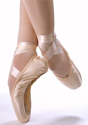
A Soccer Player s Journey to Reducing Ankle Injuries Through Pilates
 A Soccer Player s Journey to Reducing Ankle Injuries Through Pilates Roshan F. Rodriguez 18 NOV 2016 2016 Course Herndon, VA Abstract With a pitch even larger than a regulation NFL field and few substitutions
Foot. Dr. Heba Kalbouneh Associate Professor of Anatomy and Histology
 Foot Dr. Heba Kalbouneh Associate Professor of Anatomy and Histology Dorsum of the Foot Sole of the Foot Plantar aponeurosis It is a triangular thickening of deep fascia in the sole of the foot Attachments: