통증물리치료학및 실습 CH 10. 근육및인대손상재활. Gachon University Department of Physical Therapy. Hwi-young Cho, PT, PhD
|
|
|
- Walter Flowers
- 5 years ago
- Views:
Transcription


1 통증물리치료학및 실습 CH 10. 근육및인대손상재활 Gachon University Department of Physical Therapy Hwi-young Cho, PT, PhD
2 Sprain & Strain 4B_qz6c Sprain Ligament Strain Muscle & Tendon

3 Sprain An injury involving the stretching or tearing of a ligament (tissue that connects bone to bone) or a joint capsule, which help provide joint stability. 1 or more ligaments can be injured at the same time Severity of injury extent of injury and number of ligaments involved

4 Symptom Sprain pain, swelling, the inability to move a limb, popping sound, difficulty using the affected limb Severe damage Cause joint instability
5 Severity of Sprain & Strain Grade I (mild) Some stretching or minor tearing of a ligament or muscle. Grade II (moderate) A ligament or muscle that is partially torn but still intact. Grade III (severe) The ligament or muscle is completely torn, resulting in joint instability.


6
 : look at surrounding soft tissues and the ligament http://www.clinichq.co.")
7 Sprain Diagnosis Physical examination based on the clinical presentation and method of injury X-ray : ensure that there is no fracture Magnetic resonance imaging (MRI) : look at surrounding soft tissues and the ligament

8 Risk factor of Sprain Sudden movement or twist Fall down Blow to body that forces a joint out of its normal position and stretches or tears the ligament supporting that joint

9 Where Do Sprains Usually Occur? Most common site the ankle Frequently occurred site the wrist sprain to the thumb common in skiing and other sports

10 Strain Involve the stretching or tearing of a musculo-tendinous (muscle and tendon) structure Acute strain By a direct blow to the body, overstretching or excessive muscle contraction Chronic strain The result of overuse - prolonged, repetitive movement of muscles and tendons
11 Strain 급성염좌 (Acute strain) Occurs at the junction where the muscle is becoming a tendon. When a muscle is stretched and suddenly contracts, as with running or jumping. Symptom : pain, muscle spasm, loss of strength, and limited range of motion. 만성염좌 (Chronic strain) Gradually build up from overuse or repetitive stress, resulting in tendinitis (inflammation of a tendon). Ex: Tennis elbow, Golfer elbow

12 Strain Severe Strain - muscle or tendon is partially or completely ruptured, leaving person incapacitated. Moderate Strain - muscle or tendon is overstretched and slightly torn, leaving some muscle functions lost. Mild Strain - muscle or tendon stretched or pulled slightly

13 What Causes a Strain? Heavy work Sudden movement Repeated & persistent activity Twisting or pulling a muscle or tendon Acute or chronic: recent trauma or result of overuse


14 Where Do Strains Usually Occur? Common sites: Back & Hamstring Calf, Foot, Hand and Forearm

15 Signs and Symptoms of Strain Muscle pain and tenderness, especially after an activity that stretches or violently contracts the muscle -- Pain usually increases when you move the muscle but is relieved by rest. Muscle swelling, discoloration or both Muscle cramp or spasm Either a decrease in muscle strength or (in Grade III strains) a complete loss of muscle function A pop in the muscle at the time of injury A gap, dent or other defect in the normal outline of the muscle (in Grade III strain)
16 Repetitive Strain Injuries Carpal tunnel syndrome (CTS), tendonitis, and many of the ergonomic injuries result from straining muscles or ligaments. Workplace set up for person is the first step. Remember the rule of 90s for office operations (knees at 90 degrees, back/legs at 90 degrees, elbows at rest and at 90 degrees with arms). Job rotation is another method to reduce job stress. Take stretch breaks as needed. Exercise and stretch to help with blood flow and keep muscles loose.


17 TX for Sprains and Strains RICE Therapy
18 1. Rest Take a break from normal activities This is the easiest of the 4 first aid measures but is often the hardest to implement. For 48 hours
19 2. Ice Apply a cold pack as soon as possible after the injury. Cold therapy has two benefits: Reduces swelling Relieves pain Method: Apply cold therapy to the injured area for 20 minutes at a time. Remove the cold pack for at least 30 minutes to allow the skin to rewarm.
20 3. Compression Use compression when elevating a sprain or strain during early treatment. Wearing an elastic compression bandage for at least 2 days will reduce swelling.
21 4. Elevation Keep the injured part elevated above the level of the heart to reduce swelling Keep the affected area higher than your heart if possible. This is another trick to help reduce swelling and inflammation. Try placing a couple of pillows under the injured arm or leg.

22 Proper Lifting Procedures Plan the lift. Test load before lifting. Place feet shoulder - width apart close to object. Bend the knees. Get a secure grip. Lift with legs, keeping the back straight. Lift evenly and slowly - no jerky motions. Keep load as close to the body as possible.
23 Ankle Sprain Developed for Physical Therapy Students by Hwi-young Cho, Ph.D, P.T. Gachon University College of Health-Science Department of Physical therapy 2014 Second semester

24 Mini Case A basketball player was getting in position for a rebound when he stepped on another player s foot and rolled his ankle. X-rays did not reveal a fracture, but the player left the game on crutches.
25 Approaching the Problem Differential? (break, sprain, etc.) Tissue changes? (swelling, warmth, ecchymosis) Pain can patient bear weight? ROM decreased? Neuro deficits? Treatment? Don t just look at the ankle think how it affects the rest of the patient s body also
26 Joint stability Main components 1. Shape of Bones 2. Ligaments 3. Strength of muscles

 ligament")

27 Anatomy 26 bones of the foot Plus 2 sesamoid bones under great toe for weight bearing and balance 33 joints Medial longitudinal arch Calcaneous, talus, navicular, first 3 cuneforms, first 3 metatarsals Strengthened by calcaneonavicular (Spring) ligament Lateral longitudinal arch Calcaneous, cuboid, 4 th and 5 th metatarsals Weight bearing Transverse arches Weight bearing and springing off with foot

28 Anterior aspect of the right ankle skeleton and superior aspect of the foot skeleton muscle/anatomysurgical.htm

29 Posterior aspect of the right ankle skeleton and superior aspect of the foot skeleton muscle/anatomysurgical.htm


30 Soleus and Plantaris muscles and the contents of the popliteal fossa (gastrocnemius muscle removed)

31 Overview The distal end of the The distal end of the fibula forms the tibia forms the lateral malleolus medial malleolus

32 Anterior Ankle Tibiofibular Joint

33 Medial Ankle Joint

34 Posterior Ankle Tibiofibular Joint

35 Lateral Ankle Joint

36 Superior aspect of the right tarsus with ligaments
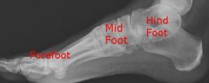
37 The foot is divided into 3 general regions:
38 Hindfoot The talus & the calcaneus (or heel bone). The two long bones of the lower leg, the tibia and fibula, are connected to the top of the talus to form the ankle. Connected to the talus at the subtalar joint, the calcaneus, the largest bone of the foot, is cushioned inferiorly by a layer of fat
39 Midfoot The five irregular bones the cuboid, navicular, and 3 cuneiform bones 3 cuneiform bones: form the arches of the foot which serves as a shock absorber Connected to the hind- and fore-foot by muscles and the plantar fascia. Tarsometatarsal jt.
 jt. Proximal interphalangeal (PIP) jt.")
40 Forefoot 5 toes and the corresponding five proximal long bones forming the metatarsus 5 metatarsals 5 proximal phalanges 4 middle phalanges 5 distal phalanges Metatarsophalangeal (MTP) jt. Proximal interphalangeal (PIP) jt. Distal interphalangeal (DIP) jt.

41

42 Joints in the Ankle
 Joined at both proximal & distal tibiofibular joints Ligaments and a strong, dense interosseus membrane between tibia & fibula shafts provide support Minimal")
43 Tibiofibular joint Syndesmotic jt. ( 인대결합관절 ) Joined at both proximal & distal tibiofibular joints Ligaments and a strong, dense interosseus membrane between tibia & fibula shafts provide support Minimal movement possible Distal joint becomes sprained occasionally in heavy contact sport
")
44 Talocrural joint (ankle jt.) Hinge jt. Talus, distal tibia & distal fibula Dorsiflexion-plantarflexion 50 degrees of plantar flexion 15 to 20 degrees of dorsiflexion Greater range of dorsiflexion with knee flexed (reduces GCM tension) Fibula rotates 3 to 5 degrees externally with ankle dorsiflexion & 3 to 5 degrees internally during plantarflexion Syndesmosis jt. widen by 1 to 2 mm during full dorsiflexion

45 Subtalar & transverse tarsal joint Inversion & eversion Combined movement of 20 to 30 degrees of inversion 5 to 15 degrees of eversion

46 Metatarsophalangeal joint Phalanges join metatarsal Great toe metatarsophalangeal (MP) jt flexes 45 degrees & extends 70 degrees MP jt of the four lesser toes 40 degrees of flexion 40 degrees of extension Also abduct & adduct minimally
47 Interphalangeal (IP) joint Greater toe interphalangeal (IP) joint flexes from 0 degrees of full extension to 90 degrees of flexion PIP jt. in lesser toes flexes from 0 degrees of extension to 35 degrees of flexion DIP jt. flexes 60 degrees & extend 30 degrees Much variation from joint & from person to person
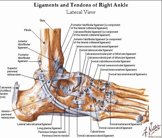
48 Anatomy-Lateral Ligaments
49 LATERAL LIGAMENTS
50 Lateral ligament = Lateral collateral ligament of the ankle Division 1 The anterior talofibular ligament - extends anteromedially from the anterior margin of the fibular malleolus to the neck of the talus. 2 The posterior talofibular ligament - extends almost horizontally from the lateral malleolar fossa to the lateral tubercle of the talus. 3 The calcaneofibular ligament - is a long cord which passes from a depression anterior to the apex of the fibular malleolus to a tubercle on the lateral calcaneal surface. It is crossed by the tendons of the peroneus longus and brevis.

51 Anatomy-Medial Ligaments

52 MEDIAL ANKLE
53 Deltoid ligament = Medial collateral ligament of the ankle Superficial fibers : - The most anterior (tibionavicular) fibers pass forward to be inserted into the tuberosity of the navicular bone, and immediately behind this they blend with the medial margin of the plantar calcaneonavicular ligament (spring ligament). - The middle (tibiocalcaneal) fibers descend almost perpendicularly to be inserted into the whole length of the sustentaculum tali of the calcaneum - The posterior fibers (posterior tibiotalar) pass backward and laterally to be attached to the medial side of the talus, and its medial tubercle. Deep fibers : - The deep fibers (anterior tibiotalar) are attached to the anterior part of medial surface of the talus. The deltoid ligament is crossed by the tendons of the tibialis posterior and Flexor digitorum longus.

54 PALPATION 1 Anterior Inferior Tibiofibular Ligament 2 Anterior Talofibular 3 Calcaneofibular Lig. 4 Base of 5 th Metatarsal

55 Anatomy of Foot/Ankle Talus is wider anteriorly Medial malleolus only comes down over 1/3 of the talus and is more anterior Lateral malleolus covers entire talus Inversion > eversion Des Moines University OMM II handouts, August 12, 2002 May 15, 2003 OUCOM Session 7 Lower Extremity lecture
56 Biomechanics In dorsiflexion foot everts, toeing out Most stable position closed packed position In plantarflexion foot inverts, toeing in Ligaments less taut Joint more vulnerable to injury
57 Muscles associated with ankle motion - 1 Plantar-Flexion Gastrocnemius Soleus Plantaris Peroneus Longus & Brevis Tibialis Posterior Flexor Hallucis Longus Flexor Digitorum Longus Dorsi-Flexion Tibialis Anterior Extensor Digitorum Longus Extensor Hallucis Longus Peroneus Tertius
58 Muscles associated with ankle motion - 2 Inversion, Adduction & Supination Tibialis Posterior Tibialis Anterior Flexor Digitorum Longus Flexor Hallucis Longus Extensor Hallucis Longus Eversion, Abduction & Pronation Peroneus Longus Peroneus Brevis Peroneus Tertius Extensor Digitorum Longus
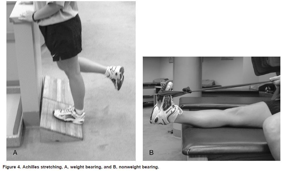
59 Cause of Ankle Sprain 85% are due to inversion Deltoid ligament is stronger than the lateral ligaments Anterior tibiotalar, tibiocalcaneal, tibionavicular, and posterior tibiotalar ligaments Lateral malleolus is longer than medial malleolus Axis of talo-crural joint In plantar flexion, ankle naturally inverts In dorsiflexion, ankle is very stable
60 Pathoanatomy and Mechanisms of Injury The most common mechanism of injury is a combination of plantar flexion and inversion. The lateral stabilizing ligaments, which include the anterior talofibular, calcaneofibular and posterior talofibular ligaments. The anterior talofibular ligament is the most easily injured. The posterior talofibular ligament is the strongest of the lateral complex and is rarely injured in an inversion sprain.

61 Mechanism of injury high ankle sprain
62 Cause OUCOM CORE OMM curriculum session 7
63 Grading Grade I: anterior talofibular ligament (ATF) Grade II: ATF plus calcaneofibular ligament (CF) Grade III: ATF plus CF plus posterior talofibular ligament
64 Ligaments used in Grading Des Moines University OMM II handouts, August 12, 2002 May 15, 2003 OUCOM Session 7 Lower Extremity lecture

65 Diagnosis History of trauma Swelling/discoloration Pain/tenderness Eversion restriction Anterior drawer test for ankle X-ray es/ankle.jpg
66 PAIN RESPONSE OF DAMAGED TISSUE 1. Damaged muscle and ligaments are painful when stretched. 2. Damaged muscle is painful to contract. 3. Both structures are painful if palpated at the site of tear.

67 DX: The anterior drawer test and the inversion stress test The inversion stress test can be used to assess the integrity of the calcaneofibular ligament. The anterior drawer test can be used to assess the integrity of the anterior talofibular ligament.
68 Ottawa Ankle Rules Radiographs should be obtained to rule out fracture when a patient presents (within 10 days of injury) with bone tenderness in the posterior half of the lower 6 cm (2.5 in) of the fibula or tibia or an inability to bear weight immediately after the injury. Bone tenderness over the navicular bone or base of the fifth metatarsal is an indication for radiographs. Anteroposterior, lateral and mortise radiographs should be obtained after the initial physical examination. The mortise projection is an anteroposterior view obtained with the leg internally rotated 15 to 20 degrees. 이러한항목중한가지라도해당되는것이있다면, 즉시병원에서 X-ray 를통해발목골절및이학적검사를통해진단을받아야함.
69
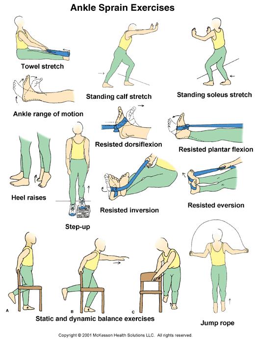
70 Functional Rehabilitation Prolonged immobilization of ankle sprains is a common treatment error. Functional stress stimulates the incorporation of stronger replacement collagen. The four components of rehabilitation are: 1. Range-of-motion rehabilitation 2. Progressive muscle-strengthening exercises 3. Proprioceptive training 4. Activity-specific training
71 Range of Motion Achilles tendon stretch, nonweight-bearing. Use a towel to pull foot toward face. Pain-free stretch for 15 to 30 seconds; perform five repetitions; repeat three to five times a day. Maintain extremity in a nongravity position with compression. Achilles tendon stretch, weight-bearing. Stand with heel on floor and bend at knees. Pain-free stretch for 15 to 30 seconds; perform five repetitions; repeat three to five times a day. Alphabet exercises, Move ankle in multiple planes of motion by drawing letters of alphabet (lower case and upper case). Repeat four to five times a day. Exercises can be performed in conjunction with cold therapy.

72
73 Muscle Strengthening Isometric exercises, Resistance can be provided by immovable object (wall or floor) or contralateral foot. For each exercise, hold 5 seconds; do 10 repetitions; repeat three times a day. Strengthening exercises should only be done in positions that do not cause pain. Plantar flexion, Push foot downward (away from head). Dorsiflexion, Pull foot upward (toward head). Inversion, Push foot inward (toward midline of body). Eversion, Push foot outward (away from midline of body). For each exercise, hold 1 second for concentric component and perform eccentric component over 4 seconds; do three sets of 10 repetitions; repeat two times a day.


74 Muscle-Strengthening Exercises FIGURE 9. Use of elastic tubing in strengthening exercises for eversion. FIGURE 8. Achilles tendon stretching using a towel.


75 Muscle Strengthening Toe curls Marble pickups Toe curls and marble pickups, Place foot on a towel; then curl toes, moving the towel toward body. Use toes to pick up marbles or other small object. Two sets of 10 repetitions; repeat two times a day. Toe curls can be done throughout the day, at work or at home. Toe raises, heel walks and toe walks, Lift body by rising up on toes. Walk forward and backward on toes and heels. Three sets of 10 repetitions; repeat two times a day; progress walking as tolerated. Strengthening can occur from using the body as resistance in weight- bearing position.


76 Muscle-Strengthening Exercises FIGURE 11. Single-leg wobble board exercise to increase proprioception. FIGURE 10. Single-leg toe raises done on a step.
77 Range of Motion Range of motion must be regained before functional rehabilitation is initiated. Regardless of weight-bearing capacity, Achilles tendon stretching should be instituted within 48 to 72 hours after the ankle injury because of the tendency of tissues to contract following trauma. Once range of motion is attained, and swelling and pain are controlled, the patient is ready to progress to the strengthening phase of rehabilitation.
78 Rehabilitation/Strengthening
79 Training for Return to Activity When walking a specified distance is no longer limited by pain, the patient may progress to a regimen of 50 percent walking and 50 percent jogging. When this can be done without pain, jogging eventually progresses to forward, backward and pattern running. Circles and figure-eights are commonly employed for pattern running. Although these routines are time-consuming, they represent the final phase and are essential for the recovery of ankle stability.

80
81
82 Ankle Taping 실제운동경기도중 Taping 시행하는예 Ankle Knee
83 Ankle Taping Non-Elastic Tape 하는방법 h?v=u1tu72odu6i h?v=0lyatixamge h?v=bzrydh6ruxe h?v=zb4l7wlpmda Elastic Tape 하는방법 h?v=elsu25gow0i h?v=kje9nobzgr4 h?v=xnpy0_s5rnm h?v=kp1qhc4ancu h?v=ioc7ub7ew9c


84 Ankle Taping


85
Main Menu. Ankle and Foot Joints click here. The Power is in Your Hands
 1 The Ankle and Foot Joints click here Main Menu Copyright HandsOn Therapy Schools 2009 K.8 http://www.handsonlineeducation.com/classes/k8/k8entry.htm[3/27/18, 1:40:03 PM] Ankle and Foot Joint 26 bones
The Lower Limb VII: The Ankle & Foot. Anatomy RHS 241 Lecture 7 Dr. Einas Al-Eisa
 The Lower Limb VII: The Ankle & Foot Anatomy RHS 241 Lecture 7 Dr. Einas Al-Eisa Ankle joint Synovial, hinge joint Allow movement of the foot in the sagittal plane only (1 degree of freedom): dorsiflexion:
Clarification of Terms
 Clarification of Terms The plantar aspect of the foot refers to the role or its bottom The dorsal aspect refers to the top or its superior portion The ankle and foot perform three main functions: 1. shock
Copyright 2004, Yoshiyuki Shiratori. All right reserved.
 Ankle and Leg Evaluation 1. History Chief Complaint: A. What happened? B. Is it a sharp or dull pain? C. How long have you had the pain? D. Can you pinpoint the pain? E. Do you have any numbness or tingling?
Anatomy of Foot and Ankle
 Anatomy of Foot and Ankle Surface anatomy of the ankle & foot Surface anatomy of the ankle & foot Medial orientation point medial malleous sustentaculum tali tuberosity of navicular TA muscle TP muscle
Anatomy and evaluation of the ankle.
 Anatomy and evaluation of the ankle www.fisiokinesiterapia.biz Ankle Anatomical Structures Tibia Fibular Talus Tibia This is the strongest largest bone of the lower leg. It bears weight and the bone creates
Dr Nabil khouri MD. MSc. Ph.D
 Dr Nabil khouri MD. MSc. Ph.D Foot Anatomy The foot consists of 26 bones: 14 phalangeal, 5 metatarsal, and 7 tarsal. Toes are used to balance the body. Metatarsal Bones gives elasticity to the foot in
Prevention and Treatment of Injuries. Anatomy. Anatomy. Tibia: the second longest bone in the body
 Prevention and Treatment of Injuries The Ankle and Lower Leg Westfield High School Houston, Texas Anatomy Tibia: the second longest bone in the body Serves as the principle weight-bearing bone of the leg.
ANKLE PLANTAR FLEXION
 ANKLE PLANTAR FLEXION Evaluation and Measurements By Isabelle Devreux 1 Ankle Plantar Flexion: Gastrocnemius and Soleus ROM: 0 to 40-45 A. Soleus: Origin: Posterior of head of fibula and proximal1/3 of
BLUE SKY SCHOOL OF PROFESSIONAL MASSAGE AND THERAPEUTIC BODYWORK Musculoskeletal Anatomy & Kinesiology KNEE & ANKLE MUSCLES
 BLUE SKY SCHOOL OF PROFESSIONAL MASSAGE AND THERAPEUTIC BODYWORK Musculoskeletal Anatomy & Kinesiology KNEE & ANKLE MUSCLES MSAK201-I Session 3 1) REVIEW a) THIGH, LEG, ANKLE & FOOT i) Tibia Medial Malleolus
Understanding Leg Anatomy and Function THE UPPER LEG
 Understanding Leg Anatomy and Function THE UPPER LEG The long thigh bone is the femur. It connects to the pelvis to form the hip joint and then extends down to meet the tibia (shin bone) at the knee joint.
بسم هللا الرحمن الرحيم
 بسم هللا الرحمن الرحيم Laboratory RHS 221 Manual Muscle Testing Theory 1 hour practical 2 hours Dr. Ali Aldali, MS, PT Department of Physical Therapy King Saud University Talocrural and Subtalar Joint
Section Three: The Leg, Ankle, and Foot Lecture: Review of Clinical Anatomy, Patterns of Dysfunction and Injury, and
 Section Three: The Leg, Ankle, and Foot Lecture: Review of Clinical Anatomy, Patterns of Dysfunction and Injury, and Treatment Implications for the Leg, Ankle, and Foot Levels I and II Demonstration and
Bones = phalanges 5 metatarsals 7 tarsals
 The Foot (Bones) Bones = 26 14 phalanges 5 metatarsals 7 tarsals Toes (Phalanges) Designed to give wider base for balance and propelling the body forward. 1st toe (Hallux) Two sesamoid bones located under
Sky Ridge Medical Center, Aspen Building Ridgegate Pkwy., Suite 309 Lone Tree, Colorado Office: Fax:
 ANKLE SPRAIN What is the ATFL? The ankle joint is made up of the tibia, fibula (bones in the lower leg) and the talus (bone below the tibia and fibula). Ligaments in the ankle connect bone to bone and
The Leg. Prof. Oluwadiya KS
 The Leg Prof. Oluwadiya KS www.oluwadiya.sitesled.com Compartments of the leg 4 Four Compartments: 1. Anterior compartment Deep fibular nerve Dorsiflexes the foot and toes 2. Lateral Compartment Superficial
Therapeutic Foot Care Certificate Program Part I: Online Home Study Program
 Therapeutic Foot Care Certificate Program Part I: Online Home Study Program 1 Anatomy And Terminology Of The Lower Extremity Joan E. Edelstein, MA, PT, FISPO Associate Professor of Clinical Physical Therapy
 P R E S E N T S Dr. Mufa T. Ghadiali is skilled in all aspects of General Surgery. His General Surgery Services include: General Surgery Advanced Laparoscopic Surgery Surgical Oncology Gastrointestinal
Recognizing common injuries to the lower extremity
 Recognizing common injuries to the lower extremity Bones Femur Patella Tibia Tibial Tuberosity Medial Malleolus Fibula Lateral Malleolus Bones Tarsals Talus Calcaneus Metatarsals Phalanges Joints - Knee
Introduction. The primary function of the ankle and foot is to absorb shock and impart thrust to the body during walking.
 The ankle 1 Introduction The primary function of the ankle and foot is to absorb shock and impart thrust to the body during walking. OSTEOLOGRY The term ankle refers primarily to the talocrural joint,
5.1 Identify, describe the attachments of and deduce the actions of the muscles of the thigh:
 5.1 Identify, describe the attachments of and deduce the actions of the muscles of the thigh: Anterior group Proximal attachment Distal attachment Sartorius ASIS» Upper part of shaft tibia (middle surface)»
Evidence-Based Examination of the Foot Presented by Alexis Wright, PT, PhD, DPT, FAAOMPT Practice Sessions/Skill Check-offs
 Evidence-Based Examination of the Foot Presented by Alexis Wright, PT, PhD, DPT, FAAOMPT Practice Sessions/Skill Check-offs Module Five: Movement Assessment of the Foot/Ankle (1 hour CEU Time) Skilled
Ankle Tendons in Athletes. Laura W. Bancroft, M.D.
 Ankle Tendons in Athletes Laura W. Bancroft, M.D. Outline Protocols Normal Anatomy Tendinopathy, partial and complete tears Posterior tibial, Flexor Hallucis Longus, Achilles, Peroneal and Anterior Tibial
Caring For Your Lateral Ankle Middlebury College
 Caring For Your Lateral Ankle Sprain @ Middlebury College ** severe sprains or medial (inner side of ankle) sprains may require a different program Anatomy, Pathology, and Classification of Ankle Sprains
MEDIAL HEAD GASTROCNEMIUS TEAR (Tennis Leg)
") MEDIAL HEAD GASTROCNEMIUS TEAR (Tennis Leg) Description Expected Outcome Medial head gastrocnemius tear is a strain of the inner part (medial head) of the major calf muscle (gastrocnemius muscle). Muscle
MEDIAL HEAD GASTROCNEMIUS TEAR (Tennis Leg) Description Expected Outcome Medial head gastrocnemius tear is a strain of the inner part (medial head) of the major calf muscle (gastrocnemius muscle). Muscle
ANKLE JOINT ANATOMY 3. TALRSALS = (FOOT BONES) Fibula. Frances Daly MSc 1 CALCANEUS 2. TALUS 3. NAVICULAR 4. CUBOID 5.
 Fibula. Frances Daly MSc 1 CALCANEUS 2. TALUS 3. NAVICULAR 4. CUBOID 5.") ANKLE JOINT ANATOMY The ankle joint is a synovial joint of the hinge type. The joint is formed by the distal end of the tibia and medial malleolus, the fibula and lateral malleolus and talus bone. It is
ANKLE JOINT ANATOMY The ankle joint is a synovial joint of the hinge type. The joint is formed by the distal end of the tibia and medial malleolus, the fibula and lateral malleolus and talus bone. It is
Review relevant anatomy of the foot and ankle. Learn the approach to examining the foot and ankle
 Objectives Review relevant anatomy of the foot and ankle Learn the approach to examining the foot and ankle Learn the basics of diagnosis and treatment of ankle sprains Overview of other common causes
Physical Examination of the Foot & Ankle
 Inspection Standing, feet straight forward facing toward examiner Swelling Deformity Flatfoot (pes planus and hindfoot valgus) High arch (pes cavus and hindfoot varus) Peek-a-boo heel Varus Too many toes
Everything. You Should Know. About Your Ankles
 Everything You Should Know About Your Ankles How Your Ankle Works The ankle joint is a hinge type joint that participates in movement and is involved in lower limb stability. There are 2 types of motions
Pelvic cavity. Gross anatomy of the lower limb. Walking. Sándor Katz M.D.,Ph.D.
 Pelvic cavity. Gross anatomy of the lower limb. Walking. Sándor Katz M.D.,Ph.D. Lower limb Pelvic girdle Free lower extremity Hip bone Definitive fusion of the Y- shaped growth plate occurs 16th -18th
SURGICAL AND APPLIED ANATOMY
 Página 1 de 9 Copyright 2001 Lippincott Williams & Wilkins Bucholz, Robert W., Heckman, James D. Rockwood & Green's Fractures in Adults, 5th Edition SURGICAL AND APPLIED ANATOMY Part of "47 - ANKLE FRACTURES"
Joints and muscles of the foot. Architecture of the foot. Sándor Katz M.D.,Ph.D.
 Joints and muscles of the foot. Architecture of the foot. Sándor Katz M.D.,Ph.D. Ankle (talocrural) joint type: hinge Talocrural joint - medial collateral ligament Medial collateral = deltoid ligament
Leg. Dr. Heba Kalbouneh Associate Professor of Anatomy and Histology
 Leg Dr. Heba Kalbouneh Associate Professor of Anatomy and Histology Skin of the Leg Cutaneous Nerves Medially: The saphenous nerve, a branch of the femoral nerve supplies the skin on the medial surface
Hip joint Type: Articulating bones:
 Ana (242 ) Hip joint Type: Synovial, ball & socket Articulating bones: Formed between head of femur and lunate surface of acetabulum of hip bone. Capsule: it is a strong fibrous sleeve connecting the articulating
Feet First. Michael K. Cooper, DO FACOFP Family Practice/OMM St John Clinic - Claremore OOA 2018 Annual Convention
 Feet First Michael K. Cooper, DO FACOFP Family Practice/OMM St John Clinic - Claremore OOA 2018 Annual Convention Disclaimer I have no conflict of interest. I am not on any pharmaceutical company payroll
~, /' ~::'~ EXTENSOR HALLUCIS LONGUS. Leg-anterolateral :.:~ / ~\,
 TIBIALIS ANTERIOR Lateral condyle of tibia, upper half of lateral surface of tibia, interosseous membrane Medial side and plantar surface of medial cuneiform bone, and base of first metatarsal bone Dorsiflexes
ANKLE SPRAINS. Explanation. Causes. Symptoms
 ANKLE SPRAINS Explanation Ankle sprains occur when ligaments in the ankle are partially or completely torn due to sudden stretching, either laterally or medially, or when the ankle is suddenly twisted
Copyright 2012 by The McGraw-Hill Companies, Inc. All rights reserved. McGraw-Hill/Irwin
 CHAPTER 8: THE LOWER EXTREMITY: KNEE, ANKLE, AND FOOT KINESIOLOGY Scientific Basis of Human Motion, 12 th edition Hamilton, Weimar & Luttgens Presentation Created by TK Koesterer, Ph.D., ATC Humboldt State
Outline. Ankle/Foot Anatomy Ankle Sprains Ottawa Ankle Rules DDx: The Sprain That Wasn t
 Ankle Injuries Outline Ankle/Foot Anatomy Ankle Sprains Ottawa Ankle Rules DDx: The Sprain That Wasn t Anatomy: Ankle Mortise Bony Anatomy Lateral Ligament Complex Medial Ligament Complex Ankle Sprains
The Dance Hall by Vincent van Gogh,1888
 The Dance Hall by Vincent van Gogh,1888 Articulations of the pelvic girdle Lumbosacral joints, sacroiliac joints & pubic symphysis The remaining joints of the lower limb Hip joint Knee joint Tibiofibular
Managing Tibialis Posterior Tendon Injuries
 Managing Tibialis Posterior Tendon Injuries by Thomas C. Michaud, DC Published April 1, 2015 by Dynamic Chiropractic Magazine Tibialis posterior is the deepest, strongest, and most central muscle of the
The Lower Limb VI: The Leg. Anatomy RHS 241 Lecture 6 Dr. Einas Al-Eisa
 The Lower Limb VI: The Leg Anatomy RHS 241 Lecture 6 Dr. Einas Al-Eisa Muscles of the leg Posterior compartment (superficial & deep): primary plantar flexors of the foot flexors of the toes Anterior compartment:
Ankle and Foot Orthopaedic Tests Orthopedics and Neurology DX 612
 Ankle and Foot Orthopaedic Tests Orthopedics and Neurology DX 612 James J. Lehman, DC, MBA, DABCO University of Bridgeport College of Chiropractic Ankle & Foot Anatomy Stability of the ankle is dependent
Ankle Sprains and Their Imitators
 Ankle Sprains and Their Imitators Mark Halstead, MD Dr. Mark Halstead is the Associate Professor of the Departments of Orthopedics and Pediatrics at Washington University School of Medicine; Director of
CHAPTER 8: THE BIOMECHANICS OF THE HUMAN LOWER EXTREMITY
 CHAPTER 8: THE BIOMECHANICS OF THE HUMAN LOWER EXTREMITY _ 1. The hip joint is the articulation between the and the. A. femur, acetabulum B. femur, spine C. femur, tibia _ 2. Which of the following is
First & second layers of muscles of the sole
 The FOOT First & second layers of muscles of the sole introduction The muscles acting on the foot can be divided into two distinct groups; extrinsic and intrinsic muscles. The extrinsic muscles arise from
CHAPTER 80 BASIC CONSIDERATIONS
 Página 1 de 32 Copyright 2001 Lippincott Williams & Wilkins Loeser, John D. Bonica's Management of Pain, 3rd Edition CHAPTER 80 BASIC CONSIDERATIONS Part of "CHAPTER 80 - Pain in the Leg, Ankle, and Foot"
CHAPTER 17. The Foot, Ankle, and Lower Leg KEY TERMS OBJECTIVES
 CHAPTER 17 The Foot, Ankle, and Lower Leg KEY TERMS Achilles tendon anterior compartment compartment syndrome cramp deep posterior compartment extrinsic muscles intrinsic muscles lateral longitudinal arch
LATERAL LIGAMENT SPRAIN OF THE ANKLE
 MUSCULOSKELETAL YOUR GUIDE TO LATERAL LIGAMENT SPRAIN OF THE ANKLE An IPRS Guide to provide you with exercises and advice to ease your condition Contents The ankle joint..................................................
ANKLE SPRAIN, ACUTE. Description
 Description ANKLE SPRAIN, ACUTE An acute ankle sprain involves the stretching and tearing of one or more ligaments in the ankle. A two-ligament sprain causes more disability than a single-ligament sprain.
FUNCTIONAL INJURY PREVENTION EXERCISES Part 3. The Ankle Complex
 FUNCTIONAL INJURY PREVENTION EXERCISES Part 3 The Ankle Complex Talk to any athlete and ask them if they have ever sprained their ankle. I would say, about 90% will tell you they have at least rolled their
BIOMECHANICS OF ANKLE FRACTURES
 BIOMECHANICS OF ANKLE FRACTURES William R Reinus, MD MBA FACR Significance of Ankle Fractures Most common weight-bearing Fx 70% of all Fxs Incidence is increasing Bimodal distribution Men 15-24 Women over
X-Ray Rounds: (Plain) Radiographic Evaluation of the Ankle.
 Radiographic Evaluation of the Ankle.") X-Ray Rounds: (Plain) Radiographic Evaluation of the Ankle www.fisiokinesiterapia.biz Anatomy Complex hinge joint Articulations among: Fibula Tibia Talus Tibial plafond Distal tibial articular surface
X-Ray Rounds: (Plain) Radiographic Evaluation of the Ankle www.fisiokinesiterapia.biz Anatomy Complex hinge joint Articulations among: Fibula Tibia Talus Tibial plafond Distal tibial articular surface
Introduction to Anatomy. Dr. Maher Hadidi. Laith Al-Hawajreh. Mar/25 th /2013
 Introduction to Anatomy Dr. Maher Hadidi Laith Al-Hawajreh 22 Mar/25 th /2013 Lower limb - The leg The skeleton of the leg is formed by two bones: 1) Medial: Tibia 2) Lateral: Fibula The two bones are
Foot and Ankle Conditioning Program
 Foot and Ankle Conditioning Program Purpose of Program After an injury or surgery, an exercise conditioning program will help you return to daily activities and enjoy a more active, healthy lifestyle.
Balanced Body Movement Principles
 Balanced Body Movement Principles How the Body Works and How to Train it. Module 3: Lower Body Strength and Power Developing Strength, Endurance and Power The lower body is our primary source of strength,
SMALL GROUP SESSION 16 January 8 th or 10 th Shoulder pain case/ Touch workshop/ Upper and Lower Extremity Examination
 SMALL GROUP SESSION 16 January 8 th or 10 th Shoulder pain case/ Touch workshop/ Upper and Lower Extremity Examination Suggested Readings: Opatrny L. The Healing Touch. Ann Int Med 2002; 137:1003. http://www.annals.org/cgi/reprint/137/12/1003.pdf
Achilles Tendon Rupture
 43 Thames Street, St Albans, Christchurch 8013 Phone: (03) 356 1353 Website: philip-bayliss.com Achilles Tendon Rupture Summary Achilles tendon ruptures commonly occur in athletic individuals in their
Musculoskeletal Ultrasound Technical Guidelines. VI. Ankle
 European Society of MusculoSkeletal Radiology Musculoskeletal Ultrasound Technical Guidelines VI. Ankle Ian Beggs, UK Stefano Bianchi, Switzerland Angel Bueno, Spain Michel Cohen, France Michel Court-Payen,
THE LOWER EXTREMITY EXAM FOR THE FAMILY PRACTITIONER
 THE LOWER EXTREMITY EXAM FOR THE FAMILY PRACTITIONER Melinda A. Scott, D.O. Orthopedic Associates of Dayton Board Certified in Primary Care Sports Medicine GOALS Identify landmarks necessary for exam of
A Patient s Guide to Ankle Anatomy
 A Patient s Guide to Ankle Anatomy Pond View Professional Park 301 Professional View Drive Freehold, NJ 07728 Phone: 732-720-2555 DISCLAIMER: The information in this booklet is compiled from a variety
OTM Lecture Gait and Somatic Dysfunction of the Lower Extremity
 OTM Lecture Gait and Somatic Dysfunction of the Lower Extremity Somatic Dysfunction Tenderness Asymmetry Range of Motion Tissue Texture Changes Any one of which must be present to diagnosis somatic dysfunction.
What Happens to the Paediatric Flat Foot? Peter J Briggs Freeman Hospital Newcastle upon Tyne
 What Happens to the Paediatric Flat Foot? Peter J Briggs Freeman Hospital Newcastle upon Tyne We don t know!! Population Studies 2300 children aged 4-13 years Shoe wearers Flat foot 8.6% Non-shoe wearers
Surgery-Ortho. Fractures of the tibia and fibula. Management. Treatment of low energy fractures. Fifth stage. Lec-6 د.
 Fifth stage Lec-6 د. مثنى Surgery-Ortho 28/4/2016 Indirect force: (low energy) Fractures of the tibia and fibula Twisting: spiral fractures of both bones Angulatory: oblique fractures with butterfly segment.
A Patient s Guide to Ankle Anatomy
 A Patient s Guide to Ankle Anatomy 1436 Exchange Street Middlebury, VT 05753 Phone: 802-388-3194 Fax: 802-388-4881 cvo@champlainvalleyortho.com DISCLAIMER: The information in this booklet is compiled from
1. A worker falls from a height and lands on his feet. Radiographs reveal a fracture of the sustentaculum tali. The muscle passing immediately
 1. A worker falls from a height and lands on his feet. Radiographs reveal a fracture of the sustentaculum tali. The muscle passing immediately beneath it that would be adversely affected is the: fibularis
Foot and Ankle Conditioning Program
 Prepared for: Prepared by: Purpose of Program After an injury or surgery, an exercise conditioning program will help you return to daily activities and enjoy a more active, healthy lifestyle. Following
Key Points for Success:
 ANKLE & FOOT 1 2 All of the stretches described in this chapter are detailed to stretch the right side. Key Points for Success: Keep your movements slow and precise. Breathe in before you move and breathe
A Patient s Guide to Ankle Anatomy
 A Patient s Guide to Ankle Anatomy 245 North College Lafayette, LA 70506 Phone: 337.232.5301 Fax: 337.237.6504 DISCLAIMER: The information in this booklet is compiled from a variety of sources. It may
Lateral Collateral Ligament Sprain
 What is lateral collateral ligament sprain? Lateral Collateral Ligament Sprain A sprain is a joint injury that causes a stretch or tear in a ligament, a strong band of tissue connecting one bone to another.
ACHILLES TENDON RUPTURE
 ACHILLES TENDON RUPTURE Description Expected Outcome Achilles tendon rupture is a complete tear of the Achilles tendon. This tendon, sometimes called the heel cord, is the tendon attachment of the calf
Contents The Ankle Joint What is a sprained ankle? What treatment can I receive? Exercises Introduction Please take note of the following
 Contents The Ankle Joint................................ 3 What is a sprained ankle?.................... 4 MUSCULOSKELETAL YOUR GUIDE TO ANKLE SPRAINS An IPRS Guide to provide you with exercises and advice
Anatomy MCQs Week 13
 Anatomy MCQs Week 13 1. Posterior to the medial malleolus of the ankle: The neurovascular bundle lies between Tibialis Posterior and Flexor Digitorum Longus The tendon of Tibialis Posterior inserts into
Practical 1 Worksheet
 Practical 1 Worksheet ANATOMICAL TERMS 1. Use the word bank to fill in the missing words. reference side stand body arms palms anatomical forward All anatomical terms have a(n) point which is called the
Dr. Abigail R. Hamilton, MD
 ACHILLES TENDINITIS Dr. Abigail R. Hamilton, MD ANATOMY The Achilles tendon is a strong tendon that connects the calf muscles to the heel. When the calf muscles contract, they pull on the Achilles tendon
Foot and Ankle Conditioning Program
 Foot and Ankle Conditioning Program Studies show, an exercise conditioning program will help you return to daily activities and enjoy a more active, healthy lifestyle. A program of this nature, focused
Sports Injuries of the Foot and Ankle. Mark McEleney, MD University of Iowa College of Medicine Refresher Course for the Family Physician 4/4/2018
 Sports Injuries of the Foot and Ankle Mark McEleney, MD University of Iowa College of Medicine Refresher Course for the Family Physician 4/4/2018 I. Objectives A. By the end of the lecture attendees will
Anatomy. Anatomy deals with the structure of the human body, and includes a precise language on body positions and relationships between body parts.
 Anatomy deals with the structure of the human body, and includes a precise language on body positions and relationships between body parts. Proper instruction on safe and efficient exercise technique requires
Dr. Gene Desepoli Anterolateral Shin Splints Summary Treatment Sheet
 Dr. Gene Desepoli Anterolateral Shin Splints Summary Treatment Sheet Pathology: Anterolateral shin splints results from strain to the tibialis anterior muscle from eccentric overuse, running on hard ground
Ankle and hindfoot Note medial malleolus, lateral malleolus, inferior tibiofibular joint, talocrural joint and subtalar joint form the 3 joint complex
 Session 4 Look at the ankle (talocrural joint) and the subtalar joint (hind foot) Anatomy of the joints Muscles and how the joints move (biomechanics) Structure of tendons and Achilles tendinitis Some
Achilles Tendonitis and Tears
 Achilles Tendonitis and Tears The Achilles tendon is an important structure for normal ankle motion and normal function, even for daily activities such as walking. Achilles tendonitis can occur in patients
Biokinesiology of the Ankle Complex
 Rehabilitation Considerations Following Ankle Fracture: Impact on Gait & Closed Kinetic Chain Function Disclosures David Nolan, PT, DPT, MS, OCS, SCS, CSCS I have no actual or potential conflict of interest
Index. Clin Sports Med 23 (2004) Note: Page numbers of article titles are in boldface type.
 Note: Page numbers of article titles are in boldface type.") Clin Sports Med 23 (2004) 169 173 Index Note: Page numbers of article titles are in boldface type. A Achilles enthesopathy, calcaneal spur with, 133 clinical presentation of, 135 136 definition of, 131
Clin Sports Med 23 (2004) 169 173 Index Note: Page numbers of article titles are in boldface type. A Achilles enthesopathy, calcaneal spur with, 133 clinical presentation of, 135 136 definition of, 131
ANKLE FRACTURES. Contents The Ankle Joint... 3
 Contents The Ankle Joint................................................. 3 What is a fractured ankle?........................................ 4 YOUR GUIDE TO ANKLE FRACTURES What treatment can I receive?.....................................
Scar Engorged veins. Size of the foot [In clubfoot, small foot]
![Scar Engorged veins. Size of the foot [In clubfoot, small foot]](/thumbs/78/77722241.jpg "Scar Engorged veins. Size of the foot [In clubfoot, small foot]") 6. FOOT HISTORY Pain: Walking, Running Foot wear problem Swelling; tingly feeling Deformity Stiffness Disability: At work; recreation; night; walk; ADL, Sports Previous Rx Comorbidities Smoke, Sugar, Steroid
6. FOOT HISTORY Pain: Walking, Running Foot wear problem Swelling; tingly feeling Deformity Stiffness Disability: At work; recreation; night; walk; ADL, Sports Previous Rx Comorbidities Smoke, Sugar, Steroid
Anatomy & Physiology. Muscles of the Lower Limbs.
 Anatomy & Physiology Muscles of the Lower Limbs http://www.ishapeup.com/musclecharts.html Muscles of the Lower Limbs Among the strongest muscles in the body. Because pelvic girdle is composed of heavy,
17/10/2017. Foot and Ankle
 17/10/2017 Alicia M. Yochum RN, DC, DACBR, RMSK Foot and Ankle Plantar Fasciitis Hallux Valgus Deformity Achilles Tendinosis Posterior Tibialis Tendon tendinopathy Stress Fracture Ligamentous tearing Turf
METATARSAL FRACTURE (Including Jones and Dancer s Fractures)
") METATARSAL FRACTURE (Including Jones and Dancer s Fractures) Description Possible Complications Metatarsal fracture is a broken bone (fracture) in the middle Nonunion (fracture does not heal, particularly
METATARSAL FRACTURE (Including Jones and Dancer s Fractures) Description Possible Complications Metatarsal fracture is a broken bone (fracture) in the middle Nonunion (fracture does not heal, particularly
Anatomy of the lower limb
 Anatomy of the lower limb Arches & sole of the foot Dr. Hayder ARCHES OF THE FOOT The foot as a mechanical unit performs two major functions: - It acts as a pliable platform to support the body weigh during
What is the most frequently sprained ligament with inversion ankle sprains? 1/30/2014
 What is the most frequently sprained ligament with inversion ankle sprains? A. Anterior Talofibular B. Anterior Tibiofibular C. Calcaniofibular D. Posterior Talofibular E. Deltoid Lateral ligaments of
Foot. Dr. Heba Kalbouneh Associate Professor of Anatomy and Histology
 Foot Dr. Heba Kalbouneh Associate Professor of Anatomy and Histology Dorsum of the Foot Sole of the Foot Plantar aponeurosis It is a triangular thickening of deep fascia in the sole of the foot Attachments:
Foot and Ankle Complaints.
 Foot and Ankle Complaints www.fisiokinesiterapia.biz INTRODUCTION Anatomy and Function Foot Ankle Common complaints Common diagnoses FOOT AND ANKLE ANATOMY 26 bones and 2 sesamoids Forefoot Metatarsals
Ankle Rehabilitation with Wakefield Sports Clinic
 Ankle Rehabilitation with Wakefield Sports Clinic With Michael Woodcock Adelaide 36ERS & Wakefield Sports Clinic Physiotherapist The ankle joint is one of the major weight bearing structures in the body.
CASE ONE CASE ONE. RADIAL HEAD FRACTURE Mason Classification. RADIAL HEAD FRACTURE Mechanism of Injury. RADIAL HEAD FRACTURE Imaging
 CASE ONE An eighteen year old female falls during a basketball game, striking her elbow on the court. She presents to your office that day with a painful, swollen elbow that she is unable to flex or extend
Ankle Injuries: Anatomical and Biomechanical Considerations Necessary for the Development of an Injury Prevention Program
 0196-6011 /80/0103-0171$02.00/0 THE JOURNAL OF ORTHOPAEDIC AND SPORTS PHYSICAL THERAPY Copyright O 1980 by The Orthopaedic and Sports Medicine Sections of the American Physical Therapy Association Ankle
Topic 7: Hip and pelvis. Parts of the hip. Parts of the femur
 Topic 7: Hip and pelvis Parts of the hip Parts of the femur Classifying the hip joint Ball and socket Synovial Multiaxial Movements of the hip: Abduction/adduction Flexion/extension Medial/lateral rotation
Sprains. Initially the ankle is swollen, painful, and may turn eccyhmotic (bruised). The bruising, and the initial swelling, is due to ruptured
. The bruising, and the initial swelling, is due to ruptured") Sprains Introduction An ankle sprain is a common injury and usually results when the ankle is twisted, or inverted. The term sprain signifies injury to the soft tissues, usually the ligaments, of the ankle.
Sprains Introduction An ankle sprain is a common injury and usually results when the ankle is twisted, or inverted. The term sprain signifies injury to the soft tissues, usually the ligaments, of the ankle.
Posterior Tibialis Tendon Dysfunction & Repair
 1 Posterior Tibialis Tendon Dysfunction & Repair Surgical Indications and Considerations Anatomical Considerations: The posterior tibialis muscle arises from the interosseous membrane and the adjacent
The University Of Jordan Faculty Of Medicine FOOT. Dr.Ahmed Salman Assistant Prof. of Anatomy. The University Of Jordan
 The University Of Jordan Faculty Of Medicine FOOT Dr.Ahmed Salman Assistant Prof. of Anatomy. The University Of Jordan Tarsal Tunnel Syndrome Due to compression of Tibial nerve as it travels through the
WRIST SPRAIN. Description
 WRIST SPRAIN Description Other sports, such as skiing, bowling, pole vaulting Wrist sprain is a violent overstretching and tearing of one Poor physical conditioning (strength and flexibility) or more ligaments