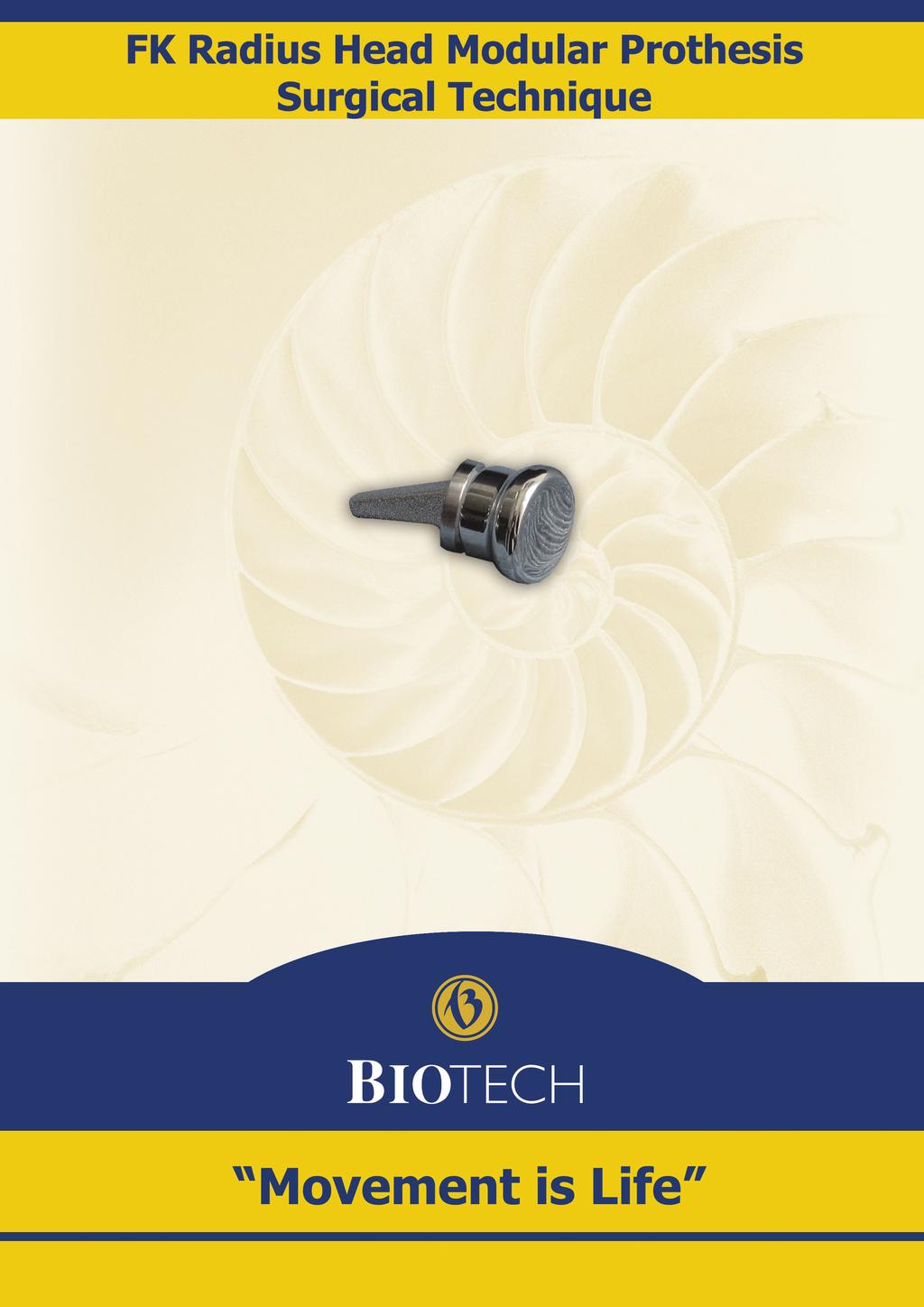
FK radius head modular prosthesis Surgical Technique
|
|
|
- Hope Freeman
- 5 years ago
- Views:
Transcription
1

 The patient has to lie on his back on a normal table. Put the limb on an arm table that is permeable by X-Ray beam.")

2 1. Operation Indication : FK radius head modular prosthesis Surgical Technique Comminuted, non-reconstructable break of radius head, Manson III, IV. Contraction restricting pronation and supination movements due to a former fracture that caused deformity or particular missing of the radius head. Terrible triad: elbow luxation, comminuted fracture of radius head, fracture of processus coronoideus which caused instability. 2. Operation in tourniquet: (In case of adults it is 300 hgmm.) The patient has to lie on his back on a normal table. Put the limb on an arm table that is permeable by X-Ray beam. The limb has to be sterilized from the tourniquet cuffs until the fingertips then left free isolated. 3. Direction of the surgical incision: The incision starts along the line of epicondyle radial humerus, above the radius head and along it in a distance of 5 7 cm. Do not cut ventrally from the epicondyle, because the deep branch of the n. radial can be damaged! The best position to access the radius head is the fascia loculum that is located between the anconeus and extensor carpal. It is useful to cut the ligamentum anulare slightly aslant, to make it easier to sew it together during the reconstruction works. Picture 1 Before the Resection Hohmann lifters have to be placed around the radial neck. Be careful and avoid the rough movements in order to save the branches of the n.radialis profound that are located around the neck in the supinator canal. Resection has to be performed by oscillating saw and blade in perpendicular direction to the longitudinal axis of the radius (Picture 2) We determine the ideal height of the resection with trial prosthesis ( xx). Always choose the shortest size! (Picture 3) Picture 2 Picture 3 1
 using them in rising size sequence until we get to")


.")
3 4. Preparation of the implant s location: The opening and rasping process of the intramedullary canal has to be made by the rasps series ( x) using them in rising size sequence until we get to the maximum size while taking into consideration the thickness of the bone (Picture 5-6). The form of the slant nook on the rasp matches the 10 degree axle position of the neck. Its square based pyramid shape increases the rotational stability. The nib of the rasp has to be pointed towards the stretched thumb during the rasping process (Picture 7). Control the elbow s stability, its adequate valgity, its free movement and the size of the implant by using the trial prosthesis. - The instrument No is for insertion of trial prosthesis (Picture 8). Picture 5 Picture 6 Picture 7 Picture 8 5. Choosing and assembling the implant: 1) Choose the head with the adequate length and size (measured) that is packed and sterilized together with polyethylene inlay (Picture 9). Picture 9 2) Choose the sterilized stem that is going to be placed into the bone hole. Its size must be the same as the one of the rasp used for the last. (Picture 10) Picture 10 3) Flip it together by using the special pincer ( ) (Picture 11). 2
 (Picture 12). 6.")


4 Picture 11 There is a special instrument on the instrument tray that can be used to separate the two components ( ) (Picture 12). 6. Insertion of the assembled implant: Picture 12 Check whether the assembled modular biarticular prosthesis can rotate freely before the insertion. Keep the elbow in varization and semiflectal position and insert the prosthesis into the radius. Pay attention to the right direction of the stem angle (Picture 13-14)! Stave in the prosthesis with the tool that was produced especially for this purpose ( ) (Picture 15). Picture 13 Picture 14 3


 Picture 17")
5 Picture 15 Picture Functional test: The test has to be performed both in stretched and bended elbow as well. Control if the rotation is free or not! The head stays normally in its location, so exactly in the opposite position to the capitulum humerus. (Picture 17-20) Picture 17 Picture 18 Picture 19 Picture 20 4

.")


6 8. Closing of the surgical incision: If it shows luxation tendency, control it after the lig.anulare suture. If it still shows dislocation tendency, the joint must be investigated as the reason could be either interpositum in the Humero-ulnar joint, or the break of the ulna! (Picture 21-22) Picture 21 Picture 22 Joint drainage with Ch Redon drain. Fascia and subcutaneously and after skin suture (Picture 23-24). Picture 23 Picture 24 The limb has to be fixed with a square form upper arm plaster only until the scar recovers or in case of close cooperation it doesn t have to be plastered at all. Long term fixation (maximum 3 weeks) may be needed in case of dislocation tendency, but it may cause restriction in the elbow movement. Redon must be removed after hours. 9. Aftercare: Exercise: Elbow stretching and bending until the pronatio and supinatio pain boundary with the help of gymnastics expert and with controlled active exercises. 10. X-Ray controls: 1. Intraoperational functional test with image enhancement. (Picture 25-28) Picture 25 Picture 26 5


7 Picture 27 Picture Post operational closing image. 3. Control before leaving the hospital. 4. Control in four weeks following the operation. 5. Control in four months following the operation. 6. Control in every 2 years. Besides of the normal recommended controls, radiological investigation is needed every time when the patient has complaints, or if there is a suspicion of prosthesis luxation or other complication occurs (unexpected movement restriction, inflection, pain that could be sign of the slack of prosthesis). 6

8 Instrument set Instrument tray Radius Head Rasp S Radius Head Rasp M Radius Head Rasp L Picker Hammer Radius Head Rasp XL Introducer Radius Head Implant Forceps Assembler Radius Head Extractor Forceps Radius Head Trial 21 S Radius Head Trial 21 M Radius Head Trial 21 L Radius Head Trial 24S Radius Head Trial 24M Radius Head Trial 24L Radius Head Trial 24 XL Radius Head Trial 24 XXL Radius Head Trial 21 XL
Terrible triad of the elbow
 Terrible triad of the elbow Vasu Pai ? Terrible triad of the elbow" Posterior dislocation of the elbow + Fractures of the radial head + Fracture of coronoid process Uncommon injury 5% of dislocation Problems
Modular Ulnar Head surgical technique. Transforming Extremities
 First Choice Modular Ulnar Head surgical technique Transforming Extremities instrumentation Head and Collar Trials Assembly Pad Starter Awl Trial Extractor Osteotomy Guide Stem Trials Implant Impactor
Bipolar Radial Head System
 Bipolar Radial Head System Katalyst Surgical Technique DESCRIPTION The Katalyst Telescoping Bipolar Radial Head implant restores the support and bearing surface of the radial head in the face of fracture,
Integra. Modular Radial Head System SURGICAL TECHNIQUE
 Integra Modular Radial Head System SURGICAL TECHNIQUE Table of Contents System Overview...2 Indications and Contraindications... 3 Modular Radial Head Implant Technique...4 Component Dimensions...8 Implant
Elbow, forearm injuries. K. Fekete
 Elbow, forearm injuries K. Fekete 1. Outline: Fractures of the elbow Dislocation of the elbow Fractures of the forearm Special injuries 2. ANATOMY 3. Lennard Funk Anatomical reminder Three joints: Humero-ulnar
UDHT08.1.qxd:UDHT /03/08 17:14 Page 1. Surgical. Technique. Elbow Prosthesis. RHS Radial Head System.
 UDHT08.1.qxd:UDHT08.1 13/03/08 17:14 Page 1 Surgical Technique Elbow Prosthesis RHS Radial Head System www.tornier.com UDHT08.1.qxd:UDHT08.1 13/03/08 17:14 Page 2 TABLE OF CONTENTS DESIGN RATIONALE INDICATIONS
the shape, the size, the fit Ascension Modular Radial Head
 the shape, the size, the fit Ascension Modular Radial Head WW anatomicdesign stem and head sizes to fit your indications and patient anatomy articular friendly shape reduces edge loading on the capitellum
Integra. Katalyst Bipolar Radial Head System SURGICAL TECHNIQUE
 Integra Katalyst Bipolar Radial Head System SURGICAL TECHNIQUE Surgical Technique As the manufacturer of this device, Integra does not practice medicine and does not recommend this or any other surgical
Integra. Modular Radial Head System SURGICAL TECHNIQUE
 Integra Modular Radial Head System SURGICAL TECHNIQUE Table of Contents System Overview...2 Indications and Contraindications... 3 Modular Radial Head Implant Technique...4 Component Dimensions...8 Implant
Surgical. Technique CRF II. Radial Head Prosthesis
 Surgical Technique CRF II CONTENTS CONTENTS 1. OVERVIEW 2. INDICATIONS 1. Acute Trauma 2. Trauma Sequalae 3. DESIGN RATIONALE 4. SURGICAL TECHNIQUE 1. Preoperative assessment 2. Patient positioning 3.
RADIOGRAPHY OF THE ELBOW & HUMERUS
 RADIOGRAPHY OF THE ELBOW & HUMERUS Patient Position: ELBOW AP Projection in same plane Part Position: Hand in ; patient Centered to Humeral epicondyles Central Ray: Structures Shown: AP Elbow Criteria
SURGICAL TECHNIQUE GUIDE
 SURGICAL TECHNIQUE GUIDE As described by: Jorge L. Orbay, M.D. Miami Hand & Upper Extremity Institute Miami, Florida. 1 ELBOW LANDMARKS With the elbow flexed 90 0, palpate and mark the lateral epicondyle.
rhead System Extended stems Operative technique
 rhead System Extended stems rhead System rhead System Extended stems Contents 1. Indications and contraindications... 3 2..... 4 The initial incision...4 Capsular exposure...5 Using the radial head resection
rhead System Radial Head Arthroplasty Operative technique
 rhead System Radial Head Arthroplasty Operative technique rhead System Operative technique rhead System Radial Head Arthroplasty Contents 1. Indications and contraindications... 3 2. Operative technique....
OPERATIVE TECHNIQUE. Intermediate. elbow prosthesis. mobile radial cup cemented radial stem. Orthopaedic Implants
 OPERATIVE TECHNIQUE Intermediate elbow prosthesis mobile radial cup cemented radial stem www.aston-medical.com Orthopaedic Implants I ntroduc tion The Evolutive prosthesis has been designed as the culmination
KATALYST. Bipolar Radial Head System. Surgical Technique. orthopedics. KATALYST English. PRODUCTS FOR SALE IN EUROPE, MIDDLE-EAST and AFRICA ONLY
 KATALYST Bipolar Radial Head System KATALYST English Surgical Technique orthopedics UPPER extremity PRODUCTS FOR SALE IN EUROPE, MIDDLE-EAST and AFRICA ONLY KATALYST Introduction Description The Katalyst
Chapter 4: Forearm 4.3 Forearm shaft fractures, transverse (12-D/4)
") AO Manual of ESIN in children s fractures Chapter 4: Forearm 4.3 Forearm shaft fractures, transverse (12-D/4) Title AO Manual of ESIN in children Subtitle Elastic stable intramedullary nailing (ESIN) Author
AO Manual of ESIN in children s fractures Chapter 4: Forearm 4.3 Forearm shaft fractures, transverse (12-D/4) Title AO Manual of ESIN in children Subtitle Elastic stable intramedullary nailing (ESIN) Author
The Muscular System. Chapter 10 Part C. PowerPoint Lecture Slides prepared by Karen Dunbar Kareiva Ivy Tech Community College
 Chapter 10 Part C The Muscular System Annie Leibovitz/Contact Press Images PowerPoint Lecture Slides prepared by Karen Dunbar Kareiva Ivy Tech Community College Table 10.9: Muscles Crossing the Shoulder
Total Knee Original System Primary Surgical Technique
 Surgical Procedure Total Knee Original System Primary Surgical Technique Where as a total hip replacement is primarily a bony operation, a total knee replacement is primarily a soft tissue operation. Excellent
MCQWeek2. All arise from the common flexor origin. The posterior aspect of the medial epicondyle is the common flexor origin.
 MCQWeek2. 1. Regarding superficial muscles of anterior compartment of the forearm: All arise from the common flexor origin. The posterior aspect of the medial epicondyle is the common flexor origin. Flexor
E-CENTRIX. Ulnar Head Replacement SURGICAL TECHNIQUE
 E-CENTRIX Ulnar Head Replacement SURGICAL TECHNIQUE E-CENTRIX ulnar head REPLACEMENT surgical technique as described by GRAHAM KING, MD University of Western Ontario London, Ontario, Canada E-CENTRIX ulnar
modular RADIAL HEAD E VOLVE
 E VOLVE modular RADIAL HEAD surgical technique 1-5 in situ assembly 6-7 alternate O.R. back table assembly 8 Proper surgical procedures and techniques are the responsibility of the medical professional.
Ascension. Silicone MCP surgical technique. surgical technique Ascension Silicone MCP
 Ascension Silicone MCP surgical technique WW 2 Introduction This manual describes the sequence of techniques and instruments used to implant the Ascension Silicone MCP (FIGURE 1A). Successful use of this
GENERIC, LOGIC, INTEGRALE TO.H.GB.011/1.0
 Surgical technique mechanical instrumentation GENERIC, LOGIC, INTEGRALE TO.H.GB.011/1.0 2 Pre-surgical planning By means of radiological assessment and templates, it is possible to: - determine the position
Distal Ulnar Locking Plate
 INDEX Indications Patient Position Surgical Technique - Step 1 Approach - Step 2 Plate Contouring - Step 3 Fracture Reduction - Step 4 Distal Plate Fixation - Step 5 Confirm Proper Reconstruction - Step
Elbow Joint Anatomy ELBOW ANATOMY, BIOMECHANICS. Bone Anatomy. Bone Anatomy. Property of VOMPTI, LLC
 ELBOW ANATOMY, BIOMECHANICS AND PATHOLOGY Kristin Kelley, DPT, OCS, FAAOMPT Elbow Joint Anatomy Joint articulations Humeroulnar Radiohumeral Radioulnar (proximal and distal) Orthopaedic Manual Physical
The Bio-Modular Choice Shoulder System,
 Surgical Technique The Bio-Modular Choice Shoulder System, designed for both total and hemiarthroplasty of the shoulder, has enjoyed nearly two decades of clinical success. The variety of head types and
Motec. Wrist Joint Arthrodesis Metacarpal Taper and Radius Connector
 Motec Wrist Joint Arthrodesis Metacarpal Taper and Radius Connector Motec Wrist Joint Arthrodesis The Motec Wrist Joint Arthrodesis has been developed as a part of the Motec Wrist Prosthesis family to
UNI-Elbow System Radio-Capitellar Replacement. Operative technique
 UNI-Elbow System Radio-Capitellar Replacement Operative technique 0798-2016 UniElbow Op tech (EMS-ST-1) - proof 3 UNI-Elbow System Operative technique UNI-Elbow System Radio-Capitellar Replacement Contents
RADIAL HEAD PROSTHESIS SYSTEM For primary and revision joint replacement of the radial head
 RADIAL HEAD PROSTHESIS SYSTEM For primary and revision joint replacement of the radial head This publication is not intended for distribution in the USA. SURGICAL TECHNIQUE Warning This description alone
#12. Joint نبيل خوري
 #12 30 Anatomy Joint هيام الر جال 9/10/2015 نبيل خوري Salam Awn Some notes before starting : ** Not all slides are included, so I recommend having a look at the slides beside this sheet ** If you find
OSKAR Radial Head Prosthesis
 Technique Guide OSKAR Radial Head Prosthesis DESCRIPTION The OSKAR Radial Head Implant is a pliable, onepiece intramedullary-stemmed cuffed implant designed to help preserve the joint space and relationships
Radial Head Prosthesis System
 For Primary and Revision Joint Replacement of the Radial Head Radial Head Prosthesis System Surgical Technique TABLE OF CONTENTS INTRODUCTION Radial Head Prosthesis System 2 Indications and Contraindications
Lecture 9: Forearm bones and muscles
 Lecture 9: Forearm bones and muscles Remember, the region between the shoulder and the elbow = brachium/arm, between elbow and wrist = antebrachium/forearm. Forearm bones : Humerus (distal ends) Radius
Fractures and dislocations around elbow in adult
 Lec: 3 Fractures and dislocations around elbow in adult These include fractures of distal humerus, fracture of the capitulum, fracture of the radial head, fracture of the olecranon & dislocation of the
Distal Humerus surgical technique
 Distal Humerus surgical technique Distal Humerus surgical technique MUTARS was developed in co-operation with Prof. Dr. W. Winkelmann, and Prof. Dr. G. Gosheger, Clinic and Polyclinic for General Orthopedics
RADIOGRAPHY OF THE WRIST
 RADIOGRAPHY OF THE WRIST Patient Position: WRIST PA Projection, elbow in same plane Part Position: Hand ; fingers centered to IR Central Ray: Structures Shown: NOTE: Optional AP projection best demonstrates
Motec. Wrist Joint Arthrodesis Metacarpal Nail and Radius Connector
 Motec Wrist Joint Arthrodesis Metacarpal Nail and Radius Connector Motec Wrist Joint Arthrodesis The Motec Wrist Joint Arthrodesis has been developed as a part of the Motec Wrist Prosthesis family to enable
LOCKING TEP LOCKING TITANIUM ELASTIC PIN INTRAMEDULLARY NAIL
 LOCKING TEP LOCKING TITANIUM ELASTIC PIN INTRAMEDULLARY NAIL ... Index -3 3-8 8 9 9 0 7 Introduction Features Indicatiıons Surgical Technique Femoral Surgical Technique Tibial Surgical Technique Ulna Radius
Locking Radial Head Plates
 Locking Radial Head Plates Locking Radial Head Plates Since 1988, Acumed has been designing solutions to the demanding situations facing orthopaedic surgeons, hospitals and their patients. Our strategy
SURGICAL TECHNIQUE GUIDE
 SURGICAL TECHNIQUE GUIDE As described by: Jorge L. Orbay, M.D. Miami Hand Institute Miami, Florida. System Overview The ALIGN Radial Head System is designed to restore the kinematics of the native radial
Fascial Compartments of the Upper Arm
 Fascial Compartments of the Upper Arm The upper arm is enclosed in a sheath of deep fascia and has two fascial septa: 1- Medial fascial septum (medial intermuscular septum): attached to the medial supracondylar
2.7 mm/3.5 mm Variable Angle LCP Elbow System DJ9257-B 1
 2.7 mm/3.5 mm Variable Angle LCP Elbow System DJ9257-B 1 System overview Simply complete: A comprehensive system, consisting of five (5) distal humerus plates and three (3) types of olecranon plates Implant
Motec. Wrist Joint Arthrodesis Straight Double Taper
 Motec Wrist Joint Arthrodesis Straight Double Taper Motec Wrist Joint Arthrodesis The Motec Wrist Joint Arthrodesis has been developed as a part of the Motec Wrist Prosthesis family to enable easy conversion
Forearm Fracture Solutions. Product Overview
 Forearm Fracture Solutions Product Overview Acumed Forearm Fracture Solutions Acumed Forearm Fracture Solutions includes plating and rodding systems with a range of diaphyseal radius and ulna fracture
Humerus Block. Discontinued December 2016 DSEM/TRM/0115/0296(1) Surgical Technique. This publication is not intended for distribution in the USA.
 Surgical Technique. This publication is not intended for distribution in the USA.") Humerus Block Surgical Technique Discontinued December 2016 DSEM/TRM/0115/0296(1) This publication is not intended for distribution in the USA. Instruments and implants approved by the AO Foundation. Contents
Humerus Block Surgical Technique Discontinued December 2016 DSEM/TRM/0115/0296(1) This publication is not intended for distribution in the USA. Instruments and implants approved by the AO Foundation. Contents
Olecranon Osteotomy Nail. For simple fractures and osteotomies of the olecranon.
 Olecranon Osteotomy Nail. For simple fractures and osteotomies of the olecranon. Technique Guide Discontinued June 2016; AVAILABLE FOR IMPLANT REMOVAL PURPOSES ONLY DSEM/TRM/0517/0843 Table of Contents
Radial Head Fractures Save or Replace?
 Radial Head Fractures Save or Replace? Current Solutions in Orthopedic Trauma Sepember 19, 2015 Jorge L. Orbay MD. Disclosure Skeletal Dynamics Elbow Joint Ulno-Humeral and Radio-Capitellar The key joint
Silicone Finger Implant
 Silicone Finger Implant Manufactured from high tear resistant implant grade silicone Surgical Technique Eleven, evenly scaled sizes for comprehensive anatomical fit Simple, precise instrumentation Designed
Ascension. MCP surgical technique. surgical technique Ascension MCP PyroCarbon Total Joint
 Ascension MCP surgical technique PyroCarbon Total Joint WW 1.0 Table of Contents 2.0 Introduction............................ 2 3.0 Ascension MCP Implants................ 2 4.0 Instrumentation for MCP
Anatomical Shoulder Glenoid. Surgical Technique
 Anatomical Shoulder Glenoid Surgical Technique Anatomical Shoulder Glenoid Surgical Technique 3 Table of Contents Glenoid Preparation Surgical Steps 4 Anatomical Shoulder Glenoid 4 Glenoid Components
Elbow Elbow Anatomy. Flexion extension. Pronation Supination. Anatomy. Anatomy. Romina Astifidis, MS., PT., CHT
 Elbow Elbow Anatomy Romina Astifidis, MS., PT., CHT Curtis National Hand Center Baltimore, MD October 6-8, 2017 Link between the arm and forearm to position the hand in space Not just a hinge Elbow = 70%
Elbow Exercise Program
 Elbow Exercise Program Name: Date: Diagnosis: Date of Surgery: 1. Deep Friction Massage deep transverse friction across area of elbow that is sore. 5 minutes, several times daily. 2. Grip grip apparatus,
Surgical Technique. *smith&nephew POLARSTEM Cementless Stem System
 Surgical Technique *smith&nephew POLARSTEM Cementless Stem System POLARSTEM Cementless Stem System Contents Introduction... 3 Indications... 4 Contraindications... 4 Case Studies... 5 Preoperative Planning...
Cubital Tunnel Syndrome
 Disclaimer This movie is an educational resource only and should not be used to manage Orthopaedic Health. All decisions about must be made in conjunction with your Physician or a licensed healthcare provider.
BIOTECH FUTURE KNEE Minimal Invasive (and classic) Surgical Technique
 Surgical Technique") BIOTECH FUTURE KNEE Minimal Invasive (and classic) Surgical Technique Page No. Product description 2. Pre-operational planning 3. 1st step Femoral resection 4. P/S Femoral component 7. 2nd step Proximal
BIOTECH FUTURE KNEE Minimal Invasive (and classic) Surgical Technique Page No. Product description 2. Pre-operational planning 3. 1st step Femoral resection 4. P/S Femoral component 7. 2nd step Proximal
Anatomical Considerations Regarding the Posterior Interosseous Nerve During Posterolateral Approaches to the Proximal Part of the Radius *
 Anatomical Considerations Regarding the Posterior Interosseous Nerve During Posterolateral Approaches to the Proximal Part of the Radius * BY THOMAS DILIBERTI, M.D., MICHAEL J. BOTTE, M.D., AND REID A.
CemtA Stem. Surgery Manual. cemented femoral Stem. veterinary implants
 CemtA Stem cemented femoral Stem Surgery Manual veterinary implants CemtA Stem use only with bonecement Size 1 Size 2 #4 #5 #6 #7 #8 Size Range 2 1: 1: veterinary implants Surgery Manual Guidelines for
Elbow Problems.
 Elbow Problems www.fisiokinesiterapia.biz Anatomy Hinged joint formed by humerus and ulna produces flexion and extension Rotation producing pronation and supination from radial head and humerus Assessment
Cemented femoral stem - type CSC
 Cemented femoral stem - type CSC Cemented Femoral Hip Joint Components ARTHROPLASTY Implant Description Preface The cemented femoral stem type CSC with centralizer was designed using the latest knowledge
REVISION KNEE PROSTHESIS ROTARY PIVOT TOTAL KNEE REPLACEMENT. w w w. a s t o n - m e d i c a l. c o m
 OPERATING TECHNIQUE REVISION KNEE PROSTHESIS ROTARY PIVOT TOTAL KNEE REPLACEMENT w w w. a s t o n - m e d i c a l. c o m I N T R O D U C T I O N O D U C T I O N Parameters to be taken into account: - Failure
AcUMEDr. FoREARM ROD SYSTEM
 AcUMEDr FoREARM ROD SYSTEM FoREARM ROD SYSTEM Since 1988 Acumed has been designing solutions to the demanding situations facing orthopedic surgeons, hospitals and their patients. Our strategy has been
A Patient s Guide to Adult Forearm Fractures
 A Patient s Guide to Adult Forearm Fractures Orthopedic and Sports Medicine 825 South 8th Street, #550 Minneapolis, MN 55404 Phone: 612-333-5000 Fax: 612-333-6922 1 DISCLAIMER: The information in this
Hip Replacement - Anterior
 Hip Replacement - Anterior Anterior hip replacement surgery is an alternative to hip replacements where the surgeon accesses the hip joint from the side or through the buttocks. The anterior procedure
MLT Muscle(s) Patient Position Therapist position Stabilization Limb Position Picture Put biceps on slack by bending elbow.
 Patient Position Therapist position Stabilization Limb Position Picture Put biceps on slack by bending elbow.") MLT Muscle(s) Patient Position Therapist position Stabilization Limb Position Picture Put biceps on slack by bending elbow. Pectoralis Minor Supine, arm at side, elbows extended, supinated Head of Table
MLT Muscle(s) Patient Position Therapist position Stabilization Limb Position Picture Put biceps on slack by bending elbow. Pectoralis Minor Supine, arm at side, elbows extended, supinated Head of Table
THE HUMERUS 20 THE HUMERUS* CROSS SECTION CROSS SECTION SUPERIOR VIEW
 20 THE HUMERUS* CROSS SECTION CROSS SECTION SUPERIOR VIEW The marrow canal of the humerus is funnel-shaped. Its successful pinning is influenced by many factors. With a few exceptions, the entire humerus
The arm: *For images refer back to the slides
 The arm: *For images refer back to the slides Muscles of the arm: deltoid, triceps (which is located at the back of the arm), biceps and brachialis (it lies under the biceps), brachioradialis (it lies
Joints of the upper limb II
 Joints of the upper limb II Prof. Abdulameer Al-Nuaimi E-mail: a.al-nuaimi@sheffield.ac.uk E. mail: abdulameerh@yahoo.com Elbow joint The elbow joint is connecting the upper arm to the forearm. It is classed
2.4 mm LCP Radial Head Plates. Part of the Synthes LCP Distal Radius Plate System.
 2.4 mm LCP Radial Head Plates. Part of the Synthes LCP Distal Radius Plate System. Technique Guide Instruments and Implants approved by the AO Foundation Table of Contents Introduction 2.4 mm LCP Radial
Silicone PIP, MCP & MCP-X (PreFlex)
") Silicone PIP, MCP & MCP-X (PreFlex) Finger Joint Arthroplasty Operative Technique Silicone PIP Silicone MCP Silicone PreFlex (MCP-X) Stryker Disclaimer This publication sets forth detailed recommended
Silicone PIP, MCP & MCP-X (PreFlex) Finger Joint Arthroplasty Operative Technique Silicone PIP Silicone MCP Silicone PreFlex (MCP-X) Stryker Disclaimer This publication sets forth detailed recommended
NEUFLEX MCP/PIP FINGER JOINT IMPLANT SYSTEMS SURGICAL TECHNIQUE. This publication is not intended for distribution in the USA.
 NEUFLEX MCP/PIP FINGER JOINT IMPLANT SYSTEMS This publication is not intended for distribution in the USA. SURGICAL TECHNIQUE MCP SURGICAL TECHNIQUE Summary provided by designing surgeon Arnold-Peter C.
Hand and wrist emergencies
 Chapter1 Hand and wrist emergencies Carl A. Germann Distal radius and ulnar injuries PEARL: Fractures of the distal radius and ulna are the most common type of fractures in patients younger than 75 years.
Hands PA; Obl. Lat.; Norgaard s Thumb AP; Lat. PA. PA; Lat.: Obls.; Elongated PA with ulnar deviation
 Projections Region Basic projections Additional / Modified projections Upper Limbs Hands PA; Obl. Lat.; Norgaard s Thumb ; Lat. PA Fingers PA; Lat. Wrist PA; Lat. Obls. Scaphoid Lunate Trapezium Triquetral
URSA HEMI-SHOULDER ARTHROPLASTY B I O T E K
 URSA HEMI-SHOULDER ARTHROPLASTY SURGICAL TECHNIQUE B I O T E K 2 Surgical Position Once general anesthesia has been satisfactorily induced, or a supraclavicular nerve block has been given, the patient
WINSTA-R. Distal Radius System
 Distal Radius System Table of Contents Introduction WINSTA-R System 2 Indication 2 Surgical Technique Palmar Access for Radius Plate 3 Dorsal Access for Radius Plate 3 Positioning of the Radius Plate
LCP Distal Humerus Plates
 The anatomic fixation system for the distal humerus with angular stability Surgical technique LCP Locking Compression Plate Contents Indications and contraindications 2 Implants 3 Instruments 5 Preparation
Nerve Injury. 1) Upper Lesions of the Brachial Plexus called Erb- Duchene Palsy or syndrome.
 Upper Lesions of the Brachial Plexus called Erb- Duchene Palsy or syndrome.") Nerve Injury - Every nerve goes to muscle or skin so if the nerve is injured this will cause paralysis in the muscle supplied from that nerve (paralysis means loss of function) then other muscles and other
Nerve Injury - Every nerve goes to muscle or skin so if the nerve is injured this will cause paralysis in the muscle supplied from that nerve (paralysis means loss of function) then other muscles and other
Surgical Technique. Distal Humerus Locking Plate
 Surgical Technique Distal Humerus Locking Plate PERI-LOC Locked Plating System Distal Humerus Locking Plate Surgical Technique Table of Contents Introduction...2 Indications...3 Plate Features...3 Patient
Ligaments of Elbow hinge: sagittal plane so need lateral and medial ligaments
 Ligaments of Elbow hinge: sagittal plane so need lateral and medial ligaments Ulnar Collateral ligament on medial side; arising from medial epicondyle and stops excess valgus movement (lateral movement)
Anterior Approach Surgical Technique. Paragon Stem System. enabling people to enjoy life
 Anterior Approach Surgical Technique Paragon Stem System enabling people to enjoy life Contents Pre-Operative Planning... 2 Suggested Templating Method... 2 Surgical Technique... 3 Surgical Approach...
Instrument set for endoscopically assisted decompression of the ulnar nerve. Arthroscopy
 Instrument set for endoscopically assisted decompression of the ulnar nerve Arthroscopy Instrument set for endoscopically assisted decompression of the ulnar nerve Introduction The compression of the ulnar
The Biomechanics of the Human Upper Extremity-The Elbow Joint C. Mirzanli Istanbul Gelisim University
 The Biomechanics of the Human Upper Extremity-The Elbow Joint C. Mirzanli Istanbul Gelisim University Structure of The Elbow Joint A simple hinge joint, actually categorized as a trochoginglymus joint
The Wrist Fusion Set. Stainless Steel and Titanium TECHNIQUE GUIDE. Instruments and implants approved by the AO Foundation
 The Wrist Fusion Set Stainless Steel and Titanium TECHNIQUE GUIDE Instruments and implants approved by the AO Foundation Three Plate Options Stainless Steel or Titanium* Standard Bend Stainless Steel [242.510]
Case Presentation: Comminuted Radial Head Fracture
 11/28/2017 Current Solutions in Orthopaedic Trauma Case Presentation: Comminuted Radial Head Fracture Melvin P. Rosenwasser, MD Robert E. Carroll Professor of Surgery of the Hand Chief, Orthopaedic Hand
Transfemoral Amputation
 Transfemoral Amputation Pre-Op: 42 year old male who sustained severe injuries in a motorcycle accident. Note: he is a previous renal transplant recipient and is on immunosuppressive treatments. His injuries
Nerves of the upper limb Prof. Abdulameer Al-Nuaimi. E. mail:
 Nerves of the upper limb Prof. Abdulameer Al-Nuaimi E-mail: a.al-nuaimi@sheffield.ac.uk E. mail: abdulameerh@yahoo.com Brachial plexus Median nerve After originating from the brachial plexus in the axilla,
RegJoint. Instructions for use
 RegJoint Instructions for use RegJoint is indicated for arthroplasties in small joints in hand and foot for patients suffering of rheumatoid arthritis or osteoarthritis. The specific target joints are
Types of Plates 1. New Dynamic Compression Plate: Diaphyseal fracture: Radius, Ulna, Humerus, Rarely tibia
 Types of Plates 1. New Dynamic Compression Plate: DCP Diaphyseal fracture: Radius, Ulna, Humerus, Rarely tibia 1. Undercut adjacent to the holes low contact: less stress shield 2. Undercut at the undersurface
Chapter 8. The Pectoral Girdle & Upper Limb
 Chapter 8 The Pectoral Girdle & Upper Limb Pectoral Girdle pectoral girdle (shoulder girdle) supports the arm consists of two on each side of the body // clavicle (collarbone) and scapula (shoulder blade)
PediFlex Advanced Elastic Stable Intramedullary Nails SURGICAL TECHNIQUE
 PediFlex Advanced Elastic Stable Intramedullary Nails SURGICAL TECHNIQUE 2 2 TABLE OF CONTENTS System Overview Indications... 4 Implant Selection... 4 Surgical Technique Product Information Inserter Assembly
Presidency General Hospital, Calcutta
 CONGENITAL ANOMALY OF HAND: " MIRROR HAND " By M. MUKERJI, F.R.C.S. Presidency General Hospital, Calcutta Case Report.--S. B., aged 4 months, was born with eight fingers and no thumb on the left hand and
Terms of Movements by Prof. Dr. Muhammad Imran Qureshi
 Terms of Movements by Prof. Dr. Muhammad Imran Qureshi Three systems of the body work in coordination to perform various movements of the body. These are: A System of Bones (Osteology), A System of Muscles
3.5 mm LCP Hook Plate
 Part of the DePuy Synthes Locking Compression Plate (LCP ) System 3.5 mm LCP Hook Plate Surgical Technique Table of Contents Introduction 3.5 mm LCP Hook Plate 2 AO Principles 4 Indications 5 Clinical
region of the upper limb between the shoulder and the elbow Superiorly communicates with the axilla.
 1 region of the upper limb between the shoulder and the elbow Superiorly communicates with the axilla. Inferiorly, a number of important structures pass between arm & forearm through cubital fossa. 2 medial
Instrument and Implant for wrist fracture
 Instrument and Implant for wrist fracture Jansri Janpanya Product specialist The Bangkok Unitrade Co,.ltd. Objectives Type of LCP for distal radius Fx. The new LCP design for distal radius Fx. Have knowledge
Integra. First Choice DRUJ System Partial Ulnar / Modular Ulnar Head Implant SURGICAL TECHNIQUE
 Integra First Choice DRUJ System Partial Ulnar / Modular Ulnar Head Implant SURGICAL TECHNIQUE Integra First Choice DRUJ System System Overview The Integra First Choice DRUJ System was developed through
Foot & Ankle. Smart Toe II. Intramedullary Implant. Operative Technique. Foot & Ankle
 Foot & Ankle Smart Toe II Intramedullary Implant Operative Technique Foot & Ankle Smart Toe This publication sets forth detailed recommended procedures for using Stryker Osteosynthesis devices and instruments.
Wright Medical Technology, Inc Cherry Road Memphis, TN
 Wright Medical Technology, Inc. 1023 Cherry Road Memphis, TN 38117 800 238 7117 901 867 9971 www.wmt.com Wright Medical EMEA Atlas Arena, Australia Building Hoogoorddreef 7 1101 BA Amsterdam the Netherlands