Posterior Slipped Capital Femoral Epiphysis Joseph Junewick, MD FACR
|
|
|
- Patrick Reeves
- 5 years ago
- Views:
Transcription
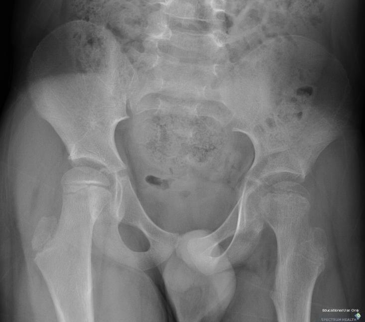
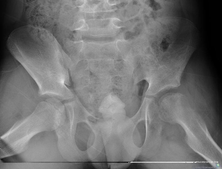
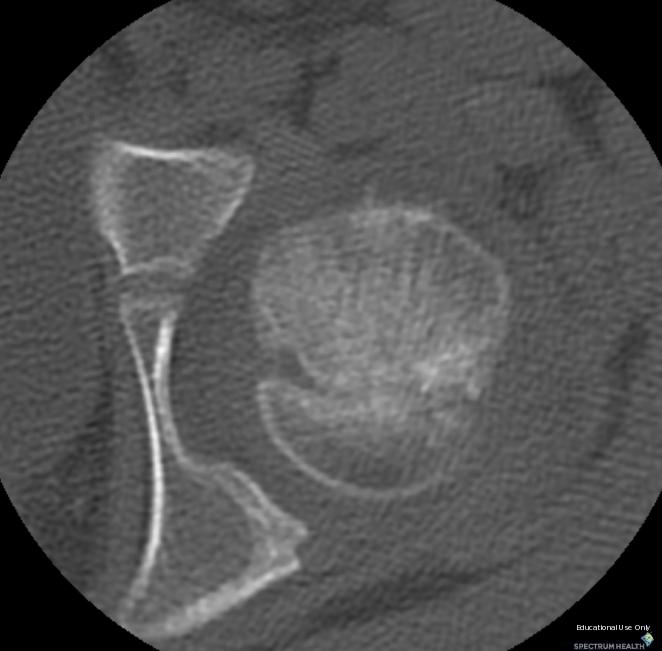
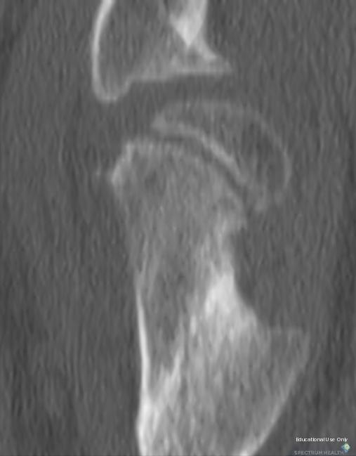
1 Posterior Slipped Capital Femoral Epiphysis Joseph Junewick, MD FACR 08/11/2010 History 6 year old male with intermittent hip pain for several months, acutely worsened after climbing the sand dunes. Diagnosis Posterior Slipped Capital Femoral Epiphysis-Probable Stickler Syndrome Discussion Slipped capital femoral epiphysis (SCFE) is a relatively atraumatic fracture through the proximal femoral physis. SCFE is the most common hip malady in adolescents, affecting males more than females and African-Americans more than caucasions. SCFE prior to adolescence suggests underlying process such as malnutrition, endocrinopathy, developmental dysplasia, and coxa valga. Pathophysiology is probably related to oblique orientation of the physis and increased body weight and activity (particularly abduction, external rotation and extension) during adolescence. Valgus displacement often presents with a relatively normal appearance on anteroposterior radiographs. Valgus SCFE may be associated with obesity, coxa valga, hypopituitarism, and Stickler syndrome. Posterolateral displacement of the femoral epiphysis makes in situ fixation of valgus SCFE more difficult, due to the necessity of a more medial starting point. Stickler syndrome is connective tissue disease characterized by midface hypoplasia, cleft palate, myopia, sensorineural hearing loss, joint hypermobility, and epiphyseal dysplasia (short stature). Radiographically there is mild to moderateflattening of the vertebral bodies, undermodeling of the long bones with broad epiphyses (particularly the femora and tibiae), and premature arthropathy. Stickler syndrome is an autosomal dominant with marked intrafamilial and mutation-dependent variability; the molecular defect is related to the COL2A1 gene. Findings CR-Neutral and abduction views show coxa valga and slipped left capital femoral epiphysis. Also note that the height of the femoral epiphyses is decreased. CT-Axial and sagittal images show the femoral head to be posterior relative to the neck. Note the buttressing posteriorly at the neck near the physis related to attempted healing, indicating an acute on chronic process. Reference Boles CA, el-khoury GY. Slipped capital femoral epiphysis. RadioGraphics (1997); 17: Shank CF, Thiel EJ, Klingele KE. Valgus Slipped Capital Femoral Epiphysis: Prevalence, Presentation, and Treatment Options. J of Pediat Orthop (2010); 30(2): Baba T, Shitoto K. Stickler syndrome associated with slippled capital femoral epiphysis. Eur J Orthop Surg Traumatol (2010); 20:
2
3
4
5
6 Sponsored By Disclaimer This teaching site is partially funded by an educational grant from GE Healthcare and Advanced Radiology Services, PC. The material on this site is independently controlled by Advanced Radiology Services, PC, and GE Healthcare and Spectrum Health have no influence over the content of this site Content Download Agreement The cases and images on this website are owned by Spectrum Health. Permission is granted (for nonprofit educational purposes) to download and print materials to distribute for the purpose of facilitating the education of health professionals. The authors retain all rights to the material and users are requested to acknowledge the source of the material. Site Disclaimer This site is developed to reach healthcare professionals and medical students. Nothing this site should be considered medical advice. Only your own doctor can help you make decisions about your medical care. If you have a specific medical question or are seeking medical care, please contact your physician. The information in this website is provided for general medical education purposes only and is not meant to substitute for the independent medical judgment of a physician relative to diagnostic and treatment options of a specific medical condition. The viewpoints expressed in these cases are those of the authors. They do not represent an endorsement. In no event will Advanced Radiology Associates, PC, Spectrum Health Hospitals (Helen Devos Children's Hospital) or GE Healthcare be liable for any decision made or action taken in reliance upon the information provided through this website.
Spinal LCH Joseph Junewick, MD FACR
 Spinal LCH Joseph Junewick, MD FACR 05/16/2009 History 16 year old female with multiply recurrent Langerhans Cell Histiocytosis now with severe left sided neck pain. Diagnosis Langerhans Cell Histiocytosis
Diskitis Joseph Junewick, MD FACR
 Diskitis Joseph Junewick, MD FACR 09/20/2010 History 2 year old with fever, back pain and elevated sedimentation rate. Diagnosis Diskitis Discussion Diskitis is an inflammatory process of the intervertebral
Chiari III Joseph Junewick, MD FACR
 Chiari III Joseph Junewick, MD FACR 07/02/2010 History Newborn with suboccipital mass. Diagnosis Chiari III Additional Clinical Surgery-Skin covered suboccipital cystic mass confined by the dura. Pathology-Leptomeningeal
Atlanto-occipital Dislocation Joseph Junewick, MD FACR
 Atlanto-occipital Dislocation Joseph Junewick, MD FACR 09/23/2009 History 12 year old male restrained back seat passenger in a car hit by a snowplow. Diagnosis Atlanto-occipital Dislocation Discussion
Testicular Microlithiasis related to McCune-Albright Syndrome Joseph Junewick, MD FACR
 Testicular Microlithiasis related to McCune-Albright Syndrome Joseph Junewick, MD FACR 04/25/2010 History 12 year old with McCune-Albright syndrome. Diagnosis Testicular Microlithiasis related to Mcune-Albright
Transverse Dural Sinus Thrombosis Joseph Junewick, MD FACR
 Transverse Dural Sinus Thrombosis Joseph Junewick, MD FACR 03/19/2010 History Child with headache and otomastoiditis. Diagnosis Dural venous thrombosis secondary to mastoiditis Discussion The cerebral
Ulcerative Colitis Joseph Junewick, MD FACR
 Ulcerative Colitis Joseph Junewick, MD FACR 06/04/2010 History 16 year old male with hematochezia and anemia. Diagnosis Ulcerative Colitis Additional Clinical History of ulcerative colitis. Discussion
Chance Fracture Joseph Junewick, MD FACR
 Chance Fracture Joseph Junewick, MD FACR 08/02/2010 History Restrained teenager involved in motor vehicle accident. Diagnosis Chance Fracture (Hyperflexion-Distraction Injury) Discussion Chance-type spinal
Term Hypoxic Ischemic Injury Joseph Junewick, MD FACR
 Term Hypoxic Ischemic Injury Joseph Junewick, MD FACR 08/11/2010 History Term infant with perinatal distress and attempted forceps delivery. Diagnosis Term Hypoxic Ischemic Injury Discussion Encephalopathy
Paraspinal Venous Malformation Joseph Junewick, MD FACR
 Paraspinal Venous Malformation Joseph Junewick, MD FACR 06/04/2010 History 2 year old with history of fall. Rule out spinal injury. Diagnosis Paraspinal Venous Malformation Additional Clinical CT of the
Vein of Galen Malformation Joseph Junewick, MD FACR
 Vein of Galen Malformation Joseph Junewick, MD FACR 04/14/2018 History Midline cystic intracranial mass on prenatal ultrasound. Diagnosis Vein of Galen Malformation Discussion In normal fetal development,
Pleural Empyema Joseph Junewick, MD FACR
 Pleural Empyema Joseph Junewick, MD FACR 03/19/2010 History Teenager with persistent fever and cough. Pneumonia diagnosed 1 week ago. Diagnosis Pleural Empyema Additional Clinical Surgery-Clear fluid with
Radiation Pneumonitis Joseph Junewick, MD FACR
 Radiation Pneumonitis Joseph Junewick, MD FACR 03/19/2010 History 16 year old with history of relapsed stage IV-A Hodgkin disease. Prior pulmonary involvement was irradiated. Diagnosis Radiation Pneumonitis
Scrofula Joseph Junewick, MD FACR
 Scrofula Joseph Junewick, MD FACR 06/20/2012 History 4 year old male with refractory cervical adenopathy Diagnosis Scrofula Additional Clinical Positive PPD skin test. Discussion Scrofula refers to tuberculous
Tuberculous Meningitis Joseph Junewick, MD FACR
 Tuberculous Meningitis Joseph Junewick, MD FACR 08/11/2010 History 14 month old with fever and increasing lethargy. Diagnosis Tuberculous Meningitis Additional Clinical Grandmother with active tuberculosis.
Presacral Neuroblastoma Joseph Junewick, MD FACR
 Presacral Neuroblastoma Joseph Junewick, MD FACR 01/12/2010 History 16 month old male with irritability. Diagnosis Presacral Neuroblastoma Additional Clinical Initial US to evaluate for intussusception
Neuroblastoma Joseph Junewick, MD FACR
 Neuroblastoma Joseph Junewick, MD FACR 03/18/2011 History 15 month old with anemia. Diagnosis Neuroblastoma Discussion Neuroblastic tumors derive from primordial neural crest cells destined for sympathetic
Pituitary Macroadenoma Joseph Junewick, MD FACR
 Pituitary Macroadenoma Joseph Junewick, MD FACR 08/13/2010 History 12 year old female with headache and visual disturbance. Diagnosis Pituitary Macroadenoma Additional Clinical Markedly elevated growth
Bilateral Retinoblastoma Joseph Junewick, MD FACR
 Bilateral Retinoblastoma Joseph Junewick, MD FACR 06/11/2010 History 17 month old adopted female with proptosis. Diagnosis Bilateral Retinoblastoma Discussion Retinoblastoma is the most common pediatric
Retroperitoneal Teratoma Heather Borders, MD
 Retroperitoneal Teratoma Heather Borders, MD 03/04/2012 History Newborn with congenitally diagnosed mass. No other clinical symptoms. Diagnosis Retroperitoneal Teratoma; Immature teratoma, grade 1, with
Thymic Involvement in Chronic Granulomatous Disease of Childhood
 Thymic Involvement in Chronic Granulomatous Disease of Childhood Joseph Junewick, MD FACR 07/16/2010 History 3 year old male with multifocal osteomyelitis. Diagnosis Thymic Involvement in Chronic Granulomatous
Fallopian tube torsion and paratubal cyst Heather Borders, MD
 Fallopian tube torsion and paratubal cyst Heather Borders, MD 01/24/2012 History 13 year old female with one week of pelvic pain Diagnosis Fallopian tube torsion with paratubal cyst Additional Clinical
Gastrointestinal Hemangiomatosis Joseph Junewick, MD FACR
 Gastrointestinal Hemangiomatosis Joseph Junewick, MD FACR 03/06/2010 History 3 month old with protuberant abdomen and anemia. Diagnosis Gastrointestinal Hemangiomatosis Discussion Gastrointestinal hemangiomatosis
Case Report Valgus Slipped Capital Femoral Epiphysis in Patient with Hypopituitarism
 Hindawi Case eports in Orthopedics Volume 2017, Article ID 8981250, 4 pages https://doi.org/10.1155/2017/8981250 Case eport Valgus Slipped Capital Femoral Epiphysis in Patient with Hypopituitarism Yoshihiro
SC FE. Slipped Capital Femoral Epiphysis SPR
 SC FE Slipped Capital Femoral Epiphysis SPR Disclosures Lecture specific-nothing to disclose. Dr. Taragin is a member of the medical advisory board of Carestream Health. SAM Goal SCFE = SCIFI PRESENTATION
Kordelle J, M.D. 1), Millis M, M.D. 2), Jolesz FA, M.D. 1), Kikinis R, M.D. 1), Richolt JA, M.D. 3)
, Millis M, M.D. 2), Jolesz FA, M.D. 1), Kikinis R, M.D. 1), Richolt JA, M.D. 3)") Three-dimensional analysis of the proximal femur in patients with slipped capital femoral epiphysis based on computed tomography. Kordelle J, M.D. 1), Millis M, M.D. 2), Jolesz FA, M.D. 1), Kikinis R,
Three-dimensional analysis of the proximal femur in patients with slipped capital femoral epiphysis based on computed tomography. Kordelle J, M.D. 1), Millis M, M.D. 2), Jolesz FA, M.D. 1), Kikinis R,
KNIEST DYSPLASIA NATURAL HISTORY
 KNIEST DYSPLASIA NATURAL HISTORY Richard M. Pauli, M.D., Ph.D., Midwest Regional Bone Dysplasia Clinics revised 8/2009 INTRODUCTION: The following summary of the medical expectations in Kniest Dysplasia
Case series - Slipped capital femoral epiphysis in three members of same family as an unusual presentationn
 Slipped capital femoral epiphysis in three members of same family Case Series ISSN: 2394-0026 (P) Case series - Slipped capital femoral epiphysis in three members of same family as an unusual presentationn
A comparison of varus and valgus slipped capital femoral epiphysis: A case series
 case Case report Series Meiling et al. 1 peer Reviewed open OPEN ACCESS A comparison of varus and valgus slipped capital femoral epiphysis: A case series James B. Meiling, W. Paul Bowman, Matthew E. Mayfield
Stickler syndrome. Geert Mortier, MD, PhD Center for Medical Genetics Ghent University Hospital Ghent, Belgium
 Stickler syndrome Geert Mortier, MD, PhD Center for Medical Genetics Ghent University Hospital Ghent, Belgium Third European Course in Clinical Dysmorphology Rome, November 20-21, 2009 Stickler syndrome
Hip Biomechanics and Osteotomies
 Hip Biomechanics and Osteotomies Organization Introduction Hip Biomechanics Principles of Osteotomy Femoral Osteotomies Pelvic Osteotomies Summary Inroduction Osteoarthritis is very prevalent Primary OA
Lower Extremity Alignment: Genu Varum / Valgum
 Lower Extremity Alignment: Genu Varum / Valgum Arthur B Meyers, MD Nemours Children s Hospital & Health System Associate Professor of Radiology, University of Central Florida Clinical Associate Professor
11/4/2018 SUBTLETIES OF LOWER EXTREMITY TRAUMA IMAGING SPEAKER DISCLOSURES
 SUBTLETIES OF LOWER EXTREMITY TRAUMA IMAGING Charles S. Resnik, M.D. Professor of Radiology University of Maryland School of Medicine Upon completion of this presentation, participants will be better able
Acquired Hip Disorders in Children and Adolescents. Sarah D. Bixby Department of Radiology Boston Children s Hospital Boston, MA
 Acquired Hip Disorders in Children and Adolescents Sarah D. Bixby Department of Radiology Boston Children s Hospital Boston, MA Don t Miss Acquired Hip Disorders SCFE Posterior Hip Dislocation Osteoid
Pediatric Orthopedics in Your Office. Laurel Saliman, MD Pediatric Orthopedic Surgeon Swedish Pediatric Specialty Care
 Pediatric Orthopedics in Your Office Laurel Saliman, MD Pediatric Orthopedic Surgeon Swedish Pediatric Specialty Care Overview for 20 minute whirlwind Clavicle Distal radius fractures Finger fractures
Childhood hip conditions. Belen Carsi Paediatric Orthopaedic Consultant
 Childhood hip conditions Belen Carsi Paediatric Orthopaedic Consultant Developmental Dysplasia of the Hip Legg-Calve-Perthes disease Slipped Capital femoral epiphysis Limp Arthritis Developmental Dysplasia
Friday Teaching. Bones
 Friday Teaching Bones Regarding slipped femoral capital epiphysis It represents Salter Harris type V injury 20% are bilateral There is slight widening of the joint space Slip is typically posteromedial
Stephanie W. Mayer, MD. Director of Child and Young Adult Hip Preservation Sports Medicine Center Children s Hospital Colorado
 Stephanie W. Mayer, MD Director of Child and Young Adult Hip Preservation Sports Medicine Center Children s Hospital Colorado University of Colorado Sports Medicine Assistant Team Physician, Colorado Avalanche
4/28/2010. Fractures. Normal Bone and Normal Ossification Bone Terms. Epiphysis Epiphyseal Plate (physis) Metaphysis
 Metaphysis") Fractures Normal Bone and Normal Ossification Bone Terms Epiphysis Epiphyseal Plate (physis) Metaphysis Diaphysis 1 Fracture Classifications A. Longitudinal B. Transverse C. Oblique D. Spiral E. Incomplete
Fractures Normal Bone and Normal Ossification Bone Terms Epiphysis Epiphyseal Plate (physis) Metaphysis Diaphysis 1 Fracture Classifications A. Longitudinal B. Transverse C. Oblique D. Spiral E. Incomplete
The Surgical Management of Rickets & Osteogenesis Imperfecta
 The Surgical Management of Rickets & Osteogenesis Imperfecta Dr Greg Firth Chris Hani Baragwanath Academic Hospital Department of Orthopaedics University of the Witwatersrand Rickets Inadequate mineralization
Spondyloperipheral dysplasia
 JOSEF VANEK Journal of Medical Genetics, 1983, 20, 117-121 From the Clinic of Orthopaedics, Medical Faculty of Charles University, County Hospital, Plzeh, Czechoslovakia. SUMMARY Skeletal dysplasia with
INFANTILE COXA VARA. Case 1 SUMMARY INTRODUCTION CASE HISTORY
 Med. J. Malaysia Vol. 41 No. 3'September 1986 INFANTILE COXA VARA K. S. OH ILLON SUMMARY Infantile or developmental coxa vara is a relatively infrequent localised dysplasia of unknown etiology which usually
The condition occurs when the proximal femur repeatedly comes into contact with the native acetabular rim during normal hip range of motion.
 RIM SYNDROME [femoroacetabular impingement] It has been suggested to be a preosteoarthritic mechanism. The condition occurs when the proximal femur repeatedly comes into contact with the native acetabular
Valgus osteotomy by external fixation for treatment for developmental coxa vara
 Strat Traum Limb Recon (2013) 8:161 167 DOI 10.1007/s11751-013-0178-3 ORIGINAL ARTICLE Valgus osteotomy by external fixation for treatment for developmental coxa vara Hany Hefny Elhussein Mohamed Elmoatasem
Bilateral hip pain with right proximal femoral lesion
 Bilateral hip pain with right proximal femoral lesion Legg-Calve-Perthes Idiopathic osteonecrosis of the femoral head epiphysis during childhood First described by Arthur Thorton Legg in 1909 and published
Cam type Femoroacetabular Impingement associated with Marker for Hyperandrogenism in Women
 Cam type Femoroacetabular Impingement associated with Marker for Hyperandrogenism in Women Andrew B. Wolff, MD a Torie Plowden, MD b Alexandra Napoli, BA a Benjamin McArthur, MD a Erin F. Wolff, MD b a
Ultrasound Evaluation of Pavlik Harness in Treatment of Infants with Developmental Dysplasia of the Hip: Prone Axial Approach to Harness in Situ
 Ultrasound Evaluation of Pavlik Harness in Treatment of Infants with Developmental Dysplasia of the Hip: Prone Axial Approach to Harness in Situ C Fernández, MD; M Guasp, MD; J Gómez Fernández-Montes,
Treatment Outcomes of Triplane and Tillaux Fractures of the Ankle in Adolescence
 Original Article Clinics in Orthopedic Surgery 2010;2:34-38 doi:10.4055/cios.2010.2.1.34 Treatment Outcomes of Triplane and Tillaux Fractures of the Ankle in Adolescence Jung Ryul Kim, MD, Kwang Hun Song,
Multiapical Deformities p. 97 Osteotomy Concepts and Frontal Plane Realignment p. 99 Angulation Correction Axis (ACA) p. 99 Bisector Lines p.
 p. 99 Bisector Lines p.") Normal Lower Limb Alignment and Joint Orientation p. 1 Mechanical and Anatomic Bone Axes p. 1 Joint Center Points p. 5 Joint Orientation Lines p. 5 Ankle p. 5 Knee p. 5 Hip p. 8 Joint Orientation Angles
Normal Lower Limb Alignment and Joint Orientation p. 1 Mechanical and Anatomic Bone Axes p. 1 Joint Center Points p. 5 Joint Orientation Lines p. 5 Ankle p. 5 Knee p. 5 Hip p. 8 Joint Orientation Angles
Femoral Acetabular Impingement 10/22/2016
 Femoral Acetabular Impingement 10/22/2016 Disclosures No Disclosures to report Questions Does FAI lead to early development of osteoarthritis? Is surgical correction an effective treatment for FAI? Who
Society for Pediatric Radiology 2015 Hands on Session. DDH: Pitfalls and Practical Tips
 Society for Pediatric Radiology 2015 Hands on Session DDH: Pitfalls and Practical Tips Michael A. DiPietro, M.D. John F. Holt Collegiate Professor of Radiology Professor of Pediatrics and Communicable
Other Hip Disorders: Congenital (Developmental) & Idiopathic 이대목동병원 윤여헌
 & Idiopathic 이대목동병원 윤여헌") Other Hip Disorders: Congenital (Developmental) & Idiopathic 이대목동병원 윤여헌 Children s hip disorders Congenital & developmental disorders Developmental hip dysplasia (dislocation) of the hip Developmental
Other Hip Disorders: Congenital (Developmental) & Idiopathic 이대목동병원 윤여헌 Children s hip disorders Congenital & developmental disorders Developmental hip dysplasia (dislocation) of the hip Developmental
A case with oto-spondylo-mega-epiphyseal-dysplasia (OSMED): the clinical recognition and differential diagnosis
: the clinical recognition and differential diagnosis") The Turkish Journal of Pediatrics 2011; 53: 346351 Case A case with otospondylomegaepiphysealdysplasia (OSMED): the clinical recognition and differential diagnosis Kadri Karaer 1, Rasim Özgür Rosti 1,
The Turkish Journal of Pediatrics 2011; 53: 346351 Case A case with otospondylomegaepiphysealdysplasia (OSMED): the clinical recognition and differential diagnosis Kadri Karaer 1, Rasim Özgür Rosti 1,
Musculoskeletal Applications for CT. Tal Laor, MD Cincinnati Children s Hospital University of Cincinnati College of Medicine
 Musculoskeletal Applications for CT Tal Laor, MD Cincinnati Children s Hospital University of Cincinnati College of Medicine I have no commercial disclosures. Why CT? Complimentary to other modalities
Mohammad Ayati,M.D Department of Orthopaedics, Yazd University of Medical Science.
 IN THE NAME OF GOD Mohammad Ayati,M.D Department of Orthopaedics, Yazd University of Medical Science. Devastating injury resulting from : high-energy usually from MVC or fall from height commonly a dashboard
Case Report Pediatric Transepiphyseal Seperation and Dislocation of the Femoral Head
 Case Reports in Orthopedics Volume 2013, Article ID 703850, 4 pages http://dx.doi.org/10.1155/2013/703850 Case Report Pediatric Transepiphyseal Seperation and Dislocation of the Femoral Head Mehmet Elmadag,
PEM GUIDE CHILDHOOD FRACTURES
 PEM GUIDE CHILDHOOD FRACTURES INTRODUCTION Skeletal injuries account for 10-15% of all injuries in children; 20% of those are fractures, 3 out of 4 fractures affect the physis or growth plate. Always consider
CHAPTER 9. Summary and general discussion.
 CHAPTER 9 Summary and general discussion. Chapter 9 Chapter 1 This thesis describes different aspects of slipped capital femoral epiphysis (SCFE). Although rare, SCFE is the most common adolescent hip
Sports Medicine in your office: What not to miss!
 Sports Medicine in your office: What not to miss! 2018 Primary Care Approach to Treating the Injured Athlete May 4, 2018 John H. Wilckens, MD Associate Professor, Dept of Orthopaedic Surgery Disclosures
Premature Closure of the Physeal Plate after Treatment of a Slipped Capital Femoral Epiphysis. Chin-En Chen, MD, Jih-Yang Ko, MD, Ching-Jen Wang, MD
 Original Article 811 Premature Closure of the Physeal Plate after Treatment of a Slipped Capital Femoral Epiphysis Chin-En Chen, MD, Jih-Yang Ko, MD, Ching-Jen Wang, MD Background: Treatment of a slipped
Non-inflammatory joint pain
 Non-inflammatory joint pain Lawrence Owino Okong o, Mmed (UoN); Mphil. (UCT). Lecturer, Department of Paediatrics and Child Health, University of Nairobi. Paediatrician/ Rheumatologist. INTRODUCTION Musculoskeletal
Femoroacetabular impingement in adolescents and young adults an update
 U N I V E R S I T E T E T I B E R G E N Femoroacetabular impingement in adolescents and young adults an update Lene Bjerke Laborie, MD, PhD Paediatric Radiology Department, Haukeland University Hospital
Physeal injuries of the ankle joint constitute 11% of all
 ORIGINAL ARTICLE Outcome of Physeal and Epiphyseal Injuries of the Distal Tibia With Intra-Articular Involvement Savvas P. Nenopoulos, MD, Vasilios A. Papavasiliou, MD, and Athanasios V. Papavasiliou,
Pocket Guide. Version 4.1: Fracture reduction and deformity correction software
 Pocket Guide www.spatialframe.com Version 4.1: Fracture reduction and deformity correction software Shoulder bolt Master tab Strut 5 Strut 1 Strut 6 Strut 4 Strut 2 Strut 3 ID band Figure 1 Frame assembly
= Developmental disorders of chondro-osseous tissue
 = Developmental disorders of chondro-osseous tissue Common Orthopedic Problems Dwarfism Short Stature Pathologic Short Stature Normal Variant Short Stature Proportionate Short Stature Midget Endocrine/nutritional
1/15/2012. Femoroacetabular impingement. Femoroacetabular Impingement FAI. Femoroacetabular impingement. Femoroacetabular impingement
 Femoroacetabular Impingement FAI Previously known as Acetabular rim syndrome Cervicoacetabular impingement Dr. Tudor H Hughes, M.D., FRCR Department of Radiology University of California School of Medicine
EVOS MINI with IM Nailing
 Case Series Dr. John A. Scolaro EVOS MINI with IM Nailing A series of studies Introduction Intramedullary nailing has become the standard for many long bone fractures. Fracture reduction prior to nail
Anatomical analysis and preoperative planning of correctional osteotomies in patients with Slipped Capital Femoral Epiphysis (SCFE).
.") Anatomical analysis and preoperative planning of correctional osteotomies in patients with Slipped Capital Femoral Epiphysis (SCFE). Kordelle J, MD 1), Mamisch C 2), Kikinis R, MD 2), Seipel R, MD 3),
Anatomical analysis and preoperative planning of correctional osteotomies in patients with Slipped Capital Femoral Epiphysis (SCFE). Kordelle J, MD 1), Mamisch C 2), Kikinis R, MD 2), Seipel R, MD 3),
DYSPLASIA EPIPHYSIALIS MULTIPLEX IN THREE SISTERS
 DYSPLASA EPPHYSALS MULTPLEX N THREE SSTERS \\T WAUGH, LONDON, ENGLAND From the Orthopaedic Department, King s College Hospital, London Dysplasia epiphysialis multiplex, first described by Fairbank (1947),
Change in Femur Shape during Postnatal Development and Growth of C57BL/6 Mice
 Change in Femur Shape during Postnatal Development and Growth of C57BL/6 Mice Introduction: Disorders of skeletal development, including hip dysplasia, slipped capital femoral epiphysis, and Legg-Calve-Perthes
Baseline Thickness of Proximal Femoral Epiphysis in the Diagnosis of Hip Diseases in Infants and Children in Nigeria
 International Journal of Surgical Research 2014, 3(2): 22-26 DOI: 10.5923/j.surgery.20140302.02 Baseline Thickness of Proximal Femoral Epiphysis in the Diagnosis of Hip Diseases in Infants and Children
TRUMATCH PERSONALIZED SOLUTIONS with the SIGMA High Performance Instruments
 TRUMATCH PERSONALIZED SOLUTIONS with the SIGMA High Performance Instruments Resection Guide System SURGICAL TECHNIQUE RESECTION GUIDE SURGICAL TECHNIQUE The following steps are an addendum to the SIGMA
Non-arthritic anterior hip pain in the younger patient: examination and intervention strategies
 Non-arthritic anterior hip pain in the younger patient: examination and intervention strategies Melodie Kondratek, PT, DScPT, OMPT Bryan Kuhlman, PT, DPT, OMPT Oakland University Orthopedic Spine and Sports
Pediatric Tibia Fractures Key Points. Christopher Iobst, MD
 Pediatric Tibia Fractures Key Points Christopher Iobst, MD Goals Bone to heal Return to full weight bearing Acceptable alignment rule of 10s 10 degrees of varus 8 degrees of valgus 12 degrees of procurvatum
Where to Draw the Line:
 Where to Draw the Line: Anatomical Measurements Used to Evaluate Patellofemoral Instability Murray Grissom, MD 1 Bao Do, MD 2 Kathryn Stevens, MD 2 1 Santa Clara Valley Medical Center, San Jose, CA 2 Stanford
Medial circumflex artery Lateral circumflex artery
 Femoral Head Fractures: A Critical But Frequently Missed Injury Susanna C. Spence MD Manickam Kumaravel MBBS University of Texas Health Science Center at Houston Background Femoral head fractures: A complication
Slipped Capital Femoral Epiphysis (SCFE)
") Slipped Capital Femoral Epiphysis (SCFE) DR MUMTAZ HUSSAIN Senior Registrar DR ABDUL LATIF SAMI (Associate Professor) Head of Department Pediatric Orthopedics Introduction and Definition Epidemiology and
Slipped Capital Femoral Epiphysis (SCFE) DR MUMTAZ HUSSAIN Senior Registrar DR ABDUL LATIF SAMI (Associate Professor) Head of Department Pediatric Orthopedics Introduction and Definition Epidemiology and
The Hip in Stickler Syndrome
 Journal of Pediatric Orthopaedics 21:657 663 2001 Lippincott Williams & Wilkins, Inc., Philadelphia The Hip in Stickler Syndrome * Peter S. Rose, M.D., Nicholas U. Ahn, M.D., * Howard P. Levy, M.D., Ph.D.,
I have no financial relationships related to disclose
 Evaluation of a child with a limp / Slipped Capital Femoral Epiphysis (SCFE) Lee S. Segal, MD Chief, Division of Pediatric Orthopedics American Family Children s Hospital Department of Orthopedics and
Slipped capital femoral epiphysis
 : Diagnosis and Management DAVID PECK, MD, Providence Athletic Medicine, Novi, Michigan Slipped capital femoral epiphysis is the most common hip disorder in adolescents, and it has a prevalence of 10.8
Running Injuries in Children and Adolescents
 Running Injuries in Children and Adolescents Cook Children s SPORTS Symposium July 2, 2014 Running Injuries Overuse injuries Acute injuries Anatomic conditions 1 Overuse Injuries Pain that cannot be tied
Publications of Mr Nigel Kiely FRCS
 Publications of Mr Nigel Kiely FRCS Peer Reviewed McFarlane J, Knight T, Sinha A, Cole T, Kiely N, Freeman R. Exostoses, enchondromatosis and metachondromatosis; diagnosis and management. Acta Orthop Belg.
3/18/18. Adolescent Hip Injuries. Adolescents with Hip Injuries DISCLOSURES
 Adolescent Hip Injuries Henry Bone Ellis, Jr., MD DFW Sports Medicine Symposium March 24, 2018 DISCLOSURES Royalties and stock options Consulting income Smith and Nephew Other support Research on Osteochondritis
Diversities.of.neck4shaft.angle.of.proximal.femur.in.patients.with. spinа bifida
 17.03.2017 Diversities.of.neck4shaft.angle.of.proximal.femur.in.patients.with. spinа bifida Ivanov S.V.,.Baindurashvili A.G.,.Kenis V.M. Turner.Scientific.and.Research.Institute.for.Children's.Orthopedics.(Saint4Petersburg,.Russia).
Index. orthopedic.theclinics.com. Note: Page numbers of article titles are in boldface type.
 Index Note: Page numbers of article titles are in boldface type. A Acetabular fractures thromboembolic disease after, 341 Achilles tendon rupture ACL. See Anterior cruciate ligament (ACL) Adolescent idiopathic
Case Report Reverse Segond Fracture Associated with Anteromedial Tibial Rim and Tibial Attachment of Anterior Cruciate Ligament Avulsion Fractures
 Hindawi Case Reports in Orthopedics Volume 2017, Article ID 9637153, 4 pages https://doi.org/10.1155/2017/9637153 Case Report Reverse Segond Fracture Associated with Anteromedial Tibial Rim and Tibial
Chronic patellar dislocation in adults
 CASE STUDY 11 Chronic patellar dislocation in adults What are the reasons for chronic dislocation? Which is the best imaging modality for documentation? How can we treat it? Table CS11 Patellofemoral joint
Original Report. The Reverse Segond Fracture: Association with a Tear of the Posterior Cruciate Ligament and Medial Meniscus
 Eva M. Escobedo 1 William J. Mills 2 John. Hunter 1 Received July 10, 2001; accepted after revision October 1, 2001. 1 Department of Radiology, University of Washington Harborview Medical enter, 325 Ninth
Brothers with genu recurvatum
 Short Communication Brothers with genu recurvatum Naoto Saito a, *, Keiji Tensyo b, Hiroshi Horiuchi b, Kaoru Aoki b, Seneki Kobayashi b, Hiroyuki Kato b, and Tomoki Kosho c adepartment of Applied Physical
Commonly Missed Injuries of the Extremities
 Commonly Missed Injuries of the Extremities Dr. Tudor H. Hughes M.D., FRCR Department of Radiology University of California School of Medicine San Diego, California 1. Base of skull 2. Odontoid process
Adult Hip Radiography: Lines and angles
 Adult Hip Radiography: Lines and angles Poster No.: P-0209 Congress: ESSR 2017 Type: Educational Poster Authors: A. L. Proença, A. P. Caetano, L. Bogalho; Lisbon/PT Keywords: Education and training, Education,
Surgical Technique VPLWK QHSKHZ 1$126 1HFN 3UHVHUYLQJ +LS 6WHP 1716-e_NANOS_OPT.indd :27
 Surgical Technique NANOS Neck Preserving Hip Stem Table of Contents Introduction... 3 Development/Concept... 4 Indications/Contraindications... 5 Preoperative Planning... 5 Surgical Technique... 6 Prosthesis
5/31/2018. Ipsilateral Femoral Neck And Shaft Fractures. Ipsilateral Neck-Shaft Fractures Introduction. Ipsilateral Neck-Shaft Fractures Introduction
 Ipsilateral Femoral Neck And Shaft Fractures Exchange Nailing For Non- Union Donald Wiss MD Cedars-Sinai Medical Center Los Angeles, California Introduction Uncommon Injury Invariably High Energy Trauma
Overgrowth of the femoral neck after hip fractures in children
 Kuo et al. Journal of Orthopaedic Surgery and Research (2016) 11:50 DOI 10.1186/s13018-016-0387-9 RESEARCH ARTICLE Open Access Overgrowth of the femoral neck after hip fractures in children Feng-Chih Kuo
Epiphysiodesis of the greater trochanter in Legg-Calvé-Perthes disease : The importance of timing
 Acta Orthop. Belg., 2006, 72, 309-313 ORIGINAL STUDY Epiphysiodesis of the greater trochanter in Legg-Calvé-Perthes disease : The importance of timing Alexander VAN TONGEL, Guy FABRY From the University
Case Presentations The Child with a Limp
 Case Presentations The Child with a Limp Douglas G. Armstrong, M.D. Professor, PennState Hershey College of Medicine Division Head, Pediatric Orthopaedics Dept. of Orthopaedics and Rehabilitation PennState
DISLOCATION AND FRACTURES OF THE HIP. Dr Károly Fekete
 DISLOCATION AND FRACTURES OF THE HIP Dr Károly Fekete 1 OUTLINE Epidemiology Incidence Anatomy Patient s examination, clinical symptons Diagnosis Classification Management Special complications 2 EPIDEMIOLOGY,
A novel method for assessing postoperative femoral head reduction in developmental dysplasia of the hip
 J Child Orthop (2014) 8:319 324 DOI 10.1007/s11832-014-0600-5 ORIGINAL CLINICAL ARTICLE A novel method for assessing postoperative femoral head reduction in developmental dysplasia of the hip Anthony Cooper
P V S MEMORIAL HOSPITAL LTD.
 SHOULDER XRAYS Instability Series o True AP (Grashey s) o Axillary o Stryker Notch view o True AP in Internal rotation o Scapular Y view o West Point view for Bony Bankart ( looks like modif axillary view)
Successful Pavlik treatment in late-diagnosed developmental dysplasia of the hip
 International Orthopaedics (SICOT) (2012) 36:1661 1668 DOI 10.1007/s00264-012-1587-5 ORIGINAL PAPER Successful Pavlik treatment in late-diagnosed developmental dysplasia of the hip Michiel A. J. van de